Effect of Long-Term Anti-CD4 or Anti-CD8 Treatment on the Development
- 格式:pdf
- 大小:500.48 KB
- 文档页数:13
人参皂苷G-Rh2通过调节脂质代谢与免疫改善小鼠代谢综合征①黄容容②张子君解梦园苟静李晴马坤向明(华中科技大学同济医学院药学院生物药学系,武汉430030)中图分类号R967文献标志码A文章编号1000-484X(2022)09-1030-07[摘要]目的:研究人参皂苷G-Rh2对高脂饮食(HFD)诱导小鼠代谢综合征(MS)的治疗效果和作用机制。
方法:从小鼠体质量、胰岛素敏感性、血脂水平和肝脏功能方面评价G-Rh2对MS小鼠的治疗效果,广靶脂质代谢组学检测肝脏中脂质代谢物变化,ELISA检测血清中TNF-α、IL-6水平,流式细胞术检测脾脏、胸腺中CD4+T、CD8+T细胞、NK细胞和骨髓来源的抑制性细胞(MDSCs)数量。
结果:G-Rh2明显减少MS小鼠的体质量和脂肪量,提高胰岛素敏感性,降低血脂总胆固醇(TC)、游离脂肪酸(NEFA)和低密度脂蛋白胆固醇(LDL-C)水平,防止肝脏脂质堆积。
G-Rh2改变肝脏中脂质代谢模式,下调74种脂质代谢物,主要包括三酰甘油(TGs)和磷脂酰胆碱(PCs)两大类。
G-Rh2降低血清中TNF-α和IL-6含量,平衡脾脏中CD4+T/MDSCs比例,减少肝脏中炎症细胞浸润。
结论:G-Rh2对HFD诱导的小鼠MS有治疗作用,通过调节肝脏脂质代谢和抑制炎症双重途径实现。
[关键词]代谢综合征;人参皂苷G-Rh2;脂质代谢;免疫Ginsenoside Rh2ameliorates metabolic syndrome in high-fat diet induced obese mice by regulating lipid metabolism and immunityHUANG Rongrong,ZHANG Zijun,XIE Mengyuan,GOU Jing,LI Qing,MA Kun,XIANG Ming.Department of Biological Medicine,School of Pharmacy,Tongji Medical College,Huazhong University of Science and Technology,Wuhan430030,China[Abstract]Objective:To explore the therapeutic effect and mechanism of G-Rh2on metabolic syndrome(MS)in high-fat diet(HFD)induced obese mice.Methods:The therapeutic effect of G-Rh2on MS mice was evaluated by body weight,insulin sensi⁃tivity,blood lipid level and liver function.The changes of lipid metabolites in liver were investigated by non-targeted lipid metabolo⁃mics technology.ELISA kits were used to detect TNF-αand IL-6in serum.Immune cells CD4+T,CD8+T,NK cells and bone marrow-derived suppressor cells(MDSCs)in spleen and thymus were detected by flow cytometer.Results:G-Rh2significantly reduced body weight,and serum lipids level especially TC,NEFA,LDL-C of MS mice.It simultaneously alleviated insulin resistance and protected liver from steatohepatitis.Further,G-Rh2down-regulated74lipid metabolites in liver,mainly including triglycerides(TGs)and phos⁃phatidylcholine(PCs),which suggested G-Rh2changed the liver metabolism pattern.On the other hand,G-Rh2reduced the inflam⁃matory cells infiltration in liver and TNF-α,IL-6in serum.And it balanced the ratio of CD4+T cells/MDSCs in spleen,which played an anti-inflammatory effect.Conclusion:G-Rh2has a therapeutic effect on MS in HFD induced obese mice by regulating liver lipid metabolism and inhibiting regional inflammation.[Key words]Metabolic syndrome;Ginsenoside Rh2;Lipid metabolism;Immunity代谢综合征(metabolic syndrome,MS)是多种代谢紊乱症候群在同一个体聚集的慢性疾病,表现为高血糖、高血压、血脂异常、腹部肥胖等,易诱发糖尿病、脂肪肝病以及心血管疾病[1-2]。
Genes, autoimmunity and pathogenesis of rheumatic heart diseaseL Guilherme1, KF Köhler1, E Postol1, J Kalil21 Heart Institute (InCor), School of Medicine, University of São Paulo, São Paulo;Institute for Immunology Investigation, National Institute for Science and Technology, University of São Paulo, São Paulo, Brazil2 Heart Institute (InCor), School of Medicine, University of São Paulo, São Paulo;Institute for Immunology Investigation, National Institute for Science and Technology, University of São Paulo, São Paulo;Clinical Immunology and Allergy Division, School of Medicine, University of São Paulo, São Paulo, BrazilClick here for correspondence address and emailAbstractPathogenesis of rheumatic heart disease (RHD) remains incompletely understood. Several genes associated with RHD have been described; most of these are involved with immune responses. Single nucleotide polymorphisms in a number of genes affect patients with RHD compared to controls. Molecular mimicry between streptococcal antigens and human proteins, including cardiac myosin epitopes, vimentin and other intracellular proteins is central to the pathogenesis of RHD. Autoreactive T cells migrate from the peripheral blood to the heart and proliferate in the valves in response to stimulation with specific cytokines. The types of cells involved in the inflammation as well as different cytokine profiles in these patients are being investigated. High TNF alpha, interferon gamma, and low IL4 are found in the rheumatic valve suggesting an imbalance between Th1 and Th2 cytokines and probably contributing to the progressive and permanent valve damage. Animal model of ARF in the Lewis rat may further contribute towards understanding the ARF.Keywords: Autoimmunity, rheumatic Heart disease, susceptibility genesHow to cite this URL:Guilherme L, Köhler KF, Postol E, Kalil J. Genes, autoimmunity and pathogenesis of rheumatic heart disease. Ann Pediatr Card [serial online] 2011 [cited 2012 Nov 2];4:13-21. Availablefrom: /text.asp?2011/4/1/13/79617Rheumatic heart disease (RHD) is still a major health problem in several countries due to the heart lesions that follow a rheumatic fever (RF) episode in 30-45% of patients. The incidence of acute RF (ARF) in some developing countries exceeds 50 per 100,000 children. The worldwide prevalence of RHD is at least 15.6 million cases, and this disease is responsible for around 233,000 deaths/year. [1] RHD results from autoimmune reactions triggered by an untreated S. pyogenes throat infection leading to severe valvular damage in genetically susceptible individuals. Recurrences of ARF play an important role in the worsening of valvularlesionss. [2],[3]In the present review, we focus on genetic susceptibility factors, their role in the development of RF and RHD, and the mechanisms that lead to autoimmune reactions and permanent valvular damage. Animal models of the disease will also be discussed, as will prospective vaccines for the prevention of RF and RHD.Innate and Adaptive Immune Responses : A Brief ReviewProtection against pathogens in the humans relies on complex interactions between innate and adaptive immunity. Most of the pathogens that enter the body are recognized initially by the innate immune system. [4]This defense mechanism is not antigen-specific and is instead focused on the recognition of a limited number of specific patterns that are shared by groups of pathogens (pathogen-associated molecular patterns - PAMPs) by pattern recognition receptors (PRRs) in the host. These PRRs can be soluble in human serum or cell-associated. [5],[6]The molecules that initiate the complement cascade are examples of soluble PRRs. The complement system is part of the innate immune system and consists of many proteins involved in a cascade of proteolysis and protein complex assembly that culminates in the elimination of invading pathogens. [6] Several components of the bacterial cell surface combine with PRRs such as Ficolin family of proteins, or Mannan binding lectins (MBL). These complexes, in turn combine with serine proteases and lead to complement activation via lectin pathway resulting in opsonophagocytosis of the invading pathogen, apoptosis, or modulation of inflammation. [7],[8],[9],[10]Toll-like receptors (TLRs) are pivotal cell-associated PRRs in the innate immune system. These receptors are capable of recognizing a wide spectrum of organisms, including viruses, bacteria and other parasites, and are classified into different groups based on their localization (cell surface or intracellular) and the type of PAMPs they recognize. TLR activation leads to the production of proinflammatory cytokines that enable macrophages and dendritic cells (DC) to eliminate invading pathogens. DCs can stimulate T cell expansion and differentiation, initiating an adaptive immune response. [4] The molecules produced during the innate immune response act as signals to activate adaptive immune responses. Antigen presenting cells (APCs), such as DCs, are activated and express costimulatory (CD80 and CD86) and MHC molecules on their cell surface that enable these cells to present processed antigens to T cells through the T cell receptor (TCR). Class I MHC molecules, such as HLA-A, -B and -C, present peptides derived from intracellular pathogens to CD8 + T cells, while class II MHC molecules, such as HLA-DR, -DQ and -DP, present peptides derived from extracellular pathogens to CD4 + T cells, which secrete a wide range of cytokines and have both effector and regulatory roles. Cytokines such as TNF-a and IFN-g act at the site of infection and can affect pathogensurvival and control the immune response. Activation of CD4 + T cells not only leads to the expansion of CD4 + effector cells, but also can promote the expansion and differentiation of antigen-specific CD8 + T cells and B cells. [4]CytokinesCytokines seem to play a pivotal role in the activation of immunological and inflammatory responses in RF. It has been shown that peripheral blood mononuclear cells (PBMC) from children with RF produce more TNF-α than heal thy controls. [20] Moreover, interleukin-6 (IL-6) and TNF-α are considered inducers of the acute phase of RF and are strongly correlated with C-reactive protein. [21],[22]TNF-α is a proinflammatory cytokine that has been associated with the severity of different autoimmune and inflammatory diseases. The gene that encodes this cytokine is located within the MHC region on chromosome 6p21.3. This region is highly polymorphic, and the TNF-alpha gene also contains a large number of polymorphisms. [23] Some of these were investigated in RF/RHD patients in different countries. An SNP in the promoter region of TNF-alpha (-308A) was associated with susceptibility to RHD in Mexico, Turkey, Brazil, and Egypt. [21],[22],[23],[24],[25],[26] The TNF-alpha -238G allele was also associated with RHD in Mexican and Brazilian patients. [24],[25] The TNF-alpha gene has a proinflammatory effect and is probably associated with the exacerbation of the inflammatory response in RF/RHD patients who present with high serum TNF-α levels[20],[22],[27],[28] and large numbers of TNF-α-producing cells in the throat and valves. [29]Polymorphisms in other cytokine genes have also been investigated and seem to be involved with the disease. These include TGF beta1, [30],[31] interleukin-1 receptor antagonist (IL1Ra), [32] interleukin 10, [21] amongst others. In Taiwanese RHD patients, both the -509T and 869T alleles of TGF beta 1 were found to be risk factors for the development of valvular RHD lesions. [30] Similar results were found in Egyptian patients. [31] RHD patients from Egypt and Brazil with severe carditis showed low frequencies of allele 1 for IL1Ra, suggesting the absence of control of the inflammatory process. [21],[32]Interleukin-10 (IL-10) is one of the most important anti-inflammatory cytokines, together with TGF-β and IL-35. It is produced by activated immune cells, especially monocytes/macrophages and T cell subsets, including regulatory T cells (Tr1 and Treg) and Th1 cells. IL-10 acts through a transmembrane receptor complex, and regulates the functions of many different immune cells. [33]The gene encoding human IL-10 is located on chromosome 1. A large number of polymorphisms have been identified in the IL-10 gene promoter. The genotype IL-10 -1082G/A that is overrepresented in RHD Egyptian patients is associated with the development of multiple valvular lesions (MVL) and with the severity of RHD. [21] More recently, polymorphism in cytotoxic T cell Lymphocyte antigen 4 (CTLA-4), which is a negative regulator of T cell proliferation has also been shown in Turkish RHD patients.[34]Human Leucocyte AntigenHuman leucocyte antigen (HLA) molecules are encoded by the HLA genes (-A, -B, -C, -DR, -DQ and -DP), which are located on the short arm of human chromosome 6. Early studies of susceptibility for RF/RHD pointed out the association of the disease with several HLA class II alleles, mainly those encoded by the DR and DQ genes.Several HLA class II alleles were described in locations around the world [Figure 2]. [35],[36]Figure 2: RF/RHD associated HLA class II alleles:distribution around the world Several identifiedalleles by serology in the 80`s and/or molecularbiology after the 90`s were shown. Americas: (USA,Mexico, Martinique, South of Brazil); Asia:(Paquistan-Kashmir, North India, Latvia, Japan,South China), United Arab Emirates: (Saudi Arabia)Europe-Asia: (Turkey), Africa: (South Africa andEgypt)Click here to viewThe HLA-DR7 allele that was found in Brazilian, Turkish, Egyptian, and Latvian patients could be considered the HLA class II gene that is most consistently associated with RF/RHD. HLA-DR4 and -DR7 are associated with HLA-DR53 [Figure 2]. In addition, the association of HLA-DR7 with some HLA-DQB or -DQA alleles may be related to the development of multiple valvular lesions (MVL) in RHD patients in Egypt and in Latvia. [37],[38],[39]The molecular mechanism by which HLA class II molecules confer susceptibility to autoimmune diseases is not clear. As mentioned above, the role of HLA class II molecules is to present antigens to the TCR, leading to the recruitment of large numbers of CD4 + T cells that specifically recognize antigenic peptides from extracellular pathogens and the activation of adaptive immune responses. Therefore, the associated alleles probably encode molecules that facilitate the presentation of some streptococcal peptides to T cells that later trigger autoimmune reactions mediated by molecular mimicry.In summary, several alleles of the HLA class II genes appear to be the dominant contributors to the development of RF and RHD. Polymorphisms (SNPs and variable number of tandem repeat sequences of nucleotides) in genes involved with inflammatory responses and host defenses against pathogens that are associated with disease probably contribute to the development of valvular lesions and can determine the type of rheumatic valvular lesions(stenosis, regurgitation, or both) that occur in RHD patients.Rheumatic Fever and Rheumatic Heart Disease areMediated by Molecular Mimicry Between S.Pyogenes andHuman ProteinsMolecular mimicry between components of B hemolyticus streptococci and human heart tissues is the central problem in the pathogenesis of ARF and RHD. The precise mechanisms are being investigated for many years, and some real progress in the understanding of the pathogenesis is occurring slowly. [40],[41],[42],[43]Both T and B lymphocytes can recognize pathogenic and self antigens via four different types of molecular mimicry: (1) identical amino acid sequences, (2) homologous but non-identical sequences, (3) common or similar amino acid sequences of different molecules (proteins, carbohydrates) and (4) structural similarities between the microbe or environmental agent and its host.[43],[44] Autoimmune diseases from molecular mimicry may be facilitated because of the phenomena of epitope spreading and TCR degeneracy. Epitope spreading is the mechanism by which an ongoing immune response leads to reactivity against epitopes that are distinct from the original disease-inducing epitope [43],[45] and degeneracy of TCRs, which allows the recognition of a broad spectrum of antigens (self and microbial antigens) by the same T lymphocyte through it`s receptor. [41],[42],[46]The M protein is the most important antigenic structure of the S. pyogenes and shares structural homology with a-helical coiled-coil human proteins such as cardiac myosin, tropomyosin, keratin, laminin, vimentin and several valvular proteins. [40],[41],[42],[47]Several studies done in the last 50 years described the presence of cross-reactivity between human proteins and streptococcal antigens recognized by antibodies. [40] Among these human proteins, cardiac myosin and vimentin seem to be the major targets of cross-reactive reactions, along with other intracellular valvular proteins. N-acetyl ß-D-glucosamine, a polysaccharide present in streptococcal cell wall, induces cross-reactivity against laminin, an extracellular matrix alpha helical coiled-coil protein present in the valves. [40] By using affinity purified anti-myosin antibodies, Cunningham΄s group identified a five amino acid (Gln-Lys-Ser-Lys-Gln) epitope of the N-terminal M proteins of serotypes 5 and 6 (M5, M6) as being cross-reactive with cardiac myosin. [40]The interplay of humoral and cellular immune responses in RHD was recently demonstrated by Cunningham΄s group through two elegant studies. They showed that, in rheumatic carditis, antibodies that cross-react with streptococcal and human proteins bind to the endothelial surface and upregulate the adhesion molecule VCAM-1 [48] , leading to inflammation, cellular infiltration and valve scarring. [49] These data suggest that ARF may result from initial antibody mediated damage that later may be perpetuated by cell mediated inflammation. [50]Although antibodies in the sera of RF/RHD patients cross-react with several human proteins, we demonstrated that rheumatic heart disease lesions are mediated mainly by inflammatory cells and CD4 + T lymphocytes. [51]Studies performed in the last 25 years showed that CD4 + T cells are the major effectors of autoimmune reactions in the heart tissue in RHD patients. [51],[52],[53] The in vitro reactivity of peripheral T cells from RHD patients was evaluated in an early study that showed that these cells were able to recognize a 50- to 54-kDa myocardial protein fraction indicating autoreactivity to heart antigens, which was probably caused by streptococcal infection. [54] The role of T cells in the pathogenesis of RF and RHD was demonstrated through the analysis of heart-tissue infiltrating T cell clones. We demonstrated for the first time that M5 protein peptides (residues 81-96 and 83-103) displayed cross-reactivity with valvular proteins by molecular mimicry. [51] We also showed that valve-infiltrating T cells recognized cardiac myosin peptides by molecular mimicry and epitope spreading mechanisms. [55]These immunodominant M5 epitopes were preferentially recognized by peripheral T lymphocytes from RHD patients, when compared with normal individuals, mainly in the context of HLA-DR7. [56] These results suggested that autoreactive T cells migrate from the periphery to the site of heart lesions. Similarly, Yoshinaga et al. [57] reported that T cell lines derived from heart valve specimens and PBMC from RF and RHD patients react with cell wall and membrane streptococcal antigens. These lymphocytes, however, did not cross-react with the M protein or mammalian cytoskeletal proteins. [57]Recently, two studies demonstrated mimicry between cardiac myosin and the streptococcal M protein and pointed out different patterns of T cell antigen cross-recognition. One of them focused on peripheral T cell clones from one patient with RHD, which recognized different alpha helical coiled-coil proteins, such as the streptococcal M protein, myosin, laminin and tropomyosin. [58]The other study focused on the reactivity of intralesional T cell clones derived from myocardium and valvular tissue of six RHD patients against cardiac myosin, the streptococcal M5 protein and valve-derived proteins. A high frequency of reactive T cell clones was found (63%). These T cells displayed three patterns of cross-reactivity: (1) cardiac myosin and valve-derived proteins; (2) cardiac myosin and streptococcal M5 peptides; and (3) cardiac myosin, streptococcal M5 peptides and valve-derived proteins.[55]Using a proteomics approach, we showed that T cells recognize vimentin, further supporting the role of this protein as a putative autoantigen involved in rheumatic lesions. In addition, we identified myocardial and valvular autoantigens that were recognized by heart-infiltrating and peripheral T cells from RF/RHD patients. Novel heart tissue proteins were identified, including disulfide isomerase ER-60 precursor (PDIA3) protein and a 78-kDa glucose-regulated protein precursor (HSPA5). [59] However, their role in RHD pathogenesis and other autoimmune diseases is not clear.As mentioned above, both epitope spreading and the degeneracy of T cell receptors contributed to the amplification of cross-reactivity that leads to tissue damage.By using a molecular approach, we evaluated Vb chain usage by TCRs and the degree of clonality of heart-tissue infiltrating T cells. In RHD, the autoreactive T lymphocytes that infiltrate both the myocardium and the valves were identified in oligoclonal expansions by analyzing their TCRs. A high number of T cell oligoclonal expansions were found in the valvular tissue, indicating that specific and cross-reactive T cells migrate to the valves. [49]An effective immune response depends on cytokine production. CD4 + T helper cells are crucial regulators of the adaptive immune response. Antigen-activated CD4 + T cells become polarized toward a Th1 or Th2 phenotype based on the cytokines they secrete. Th1 cells are involved with cellular immune response and produce IL-2, IFN-γ and TNF-α. Th2 cells mediate humoral and allergic immune responses and produce IL-4, IL-5 and IL-13. A new lineage of CD4 + T cells (Th17) was recently described and is characterized by the production of IL-17. In vitro studies indicated a proinflammatory function for IL-17, and its expression was found to be associated with some inflammatory and autoimmune diseases. [60],[61]The role of cytokines in RF/RHD was first evaluated by examining the sera of patients and peripheral mononuclear cells stimulated by streptococcal antigens. These samples showed increased amounts of proinflammatory cytokines (IL-1, IL-6, TNF-a and IFN-g). [62] Immunohistochemistry on heart tissue (myocardium and valves) from acute and chronic RHD patients, showed a large number of mononuclear cells able to secrete TNF-a, IFN-g and the regulatory cytokine IL-10. Importantly, while a significant number of IL-4 + cells were found in the myocardium, these cells were very scarce in valve lesions in RHD patients. It is important to remember that valve damage and not myocarditis is the main problem in ARF. These observations indicated a role for balanced Th1/Th2 cytokines in healing myocarditis and in the induction of progressive and permanent valve damage. [29] IL-17 and Il-23 (Th17 cytokines) were recently analyzed by immunohistochemistry in both myocardium and valvular tissue from RHD patients. We observed a large number of IL-17 + and IL-23 + heart-tissue infiltrating cells (unpublished results), showing that these cytokines also play an important role in the development of heart lesions.Animal Models of RF/RHDHumans are unique hosts for S. pyogenes infections.Several studies (in mice, rats, hamsters, rabbits and primates) have been done to find a suitable animal model in which to examine the autoimmune process leading to RF/RHD with little success. [63] In the last decade, a model in Lewis rats has been developed that appears to be useful for the study of RF/RHD. These rats have already been used to induce experimental autoimmune myocarditis and study the pathogenesis of RF/RHD. [64],[65],[66]Immunization of Lewis rats with recombinant M6 protein induced focal myocarditis, myocyte necrosis and valvular heart lesions in three out of six animals. The disease in these animalsincluded verruca-like nodules and the presence of Anitschkow cells, which are large macrophages (also known as caterpillar cells), in mitral valves. Lymph node cells from these animals showed a proliferative response against cardiac myosin, but not skeletal myosin or actin. A CD4 + T cell line responsive to both the M protein and cardiac myosin was also obtained. Taken together, these results confirm the cross-reactivity between the M protein and cardiac myosin triggered by molecular mimicry, as observed in humans, possibly causing a break in tolerance leading to autoimmunity. [67],[68]Similarly, immunizing the Lewis rats with synthetic peptides from the conserved regions of M5 protein, or from B and C regions , or a recombinant M5 proteins have yielded focal myocarditis, infiltration with CD4+T cells, CD68 + macrophage but no typical aschoff`s nodule. [69],[70],[71],[72]Thus the Lewis Rat model could be considered a model of autoimmune valvulitis akin to ARF.Prospective Vaccines against S.PyogenesMany studies have focused on developing a vaccine against S. pyogenes in order to prevent infection and its complications. There are four anti-group A streptococci (GAS) vaccine candidates based on the M protein and eight more candidates based on other streptococci antigens, including group A CHO, C5a peptidase (SCPA), cysteine protease (Spe B), binding proteins similar to fibronectin, opacity factor, lipoproteins, Spes (super antigens) and streptococcal pili. [73]A multivalent vaccine, currently under phase II clinical trials, combines the amino acid sequences of the N-terminal portion of the M protein from the 26 most common strains of GAS in the US as a recombinant protein. [74],[75],[76]Because the C-terminal portion of the M protein is conserved among the 200 strains identified by their emm-types, vaccines based on this region are expected to provide broad coverage. The first attempt to develop a vaccine based on the C-terminal portion of the M protein was performed by Fischetti et al. [77] This vaccine was able to induce protection against S. pyogenes containing homologous (M6) and heterologous (M14) M protein, demonstrating that the use of conserved region-derived peptides could induce protection against different serotypes. [78]Conserved epitopes from the M protein have been also studied by a group from Australia, where the incidence of streptococcal infections in aboriginal communities is very high. Two synthetic peptides from the M5 protein (J8 and J14) were selected, and several formulations presented promising results. [79],[80],[81],[82] A combination of J8 and the fibronectin-binding repeats region (FNBR) of fibronectin I (SfbI) provided enhanced protection against S. pyogenes in mice. [83]We developed a vaccine epitope (StreptInCor) composed of 55 amino acid residues of the C-terminal portion of the M protein that encompasses both T and B cell protective epitopes. [84]The structural, chemical and biological properties of this peptide were evaluated and have shown that StreptInCor is a very stable molecule, an important property for a vaccine candidate. [85] Furthermore, experiments with mice showed that this construct is immunogenic and safe. [86]The greatest challenge for the development of a GAS vaccine resides in the promotion of immunity without generating cross-reactivity with human tissue. An effective and safe vaccine is still needed, most of all in developing countries.ConclusionSeveral genes involved in the control of infection and the immune response play a role in the development of RF and RHD. Some genes are associated with the innate immune response, and others with the adaptive immune response. Many of these genes are responsible for the inflammatory process and autoimmune reactions.In rheumatic carditis, antibodies that cross-react with streptococcal and human proteins upregulate adhesion molecules, leading to inflammation and increased cellular infiltration. CD4 + T cells that cross-react with heart tissue and streptococcal antigens are the major effectors of heart lesions.Large numbers of mononuclear cells that infiltrate rheumatic heart lesions produce inflammatory cytokines (TNF-a and IFN-g) and the imbalance between these cells and the IL-4-producing Th2 cells in the valve tissue might contribute to the progression and maintenance of rheumatic valvular lesions. Th17 cells also play a role in the development of autoimmunity.Several pathogenic M protein epitopes were identified that can induce cross-reactive responses against human proteins. Both epitope spreading and TCR degeneracy increase the possibility of cross-reactivity between infectious agents and self antigens.An animal model of Lewis rat displays similar heart lesions to RHD and is considered a good model of the disease.The development of a vaccine against S. pyogenes is an important goal and, given the number of ongoing studies, will probably be a reality in the near future.。
·论著·肾移植术后抗HLA-Ⅱ类抗体阳性患者与移植肾功能和长期存活的研究康颖1,贾保祥1,2,刘杰1,徐秀红 2(1. 北京诺诗康瀛医学科技有限公司移植免疫研究中心,北京100050;2. 首都医科大学附属北京友谊医院泌尿科,北京100050)【摘要】目的研究肾移植术后抗人类白细胞抗原(HLA)-Ⅱ类抗体阳性患者与移植肾功能和长期存活。
方法对2007年在首都医科大学附属北京友谊医院已检测出抗HLA-Ⅱ类抗体阳性的肾移植术后107例患者,于2010年和2011年再次检测其抗HLA-Ⅱ类抗体,并在2014年10月前后进行肾功能和长期存活观察。
结果2007年检测出107例肾移植术后抗HLA-Ⅱ类抗体阳性患者,其中19例患者肾功能正常,2010-2011年检测仍阳性,2014年检测肾功能正常。
43例患者肾功能正常,2010-2011年检测抗HLA-Ⅱ类抗体转阴性,2014年肾功能正常。
2例患者肾功能正常,2010-2011年查抗体转阴性,肾功能正常,2014年肾功能下降。
3例患者肾功能正常,2010-2011年查抗体转阴性,肾功能下降,2014年肾功能无再次下降趋势。
18例患者肾功能正常,2010-2011年查抗HLA-Ⅱ类抗体阳性,大部分患者肾功能下降,2014年时肾功能全部下降。
22例患者肾功能正常,2~3年后移植肾失功或失联。
结论抗HLA-Ⅱ类抗体对移植肾功能的影响因人而异,也可能存在保护性抗体。
【关键词】肾移植;抗HLA-Ⅱ抗体;肾功能;长期存活Anti-HLA-Ⅱantibody-positive patients with transplanted renal function and long-term survivalKang ying1,Jia baoxiang1,2,Liu jie1,Xu xiuhong2. 1. Rose Kang Ying,Beijing Medical Technology TransplantImmune Research Center,Beijing 100050,China.; 2.Beijing Friendship Hospital, Capital Medical University,Beijing100050,China.Corresponding author:Jia Baoxiang,Email:********************【Abstract】Objective This study focuses on the effect of anti HLA-Ⅱ antibody on kidney function andlong-term survival of kidney transplantation patients. Methods A total number of 107kidney transplantationpatients from Capital Medical University Affiliated Beijing Friendship Hospital, whose anti HLA class Ⅱ antibodieshad been detected to be positive and transplantation operation had been performed in 2007. In 2010-2011, these patients’HLA class Ⅱantibodies were detected again. In October 2014, these patients were detected for renalfunction and long-term survival. Results The detection results in 2007showed that 107kidney transplantationpatients had positive anti HLA-Ⅱantibody. 19patients had normal renal function with positive anti HLA-Ⅱantibody in the detection of 2010 and 2011, and normal renal function in 2014. 43 of these patients had normal renalfunction but their HLA class Ⅱ antibodies turn to be negative in 2010-2011, and they had normal kidney functionin 2014. There were 2 of these patients who had normal renal function with negative antibody in 2010-2011, but theirrenal function reduced in 2014. There were 3 patients who had renal function reduced in 2010-2011 with negative antibody, but their renal function didn’t show downward trend in 2014. A total number of 18 patients lost most renal DOI:10.3969/j.issn.2095-5332.2016.01.004 基金项目:国家火炬计划项目(2014GH010024) 通讯作者:贾保祥,Email:********************肾移植术后产生抗人类白细胞抗原(HLA)抗体,可使肾移植患者发生急性和慢性排斥,从而导致移植肾功能下降或丧失,是肾移植手术失败的重要因素之一[1],因此,肾移植术后关注和检测抗HLA抗体的产生是预测和诊断移植肾体液性排斥的重要手段。

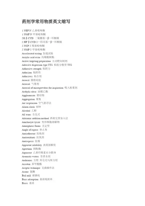
药剂学常用物质英文缩写1-NEP N-乙基吡咯酮1-NMP N-甲基吡咯酮2G-β-CYD 二葡糖基-β-环糊精2-HP-β-CYD 2-羟丙基-β-环糊精5-NCP 5-羧基吡咯酮5-NMP 5-甲基吡咯酮Accelerated testing 加速试验Acrylic acid resin 丙烯酸树酯Active targeting preparation 主动靶向制剂Adersive dispersion-type TTS 粘胶分散型TTS Adhersive strength 粘附力Adhesion 粘附性Adhesives 粘合剂Aerosil 微粉硅胶Aerosol 气雾剂Aerosol of micropowders for inspiration 吸入粉雾剂Aethylis oleas 油酸乙酯Agglomerate 聚结物Aggregation 聚集Air suspension 空气悬浮法Alarm clock 闹钟Alcohol 乙醇All-trans 全反式Alterntae addition method 两相交替加入法Amebocyte lysate 变形细胞溶解物Amorphous forms 无定型Angle of repose 休止角Antiadherent 抗粘剂Antioxidants 抗氧剂Antisepesis 防腐Apparent solubility 表现溶解度Aprotinin 抑酞酶Aquacoat 乙基纤维素水分散体Aromatic waters 芳香水剂Arrhenius 方程阿仑尼乌斯方程Ascabin 苯甲酸酯Aseptic technique 无菌操作法Azone 氮酮Ball mill 球磨机Base adsorption 基质吸附率Bases 基质Beeswax 蜂蜡Bending 弯曲力BHA 叔丁基对羟基茴香醚BHT 二叔丁基对甲酚Bioavailability 生物利用度Biochemical approach 生物学方法Biopharmaceutics 生物药剂学Biotechnology 生物技术Bond学说中等粉碎(粒径)Bound water 结合水分Breakage (Bk) 脆碎度Brij 泽、聚氧乙烯脂肪醇醚Brij 聚氧乙烯脂肪醇醚Buccal tablets 颊额片Bulk density 松密度Bulk density 松密度、堆密度Burst effect 突释效应CA 醋酸纤维素CAB 醋酸纤维素丁酸酯Cabomer 羟基乙烯共聚物Caking 结饼CAP 醋酸纤维素酞酸酯CAP 邻苯二甲酸醋酸纤维素Capillary state 毛细管状Capsules 胶囊剂Carbomer 卡波姆、羧基乙烯共聚物Carbopol 卡波普Carbopol 934 卡波普Carboxymethyl cellulose sodium 羟甲基纤维素钠Carboxymethyl starch sodium CMS-Na羧甲基淀粉纳Carboxymethylcellulose sodium CMC-Na羧甲基纤维素纳CAT 醋酸纤维素苯三酸酯CD 圆二色谱法Cellulose acetate (CA) 醋酸纤维素Cellulose acetate phthalate (CAP) 醋酸纤维素酞酸醋Central composite design (CCD) 星点设计Cera aseptical pro osse bone wax 骨蜡Ceramide 神经酰胺Cetomacrogol 聚乙二醇与十六醇缩合Chemical approach 化学方法Chewable tablets 咀嚼片Chitin 壳多糖Chitosan 壳聚糖Chronopathology 时辰病理学Chronopharmacology 时辰药理学Clausius-Clapeyron方程克劳修斯-克拉珀龙方程Clinical pharmaceutics 临床药剂学Cloud point 对聚氧乙烯型非离子表面活性剂CMC-Na 羧甲基纤维素纳CMEC 羧甲乙纤维素CMS 羧甲基淀粉CMS-Na 羧甲基淀粉钠Coadminiatration of skin Meta Inh 皮肤代谢抑制剂的合用Coadministraition of chem. P Enh 化学吸收促进剂的合用Coagulation 聚沉Coated tablets 包衣片Coating material 表材Cocoa butter 可可豆脂Cohesion 凝聚性、粘着性Cohesive strength 内聚力Cold compression method 汽压法Cold-homogenization 冷却一匀化法Colon-targeted capsules 结肠靶向胶囊剂Compactibility 成形性Complex coacervation 复凝聚法Compliance 顺应性Compressed tablets 普通片Compressibility 压缩度Compression 压缩力Compressive work 压缩功Cone and plate viscometer 圆椎平板粘度计Consistency curve 稠度曲线Controllability 可控性Controlled release tablets 控释片Controlled-release preparation 控释制剂Convective mixing 对流混合Convective transport 传递透过Coordination number 配位数Copoly (latic/glycolic) acid 聚乳酸乙醇酸共聚物Core material 表心物Cosolvency 潜溶Cosolvent 潜溶剂Coulter counter method 库尔特计数法Count basis 个数基准CP 聚羧乙烯CPVP 交联聚乙烯比咯烷酮CRacemization 外消旋作用Creams 乳青剂Creep 蠕变性Cremolphore EL 聚氧乙烯蓖麻油甘油醚Critical relative humidity (CRH) 临界相对湿度Critical relative humidity(CRH)临界相对湿度Critical velocity 临界速度Critrical micell concentration CMC临界胶束浓度Croscarmellose sodium CCNa交联羧甲基纤维素纳Croscarmellose sodium (CCNa) 交联甲基纤维素钠Crospovidone 交联聚维酮Cross-liked polyvinyl pyrrolidone PVPP交联聚维酮Crushing 粉碎Crystal form 晶型Crystal habit 晶态、晶癖、结晶习性CTS 普通栓剂Cumulative size distribution 累积分布Cutting 剪切力Cyclodextrin (CYD) 环糊精Cylinder model 圆栓体模型Cytotoxicity 细胞素DDS 药物传递系统Decoction 汤剂Degree of circularity 圆形度Degree of sphericility 球形度Delipidization 角质层去脂质化Dextrin 糊精Dialysis cell method 渗析池法Dicetyl phosphate 磷酸二鲸蜡脂Dielectric constant 介电常数Differential scanning calorimetry DSC差示扫描显热法Differential thermal analysis DTA差示热分析法Diffusive mixing 扩散混合Dilatant flow 胀性流动Diluents 稀释剂、填充剂Dimethicone (silicones) 二甲基硅油、硅油、硅酮Dimethyl sulfoxide(DMSO) 二甲基亚砜Dimethylacetamide (DMA) 二甲基乙酰胺Disinfection 消毒Disintegrants 崩解剂Disk assemble method 圆盘法Disperse medium 分散介质Disperse phase 分散相Disperse system 分散体系Dispersed phase 分散相、内相、非连续相Dispersible tablets 分散片Displacement value (DV) 置换价Distilled water 蒸馏水DL-phenylalanine ethyl acetoacetate DL苯基苯胺乙醚乙酸乙酯DLVO理论引力势能与斥力势能DME 二甲醚DMSO 二甲基亚矾DM-β-CYD 二甲基-β-环糊精Donor cell 供给宝DOPE 二油酰磷脂酰乙醇胺Dosage form 药物剂型DPPC 二棕榈酰磷脂酰胆碱Drop dentifrices 滴牙剂Drug carrier 药物载体Drug-loading rate 载药量Dry bulb temperature 干球温度DSPC 二硬脂酰磷脂酰胆碱DSPE 二硬脂酰磷脂酰乙醇胺Dumping effect 突释效应EA 乙基纤维素Ear drops 滴耳剂EC 乙基纤维素EC 毛细管电泳Effect diameter Dsk,有效径Effectiveness 有效性Effervescent disintegrants 泡腾崩解剂Effervescent tablets 泡腾片Elastic deformation 弹性变形Elastic recovery (ER) 弹性复原率Elastic work 弹性功Elasticity 弹性Electro phoresis 电泳Electroporesis 电致孔法Electuary 煎膏剂EMA 甲丙烯酸乙酯Emolphor 聚氧乙烯蓖麻油化合物Emulsifer in water method 水中乳化剂法、湿胶法Emulsifier in oil method 油中乳化剂法、干胶法Emulsion 普通乳Emulsions 乳剂Enamine 烯胺Endocytosis 内呑Endotoxin 内毒素Enteric capsules 肠溶胶囊剂Enteric coated tablets 肠溶衣片Entrapment rate 包封率Epidermis 表皮Epimerization 差向异构作用EPR效应促渗滞留作用Equilibrium solubility 平衡溶解度Equilibrium water 平衡水分Equivalent specific surface DSVEquivalent volume diameter Dv,体积等价径,球相当径Ethanol 乙醇Ethical (prescription) drug 处方药Ethycellulose (EC) 乙基纤维素Ethylcellulose EC乙基纤维素Ethylene vilnylacetate copolymer 乙烯-醋酸乙烯共聚物Ethylene vinylacetate copolymer 乙烯-醋酸乙烯共聚物Eu L, Eu S 聚甲基丙烯酸Eu RL, Eu RS 聚甲基丙烯酸酯Eu RL100, Eu RS100 甲基丙烯酸酯共聚物(不溶)Eu RL100, Eu SL100 聚丙烯酸树脂系列Eu S100, Eu L100 甲基丙烯酸共聚物(肠溶)Eudragit (E, RL, RS) 甲基丙烯酸酯共聚物Eudragit L100 甲基丙烯酸共聚物Eudragit RS100, RL100, NE30D 甲基丙烯酸酸共聚物- Eudragit S100 甲基丙烯酸共聚物EV A 乙烯-醋酸乙烯共聚物Evaporation 蒸发Excipients (adjuvants) 辅料External phase 分散介质、外相、连续相Extracts 浸膏剂Eye drop 滴眼剂Eye ointments 眼膏剂Factorial design 析因设计Fatty oils 脂肪油Feret diameter 定方向接线径Ficks第一扩散公式药材提取Fillers 填充剂Film coated tablets 薄膜衣片Film dispersion method 薄分散法Films 膜剂First-pass effect 首过效应Fliud extracts 流浸膏剂Flocculation 絮凝Flocculation value 絮凝度Flow curve 流动曲线Flow velocity 流出速度Flowability 流动性Fluid-energy mills 流能磨、气流式粉碎机Fluidity buffer 流动性缓冲剂Fluidized bed coating 流化床包衣法Free water 自由水分Freely movable liquid 自由流动液体Freon 氟氯烷烓类、氟里昂Frequency size distribution 频率分布Funicular state 索带状Fusion 融合Fusion method 热烙法Garles 含潄剂GAS 气体反溶剂技术Gas adsorption method 气体吸附法Gas antisolution GASGas permeability method 气体透过法GCP 药物临床试验管理规范Gelatin 胫胶Gelatin 明胶Gelatin glycerin 甘油胫胶Gelatinization 糊化General acid-base catalysis 广义酸碱催化Geometric diameter 几何学粒子径Geometric isomerization 几何异构Ghost cell 影细胞Glidants 助流剂GLP 药物非临床研究管理规范Glycerin 甘油Glycerins 甘油剂Glyceryl monostearate 硬脂酸、甘油酯Glycolic acid 羟基乙酸GMP 药品生产质量管理规范Granule density 颗粒密度Granules 颗粒剂Graton-Fraser模型颗粒的排列模型Group number HLB基团数Guest molecules 客分子G-β-CYD 葡糖--环糊精Half life 半衰期Handerson-Hasselbach公式解离状态、pkc、ph的关系Hard capsules 硬胶囊剂Hardness 硬度HCO 氢仪蓖麻油HEC 羟乙基纤维素HEMA 甲基丙烯酸羟乙酯HES 羟乙基淀粉Heywood diameter Dh,投影面积圆相当径Higuchi方程希古契方程Host molecules 主分子HPC 羟丙纤维素HPMA 羟丙甲丙烯酸甲酯HPMC 羟丙甲基纤维素HPMC 羟丙甲纤维素HPMCAS 醋酸羟丙甲纤维素琥珀酸酯HPMCP 羟丙甲基纤维素酞酸酯HPMCP (HP-50, HP-55) 羟丙甲纤维酸酯Humidity 湿度Hydration of stratum corneum 角质层的水化作用Hydrogel 水性凝胶Hydrophile-lipophile balance 亲水亲油平衡值Hydrotropy 助溶Hydrotropy agent 助溶剂Hydroxypropyl methylcellulose 羟丙甲纤维素Hydroxypropyl methylcellulose acetate succinate 醋酸羟丙甲纤维素琥珀酸酯Hydroxypropyl methylcellulose phthalate 羟丙甲纤维素酞酸醋Hydroxypropylcellulose (HPC) 羟丙基纤维素Hydroxypropylmethyl cellulose HPMC羟丙甲基纤维素Hygroscopicity 吸湿性Hypodermic tablets 皮下注射用片ICH 国际协调会议IDDS 植入给药系统IEC 离子交换色谱法IEF 等电点聚焦Immobile liquid 不可流动液体Impact 冲击力Impact mill 冲击式粉碎机Implant tablets 植入片Inclusion compound 包含物Industrial pharmaceutics 工业药剂学Infusion solution 输液Injection 注射液In-liquid drying 液中干燥法(乳化-溶剂挥发法)Interface polycondensation 界面缩聚法intra-arterial route 动脉内注射Intradermal (ID) route 皮内注射Intramuscular (IM) route 肌肉注射Intravenous (IV) route 静脉注射Intrinsic dissolution rate 特性溶出速率Intrinsic solubility 特性溶解度Inverse targeting 反向靶向Iontophoresis 离子渗透法IR 红外Isoclectric focusing IEF等电点聚焦Isoosmotic solution 等渗溶液Isopropylpalmitate 异丙酸棕榈酯Isostearylisostearate 异硬脂酸异硬酯Isotonic solution 等张溶液Journal of Drug Targeting 药物靶向杂志Kick学说粗粉碎(体积)Krafft point 对离子型表面活性剂而言Krummbein diameter 定方向最大径Lactic acid 乳酸Lactose 乳糖Lag time 滞留时间Large unilamellar vesicles 大单室脂质体Laurocapam 月桂氮草酮Length basis 长度基准L-HPC 低取代羟丙基纤维素Limulus lysate test 鲎试验法Liniments 搽剂Liposomes 脂质体Liquid immersion method 液浸法Liquid injection 无针液体注射器Liquid paraffin 液体石蜡Long-circulating liposome 长循环脂质体Long-circulating liposomes 长循环脂质体Long-term testing 长期试验Loo-Rigelman方程双宝血药浓度-吸收率换算Lotions 洗剂Lubricants 润滑剂LUVs 大单宝脂质体。
黄芪促进中枢神经系统再生与修复的研究进展张力【摘要】黄芪具有免疫调节、抗菌、利尿、强心等多方面药理作用.中医认为神经功能缺损通常是由肾精不足所致.黄芪作为补益中药可提高机体对创伤的修复能力,增加损伤部位血供,有利于神经再生,促进神经干细胞增殖及定向分化,从中提取的单体或有效成分对中枢神经系统再生与修复具有保护作用,形成功能性神经元,为治疗神经功能缺损疾病提供了临床应用前景.该文依据黄芪对神经再生不同作用并最终促进功能性神经再生的思路对其综述.【期刊名称】《医学综述》【年(卷),期】2014(020)003【总页数】3页(P398-400)【关键词】黄芪;中枢神经系统;再生;修复;神经干细胞;神经保护【作者】张力【作者单位】西安医学院解剖教研室,西安,710021【正文语种】中文【中图分类】R282.71中医药学对神经功能缺损疾病的治疗有较深刻的认识。
神经再生已成为当前国际神经科学和脑科学研究的热点,同时也成为中医药现代化发展的一个切入点。
近年来的研究表明,中药不仅可以保护神经细胞,提高神经元抵抗损伤的能力[1],还可以促进神经干细胞(neural stem cell,NSC)增殖及定向分化,形成功能性神经元,达到治疗神经功能缺损疾病的目的[2]。
了解中药的作用机制,有助于为中药的临床应用拓展思路。
1 黄芪的有效成分及作用《中国药典》中收载蒙古黄芪或膜荚黄芪的干燥根作为药用黄芪正品。
中国黄芪属特有植物——淡紫花黄芪可当做膜荚黄芪使用。
常见的药用黄芪及制品多为蒙古黄芪。
其注射液是经提取后制成的灭菌水溶液。
目前对黄芪药材及含君药黄芪的中成药均以黄芪皂苷Ⅳ(黄芪甲苷)作为质量评价指标[3]。
药理研究表明,黄芪注射液中不同化学成分具有不同的药理活性,主要有3种:黄芪皂苷、黄芪酮、黄芪多糖[4]。
此外,有研究发现一些中药配伍黄芪可提高机体对创伤的修复能力,增加损伤部位血供,有利于神经再生和修复[5]。
黄芪作为祖国医学常用的中药材之一,其药用历史悠久。
浅论β受体阻断药治疗青光眼的研究概况【关键词】青光眼/ 治疗;β受体阻断药青光眼是世界第二大致盲眼病。
据统计[1],2000年全世界共有6 680万青光眼患者,其中670万患者因此而双目失明;我国大约有500~600万青光眼患者,致盲人数也有50万左右。
目前治疗青光眼的药物主要是降眼压药,分五类:β受体阻断药、α受体激动药、碳酸酐酶抑制药、前列腺素类药物、拟胆碱能药。
其中β受体阻断药又是临床治疗青光眼的一线用药。
笔者主要介绍β受体阻断药治疗青光眼的研究进展。
1 噻吗洛尔噻吗洛尔是1978年被美国FDA批准第一个用于治疗青光眼的β受体阻断药,其抗青光眼机制有以下几方面:减少房水生成来降低眼压;减少Ca2+和Na+内流从而保护视网膜神经[2,3]。
在长达6个月的随机双盲、多中心临床试验中,%噻吗洛尔滴眼液降眼压达~,与1%布林佐胺滴眼液为~相似,但明显弱于1%布林佐胺、%噻吗洛尔复方制剂~。
另外一项临床试验中,噻吗洛尔降低眼压平均达(~),与拉坦前列素疗效相同(~)。
研究证实每天予%噻吗洛尔眼用凝胶液滴眼1次与每天滴眼2次的疗效相当[6, 7]。
由于噻吗洛尔能结合于虹膜黑色素,所以噻吗洛尔治疗虹膜色素沉着病人效果较差;噻吗洛尔能减少脉络膜和视神经盘血液供应,这对青光眼治疗有害。
噻吗洛尔常见局部不良反应有结膜充血、上皮点状着色、干眼症等;全身不良反应有心动过缓、心律失常、房室传导阻滞、心力衰竭、支气管痉挛;中枢神经系统不良反应有抑郁、焦虑、阳萎、疲劳、幻觉等。
2 卡替洛尔卡替洛尔降眼压机制和噻吗洛尔一样,也是通过减少房水生成来降低眼压。
临床研究从(±)kPa降至(±)kPa,优于尼普洛尔的(±)kPa至(±)kPa。
表明,拉坦前列腺素治疗3个月的青光眼患者中,卡替洛尔能进一步降低眼压。
在146例原发性青光眼及眼高压患者参与的随机双盲试验中,1%长效卡替洛尔滴眼液降眼压效果(每日1次)与1%常规卡替洛尔滴眼液(每日2次)基本相同。
药物经济学评价是基于对药品卫生成本和健康产出的全面分析,从而对药品经济性进行评价和优选的学科技术[1]。
虽然在药物经济学评价中探讨的是整体成本,但药品价格和费用是构成卫生成本的重要内容。
因此,药品价格的改变可能会显著影响经济学评价的结果。
现实中,医药价格具有易变性[2],药物经济学评价结果也会有相应的动态改变,需要及时更新经济学评价结果,以更好地支持决策。
德谷门冬双胰岛素是由70%的德谷胰岛素和30%的门冬胰岛素组成的可溶性双胰岛素制剂,于2019年在我国上市,并在2020年中完成和发表了该药对比甘精胰岛素的药物经济学研究报告[3]。
2020年底,德谷门冬双胰岛素通过国家医保谈判进入了医保用药目录[4],其医保支付价格有了明显下降。
为此,本文基于该药品新的价格水平,采作者简介:陶立波,博士,研究员。
研究方向:卫生经济、卫生政策分析。
E-mail :***************医保准入后德谷门冬双胰岛素对比甘精胰岛素治疗口服降糖药控制不佳的中国2型糖尿病患者的成本-效果陶立波1,2,王芳旭31.北京大学公共卫生学院卫生政策与管理学系,北京 100191;2. 北京大学医学部卫生政策与技术评估中心,北京 100191;3. 北京医药卫生经济研究会,北京 100012[摘要]目的:评价在德谷门冬双胰岛素医保谈判准入后,口服降糖药控制不佳的中国2型糖尿病患者应用德谷门冬双胰岛素治疗的长期成本-效果。
方法:基于IQVIA CORE 糖尿病模型,计算德谷门冬双胰岛素和甘精胰岛素治疗30年的成本和健康产出,并进行增量成本-效果分析。
研究采用卫生系统视角,成本包括降糖治疗、疾病管理及并发症治疗成本,健康产出指标采用质量调整生命年(QALYs ),贴现率使用5%。
通过概率敏感性分析评价结果的稳健性。
结果:与甘精胰岛素组比较,德谷门冬双胰岛素组的QALYs 增加了0.064年(8.285年vs 8.221年),直接总医疗成本减少77 567元(232 867元vs 310 434元)。

熊去氧胆酸与腺苷蛋氨酸治疗胆汁性肝硬化的比较分析赵越;宋宝君【摘要】目的比较熊去氧胆酸(UDCA)与腺苷蛋氨酸(SAM)治疗胆汁性肝硬化的疗效.方法采用前瞻性研究方法,选择80例原发性胆汁性肝硬化患者,随机平均分为观察组与对照组,两组都予以甘利欣、还原型谷胱甘肽、门冬氨酸钾镁、钙剂治疗,在此基础上,观察组给予UDCA胶囊治疗,对照组给予注射用丁二磺酸SAM治疗,均连续治疗观察3个月.结果治疗后,观察组的总有效率(97.5%)显著高于对照组(85.0%)(P<0.05).治疗后,观察组的血清ALT与AST值分别为(32.87±12.10)U/L 和(33.20±14.20)U/L,明显低于对照组的(51.39±13.09)U/L和(51.49±14.22)U/L(P<0.05).治疗后两组的CD4+值明显低于治疗前,且观察组明显低于对照组(P<0.05),而治疗前后两组的CD8+值均无明显变化(P>0.05).结论UDCA辅助治疗原发性胆汁性肝硬化,能减轻肝脏的损害,提高治疗有效率,疗效明显优于SAM,其作用机制可能与提高患者的细胞免疫功能有关.%Objective To compare the curative effects between ursodeoxycholic acid (UDCA) and S-adenosyl methionine (SAM) in the treatment of biliary cirrhosis.Methods A prospective study was made on 80 patients with primary biliary cirrhosis, who were divided randomly into an observation group and a control group, and the two groups were administered with Diammonium Glycyrrhizinate, reduced glutathione, Potassium Aspartate magnesium, and calcium; on the basis of this, the observation group was additionally administered with UDCA capsule and the control group with SAM-B injection. Both groups were treated and observed for three consecutive months. divided randomly into an observation group and a control group,and the two groups were administered with Diammonium Glycyrrhizinate, reduced glutathione, Potassium Aspartate magnesium, and calcium; on the basis of this, the observation group was additionally administered with UDCA capsule and the control group with SAM-B injection. Both groups were treated and observed for three consecutive months.Results After the treatment, the total effective rate in the observation group (97.5%) was significantly higher than that in the control group (85%) (P < 0.05); the serum ALT and AST values in the observat ion group were (32.87±12.10)U/L and (33.20±14.20)U/L, respectively, significantly lower than those in the control group (51.39±13.09)U/L and (51.49±14.22)U/L(P < 0.05). The CD4+ values in both groups were significantly lower than those before the treatment; the CD4+ value in the observation group were significantly lower than those in the control group (P < 0.05), whereas the CD8+ values in both groups before and after the treatment were not significantly changed (P > 0.05).Conclusion As an adjuvant medicine in the treatment of primary biliary cirrhosis, UDCAcan reduce the damage to the liver, improve the efficiency of treatment, and has better curative effect than SAM; its mechanism is probably related to the improvement of the cellular immune function of patients.【期刊名称】《西南国防医药》【年(卷),期】2017(027)003【总页数】3页(P239-241)【关键词】熊去氧胆酸;胆汁性肝硬化;肝功能;免疫功能;腺苷蛋氨酸【作者】赵越;宋宝君【作者单位】113015辽宁抚顺,抚顺市传染病医院传染科;113015辽宁抚顺,抚顺市传染病医院传染科【正文语种】中文【中图分类】R575原发性胆汁性肝硬化(PBC)以慢性肝内胆汁淤积为主要临床特征,是一种肝内细小胆管的慢性非化脓性炎症,伴有门静脉炎症性改变[1-2]。
2021年全国硕士研究生招生考试英语(一)试题及答案考试采取“一题多卷”模式,试题答案顺序不统一,请依据试题进行核对。
Section I Use of EnglishDirections:Read the following text. Choose the best word(s) for each numbered blank and mark A, B, C or D on the ANSWER SHEET. (10 points)Fluid intelligence is the type of intelligence that has to do with short-term memory and the ability to think quickly, logically, and abstractly in order to solve new problems. It_____(1)in young adulthood, levels out for a period of time, and then_____(2)starts to slowly decline as we age. But_____(3)aging is inevitable, scientists are finding that certain changes in brain function may not be.One study found that muscle loss and the_____(4)of body fat around the abdomen are associated with a decline in fluid intelligence. This suggests the_____(5)that lifestyle factors might help prevent or_____(6)this type of decline.The researchers looked at data that_____(7)measurements of lean muscle and abdominal fat from more than 4,000 middle-to-older-aged men and womenand_____(8)that data to reported changes in fluid intelligence over a six-year period. They found that middle-aged people_____(9)higher measures of abdominalfat_____(10)worse on measures of fluid intelligence as the years_____(11).For women, the association may be_____(12)to changes in immunity that resulted from excess abdominal fat; in men, the immune system did not appear tobe_____(13)It is hoped that future studies could_____(14)these differences and perhaps lead to different_____(15)for men and women._____(16)there are steps you can_____(17)to help reduce abdominal fat and maintain lean muscle mass as you age in order to protect both your physical and mental _____(18). The two highly recommended lifestyle approaches are maintaining or increasing your_____(19)of aerobic exercise and following Mediterranean-style_____(20)that is high in fiber and eliminates highly processed foods.1.【题干】1._____【选项】A.pausesB.returnC.peaksD.fades2.【题干】2._____【选项】A.alternativelyB.formallyC.accidentallyD.generally3.【题干】3._____【选项】A.whileB.sinceC.onceD.until4.【题干】4._____【选项】A.detectionB.accumulationC.consumptionD.separation5.【题干】5._____【选项】A.possibilityB.decisionC.goalD.requirement6.【题干】6._____【选项】A.delayB.ensureC.seekD.utilize7.【题干】7._____【选项】A.modifyB.supportedC.includedD.predicted8.【题干】8._____A.devotedparedC.convertedD.applied9.【题干】9._____【选项】A.withB.aboveC.byD.against10.【题干】10._____【选项】A.aboveB.managedC.scoredD.played11.【题干】11._____【选项】A.ran outB.set offD.went by12.【题干】12._____【选项】A.superiorB.attributableC.parallelD.resistant13.【题干】13._____【选项】A.restoredB.isolatedC.involvedD.controlled14.【题干】14._____【选项】A.alterB.spreadC.removeD.explain15.【题干】15._____【选项】pensationsB.symptomsC.demandsD.treatments16.【题干】16._____【选项】A.LikewiseB.MeanwhileC.ThereforeD.Instead17.【题干】17._____【选项】A.changeB.watchC.countD.take18.【题干】18._____【选项】A.well-beingB.processC.formationD.coordination19.【题干】19._____【选项】A.levelB.loveC.knowledgeD.space20.【题干】20._____【选项】A.designB.routineC.dietD.prescriptionSection II Reading Comprehension Part ADirections: Read the following four texts. Answer the questions below each text by choosing A, B, C or D. Mark your answers on the ANSWER SHEET. (40 points) How can the train operators possibly justify yet another increase to rail passenger fares? It has become a grimly reliable annual ritual: every January the cost of travelling by train rises, imposing a significant extra burden on those who have no option but to use the rail network to get to work or otherwise. This year's rise, anaverage of 2.7 per cent, may be a fraction lower than last year's, but it is still well above the official Consumer Price Index (CPI) measure of inflation.Successive governments have permitted such increases on the grounds that the cost of investing in and running the rail network should be borne by those who use it, rather than the general taxpayer. Why, the argument goes, should a car-driving pensioner from Lincolnshire have to subsidise the daily commute of a stockbroker from Surrey? Equally there is a sense that the travails of commuters in the South East, many of whom will face among the biggest rises, have received too much attention compared to those who must endure the relatively poor infrastructure of the Midlands and the North.However, over the past12 months, those commuters have also experienced some of the worst rail strikes in years. It is all very well train operators trumpeting the improvements they are making to the network, but passengers should be able to expect a basic level of service for the substantial sums they are now paying to travel. The responsibility for the latest wave of strikes rests on the unions. However, there is a strong case that those who have been worst affected by industrial action should receive compensation for the disruption they have suffered.The Government has pledged to change the law to introduce a minimum service requirement so that, even when strikes occur, services can continue to operate. This should form part of a wider package of measures to address the long-running problems on Britain's railways. Yes, more investment is needed, but passengers will not be willing to pay more indefinitely if they must also endure cramped, unreliable services, punctuated by regular chaos when timetables are changed, or planned maintenance is managed incompetently. The threat of nationalisation may have been seen off for now, but it will return with a vengeance if the justified anger of passengers is not addressed in short order.21.【题干】The author holds that this year's increase in rail passengers fares_____.【选项】A.will ease train operation's' burden.B.has kept pace with inflation.C.is a big surprise to commuters.D.remains an unreasonable measure.22.【题干】The stockbroker in 2 is used to stand for_____.【选项】A.car driversB.rail travellersC.local investorsD.ordinary taxpayers23.【题干】It is indicated in 3 that train operators_____.【选项】A.are offering compensations to commuters.B.are trying to repair relations with the unions.C.have failed to provide an adequate service.D.have suffered huge losses owing to the strikes.24.【题干】If unable to calm down passengers, the railways may have to face_____.【选项】A.the loss of investment.B.the collapse of operations.C.a reduction of revenueD.a change of ownership.25.【题干】Which of the following would be the best title for the text?【选项】A.Who Are to Blame for the Strikes?B.Constant Complaining Doesn't WorkC.Can Nationalization Bring Hope?D.Ever-rising Fares Aren't SustainableLast year marked the third year in a row of that Indonesia’s bleak rate of deforestation has slowed in pace. One reason for the turnaround may be the country's antipoverty program.In 2007, Indonesia started phasing in program that gives money to its poorest residents under certain conditions, such as requiring people to keep kids in school or get regular medical care. Called conditional cash transfers or CCTs, these social assistance programs are designed to reduce inequality and break the cycle of poverty. They're already used in dozens of countries worldwide. In Indonesia, the program has provided enough food and medicine to substantially reduce severe growth problems among children.But CCT programs don't generally consider effects on the environment. In fact, poverty alleviation and environmental protection are often viewed as conflicting goals, says Paul Ferraro, an economist at Johns Hopkins University.That's because economic growth can be correlated with environmental degradation, while protecting the environment is sometimes correlated with greater poverty. However, those correlations don't prove cause and effect. The only previous study analyzing causality, based on an area in Mexico that had instituted CCTs, supported the traditional view. There, as people got more money, some of them may have more cleared land for cattle to raise for meat, Ferraro says.Such programs do not have to negatively affect the environment, though. Ferraro wanted to see if Indonesia's poverty-alleviation program was affecting deforestation. Indonesia has the third-largest area of tropical forest in the world and one of the highest deforestation rates.Ferraro analyzed satellite data showing annual forest loss from 2008 to2012-including during Indonesia's phase-in of the antipoverty program-in 7, 468 forested villages across 15 provinces and multiple islands. The duo separated the effects of the CCT program on forest loss from other factors, like weather and macroeconomic changes, which were also affecting forest loss. With that, "we see that the program is associated with a 30 percent reduction in deforestation," Ferraro says.That's likely because the rural poor are using the money as makeshift insurance policies against inclement weather, Ferraro says. Typically, if rains are delayed,people may clear land to plant more rice to supplement their harvests. With the CCTs, individuals instead can use the money to supplement their harvests.Whether this research translates elsewhere is anybody's guess. Ferraro suggests the importance of growing rice and market access. And regardless of transferability, the study shows that what's good for people may also be good for the value of the avoided deforestation just for carbon dioxide emissions alone is more than the program costs.26.【题干】According to the first two paragraphs, CCT programs aim to_____.【选项】A.facilitate health care reform.B.help poor families get better off.C.improve local education systems.D.lower deforestation rates.27.【题干】The study based on an area in Mexico is cited to show that_____.【选项】A.cattle rearing has been a major means of livelihood for the poor.T programs have he helped preserve traditional lifestyles.C.antipoverty efforts require the participation of local farmers.D.economic growth tends to cause environmental degradation.28.【题干】In his study about Indonesia, Ferraro intends to find out_____.【选项】A.its acceptance level of CCTs.B.its annual rate of poverty alleviation.C.the relation of ccts to its forest loss.D.the role of its forests in climate change.29.【题干】According to Ferraro, the CCT program in Indonesia is most valuable in that_____.【选项】A.it will benefit other Asian countries.B.it will reduce regional inequality.C.it can protect the environment.D.it can boost grain production.30.【题干】What is the text centered on?【选项】A.The effects of a program.B.The debates over a program.C.The process of a study.D.The transferability of a study.As a historian who's always searching for the text or the image that makes usre-evaluate the past, I've become preoccupied with looking for photographs that show our Victorian ancestors smiling (what better way to shatter the image of19th-century prudery?). I've found quite a few, and- since I started posting them on Twitter-they have been causing quite stir. People have been surprised to see evidence that Victorians had fun and could, and did, laugh. They are noting that the Victorians suddenly seem to become more human as the hundred-or-so years that separate us fade away through our common experience of laughter.Of course, I need to concede that my collection of 'Smiling Victorians' makes up only a tiny percentage of the vast catalogue of photographic portraiture created between 1840 and 1900, the majority of which show sitters posing miserably and stiffly infront of painted backdrops, or staring absently into the middle distance. How do we explain this trend?During the 1840s and 1850s, in the early days of photography, exposure times were notoriously long: the daguerreotype photographic method (producing an image on a silvered copper plate) could take several minutes to complete, resulting in blurred images as sitters shifted position or adjusted their limbs. The thought of holding a fixed grin as the camera performed its magical duties was too much to contemplate, and so a non-committal blank stare became the norm.But exposure times were much quicker by the 1880s, and the introduction of the Box Brownie and other portable cameras meant that, though slow by today's digital standards, the exposure was almost instantaneous. Spontaneous smiles were relatively easy to capture by the 1890s, so we must look elsewhere for an explanation of why Victorians still hesitated to smile.One explanation might be the loss of dignity displayed through a cheesy grin.“Nature gave us lips to conceal our teeth,” ran one popular Victorian maxim, alluding to the fact that before the birth of proper dentistry, mouths were often in a shocking state of hygiene. A flashing set of healthy and clean, regular pearly whites' rare sight in Victorian society, the preserve of the super-rich (and even then, dental hygiene was not guaranteed).A toothy grin (especially when there were gaps or blackened teeth) lacked class: drunks, tramps, prostitutes and buffoonish music hall performers might gurn and grin with a smile as wide as Lewis Carroll's gum-exposing Cheshire Cat, but it was not a becoming look for properly bred persons. Even Mark Twain, a man who enjoyed a hearty laugh, said that when it came to photographic portraits there could be "nothing more damning than a silly, foolish smile fixed forever".31.【题干】According to Paragraph 1, the author's posts on Twitter. _____【选项】A.changed people's impression of the Victorians.B.highlighted social media's role in Victorian studies.C.re-evaluated the Victorians' notion of public image.D.illustrated the development of Victorian photography.32.【题干】What does author say about the Victorian portraits he has collected?_____【选项】A.They are in popular use among historians.B.They are rare among photographs of that age.C.They mirror 19th-century social conventions.D.They show effects of different exposure times.33.【题干】What might have kept the Victorians from smiling for pictures in the 1890s? _____【选项】A.Their inherent social sensitiveness.B.Their tension before the camera.C.Their distrust of new inventions.D.Their unhealthy dental condition.34.【题干】Mark Twain is quoted to show that the disapproval of smiles in pictures was_____.【选项】A.a deep-root belief.B.a misguided attitude.C.a controversial view.D.a thought-provoking idea.35.【题干】Which of the following questions does the text answer?_____【选项】A.Why did most Victorians look stern in photographs?B.Why did the Victorians start view photographs?C.What made photography develop slowly in the Victorian period?D.How did smiling in photographs become a post-Victorian norm?From the early days of broadband, advocates for consumers and web-based companies worried that the cable and phone companies selling broadband connections had the power and incentive to favor affiliated websites over their rivals. That's why there has been such a strong demand for rules that would prevent broadband providers from picking winners and losers online, preserving the freedom and innovation that have been the lifeblood of the internet.Yet that demand has been almost impossible to fill-in part because of pushback from broadband providers, anti-regulatory conservatives and the courts. A federal appeals court weighed in again Tuesday, but instead of providing badly needed resolution, it only prolonged the fight. At issue before the U. S. Court of Appeals for the District of Columbia Circuit was the latest take of the Federal Communications Commission (FCC) on net neutrality, adopted on a party-line vote in 2017. The Republican-penned order not only eliminated the strict net neutrality rules the FCC had adopted when it had a Democratic majority in 2015, but rejected the commission's authority to require broadband providers to do much of anything. The order also declared that state and local governments couldn't regulate broadband providers either.The commission argued that other agencies would protect against anti-competitive behavior, such as a broadband-providing conglomerate like AT&T favoring its own video-streaming service at the expense of Netflix and Apple TV. Yet the FCC also ended the investigations of broadband providers that imposed data caps on their rivals' streaming services but not their own.On Tuesday, the appeals court unanimously upheld the 2017 order deregulating broadband providers, citing a Supreme Court ruling from 2005 that upheld a similarly deregulatory move. But Judge Patricia Millett rightly argued in a concurring opinion that “the result is unhinged from the realities of modern broadband service,” and said Congress or the Supreme Court could intervene to "avoid trapping Internet regulation in technological anachronism."In the meantime, the court threw out the FCC's attempt to block all state rules on net neutrality, while preserving the commission's power to preempt individual statelaws that undermine its order. That means more battles like the one now going on between the Justice Department and California, which enacted a tough net neutrality law in the wake of the FCC's abdication.The endless legal battles and back-and-for at the FCC cry out for Congress to act. It needs to give the commission explicit authority once and for all to bar broadband providers from meddling in the traffic on their network and to create clear rules protecting openness and innovation online.36.【题干】There has long been concern that broadband provides would_____.【选项】A.bring web-based firms under control.B.slow down the traffic on their network.C.show partiality in treating clients.D.intensify competition with their rivals.37.【题干】Faced with the demand for net neutrality rules, the Fcc_____.【选项】A.Sticks to an out-of-date order.B.Takes an anti-regulatory stance.C.Has issued a special resolution.D.Has allowed the states to intervene.38.【题干】What can be learned about AT&T from Paragraph 3?【选项】A.It protects against unfair competition.B.It engages in anti-competitive practices.C.It is under the FCC's investigation.D.It is in pursuit of quality service.39.【题干】Judge Patricia Millett argues that the appeals court's decision_____.【选项】A.focuses on trivialities.B.conveys an ambiguous message.C.is at odds with its earlier rulings.D.is out of touch with reality.40.【题干】What does the author argue in the last paragraph?【选项】A.Congress needs to take action to ensure net neutrality.B.The FCC should be put under strict supervision.C.Rules need to be set to diversify online services.D.Broadband providers' rights should be protected.Section II Reading Comprehension Part BThe following paragraphs are given in a wrong order. For Questions 41-45, you are required to reorganize these paragraphs into a coherent article by choosing from the list A-G and filling them into the numbered boxes. Paragraphs C and F have been correctly placed. Mark your answers on ANSWER SHEET. (10 points)In the movies and on television, artificial intelligence is typically depicted as something sinister that will upend our way of life. When it comes to AI in business, we often hear about it in relation to automation and the impending loss of jobs, but in what ways is AI changing companies and the larger economy that don’t involve doom-and-mass unemployment predictions?A recent survey of manufacturing and service industries from Tata Consultancy Services found that companies currently use Al more often in computer-to-computer activities than in automating human activities. One common application? Preventing electronic security breaches, which, rather than eliminating IT jobs, actually makes those personnel more valuable to employers, because they help firms prevent hacking attempts.Here are a few other ways AI is aiding companies without replacing employees: Better hiring practicesCompanies are using artificial intelligence to remove some of the unconscious bias from hiring decisions. "There are experiments that show that, naturally, the results of interviews are much more biased than what AI does," says Pedro Domingos, author of The Master Algorithm: How the Quest for the Ultimate Learning Machine Will Remake Our World and a computer science _____(41)One company that’s doing this is called Blendoor. It uses analytics to help identify where there may be bias in the hiring process.More effective marketingSome AI software can analyze and optimize marketing email subject lines to increase open rates. One company in the UK, Phrasee, claims their software can outperform humans by up to 10 percent when it comes to email open rates. This can mean millions more in revenue. _____(42)There are“tools that help people use data, not a replacement for people,” says Patrick H. Winston, a professor of artificial intelligence and computer science at MIT.Saving customers moneyEnergy companies can use AI to help customers reduce their electricity bills saving them money while helping the environment. Companies can also optimize their own energy use and cut down on the cost of electricity. Insurance companies meanwhile, can base their premiums on AI models that more accurately access risk. "Before, they might not insure the ones who felt like a high risk or charge them too much," says Domingos, _____(43)Improved accuracyMachine learning often provides a more reliable form of statistics, which makes data more valuable," says Winston. It "helps people make smarter decisions." _____(44) Protecting and maintaining infrastructureA number of companies, particularly in energy and transportation, use AI image processing technology to inspect infrastructure and prevent equipment failure or leaks before they happen. "If they fail first and then you fix them, it's very expensive," says Domingos. _____(45)[A] I replaces the boring parts of your job. If you're doing research, you can have AI go out and look for relevant sources and information that otherwise you just wouldn't have time for.[B] One accounting firm, EY, uses an AI system that helps review contracts during an audit. This process, along with employees reviewing the contracts, is faster and more accurate.[C] There are also companies like Acquisio, which analyzes advertising performance across multiple channels like Adwords, Bing and social media and makes adjustments or suggestions about where advertising funds will yield best results.[D] You want to predict if something needs attention now and point to where it's useful for employees to go to.[E] Before, they might not insure the ones who felt like a high risk or charge them too much, or they would charge them too little and then it would cost [the company] money.[F] We're also giving our customers better channels versus picking up the phone to accomplish something beyond human scale.[G] AI looks at resumes in greater numbers than humans would be able to, and selects the more promising candidates.41.【题干】41._____.【选项】A.AB.BC.CD.DE.EF.FG.G42.【题干】42._____.【选项】A.AB.BC.CD.DE.EF.FG.G43.【题干】43._____.【选项】A.AB.BC.CD.DE.EF.FG.G44.【题干】44._____.【选项】A.AB.BC.CD.DE.EF.FG.G45.【题干】45._____.【选项】A.AB.BC.CD.DE.EF.FG.GSection III TranslationDirections:Read the following text carefully and then translate the underlined segments into Chinese. Your translation should be written neatly on the ANSWER SHEET. (10 points)World war was the watershed event for higher education in modern Western societies(46)Those societies came out of the war with levels of enrollment that had been roughly constant at 3-5% of the relevant age groups during the decades before the war. But after the war, great social and political changes arising out of the successful war against Fascism created a growing demand in European and American economies for increasing numbers of graduates with more than a secondary schooleducation.(47)And the demand that rose in those societies for entry to higher education extended to groups and social classes that had not thought of attending a university before the war. These demands resulted in a very rapid expansion of the systems of higher education, beginning in the 1960s and developing very rapidly (though unevenly) during the 1970s and 1980s.The growth of higher education manifests itself in at least three quite different ways, and these in turn have given rise to different sets of problems. There was first the rate of growth:(48)in many counties of Western Europe, the numbers of students in higher education doubled within five-year periods during the 1960s and doubled again in seven, eight or 10 years by the middle of the 1970s. Second growth obviously affected the absolute size both of systems and individual institutions. And third growth was reflected in changes in the proportion of the relevant age group enrolled in institutions of higher education.Each of these manifestations of growth carried its own peculiar problems in its wake/ For example, a high growth rate placed great strains on the existing structures of governance, of administration, and above all of socialization. When a faculty or department grows from, say, five to 20 members within three or four years,(49)and when the new staff predominantly young men and women fresh from postgraduate study, they largely define the norms of academic life in that faculty. And if the postgraduate student population also grows rapidly and there is loss of a close apprenticeship relationship between faculty members and students, the student culture becomes the chief socializing force for new postgraduate students, with consequences for the intellectual and academic life of the institution-this was seen in America as well as in France, Italy, West Germany, and Japan.(50)High growth rates increased the chances for academic innovation, they also weakened the forms and processes by which teachers and students are admitted into a community of scholars during periods of stability or slow growth. In the 1960s and 1970s,European universities saw marked changes in their governance arrangements, with empowerment of junior faculty and to some degree of students as well.46.【题干】Those societies came out of the war with levels of enrollment that had been roughly constant at 3-5% of the relevant age groups during the decades before the war.47.【题干】And the demand that rose in those societies for entry to higher education extended to groups and social classes that had not thought of attending a university before the war.48.【题干】in many counties of Western Europe, the numbers of students in higher education doubled within five-year periods during the 1960s and doubled again in seven, eight or 10 years by the middle of the 1970s.49.【题干】and when the new staff predominantly young men and women fresh from postgraduate study, they largely define the norms of academic life in that faculty.50.【题干】High growth rates increased the chances for academic innovation, they also weakened the forms and processes by which teachers and students are admitted into a community of scholars during periods of stability or slow growth.Section IV WritingPart A (10 points)【题干】Directions:A foreign friend of yours has recently graduated from college and intends to find a job in China. Write him/her an email to make some suggestions.You should write about 100 words on ANSWER SHEET 2.Do not sign your own name at the end. Use "Li Ming Open" instead.You do not need to write the address.ear friend,Hope this letter finds you well I am glad to hear you intend to find a job in China, so I would like to extend my warmest welcome as well as provide you with a few suggestions on job-hunting.First, you can start from listing 3 to 5 cities which you would like to work or live in To be more specific, rate them by location, working opportunities and prospects and, of course the city's happiness level. What's more, be prepared for the culture shock. There is a sharp contrast in how eastern people and western people work. The former prefers working individually while the latter is prone to teamwork. There is one more point that, I suppose I have to touch on: make good use of onlinejob-hunting applications, such as BOSS and 51Job.I hope you will find my humble suggestions be of help. I am looking forward to your reply. Best wishes.Yours,Li MingPart B (15 points)【题干】Directions:。
COMMON TOXICITY CRITERIA (CTC)GradeAdverse Event01234ALLERGY/IMMUNOLOGYAllergic reaction/ hypersensitivity (including drug fever)none transient rash, drugfever <38°C (<100.4°F)urticaria, drug fever≥38°C (≥100.4°F),and/or asymptomaticbronchospasmsymptomaticbronchospasm,requiring parenteralmedication(s), with orwithout urticaria;allergy-relatededema/angioedemaanaphylaxisNote: Isolated urticaria, in the absence of other manifestations of an allergic or hypersensitivity reaction, is graded in the DERMATOLOGY/SKIN category.Allergic rhinitis (including sneezing, nasal stuffiness, postnasal drip)none mild, not requiringtreatmentmoderate, requiringtreatment--Autoimmune reaction none serologic or otherevidence ofautoimmune reactionbut patient isasymptomatic (e.g.,vitiligo), all organfunction is normal andno treatment is required evidence ofautoimmune reactioninvolving a non-essential organ orfunction (e.g.,hypothyroidism),requiring treatmentother thanimmunosuppressivedrugsreversible autoimmunereaction involvingfunction of a majororgan or other adverseevent (e.g., transientcolitis or anemia),requiring short-termimmunosuppressivetreatmentautoimmune reactioncausing major grade 4organ dysfunction;progressive andirreversible reaction;long-termadministration of high-dose immuno-suppressive therapyrequiredAlso consider Hypothyroidism, Colitis, Hemoglobin, Hemolysis.Serum sickness none--present-Urticaria is graded in the DERMATOLOGY/SKIN category if it occurs as an isolated symptom. If it occurs with other manifestations of allergic or hypersensitivity reaction, grade as Allergic reaction/hypersensitivity above.Vasculitis none mild, not requiringtreatment symptomatic, requiringmedicationrequiring steroids ischemic changes orrequiring amputationAllergy/Immunology - Other (Specify, __________)none mild moderate severe life-threatening ordisablingAUDITORY/HEARINGConductive hearing loss is graded as Middle ear/hearing in the AUDITORY/HEARING category. Earache is graded in the PAIN category.External auditory canal normal external otitis witherythema or drydesquamation external otitis withmoist desquamationexternal otitis withdischarge, mastoiditisnecrosis of the canalsoft tissue or boneNote: Changes associated with radiation to external ear (pinnae) are graded under Radiation dermatitis in the DERMATOLOGY/SKIN category.Adverse Event01234Inner ear/hearing normal hearing loss onaudiometry only tinnitus or hearing loss,not requiring hearingaid or treatmenttinnitus or hearing loss,correctable with hearingaid or treatmentsevere unilateral orbilateral hearing loss(deafness), notcorrectableMiddle ear/hearing normal serous otitis withoutsubjective decrease inhearing serous otitis or infectionrequiring medicalintervention; subjectivedecrease in hearing;rupture of tympanicmembrane withdischargeotitis with discharge,mastoiditis orconductive hearing lossnecrosis of the canalsoft tissue or boneAuditory/Hearing - Other (Specify, __________)normal mild moderate severe life-threatening ordisablingBLOOD/BONE MARROWBone marrow cellularity normal for age mildly hypocellular or≤25% reduction fromnormal cellularity forage moderately hypocellularor >25 - ≤50%reduction from normalcellularity for age or >2but <4 weeks torecovery of normalbone marrow cellularityseverely hypocellular or>50 - ≤75% reductionin cellularity for age or4 - 6 weeks to recoveryof normal bone marrowcellularityaplasia or >6 weeks torecovery of normalbone marrow cellularityNormal ranges:children (≤18 years)90% cellularityaverageyounger adults (19-59)60 - 70%cellularity averageolder adults (≥60 years)50% cellularityaverageNote: Grade Bone marrow cellularity only for changes related to treatment not disease.CD4 count WNL<LLN - 500/mm3200 - <500/mm350 - <200/mm3<50/mm3 Haptoglobin normal decreased-absent-Hemoglobin (Hgb)WNL<LLN - 10.0 g/dL<LLN - 100 g/L<LLN - 6.2 mmol/L 8.0 - <10.0 g/dL80 - <100 g/L4.9 - <6.2 mmol/L6.5 - <8.0 g/dL65 - <80 g/L4.0 - <4.9 mmol/L<6.5 g/dL<65 g/L<4.0 mmol/LFor leukemia studies or bone marrow infiltrative/ myelophthisic processes, if specified in the protocol.WNL10 - <25% decreasefrom pretreatment25 - <50% decreasefrom pretreatment50 - <75% decreasefrom pretreatment≥75% decrease frompretreatmentHemolysis (e.g., immune hemolytic anemia, drug-related hemolysis, other)none only laboratoryevidence of hemolysis[e.g., direct antiglobulintest (DAT, Coombs’)schistocytes]evidence of red celldestruction and ≥2gmdecrease in hemoglobin,no transfusionrequiring transfusionand/or medicalintervention (e.g.,steroids)catastrophicconsequences ofhemolysis (e.g., renalfailure, hypotension,bronchospasm,emergencysplenectomy)Also consider Haptoglobin, Hemoglobin.Adverse Event01234Leukocytes (total WBC)WNL<LLN - 3.0 x 10⁹ /L<LLN - 3000/mm3≥2.0 - <3.0 x 10⁹ /L≥2000 - <3000/mm3≥1.0 - <2.0 x 10⁹ /L≥1000 - <2000/mm3<1.0 x 109 /L<1000/mm3For BMT studies, if specified in the protocol.WNL≥2.0 - <3.0 X 10⁹/L≥2000 - <3000/mm3≥1.0 - <2.0 x 10⁹ /L≥1000 - <2000/mm3≥0.5 - <1.0 x 10⁹ /L≥500 - <1000/mm3<0.5 x 109 /L<500/mm3For pediatric BMT studies(using age, race and sexnormal values), if specifiedin the protocol.≥75 - <100% LLN≥50 - <75% LLN≥25 - 50% LLN<25% LLNLymphopenia WNL<LLN - 1.0 x 10⁹ /L<LLN - 1000/mm3≥0.5 - <1.0 x 10⁹ /L≥500 - <1000/mm3<0.5 x 109 /L<500/mm3-For pediatric BMT studies(using age, race and sexnormal values), if specifiedin the protocol.≥75 - <100%LLN≥50 - <75%LLN≥25 - <50%LLN<25%LLNNeutrophils/granulocytes (ANC/AGC)WNL≥1.5 - <2.0 x 10⁹ /L≥1500 - <2000/mm3≥1.0 - <1.5 x 10⁹ /L≥1000 - <1500/mm3≥0.5 - <1.0 x 10⁹ /L≥500 - <1000/mm3<0.5 x 109 /L<500/mm3For BMT studies, if specified in the protocol.WNL≥1.0 - <1.5 x 10⁹ /L≥1000 - <1500/mm3≥0.5 - <1.0 x 10⁹ /L≥500 - <1000/mm3≥0.1 - <0.5 x 10⁹ /L≥100 - <500/mm3<0.1 x 109 /L<100/mm3For leukemia studies or bone marrow infiltrative/ myelophthisic process, if specified in the protocol.WNL10 - <25% decreasefrom baseline25 - <50% decreasefrom baseline50 - <75% decreasefrom baseline≥75% decrease frombaselinePlatelets WNL<LLN - 75.0 x 10⁹ /L<LLN - 75,000/mm3≥50.0 - <75.0 x 10⁹ /L≥50,000 - <75,000/mm3≥10.0 - <50.0 x 10⁹ /L≥10,000 - <50,000/mm3<10.0 x 109 /L<10,000/mm3For BMT studies, if specified in the protocol.WNL≥50.0 - <75.0 x 10⁹ /L≥50,000 - <75,000/mm3≥20.0 - <50.0 x 10⁹ /L≥20,000 - <50,000/mm3≥10.0 - <20.0 x 10⁹ /L≥10,000 - <20,000/mm3<10.0 x 109 /L<10,000/mm3For leukemia studies or bone marrow infiltrative/ myelophthisic process, if specified in the protocol.WNL10 - <25% decreasefrom baseline25 - <50% decreasefrom baseline50 - <75% decreasefrom baseline≥75% decrease frombaselineTransfusion: Platelets none--yes platelet transfusions andother measures requiredto improve plateletincrement; platelettransfusionrefractoriness associatedwith life-threateningbleeding. (e.g., HLA orcross matched platelettransfusions)For BMT studies, if specified in the protocol.none 1 platelet transfusion in24 hours2 platelet transfusions in24 hours≥3 platelet transfusionsin 24 hoursplatelet transfusions andother measures requiredto improve plateletincrement; platelettransfusionrefractoriness associatedwith life-threateningbleeding. (e.g., HLA orcross matched platelettransfusions)Also consider Platelets.Adverse Event01234 Transfusion: pRBCs none--yes-For BMT studies, if specified in the protocol.none≤2 u pRBC in 24 hourselective or planned3 u pRBC in 24 hourselective or planned≥4 u pRBC in 24 hours hemorrhage orhemolysis associatedwith life-threateninganemia; medicalintervention required toimprove hemoglobinFor pediatric BMT studies, if specified in the protocol.none≤15mL/kg in 24 hourselective or planned>15 - ≤30mL/kg in 24hours elective orplanned>30mL/kg in 24 hours hemorrhage orhemolysis associatedwith life-threateninganemia; medicalintervention required toimprove hemoglobinAlso consider Hemoglobin.Blood/Bone Marrow - Other (Specify, __________)none mild moderate severe life-threatening ordisabling CARDIOVASCULAR (ARRHYTHMIA)Conduction abnormality/ Atrioventricular heart block none asymptomatic, notrequiring treatment(e.g., Mobitz type Isecond-degree AVblock, Wenckebach)symptomatic, but notrequiring treatmentsymptomatic andrequiring treatment(e.g., Mobitz type IIsecond-degree AVblock, third-degree AVblock)life-threatening (e.g.,arrhythmia associatedwith CHF, hypotension,syncope, shock)Nodal/junctional arrhythmia/dysrhythmia none asymptomatic, notrequiring treatmentsymptomatic, but notrequiring treatmentsymptomatic andrequiring treatmentlife-threatening (e.g.,arrhythmia associatedwith CHF, hypotension,syncope, shock)Palpitations none present---Note: Grade palpitations only in the absence of a documented arrhythmia.Prolonged QTc interval (QTc >0.48 seconds)none asymptomatic, notrequiring treatmentsymptomatic, but notrequiring treatmentsymptomatic andrequiring treatmentlife-threatening (e.g.,arrhythmia associatedwith CHF, hypotension,syncope, shock)Sinus bradycardia none asymptomatic, notrequiring treatment symptomatic, but notrequiring treatmentsymptomatic andrequiring treatmentlife-threatening (e.g.,arrhythmia associatedwith CHF, hypotension,syncope, shock)Sinus tachycardia none asymptomatic, notrequiring treatment symptomatic, but notrequiring treatmentsymptomatic andrequiring treatment ofunderlying cause-Supraventricular arrhythmias (SVT/atrial fibrillation/ flutter)none asymptomatic, notrequiring treatmentsymptomatic, but notrequiring treatmentsymptomatic andrequiring treatmentlife-threatening (e.g.,arrhythmia associatedwith CHF, hypotension,syncope, shock)Syncope (fainting) is graded in the NEUROLOGY category.Vasovagal episode none-present without loss ofconsciousness present with loss of consciousness-Adverse Event01234Ventricular arrhythmia (PVCs/bigeminy/trigeminy/ ventricular tachycardia)none asymptomatic, notrequiring treatmentsymptomatic, but notrequiring treatmentsymptomatic andrequiring treatmentlife-threatening (e.g.,arrhythmia associatedwith CHF, hypotension,syncope, shock)Cardiovascular/ Arrhythmia - Other (Specify, ___________)none asymptomatic, notrequiring treatmentsymptomatic, but notrequiring treatmentsymptomatic, andrequiring treatment ofunderlying causelife-threatening (e.g.,arrhythmia associatedwith CHF, hypotension,syncope, shock) CARDIOVASCULAR (GENERAL)Acute vascular leak syndrome absent-symptomatic, but notrequiring fluid supportrespiratory compromiseor requiring fluidslife-threatening;requiring pressorsupport and/orventilatory supportCardiac-ischemia/infarction none non-specific T - waveflattening or changes asymptomatic, ST - andT - wave changessuggesting ischemiaangina without evidenceof infarctionacute myocardialinfarctionCardiac left ventricular function normal asymptomatic declineof resting ejectionfraction of ≥10% but<20% of baseline value;shortening fraction≥24% but <30%asymptomatic butresting ejection fractionbelow LLN forlaboratory or decline ofresting ejection fraction≥20% of baseline value;<24% shorteningfractionCHF responsive totreatmentsevere or refractoryCHF or requiringintubationCNS cerebrovascular ischemia is graded in the NEUROLOGY category.Cardiac troponin I (cTnI)normal--levels consistent withunstable angina asdefined by themanufacturer levels consistent with myocardial infarction as defined by the manufacturerCardiac troponin T (cTnT)normal≥0.03 - <0.05 ng/mL≥0.05 - <0.1 ng/mL≥0.1 - <0.2 ng/mL≥0.2 ng/mLEdema none asymptomatic, notrequiring therapy symptomatic, requiringtherapysymptomatic edemalimiting function andunresponsive to therapyor requiring drugdiscontinuationanasarca (severegeneralized edema)Hypertension none asymptomatic, transientincrease by >20 mmHg(diastolic) or to>150/100* if previouslyWNL; not requiringtreatment recurrent or persistentor symptomatic increaseby >20 mmHg(diastolic) or to>150/100* if previouslyWNL; not requiringtreatmentrequiring therapy ormore intensive therapythan previouslyhypertensive crisis*Note: For pediatric patients, use age and sex appropriate normal values >95th percentile ULN.Adverse Event01234Hypotension none changes, but notrequiring therapy(including transientorthostatic hypotension)requiring brief fluidreplacement or othertherapy but nothospitalization; nophysiologicconsequencesrequiring therapy andsustained medicalattention, but resolveswithout persistingphysiologicconsequencesshock (associated withacidemia and impairingvital organ function dueto tissue hypoperfusion)Also consider Syncope (fainting).Notes:Angina or MI is graded as Cardiac-ischemia/infarction in the CARDIOVASCULAR (GENERAL) category.For pediatric patients, systolic BP 65 mmHg or less in infants up to 1 year old and 70 mmHg or less in children older than 1 year of age, use two successive or three measurements in 24 hours.Myocarditis none--CHF responsive totreatment severe or refractory CHFOperative injury of vein/artery none primary suture repairfor injury, but notrequiring transfusionprimary suture repairfor injury, requiringtransfusionvascular occlusionrequiring surgery orbypass for injurymyocardial infarction;resection of organ (e.g.,bowel, limb)Pericardial effusion/ pericarditis none asymptomatic effusion,not requiring treatmentpericarditis (rub, ECGchanges, and/or chestpain)with physiologicconsequencestamponade (drainage orpericardial windowrequired)Peripheral arterial ischemia none-brief episode ofischemia managed non-surgically and withoutpermanent deficit requiring surgicalinterventionlife-threatening or withpermanent functionaldeficit (e.g.,amputation)Phlebitis (superficial)none-present--Notes:Injection site reaction is graded in the DERMATOLOGY/SKIN category.Thrombosis/embolism is graded in the CARDIOVASCULAR (GENERAL) category.Syncope (fainting) is graded in the NEUROLOGY category.Thrombosis/embolism none-deep vein thrombosis,not requiringanticoagulant deep vein thrombosis,requiring anticoagulanttherapyembolic event includingpulmonary embolismVein/artery operative injury is graded as Operative injury of vein/artery in the CARDIOVASCULAR (GENERAL) category.Visceral arterial ischemia (non-myocardial)none-brief episode ofischemia managed non-surgically and withoutpermanent deficitrequiring surgicalinterventionlife-threatening or withpermanent functionaldeficit (e.g., resection ofileum)Cardiovascular/General - Other (Specify, ______________)none mild moderate severe life-threatening ordisablingAdverse Event01234COAGULATIONNote: See the HEMORRHAGE category for grading the severity of bleeding events.DIC(disseminated intravascular coagulation)absent--laboratory findingspresent with nobleedinglaboratory findings andbleedingAlso consider Platelets.Note: Must have increased fibrin split products or D-dimer in order to grade as DIC.Fibrinogen WNL≥0.75 - <1.0 x LLN≥0.5 - <0.75 x LLN≥0.25 - <0.5 x LLN<0.25 x LLNFor leukemia studies or bone marrow infiltrative/ myelophthisic process, if specified in the protocol.WNL<20% decrease frompretreatment value orLLN≥20 - <40% decreasefrom pretreatment valueor LLN≥40 - <70% decreasefrom pretreatment valueor LLN<50 mgPartial thromboplastin time(PTT)WNL>ULN - ≤1.5 x ULN>1.5 - ≤2 x ULN>2 x ULN-Phlebitis is graded in the CARDIOVASCULAR (GENERAL) category.Prothrombin time (PT)WNL>ULN - ≤1.5 x ULN>1.5 - ≤2 x ULN>2 x ULN-Thrombosis/embolism is graded in the CARDIOVASCULAR (GENERAL) category.Thrombotic microangiopathy (e.g., thrombotic thrombocytopenic purpura/TTP or hemolytic uremic syndrome/HUS)absent--laboratory findingspresent without clinicalconsequenceslaboratory findings andclinical consequences,(e.g., CNS hemorrhage/bleeding or thrombosis/embolism or renalfailure) requiringtherapeutic interventionFor BMT studies, if specified in the protocol.-evidence of RBCdestruction(schistocytosis) withoutclinical consequencesevidence of RBCdestruction withelevated creatinine (≤3x ULN)evidence of RBCdestruction withcreatinine (>3 x ULN)not requiring dialysisevidence of RBCdestruction with renalfailure requiringdialysis and/orencephalopathyAlso consider Hemoglobin, Platelets, Creatinine.Note: Must have microangiopathic changes on blood smear (e.g., schistocytes, helmet cells, red cell fragments).Coagulation - Other (Specify, __________)none mild moderate severe life-threatening ordisabling CONSTITUTIONAL SYMPTOMSFatigue(lethargy, malaise, asthenia)none increased fatigue overbaseline, but notaltering normalactivitiesmoderate (e.g., decreasein performance statusby 1 ECOG level or20% Karnofsky orLansky) or causingdifficulty performingsome activitiessevere (e.g., decrease inperformance status by≥2 ECOG levels or 40%Karnofsky or Lansky) orloss of ability toperform some activitiesbedridden or disablingNote: See Appendix III for performance status scales.Adverse Event01234Fever (in the absence of neutropenia, where neutropenia is defined as AGC <1.0 x 10⁹/L)none38.0 - 39.0°C (100.4 -102.2°F)39.1 - 40.0°C (102.3 -104.0°F )>40.0°C (>104.0°F ) for<24hrs>40.0°C (>104.0°F ) for>24hrsAlso consider Allergic reaction/hypersensitivity.Note: The temperature measurements listed above are oral or tympanic. Hot flashes/flushes are graded in the ENDOCRINE category.Rigors, chills none mild, requiringsymptomatic treatment(e.g., blanket) or non-narcotic medication severe and/orprolonged, requiringnarcotic medicationnot responsive tonarcotic medication-Sweating(diaphoresis)normal mild and occasional frequent or drenching--Weight gain<5% 5 - <10%10 - <20%≥20%-Also consider Ascites, Edema, Pleural effusion (non-malignant).Weight gain associated with Veno-Occlusive Disease (VOD) for BMT studies, if specified in the protocol.<2%≥2 - <5%≥5 - <10%≥10% or as ascites≥10% or fluid retentionresulting in pulmonaryfailureAlso consider Ascites, Edema, Pleural effusion (non-malignant).Weight loss<5% 5 - <10%10 - <20%≥20%-Also consider Vomiting, Dehydration, Diarrhea.Constitutional Symptoms -Other(Specify, __________)none mild moderate severe life-threatening ordisablingDERMATOLOGY/SKINAlopecia normal mild hair loss pronounced hair loss--Bruising(in absence of grade 3 or 4 thrombocytopenia)none localized or independent areageneralized--Note:Bruising resulting from grade 3 or 4 thrombocytopenia is graded as Petechiae/purpura and Hemorrhage/bleeding with grade 3 or 4 thrombocytopenia in the HEMORRHAGE category, not in the DERMATOLOGY/SKIN category.Dry skin normal controlled withemollients not controlled withemollients--Erythema multiforme (e.g., Stevens-Johnson syndrome, toxic epidermal necrolysis)absent-scattered, but notgeneralized eruptionsevere or requiring IVfluids (e.g., generalizedrash or painfulstomatitis)life-threatening (e.g.,exfoliative or ulceratingdermatitis or requiringenteral or parenteralnutritional support)Flushing absent present---Hand-foot skin reaction none skin changes ordermatitis without pain(e.g., erythema, peeling)skin changes with pain,not interfering withfunctionskin changes with pain,interfering withfunction-Injection site reaction none pain or itching orerythema pain or swelling, withinflammation orphlebitisulceration or necrosisthat is severe orprolonged, or requiringsurgery-Adverse Event01234Nail changes normal discoloration or ridging(koilonychia) or pitting partial or complete lossof nail(s) or pain innailbeds--Petechiae is graded in the HEMORRHAGE category.Photosensitivity none painless erythema painful erythema erythema withdesquamation-Pigmentation changes (e.g., vitiligo)none localized pigmentationchangesgeneralizedpigmentation changes--Pruritus none mild or localized,relieved spontaneouslyor by local measures intense or widespread,relieved spontaneouslyor by systemic measuresintense or widespreadand poorly controlleddespite treatment-Purpura is graded in the HEMORRHAGE category.Radiation dermatitis none faint erythema or drydesquamation moderate to briskerythema or a patchymoist desquamation,mostly confined to skinfolds and creases;moderate edemaconfluent moistdesquamation ≥1.5 cmdiameter and notconfined to skin folds;pitting edemaskin necrosis orulceration of fullthickness dermis; mayinclude bleeding notinduced by minortrauma or abrasionNote: Pain associated with radiation dermatitis is graded separately in the PAIN category as Pain due to radiation.Radiation recall reaction (reaction following chemotherapy in the absence of additional radiation therapy that occurs in a previous radiation port)none faint erythema or drydesquamationmoderate to briskerythema or a patchymoist desquamation,mostly confined to skinfolds and creases;moderate edemaconfluent moistdesquamation ≥1.5 cmdiameter and notconfined to skin folds;pitting edemaskin necrosis orulceration of fullthickness dermis; mayinclude bleeding notinduced by minortrauma or abrasionRash/desquamation none macular or papulareruption or erythemawithout associatedsymptoms macular or papulareruption or erythemawith pruritus or otherassociated symptomscovering <50% of bodysurface or localizeddesquamation or otherlesions covering <50%of body surface areasymptomaticgeneralizederythroderma ormacular, papular orvesicular eruption ordesquamation covering≥50% of body surfaceareageneralized exfoliativedermatitis or ulcerativedermatitisAlso consider Allergic reaction/hypersensitivity.Note: Stevens-Johnson syndrome is graded separately as Erythema multiforme in the DERMATOLOGY/SKIN category.Rash/dermatitis associated with high-dose chemotherapy or BMT studies.none faint erythema or drydesquamationmoderate to briskerythema or a patchymoist desquamation,mostly confined to skinfolds and creases;moderate edemaconfluent moistdesquamation ≥1.5 cmdiameter and notconfined to skin folds;pitting edemaskin necrosis or ulcera-tion of full thicknessdermis; may includespontaneous bleedingnot induced by minortrauma or abrasionRash/desquamation associated with graft versus host disease (GVHD) for BMT studies, if specified in the protocol.None macular or papulareruption or erythemacovering <25% of bodysurface area withoutassociated symptomsmacular or papulareruption or erythemawith pruritus or otherassociated symptomscovering ≥25 - <50% ofbody surface orlocalized desquamationor other lesionscovering ≥25 - <50% ofbody surface areasymptomaticgeneralizederythroderma orsymptomatic macular,papular or vesiculareruption, with bullousformation, ordesquamation covering≥50% of body surfaceareageneralized exfoliativedermatitis or ulcerativedermatitis or bullousformationAlso consider Allergic reaction/hypersensitivity.Note: Stevens-Johnson syndrome is graded separately as Erythema multiforme in the DERMATOLOGY/SKIN category.Adverse Event01234Urticaria(hives, welts, wheals)none requiring no medication requiring PO or topicaltreatment or IVmedication or steroidsfor <24 hoursrequiring IV medicationor steroids for ≥24hours-Wound-infectious none cellulitis superficial infection infection requiring IVantibioticsnecrotizing fasciitisWound-non-infectious none incisional separation incisional hernia fascial disruptionwithout evisceration fascial disruption with eviscerationDermatology/Skin - Other (Specify, ________)none mild moderate severe life-threatening ordisablingENDOCRINECushingoid appearance (e.g.,moon face, buffalo hump,centripetal obesity,cutaneous striae)absent-present--Also consider Hyperglycemia, Hypokalemia.Feminization of male absent--present-Gynecomastia none mild pronounced or painful pronounced or painfuland requiring surgery-Hot flashes/flushes none mild or no more than 1per day moderate and greaterthan 1 per day--Hypothyroidism absent asymptomatic,TSHelevated, no therapygiven symptomatic or thyroidreplacement treatmentgivenpatient hospitalized formanifestations ofhypothyroidismmyxedema comaMasculinization of female absent--present-SIADH (syndrome ofinappropriate antidiuretichormone)absent--present-Endocrine - Other (Specify, __________)none mild moderate severe life-threatening ordisablingGASTROINTESTINALAmylase is graded in the METABOLIC/LABORATORY category.Anorexia none loss of appetite oral intake significantlydecreased requiring IV fluids requiring feeding tubeor parenteral nutritionAscites (non-malignant)none asymptomatic symptomatic, requiringdiuretics symptomatic, requiringtherapeutic paracentesislife-threateningphysiologicconsequencesColitis none-abdominal pain withmucus and/or blood instool abdominal pain, fever,change in bowel habitswith ileus or peritonealsigns, and radiographicor biopsydocumentationperforation or requiringsurgery or toxicmegacolonAlso consider Hemorrhage/bleeding with grade 3 or 4 thrombocytopenia, Hemorrhage/bleeding without grade 3 or 4 thrombocytopenia, Melena/GI bleeding, Rectal bleeding/hematochezia, Hypotension.Constipation none requiring stool softeneror dietary modification requiring laxatives obstipation requiringmanual evacuation orenemaobstruction or toxicmegacolon。
乌司他丁在婴幼儿体外循环时的抗炎和肺功能的保护作用曾庆玲;唐培佳;徐月秀;梁勇升【摘要】Objective: To explore the effect of ulinastatin on anti-inflammation and pulmonary function protection with its mechanism for infants at cardiopulmonary bypass surgery. Methods: A total of 38 infants with ventricular septal defect undergoing cardiac operation were randomly divided into 2 groups. Ulinastatin group, the patients received uliastatin 20 000 U/kg,n=20 and Control group, the patients received the same volumeof normal saline,n=18. The serum levels of TNF-α, IL-2, IL-l0 were examined at 4 time points: 5 min before skin incision (T1), immediate opening of aorta (T2), 4 hours after operation (T3) and 24 hours after operation (T4). The expressions of CD4+CD45+ T cells and CD4+Foxp3+ T cells were measured at T4. The respiratory index and oxygenation index at4 time points were compared between 2 groups. Results: Compared with Control group, Ulinastatin group had the lower levels of TNF-α, IL-2 and higher level of IL-l0 at T2, T3, T4; Ulinastatin group also had the higher oxygenation index and lower respiratory index at T2, T3, T4, allP<0.05. Ulinastatin group had less expression of CD4+CD45+ T cells (35.98 ± 3.67)% than Control group (41.94 ± 4.56)% , and more expression of CD4+Foxp3+ T cells (19.65 ± 3.45)% than Control group (6.45 ± 1.47)%,P<0.05-P<0.01. Conclusion: Ulinastatin may improve the differentiation from CD4+CD45+T cell to Foxp3+CD4+ T cell, down regulating inlfammatory response and protecting pulmonary function for infants at cardiopulmonary bypasssurgery.%目的:探讨乌司他丁对婴幼儿体外循环时的抗炎和肺功能保护作用及其机制。
Journal of Autoimmunity(1995)8,33–45Effect of Long-Term Anti-CD4or Anti-CD8 Treatment on the Development of lpr CD4 CD8 Double Negative T Cells and of the Autoimmune Syndrome in MRL-lpr/lpr MiceRamón Merino,Liliane Fossati,Masahiro Iwamoto, Satoru Takahashi,Robert Lemoine,Nabila Ibnou-Zekri, Luisella Pugliatti,Jesús Merino*and Shozo Izui Department of Pathology,Centre Médical Universitaire,University of Geneva, 1211Geneva4,Switzerland,and*Departamento de Immunología,Hospital Universitario Marqués de Valdecilla,Santander,Spain(Received13July1994and accepted14September1994)We have determined the effect of anti-CD4or anti-CD8monoclonalantibody(mAb)treatment from birth on the generation of the lprCD4 CD8 double-negative(DN)T cell subset and on the development oflupus-like autoimmune syndrome in MRL-lpr/lpr mice.Both anti-CD4and anti-CD8mAb treatments resulted in a marked inhibition of lymph-adenopathy,whereas the development of the lpr DN T cells and of thelupus-like autoimmune syndrome strikingly differed in these two groups ofmice.The treatment with anti-CD8mAb almost completely blocked theappearance of the lpr DN T cells without any significant effect on thedevelopment of lupus-like autoimmune syndrome in MRL-lpr/lpr mice.Incontrast,mice treated with anti-CD4mAb failed to develop a lupus-likesyndrome,while they still developed the lpr DN T cell subset,the predomi-nant population in their lymph nodes,although absolute numbers weremarkedly diminished.Our results support the idea that CD8+T cells are amajor source of the lpr DN T cells,and that the lpr DN T cells play a minor,if any,role in the pathogenesis of lupus-like autoimmune syndrome inMRL-lpr/lpr mice.IntroductionThe lpr mutation,first described in the MRL strain(MRL-lpr/lpr),provokes a massive enlargement of lymph nodes with the accumulation of a unique subset ofCorrespondence should be addressed to:S.Izui,Department of Pathology,C.M.U.,1211Geneva4, Switzerland.330896-8411/95/010033+13$08.00/0 1995Academic Press Limited34R.Merino et al.T cells that are phenotypically Thy-1+TCR +CD4 CD8 CD44hi,but express the B220antigen characteristic of B cells[1,2].Recently,it has been demonstrated that the lpr mutation causes defects in the Fas protein[3]due to the insertion of endogenous retrovirus in the Fas gene in MRL-lpr/lpr mice[4–6].Since the Fas protein is involved in the transduction of apoptotic signals[7–9]and is expressed in CD4+CD8+double-positive thymocytes and in activated mature T cells[10],it is conceivable that the lack of functional Fas protein in MRL-lpr/lpr mice may lead to a deficiency of the apoptotic processes in thymic and/or peripheral T cells,resulting in lymphoproliferation.However,it remains unclear how the lpr mutation results in a massive expansion of the lpr double-negative(DN)T cell subset and whether the lpr DN T cells are indeed involved in the generation of autoantibodies and the development of glomerulonephritis and systemic granulomatous arteritis in MRL-lpr/lpr mice.To address these two questions,we treated MRL-lpr/lpr mice from birth with either anti-CD4or anti-CD8monoclonal antibody(mAb),and determined the generation of the lpr DN T cells in relation to the development of autoimmune syndrome.Here we report evidence that CD8+T cells are a major source of the lpr DN T cells,and that the lpr DN T cells play a minor,if any,role in the pathogenesis of a lupus-like syndrome in MRL-lpr/lpr mice.Materials and methodsMice and in vivo treatmentMRL-lpr/lpr and MRL-+/+mice were purchased from Bomholtgard,Ltd.,Ry, Denmark.Mice were bled from the retro-orbital plexus,and the resulting sera were stored at 20 C until use.MRL-lpr/lpr mice were treated from birth(during the first24h of life)to4months of age with either anti-CD4mAb(GK-1.5;rat IgG2b) [11]or anti-CD8mAb(H-35-17.2,rat IgG2b)[12].The doses used were:i.p.0.5mg/week of each mAb from birth to1month of age,1mg/week from1to2 months of age and1.5mg/week from2to4months of age.A group of mice were similarly treated with polyclonal rat IgG prepared from pooled rat sera.Cytofluorometric analysisThe expression of different cell surface antigens was analysed using anti-Thy-1.2 (30-H12),anti-CD4(GK-1.5),anti-CD8(H35-17.2),anti-B220(RA3-3A1/6.1), anti-CD44(IM7.8.1)and anti-IgM(LO-MM-9)mAb with a FACScan(Becton Dickinson,Mountain View,CA),as described previously[13].Northern blot analysisTotal cellular RNA from lymph nodes and DN T cells,prepared as described previously[14],was extracted using the guanidine isothiocyanate/CsCl method. RNA were glyoxalated,subjected to agarose gel electrophoresis,transferred to nylon membrane,and hybridized to32P-labelled cDNA corresponding to Eta-1, c-myb,fyn and -actin,as described[14].Morphological and serological analysisWeights of axillary and inguinal lymph nodes from MRL-lpr/lpr and MRL-+/+mice were measured.Glomerulonephritis was scored on a 0to 4scale,in blind,based on the intensity and extent of histopathological changes as described previously [15].Serum levels of IgG anti-DNA,total (IgM and IgG),IgM and IgG3anti-IgG2a rheumatoid factor (RF),and IgG3were determined by ELISA as described [13,14,16].Cryoglobulins were separated from sera as described previously [17].ResultsInhibition of lymphadenopathy in MRL-lpr/lpr mice treated with anti-CD4oranti-CD8mAbMRL-lpr/lpr mice were treated from birth to 4months of age with either anti-CD4or anti-CD8mAb.E fficient elimination of corresponding T cells was documented by cytofluorometric analysis on peripheral blood mononuclear cells once a month (data not shown),and by the lack of IgG anti-HGG antibody responses after an i.v.injection of heat-aggregated HGG at 2months of age,as shown previously [18].At 4months of age,control MRL-lpr/lpr mice exhibited a marked lymphaden-opathy,with the mean weight of lymph nodes being 1.2g.In contrast,the treatment with anti-CD4or anti-CD8mAb caused an 8-or 6-fold reduction,respectively,in the weights of lymph nodes (Table 1).The treatment with polyclonal rat IgG did not cause any significant e ffects on the development of lymphadenopathy.Table 1.E ffect of anti-CD4or anti-CD8mAb treatment on the development oflymphadenopathy and the generation of the lpr DN T cells in MRL-lpr/lpr mice Mice Treatment*Lymph nodes DN CD4+CD8+sIg +lpr/lpr anti-CD4(8)†136 49‡(124 55)‡65.3 14.0§(81 44)§<1§(<1)§5.06.6§(19 14)§17.4 6.9§(22 13)§lpr/lpr anti-CD8(5)206 63(113 69)14.6 4.4(14 5)58.49.9(71 51)<1(<1)16.2 4.4(16 8)lpr/lpr rat IgG (5)980 129(1,034 123)72.0 2.0(713 89)9.1 0.3(41 6)2.7 0.4(28 3)7.1 1.8(71 21)lpr/lpr untreated (10)1,209 681(1,150 491)79.43.7(1,021 449)10.8 3.6(142 91)2.1 0.6(27 15) 4.4 2.1(65 55)+/+untreated (5)90 16(88 6)3.0 2.0(3 1)46.5 6.1(41 6)30.3 9.8(26 8)12.0 1.4(12 3)*MRL-lpr/lpr mice were treated from birth with anti-CD4,anti-CD8or rat IgG.†Number of mice analysed at 4months of age.‡Weights (mg 1SD)of lymph nodes in MRL mice;number of lymph node cells ( 10 6)in parentheses.§Mean percentages and numbers ( 10 6;in parentheses)of DN,CD4+,CD8+and surface IgM +(sIg +)cells from lymph nodes.Anti-CD4and anti-CD8treatment in MRL-lpr/lpr mice 35Prevention of the lpr DN T cell development in MRL-lpr/lpr mice treated withanti-CD8mAb,but not with anti-CD4mAbAlthough both anti-CD4and anti-CD8mAb treatments resulted in a marked inhibition of lymphadenopathy,the development of the lpr DN T cells strikingly di ffered in these two groups of mice (Table 1,Figure 1).The predominant phenotype of lymph node cells in MRL-lpr/lpr mice treated with anti-CD4mAb was the typical lpr DN T cells,which were B220+and CD44hi .Although,as a result of an inhibition of lymphadenopathy,the number of the lpr DN T cells was approximately 10times less than that of control or polyclonal rat IgG-treated MRL-lpr/lpr mice,their number was still highly significant compared with MRL-+/+mice.The lpr origin of these DN T cells was further supported by the high expression in their lymph nodes of Eta-1,c-myb and fyn mRNA (Figure 2),all of which were shown to be highly expressed by the lpr DN T cells [19–21].Analysis on purified DN T cells revealed that levels of the Eta-1,c-myb and fyn transcripts were comparable in anti-CD4mAb-treated and control mice.It is worth noting that despite almost complete depletion of CD4+T cells,compensatory increases in CD8+T cells were relatively limited.In fact,the percentage of CD8+T cells was still two times lower than that seen in MRL-+/+lymphnodes.Figure 1.Cytofluorometric analysis of lymph node cells from 4-month-old MRL-lpr/lpr mice treated from birth with anti-CD4,anti-CD8or rat IgG.Lymph node cells were first stained with anti-CD4mAb,followed by goat anti-rat IgG FITC conjugates,and then incubated with biotinylated anti-CD8mAb,followed by phycoerythrin-conjugated avidin (upper panel).Lymph node cells were first stained with anti-CD4and anti-CD8mAb,followed by FITC-labeled goat anti-rat IgG conjugates,and then incubated with biotinylated anti-Thy-1.2mAb,followed by phycoerythrin-conjugated avidin (lower panel).Note the presence of a significant percentage of CD4+Thy-1 T cells in anti-CD8mAb-treated mice.36R.Merino et al .In contrast,the appearance of the lpr DN T cells was markedly inhibited following the treatment with anti-CD8mAb (Table 1,Figure 1).The lpr DN T cell population represented only 15%of lymph node cells,and their number was only 1%of that of control MRL-lpr/lpr mice.This was reflected by a markedly limited expression in their lymph nodes of mRNA specific for the Eta-1,c-myb and fyn genes (Figure 2).The diminishment of the lpr DN T cells was accompanied by increases in the percentage of CD4+T and IgM +B cells,reaching levels comparable to or even higher than those of MRL-+/+mice.The analysis of CD4+T cells in these mice revealed the presence of two populations di ffering from the conventional CD4+T cells present in MRL-+/+lymph nodes:CD4lo Thy-1+B220+and CD4hi Thy-1 B220 (data not shown).Lack of association of the lpr DN T cell development with lupus-like autoimmunesyndrome in MRL-lpr/lpr miceWhen evaluated for renal histopathology at 4months of age,MRL-lpr/lpr mice treated with anti-CD4mAb had no histological evidence of glomerulonephritis (mean grades from eight mice 1SD;0.8 0.4)or of granulomatousarteritis 74591f2Figure 2.(A)Detection of Eta-1,c-myb and fyn mRNA in lymph nodes from anti-CD4or anti-CD8mAb-treated or control MRL-lpr/lpr mice by Northern blot analysis.RNA (20 g)from lymph nodes of three di fferent individual control MRL-lpr/lpr (lanes 1–3),anti-CD4mAb-treated MRL-lpr/lpr (lanes 4–6)and anti-CD8mAb-treated MRL-lpr/lpr (lanes 7–9)mice were probed for Eta-1,c-myb ,fyn and -actin transcripts.(B)Detection of Eta-1,c-myb and fyn mRNA in purified DN T cells from anti-CD4mAb-treated (lane 1)or control MRL-lpr/lpr mice (lane 2)by Northern blot analysis.Anti-CD4and anti-CD8treatment in MRL-lpr/lpr mice 37(Figure 3).In contrast,both anti-CD8mAb-treated and control MRL-lpr/lpr mice similarly developed severe glomerular lesions,with mean grades of 2.5( 1.0)and 2.9( 1.2),respectively.In addition,they developed systemic granulo-matous arteritis,characterized by the destruction of the arterial media and adventitia associated with a massive perivascular infiltration of mononuclear cells (Figure 3).In correlation with the renal lesions,spontaneous production of autoanti-bodies (IgG anti-DNA and total RF)was markedly limited in 4-month-old MRL-lpr/lpr mice receiving anti-CD4mAb,as compared with control and anti-CD8mAb-treated MRL-lpr/lpr mice (Figure 4).Although serum levels of IgG anti-DNA antibodies in anti-CD8mAb-treated mice were somewhatlowerFigure 3.Representative histological appearance of glomeruli (A–C)and small renal arteries (D–F)in 4-month-old control MRL-lpr/lpr (A,D),anti-CD4mAb-treated MRL-lpr/lpr (B,E)and anti-CD8mAb-treated MRL-lpr/lpr (C,F)mice.Note essentially identical glomerular and granulomatous vascular lesions in control and anti-CD8mAb-treated MRL-lpr/lpr mice,but complete absence of these two lesions in anti-CD4mAb-treated MRL-lpr/lpr mice (A–C:PAS, 200;D–F:HE, 100;all reproduced for publication at 50%).38R.Merino et al .than those in control mice (P <0.05),there were no significant di fferences in serum levels of RF (P >0.1).The anti-CD4mAb treatment similarly inhibited the production of IgM and IgG3RF.In addition,serum levels of IgG3,which correlate well with the development of lupus nephritis in MRL-lpr/lpr mice[14,22],were far less in the anti-CD4mAb-treated mice (0.660 0.420mg/ml)than in control mice (14.250 7.070mg/ml),and not higher than those in MRL-+/+mice (0.880 0.320mg/ml).Consequently,none of the anti-CD4mAb-treated mice generated measurable amounts of cryoglobulins markedly enriched in IgG3,one of the most characteristic serological abnormalities of MRL-lpr/lpr mice [17,23].Figure 4.Serum levels of IgG anti-DNA,and total (IgM and IgG),IgM and IgG3anti-IgG2a RF in 4-month-old control (Cont.),anti-CD4( -CD4)or anti-CD8mAb ( -CD8)-treated MRL-lpr/lpr mice.The results are expressed in titration units (U/ml)referring to standard curves obtained from a 4-month-old MRL-lpr/lpr serum pool.All these parameters in anti-CD4mAb-treated MRL-lpr/lpr mice were comparable to those in 4-month-old MRL-+/+mice (+/+).Anti-CD4and anti-CD8treatment in MRL-lpr/lpr mice 3940R.Merino et al.DiscussionIn the present study,we have determined the effect of anti-CD4or anti-CD8mAb treatment from birth on the generation of the lpr DN T cell subset and on the development of lupus-like autoimmune syndrome in MRL-lpr/lpr mice.Our results support the idea that,first,CD8+T cells are a major source of the lpr DN T cells, and second,the lpr DN T cells play a minor,if any,role in the pathogenesis of lupus-like autoimmune syndrome in MRL-lpr/lpr mice.Thefirst conclusion that CD8+T cells are a major source of the lpr DN T cell subset was based on thefinding that the treatment with anti-CD8mAb almost completely blocked the appearance of the lpr DN T cells,while the lpr DN T cell subset was still a predominant population in lymph nodes in mice depleted of CD4+T cells.One may argue that down-regulation of CD4expression by the anti-CD4mAb treatment may account for the presence of DN T cells,as previously reported in mice lacking the lpr mutation by a similar treatment with anti-CD4 mAb[18].However,the latter DN T cells lacked the expression of B220antigen, a unique feature of the lpr DN T cells.The lpr origin of DN T cells present in anti-CD4mAb-treated MRL-lpr/lpr mice was further supported by a high consti-tutive expression of Eta-1,c-myb and fyn mRNA,which is another feature of the lpr DN T cells[19–21].A limited compensatory increase in CD8+T cells in anti-CD4 mAb-treated mice is consistent with the idea that the lpr DN T cells are mainly derived from CD8+,but not from CD4+T cells.Our present conclusion is in agreement with recent results obtained in C3H-lpr/lpr and C3H-gld/gld mice treated with anti-CD8mAb[24]and in MHC class II-deficient MRL-lpr/lpr mice;the latter mice fail to develop CD4+T cells,but still generate the lpr DN T cells[25]. However,one cannot totally exclude the possibility that a minor fraction of the lpr DN T cells could be derived from CD4+T cells,because the anti-CD4mAb treatment markedly reduced absolute numbers of the lpr DN T cells,and limited, but significant numbers of the lpr DN T cells were still generated in anti-CD8 mAb-treated mice.One important difference between anti-CD4mAb-treated and MHC class II-deficient MRL-lpr/lpr mice is the development of lymphadenopathy.The elimi-nation of CD4+T cells by the anti-CD4mAb treatment markedly diminished lymphadenopathy,as similarly observed in MRL-lpr/lpr mice treated with anti-CD4 mAb starting from2months of age[26],while MHC class II-deficient MRL-lpr/lpr mice exhibited no diminishment of lymphadenopathy[25].A non-specific effect by the treatment with rat antibodies on the development of lymphadenopathy is unlikely,because of the lack of inhibition by treatment with polyclonal rat IgG.One possible explanation may be that the massive production of cytokines by persis-tently activated macrophages as a result of Fc receptor-mediated phagocytosis of opsonized CD4+T cells may play some role in the observed diminution of lymphadenopathy.In fact,we have recently observed that in MRL-lpr/lpr mice treated with anti-CD4mAb,serum levels of IL-6were approximately10times higher that those in control mice(Iwamoto et al.,unpublished observation),which can be a result of macrophage activation in anti-CD4mAb-treated mice.Our present conclusion on the role of CD8+cells in the generation of the lpr DN T cells is consistent with the observations made by others.First,the lpr DN T cellsAnti-CD4and anti-CD8treatment in MRL-lpr/lpr mice41 apparently expressed at least the CD8molecule in their ontogeny,as suggested by clonal deletion of I-E-reactive T cells[27,28]and by demethylation of the CD8 gene in the lpr DN T cell population[29].TCR stimulation of autoreactive CD4 CD8lo precursor thymocytes may result in their becoming DN TCR + thymocytes[30],thus being a possible source of the DN T cells normally found in the periphery[31,32];these DN T cells may abnormally expand in mice bearing the lpr mutation.However,the fact that these DN T cells do not undergo clonal deletion[32,33],unlike the lpr DN T cells,strongly contradicts this possibility. Second,recent analysis of the TCR V repertoire of the lpr DN T cells as well as CD4+and CD8+single-positive cells has suggested that the lpr DN T cells may be mostly selected on MHC class I,but not class II molecules,in a pattern similar to the CD8+population[34].Third,the lpr DN T cells appear to represent activated cytotoxic T cells in vivo,since they exhibit spontaneous cytotoxic activity and constitutively express transcripts for the perforin gene[35].Based on the results of this study and others,we propose that the lack of the functional Fas protein in mice bearing the lpr mutation may prevent apoptosis of activated CD8+T cells,which express higher levels of the Fas protein in normal mice[10],and consequently,these CD8+T cells may down-regulate the CD8 molecule,becoming DN T cells;the latter possibility was recently reported in vitro [36].This view is consistent with previousfindings in MRL-lpr/lpr(H-2b/k)and C57BL/6-lpr/lpr(H-2b)mice expressing a TCR transgene specific for the male H-Y and H-2D b antigens.These transgenic females,being unable to activate CD8+T cells expressing the transgenic TCR because of the absence of the H-Y antigen,fail to generate the lpr DN T cells,although they apparently develop functional CD4+ T cells promoting enhanced IgG autoantibody production[37,38].It should be mentioned that in anti-CD8mAb-treated MRL-lpr/lpr mice,as a result of almost complete absence of CD8+and the lpr DN T cells,we identified two phenotypes of CD4+T cells differing from the conventional CD4+T cells in terms of the expression of CD4,Thy-1and B220antigens(CD4lo Thy-1+B220+ and CD4hi Thy-1 B220 ).The CD4lo Thy-1+B220+T cells,which have already been described in mice bearing the lpr mutation[39,40],were speculated to be in a transitional stage between the CD4+and lpr DN populations.However,the very limited development of the lpr DN T cells in anti-CD8mAb-treated mice argues against this hypothesis.Regarding the new phenotype of CD4hi Thy-1 B220 T cells found in MRL-lpr/lpr mice treated with anti-CD8mAb,an age-dependent increase in a similar phenotype of CD4+T cells was recently observed in other lupus-prone mice.It is significant that this population of CD4hi T cells is CD44hi, Mel-14lo and CD45RB lo,thus corresponding to memory or activated T cell phenotype(Fossati et al.,unpublished observation).Several studies in the past have proposed a possible role for the lpr DN T cells in the pathogenesis of lupus-like autoimmune disease,although there has been no clear-cut demonstration of a cause-and-effect relationship between lymphopro-liferative disease and production of autoantibodies with pathogenic potential causing glomerulonephritis.However,we have demonstrated in the present study that mice treated with anti-CD8mAb developed lupus-like autoimmune syndrome as severe as control MRL-lpr/lpr mice,despite almost complete absence of the lpr DN T cells,while anti-CD4mAb-treated mice failed to develop the disease even in42R.Merino et al.the presence of a substantial number of the lpr DN T cells.Our present results are consistent with those obtained in anti-CD8mAb-treated C3H-lpr/lpr and C3H-gld/ gld mice[24]and in MHC class II-deficient MRL-lpr/lpr mice[25].Although one cannot exclude a role of the lpr DN T cells,since anti-CD8mAb-treated mice still developed lower,but significant numbers of the lpr DN T cells,our present results and those of others support the idea that the lpr DN T cells play a minor,if any,role in the pathogenesis of lupus-like autoimmune syndrome in MRL-lpr/lpr mice. Instead,CD4+T cells are likely to play an essential role in the generation of autoantibodies,including IgM and IgG3classes,whose production is independent of CD4+T cells in non-autoimmune mice[18],and the development of auto-immune glomerulonephritis,as is the case of other lupus-prone mice[41,42].It is particularly significant to note an active role of CD4+T cells in the production of IgG3antibodies,which can be highly nephritogenic[16,43],because of their cryoglobulin activity associated with the IgG3heavy chain constant region[17,44]. Moreover,a role of IgG3autoantibodies in the development of lupus nephritis has been supported by genetic studies on MRL-lpr/lpr mice[14,22,45].Since IgG3 production can be regulated by IFN- [46],an increased production of IFN- vs. IL-4by CD4+T cells in MRL-lpr/lpr mice(Takahashi et al.,manuscript in preparation)may account for the observed T cell dependency on IgG3production in MRL-lpr/lpr mice.It should also be mentioned that the development of granulomatous arteritis in MRL-lpr/lpr mice is highly dependent on CD4+T cells, because of its development in MRL-lpr/lpr mice treated with anti-CD8,but not with anti-CD4mAb.Since B cell-depleted C57BL/6-lpr/lpr mice still develop similar vascular lesions in the absence of autoantibody and immune complex formation [47],CD4+T cells exhibiting delayed type hypersensitivity are likely to play a critical role in the pathogenesis of granulomatous arteritis.Finally,the lack of effect of the depletion of CD8+T cells excludes any regulatory role of CD8+T cells in the development of autoimmune syndrome in MRL-lpr/lpr mice.AcknowledgementsThis work was supported by the Swiss National Foundation for Scientific Research,‘Ministerio de Educación y Ciencia’,Spain(gran no.91/377)and La Fondation Centre de Recherche Médicales Carlos et Elsie de Reuter.References1.Wofsy,D.,R.R.Hardy,and W.E.Seaman.1984.The proliferating cells in auto-immune MRL/lpr mice lack L3T4,an antigen on‘‘helper’’T cells that is involved in the response to class II major histocompatibility antigens.J.Immunol.132:2686–2689 2.Morse,H.C.,III.,W.F.Davidson,R.A.Yetter,E.D.Murphy,J.B.Roths,and R.J.Cottman.1982.Abnormalities induced by the mutant gene lpr:expansion of a unique lymphocyte subset.J.Immunol.129:2612–26153.Watanabe-Fukunaga,R.,C.I.Brannan,N.G.Copeland,N.A.Jenkins,and S.Nagata.1992.Lymphoproliferation disorder in mice explained by defects in Fas antigen that mediates apoptosis.Nature356:314–3174.Adachi,M.,R.Watanabe-Fukunaga,and S.Nagata.1993.Aberrant transcriptioncaused by the insertion of an early transposable element in an intron of the Fas antigen gene of lpr A90:1756–17605.Wu,J.,T.Zhou,J.He,and J.D.Mountz.1993.Autoimmune disease in mice due tointegration of an endogenous retrovirus in an apoptosis gene.J.Exp.Med.178:461–468 6.Chu,J.-L.,J.Drappa,A.Parnassa,and K.B.Elkon.1993.The defect in Fas mRNAexpression in MRL/lpr mice is associated with insertion of the retrotransposon,ETn.J.Exp.Med.178:723–7307.Yonehara,S.,A.Ishii,and M.Yonehara.1989.A cell-killing monoclonal antibody(anti-Fas)to a cell surface antigen co-downregulated with the receptor of tumour necrosis factor.J.Exp.Med.169:1747–17568.Itoh,N.,S.Yonehara,A.Ishii,M.Yonehara,S.-I.Mizushima,M.Sameshima,A.Hase,Y.Seto,and S.Nagata.1991.The polypeptide encoded by the cDNA for human cell surface antigen Fas can mediate apoptosis.Cell66:233–2439.Trauth,B.C.,C.Klas,A.M.J.Peters,S.Matzku,P.Möller,W.Falk,K.M.Debatin,and P.H.Krammer.1989.Monoclonal antibody-mediated tumor regression by induced of apoptosis.Science245:301–30510.Drappa,J.,N.Brot,and K.B.Elkon.1993.The Fas protein is expressed at high levelson CD4+CD8+thymocytes and activated mature lymphocytes in normal mice but not in the lupus-prone strain,MRL lpr/A90:10340–10344 11.Dialynas,D.P.,Z.S.Quan,K.A.Wall,A.Pierres,J.Qunitans,M.R.Loken,M.Pierres,and F.W.Fitch.1983.Characterization of the murine T cell surface molecule, designated L3T4,identified by monoclonal antibody GK1.5:Similarity of L3T4to the human Leu-3/T4molecule.J.Immunol.131:2445–245112.Pierres,M.,C.Goridis,and P.Golstein.1982.Inhibition of murine T cell-mediatedcytolysis and T cell proliferation by a rat monoclonal antibody immunoprecipitating two lymphoid cell surface polypeptides of94,000and180,000molecular weight.Eur.J.Immunol.12:6013.Merino,R.,M.Iwamoto,L.Fossati,P.Muniesa,K.Araki,S.Takahashi,J.Huarte,K.Yamamura,J. D.Vassalli,and S.Izui.1993.Prevention of systemic lupus erythematosus in autoimmune BXSB mice by a transgene encoding I-E -chain.J.Exp.Med.178:1189–119714.Fossati,L.,S.Takahashi,R.Merino,M.Iwamoto,J.-P.Aubry,M.Nose,C.Spach,R.Motta,and S.Izui.1993.An MRL/MpJ-lpr/lpr substrain with a limited expansion of lpr double-negative T cells and a reduced autoimmune syndrome.Int.Immunol.5:525–53215.Izui,S.,M.Higaki,D.Morrow,and R.Merino.1988.The Y chromosome fromautoimmune BXSB/MpJ mice induces a lupus-like syndrome in(NZW C57BL/6)F1 male mice,but not in C57BL/6male mice.Eur.J.Immunol.18:911–91516.Berney,T.,T.Fulpius,T.Shibata,L.Reininger,J.Van Snick,H.Shan,M.Weigert,A.Marshak-Rothstein,and S.Izui.1992.Selective pathogenicity of murine rheumatoidfactors of the cryoprecipitable IgG3subclass.Int.Immunol.4:93–9917.Abdelmoula,M.,F.Spertini,T.Shibata,Y.Gyotoku,S.Luzuy,mbert,and S.Izui.1989.IgG3is the major source of cryoglobulins in mice.J.Immunol.143:526–53218.Merino,R.,M.Iwamoto,L.Fossati,and S.Izui.1993.Polyclonal B cell activationarises from different mechanisms in lupus-prone(NZB NZW)F1and MRL/MpJ-lpr/lpr mice.J.Immunol.151:6509–651619.Patarca,R.,F.-Y.Wei,P.Singh,M.I.Morasso,and H.Cantor.1990.Dysregulatedexpression of the T cell cytokine Eta-1in CD4 8 lymphocytes during the develop-ment of murine autoimmune disease.J.Exp.Med.172:1177–118320.Mountz,J.D.,A.D.Steinberg,D.M.Klinman,H.R.Smith,and J.F.Mushinski.1984.Autoimmunity and increased c-myb transcription.Science226:1087–108921.Katagiri,T.,K.Urakawa,Y.Yamanshi,K.Semba,T.Takahashi,K.Toyoshima,T.Yamamoto,and K.Kano.1989.Overexpression of src family gene for tyrosine-kinase p59fyn in CD4 CD8 T cells of mice with a lymphoproliferative disorder.Proc.Natl.A86:10064–1006822.Takahashi,S.,M.Nose,J.Sasaki,T.Yamamoto,and M.Kyogoku.1991.IgG3production in MRL-lpr mice is responsible for development of lupus nephritis.J.Immunol.147:515–519Anti-CD4and anti-CD8treatment in MRL-lpr/lpr mice4344R.Merino et al.23.Andrews,B.S.,R.A.Eisenberg,A.N.Theofilopoulos,S.Izui,C.B.Wilson,P.J.McConahey,E.D.Murphy,J.B.Roths,and F.J.Dixon.1978.Spontaneous murine lupus-like syndromes.Clinical and immunopathological manifestations in several strains.J.Exp.Med.148:1198–121524.Giese,T.and W.F.Davidson.1994.Chronic treatment of C3H-lpr/lpr and C3H-gld/gldmice with anti-CD8monoclonal antibody prevents the accumulation of double negative T cells but not autoantibody production.J.Immunol.152:2000–201025.Jevnikar,A.M.,M.J.Grusby,and L.H.Glimcher.1994.Prevention of nephritis inmajor histocompatibility complex class II-deficient MRL-lpr mice.J.Exp.Med.179: 1137–114326.Santoro,T.J.,J.P.Portanova,and B.L.Kotzin.1988.The contribution of L3T4+Tcells to lymphoproliferation and autoantibody production in MRL-lpr/lpr mice.J.Exp.Med.167:1713–171827.Kotzin, B.L.,S.K.Babcock,and L.R.Herron.1988.Deletion of potentiallyself-reactive T cell receptor specificites in L3T4 ,Lyt-2 T cells of lpr mice.J.Exp.Med.168:2221–223028.Singer,P.A.,R.S.Balderas,R.J.Mcevilly,M.Bobardt,and A.N.Theofilopoulos.1989.Tolerance-related V clonal deletions in Normal CD4 CD8 ,TCR- / +and abnormal lpr and gld cell populations.J.Exp.Med.170:1869–187729.Tutt Landolfi,M.M.,N.Van Houten,J.Q.Russel,R.Scollay,J.R.Parnes,and R.C.Budd.1993.CD2 CD4 CD8 lymph node T lymphocytes in MRL-lpr/lpr mice are derived from a CD2+CD4+CD8+thymic precursor.J.Immunol.151:1086–1096 30.Takahama,Y.and A.Singer.1992.Post-transcriptional regulation of early T celldevelopment by T cell receptor signals.Science258:1456–146231.Guidos, C.J.,I.L.Weissman,and B.Adkins.1989.Developmental potential ofCD4 8 thymocytes.Peripheral progeny include mature CD4 8 T cells bearing alpha-beta T cell receptor.J.Immunol.142:377332.Huang,L.and I.N.Crispe.1992.Distinctive selective mechanisms govern the T cellreceptor repertoire of peripheral CD4 CD8 / T cells.J.Exp.Med.176:699–706 33.Martinez-A.C.,M.A.R.Marcos,I.M.De Alboran,J.M.Alonso,R.De Cid,G.Kroemer,and A.Coutinho.1993.Functional double-negative T cells in the periphery express T cell receptor V gene products that cause deletion of single-positive T cells.Eur.J.Immunol.23:250–25434.Herron,L.R.,R.A.Eisenberg,E.Roper,V.N.Kakkanaiah,P.L.Cohen,and B.L.Kotzin.1993.Selection of the T cell receptor repertoire in lpr mice.J.Immunol.151: 3450–345935.Hammond, D.M.,P.S.Nagarkatti,L.R.Goté, A.Seth,M.R.Hassuneh,andM.Nagarkatti.1993.Double-negative T cells from MRL-lpr/lpr mice mediate cytolytic activity when triggered through adhesion molecules and constitutively express perforin gene.J.Exp.Med.178:2225–223036.Erard,F.,M.-T.Wild,J.A.Garcia-Sanz,and G.Le Gros.1993.Switch of CD8T cellsto noncytolytic CD8 CD4 cells that make TH2cytokines and help B cells.Science 260:1802–180537.Mountz,J.D.,T.Zhou,J.Eldridge,K.Berry,and H.Blüthmann.1990.Transgenicrearranged T-cell receptor gene inhibits lymphadenopathy and accumulation of CD4 CD8 B220+T cells in lpr/lpr mice.J.Exp.Med.172:1805–181738.Zhou,T.,H.Bluethmann,J.Eldridge,K.Berry,and J.D.Mountz.1993.Origin ofCD4 CD8 B220+T cells in MRL/lpr/lpr mice.Clues for a T cell receptor transgenic mouse.J.Immunol.150:3651–366739.Asano,T.,S.Tomooka,B.A.Serushago,K.Himeno,and K.Nomoto.1988.A new Tcell subset expressing B220and CD4in lpr mice:defects in the response to mitogen and in the production of IL-2.Clin.Exp.Immunol.74:36–4040.Davignon,J.L.,L.W.Arnold,P.L.Cohen,and R. A.Eisenberg.1992.CD3expression,modulation,and signalling in T-cell subpopulations from MRL/Mp-lpr/lpr mice.J.Autoimmun.4:831–844。