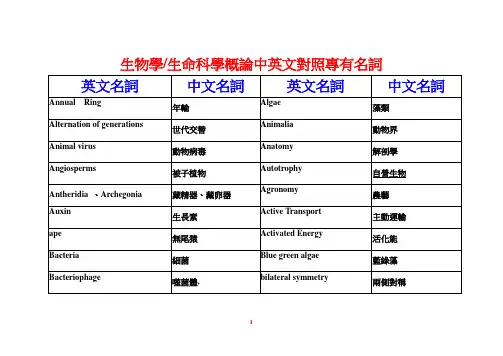
Derivation of pluripotent epiblast stem cells from mammalian embryos
- 格式:pdf
- 大小:725.65 KB
- 文档页数:6
本周氏蛋白名词解释本周氏蛋白是一种结构多样、功能复杂的细胞内蛋白质。
它在细胞生命活动中起着重要的调控作用,参与了细胞的增殖、分化、凋亡等多个生物学过程。
本周氏蛋白通常包含多个结构域,其中最常见的是PDZ结构域。
在人体中,本周氏蛋白家族成员有数百种,包括β-本周氏蛋白、γ-本周氏蛋白等。
本周氏蛋白的命名源于其最早发现的成员本周(Cooper)氏与其同事的名字。
他们在1996年首次发现的一组具有PDZ结构域的蛋白质,命名为本周氏蛋白家族。
本周氏蛋白家族的成员通过PDZ结构域与其他蛋白质结合,形成蛋白质复合物,从而调节细胞内信号传导通路。
PDZ结构域是一个约90个氨基酸残基组成的结构域,也是本周氏蛋白家族的重要特征。
PDZ结构域通常由6个α螺旋和2个β折叠片组成。
它具有特异性的蛋白质结合能力,可以与其他蛋白质上的短肽片段结合,从而调控这些蛋白质的功能。
例如,PDZ结构域可以与离子通道、细胞黏附蛋白等结合,在细胞内形成蛋白质复合物,参与离子通道的调控和细胞黏附的稳定。
本周氏蛋白在细胞生物学中的功能非常复杂。
它可以通过与其他蛋白质的相互作用,调节细胞的增殖、分化和凋亡。
例如,本周氏蛋白可以参与细胞的分裂,调节细胞周期的进程。
它还可以调控细胞的分化,促进细胞向特定细胞类型的发展。
此外,本周氏蛋白还可以参与细胞的凋亡,调节程序性细胞死亡的过程。
总的来说,本周氏蛋白是一类结构多样、功能复杂的细胞内蛋白质,通过与其他蛋白质的结合,调控细胞的增殖、分化和凋亡等多个生物学过程。
它在细胞生命活动中起着重要的调控作用,广泛参与许多生理过程的调节。
对于研究细胞生物学和疾病的发生机制具有重要意义。
【铁死亡】铜绿假单胞菌利用宿主的磷脂酰乙醇胺不饱和脂肪酸诱导支气管上皮细胞发生铁死亡——生物学背景——铁死亡(Ferroptosis)是2012年发现的新型细胞死亡方式,它是由15-脂氧合酶(15LOXes)催化多不饱和脂肪酸(PUFA)发生脂质过氧化(LPO),且生成的过氧化磷脂(hydroperoxy-PLs)无法被谷胱甘肽过氧化物酶4(GPX4)消除造成积累,从而引发的细胞死亡程序。
铁死亡与许多病理过程相关,例如脑、肾和肝损伤/疾病。
虽然15LOXes在许多真核细胞中广泛表达,但也在少数原核生物中发现。
原核生物铜绿假单胞菌(Pseudomonas aeruginosa)可以表达脂氧合酶(pLoxA),将宿主AA-PE氧化成15-氢过氧化物-AA-PE(15-HOO-AA-PE),并在人支气管上皮细胞中诱导铁死亡。
这种劫持哺乳动物的PUFA-PE和死亡程序的特点对于其引发的呼吸系统疾病的发病机制很重要,pLoxA也应成为相关疾病的新的治疗靶点。
本研究于2018年10月1日发表在J. Clin. Invest.杂志上。
——实验结果——铜绿假单胞菌从浮游生长到生物膜生长的转变与其脂氧合酶(ploxA)基因的表达增加有关。
像许多革兰氏阴性细菌一样,铜绿假单胞菌会产生并释放囊泡,其中含有许多细胞内蛋白质,包括pLoxA。
本文作者选取了野生型WT,wspF基因的灭活突变ΔwspF,不含ploxA的突变体loxA::Tn(PW3111)三种菌株,其中ΔwspF菌株具有超生物膜性质。
从图1中可以看出,诱导细胞死亡的能力ΔwspF菌株最强,且有且仅有铁死亡抑制剂Fer可以挽救回来。
通过荧光抗体染色可以看到pLoxA含量也是ΔwspF菌株最多,而且电镜结果也表明缺乏pLoxA的菌株突变体loxA::Tn成膜能力正常。
为了评估加氧酶的活性,他们还基于液相色谱-质谱联用的方法测定了WT,ΔwspF和loxA::Tn处理产生的产物,并发现比起缺乏pLoxA的菌株,ΔwspF菌株诱导生成的产物多了一个数量级,且后者引起非铁死亡效应。

植物逆境响应的分子机制植物作为一种生命体,在生长过程中难免会遭受各种不利条件的影响,比如土壤中的盐碱化、干旱、高温等环境因素。
对于植物本身而言,逆境的来临无异于是一场危机,即便是那些早已适应了恶劣环境的野生植物也无法完全摆脱逆境所带来的压力。
不过,植物也会在遭受逆境的时候出现以适应为目标的分子级反应,俗称逆境响应过程。
植物逆境响应的分子机制是什么呢?1.热休克蛋白热休克蛋白(Heat shock proteins,HSPs)是一种酶或者酶辅因子,在逆境环境下高表达,它们能够帮助植物维持正确的蛋白折叠状态、抑制错配和解聚。
逆境环境下,热休克蛋白对植物细胞的保护作用十分显著。
2.质膜脂质调节植物逆境响应过程中,受逆境条件刺激的质膜脂质会发生改变,进而导致细胞质壁的导电性能改变,或者是需要依赖于脂膜的酶等调节层级也会受到影响。
植物紧接着会启动各种反应,保证细胞膜的稳定性和完整性。
3.脱落酸脱落酸(Abscisic acid,ABA)是一种植物激素,起主要作用是抑制细胞分裂以及促进干旱等逆境环境下的植物适应。
在植物受到逆境刺激时,ABA的生产量会增加,依次来提高植物逆境抵抗能力。
4.氧化还原状态氧化还原状态同样是植物逆境响应过程的重要组成部分。
在植物细胞中,因应的代谢过程需要一定的氧化还原状态来进行调节。
在逆境环境下,产生大量有害的活性氧,会对植物细胞的氧化还原状态造成严重的影响。
植物逆境响应机制是复杂的,涉及到多种细胞层面的调节。
每一层面都有多个环节可以预测到的逆境,以便可能依次打击。
植物在反应逆境的过程中,有着复杂精细而高明的调控系统,最终保证了其在逆境环境中的生长。
细胞骨架名词解释1.细胞骨架(cytoskeleton):真核细胞中由纤维状蛋白质组成的网络系统,可分为三种:微丝、微管和中间纤维。
2.微丝(microfilament):是由肌动蛋白单体聚合而成的纤维状结构,肌动蛋白头尾相连组成微丝,具有极性。
3.中间纤维(intermediate filament, IF):10nm的纤维状蛋白结构,由于其直径介于微管和微丝之间,故称之为中间纤维。
IF蛋白由约310个aa形成的、非常保守的a-螺旋杆状区和高度可变的非螺旋端部组成。
4.微管(microtubule):管蛋白组成的管状结构,由13根原丝平行排列组成的圆柱形管状结构,原丝由α-tubulin和β-tubulin组成的异二聚体组成。
5.踏车模型:当肌动蛋白浓度高于正端临界浓度,而低于负端临界浓度时,微丝可以表现出在正端因加入肌动蛋白而延长,而在负端因肌动蛋白脱落而缩短。
6.微管组织中心(MTOC):细胞内能够起始微管成核并使之延伸的结构,微管组织中心MTOC是细胞组织微管聚合的特殊细胞器或部位。
大多数动物细胞的MTOC是中心体。
鞭毛和纤毛的MTOC是基体。
卵母细胞和植物细胞中没有中心体。
7.胞质分裂环:在有丝分裂末期,两个即将分裂的子细胞之间产生一个收缩环。
收缩环是由大量平行排列的微丝组成,由分裂末期胞质中的肌动蛋白装配而成,随着收缩环的收缩,两个子细胞被分开。
胞质分裂后,收缩环即消失。
填空题1.在细胞内微管以单管、二联体和三联体3种形式存在。
2.微管壁由13根原纤丝组成。
3.微管由管蛋白分子组成,微管的单体形式是α-tubulin和β-tubulin组成的异二聚体。
4.微管结合蛋白具有稳定微管,防止解聚,协调微管与其他细胞成分相互关系的作用。
5.中心体含有一对垂直排列的中心粒,外面被无定形结构γ-tubulin所包围。
6.基体类似于中心粒,是由9个三联管组成的小型圆柱形细胞器。
7.在细胞内参与物质运输的马达蛋白分为三类:沿微丝运动的肌球蛋白、沿微管运动的驱动蛋白和动力蛋白。
医学细胞生物学专业英语词汇* acrocentric chromosome 近端着丝粒染色体 actin 肌动蛋白 actin filament 肌动蛋白丝 actinomycin D 放线菌素D activator 活化物 active transport 主动运输 adenine 腺嘌呤 adenosine monophosphate, AMP 腺苷一磷酸, 腺苷酸 adenyl cyclase, AC 腺苷酸环化酶 adhesion plaque 黏着斑agranular endoplasmic reticulum 无颗粒内质网 Alzheimer disease 阿尔茨海默病 amino acid 氨基酸 aminoacyl site, A site 氨基酰位,A位 amitosis; direct division 无丝分裂;直接分裂 amphipathic molecule 双型性分子anaphase 后期anchoring junction 锚定连接 annular granule 孔环颗粒 anticoding strand 反编码链 antigen 抗原antiparallel 逆平行性 apoptic body 凋亡小体 apoptosis 凋亡assembly 组装aster 星体asymmetry 不对称性autolysis 自溶作用 autophagolysosome 自噬性溶酶体 autophagy 自噬作用autoradiography 放射自显影技术 autosome 常染色体 B lymphocyte B淋巴细胞bacteria 细菌 base substitution 碱基替换 belt desmosome 带状桥粒bioblast 生命小体 biological macromolecule 生物大分子 biomembrane 生物膜biotechnology 生物技术 bivalent 二价体 breakage 断裂 cadherin 钙粘连素calmodulin, CaM 钙调蛋白 cAMP 环一磷酸腺苷 cAMP-dependent protein kinase 环一磷酸腺苷依赖型蛋白激酶capping 戴帽 carrier protein 载体蛋白 cat cry syndrome 猫叫综合症cell division cycle gene CDC基因 cell 细胞 cell and molecular biology 细胞分子生物学 cell biology 细胞生物学 cell coat; glycocalyx 细胞衣;糖萼 cell culture 细胞培养 cell cycle 细胞周期cell cycle-regulating protein 细胞周期调节蛋白 cell cycle time 细胞周期时间 cell determination 细胞决定 cell differentiation 细胞分化 cell division cycle, CDC 细胞分裂周期 cell division cycle gene, CDC gene 细胞分裂周期基因 cell engineering 细胞工程 cell fractionation 细胞分级分离cell fusion 细胞融合 cell junction 细胞连接 cell line 细胞系 cell membrane; plasma membrane 细胞膜;质膜 cell plate 细胞板 cell proliferation 细胞增殖 cell recognition 细胞识别 cell surface antigen 细胞表面抗原 cell theory 细胞学说 cell strain 细胞株 cell aging 细胞衰老cell synchronization 细胞同步化 cellular oxidation 细胞氧化 cellular respiration 细胞呼吸 central granule 中央颗粒 centromere 着丝粒 chalone 抑素 channel protein 通道蛋白 chemiosmotic hypothesis 化学渗透假说chiasmata 交叉 cholesterol 胆固醇chromatid 染色单体 chromatin 染色质 chromomere 染色粒 chromosome 染色体 chromosome arm 染色体臂 chromosome banding 染色体带 chromosome disease 染色体病 chromosome engineering 染色体工程 chromosome scaffold 染色体支架 chromosome syndrome 染色体综合症 cis Golgi network 顺面高尔基网状结构 cisterna(pl. cisternae)扁平囊 clathrin 笼蛋白 clone 克隆coated pit 有被小窝 coated vesicle 包被小泡 coding strand 编码链 codon 密码子 codon degeneracy 密码子兼并性 coenzyme 辅酶 collagenfibronectin, FN 纤连蛋白 communication junction 通讯连接 complementation 互补性condensation stage 凝集期 confocal laser scanning microscope 共焦激光扫描显微镜 connexin 连接子 constitutive heterochromatin 结构异染色质continuous microtubules 极微管 converting enzyme 转变酶crista(pl. cristae)嵴 cyanine 胞嘧啶 cyclin 细胞周期素cydoeximide 放线菌酮 cytidine monophosphate, CMP 胞苷一磷酸,胞苷酸cytokinesis 细胞质分裂 cytology 细胞学 cytoplasm 细胞质 cytoplasm engineering 细胞质工程 cytoplasm substitution 细胞质代换 cytoplasmic plaque 胞质斑 cytoskeleton 细胞骨架 dark field microscope 暗视野显微镜dedifferentiation 去分化 degeneracy 兼并 deletion 缺失 density gradient centrifugation 密度梯度离心 deoxyadenosine monophosphate, dAMP 脱氧腺苷酸 deoxycytidine monophosphate, dCMP 脱氧胞苷酸 deoxyguanosine monophosphate, dGMP 脱氧鸟苷酸 deoxyribonucleic acid, DNA 脱氧核糖核酸deoxythymidine monophosphate, dTMP 脱氧胸苷酸 desmosome 桥粒 diakinesis 终变期 differential centrifugation 差速离心 differential expression 差异性表达 differentiation induction 分化诱导 differentiation inhibition 分化抑制 diplococcus pneumonia 肺炎双球菌diplotene 双线期 disassembly 去组装 DNA probe DNA探针 DNA synthesis phase DNA合成期 dosage compensation 剂量补偿 doublet 二联管 duplication 重复 effector 效应器 electric coupling 电偶联 electron microscope 电子显微镜 elementary particle 基粒 eletronfusion 电融合 elongation factor, EF 延长因子 embryonic induction 胚胎诱导作用 endocytosis 内吞作用endolysosome 内体性溶酶体 endomembrane system 内膜系统 endoplasmic reticulum, ER 内质网 enhancer 增强子 enzyme 酶 equatorial plane 赤道面eucaryotes 真核生物 euchromatin 常染色质 eukaryotic cell 真核细胞exocytosis 胞吐作用 exon 外显子 extracellular matrix, ECM 细胞外基质extrinsic; peripheral protein 外在蛋白;外周蛋白 F body 荧光小体facilitated diffusion 易化扩散 facultative heterochromatin 兼性异染色质 fibrillar component 原纤维成分 fibronectin, FN 纤粘连蛋白 fibrous actin, F-actin 纤维状肌动蛋白 flanking sequence 侧翼顺序 fluid mosaic model 液态镶嵌模型 fluorescence microscope 荧光显微镜 fluorescence recovery after 荧光漂白恢复 photobleaching, FRAPfork-initiation protein 叉起始蛋白 frameshift mutation 移码突变 free cell 游离细胞 free diffusion 自由扩散 free energy 自由能galactocerebroside 半乳糖脑苷脂 ganglioside 神经节苷脂 gap junction 间隙连接 gene 基因 gene cluster 基因簇 gene engineering 基因工程 gene expression 基因表达 gene family 基因家族 gene mutation 基因突变 genetic code 遗传密码 genetic message 遗传信息 genome 基因组 genome engineering 染色体工程 genomic DNA library 基因组DNA文库glycogen storage disease type? ?型糖原蓄积病 glycolipid 糖脂glycoprotein 糖蛋白 glycosaminoglycan, GAG 氨基聚糖 glycosylation 糖基化Golgi apparatus 高尔基器 Golgi body 高尔基体 Golgi complex 高尔基复合体granular component 颗粒成分 granular drop 脱粒 granular endoplasmic reticulum 颗粒内质网 growth factor 生长因子 GT-AG rule GT-AG法则guanine 鸟嘌呤 guanosine monophosphate, GMP 鸟苷一磷酸,鸟苷酸hemidesmosome 半桥粒 hereditary factor 遗传因子 heterochromatin 异染色质heterogeneous nuclear RNA, hnRNA 不均一核RNA heterokaryon 异核体heterophagolysosome 异噬性溶酶体 heterophagy 异噬作用 heteropyknosis 异固缩 highly repetitive sequence 高度重复序列 histone 组蛋白 holoenzyme全酶 homokaryon 同核体 housekeeping gene 管家基因 housekeeping protein管家蛋白human leukocyte antigen, HLA 人白细胞抗原 hyaluronic acid, HA 透明质酸 hybrid cell 杂交细胞 hyperdiploid 超二倍体 hypodiploid 亚二倍体immunofluorescence microscopy 免疫荧光显微镜技术 immunoglobulin 免疫球蛋白 in vitro 离体的 in vivo 体内的 inactive X hypothesis 失活X假说inborn errors of metabolism 先天性代谢缺陷病 inducer 诱导物 induction 诱导 inhibitor of mitotic factor, IMF 有丝分裂因子抑制物 initiation factor, IF 起始因子 inner membrane 内膜 inner nuclear membrane 内层核膜insertion sequence, IS 插入顺序 Integral protein 整合蛋白 integrin 整连蛋白 inter membrane space; outer chamber 膜间腔;外室 intercellular communication 细胞间通讯 intercristal space; inner chamber 嵴间腔;内室intermediate filament 中间纤维 internal membrane 内膜 internal reticular apparatus 内网器 interphase 间期 interstitial deletion 中间缺失interzonal microtubules 区间微管intracristal space 嵴内腔 intra-nucleolar chromatin 核仁内染色质intrinsic; integral protein 内在蛋白;整合蛋白 intron 内含子 inversion倒位 inverted repetitive sequence 倒位重复顺序 ionic channel 离子通道ionic coupling 离子偶联 jumping gene 跳跃基因 karyotype 核型 kinetochore 着丝点 kinetochore microtubules 动粒微管Klinefelter’s syndrome 先天性睾丸发育不全症 lagging strand 后随链 laminin, LN 层粘连蛋白 lateral diffusion 侧向扩散 leading strand 前导链 leptotene 细线期 ligand; chemical signal 配体;化学信号 light microscope 光学显微镜 linear polymer 线性多聚体 linker 连接线 liposome 脂质体 liquid crystal 液晶 lowdensity lipoprotein, LDL 低密度脂蛋白 luxury gene 奢侈基因 luxuryprotein 奢侈蛋白 lymphokine 淋巴激活素 lymphotoxin 淋巴毒素lysosome 溶酶体 major histocompatibility complex, MHC 组织相容性复合体 malignancy 恶性 matrical granule 基质颗粒 matrix 基质 matrix fibronectin, mFN 基质纤连蛋白 maturation-prompting factor, MPF 成熟促进因子 medial Golgi stack 高尔基中间囊膜 meiosis 减数分裂 membrane antigen 膜抗原 membrane carbohydrate 膜碳水化合物 membrane flow 膜流 membrane lipid 膜脂 membrane protein 膜蛋白 membrane receptor 膜受体 membranous structure 膜相结构 messenger RNA 信使核糖核酸 mesosome 中间体 metabolic coupling 代谢偶联 metacentric chromosome 中央着丝粒染色体 metaphase 中期micelle 微团 microfilament 微丝 microscopy 显微镜技术 microsome 微粒体microtrabecular lattice 微梁网格 microtubule 微管 microtubule associated protein, MAP 微管结合蛋白 microtubule organizing centers, MTOC 微管组织中心microvillus 微绒毛 middle repetitive sequence 中度重复序列 miniband 微带 missense mutation 错义突变 mitochondria 线粒体 mitosis 有丝分裂mitosis phase 有丝分裂期 mitotic apparatus 有丝分裂器 mitotic factor, MF 有丝分裂因子 mobility 流动性 model for controlling gene expression 基因表达调控模型 molecular biology 分子生物学 molecular disease 分子病monopotent cell 单能细胞 monosomy 单体性 multiple coiling model 多级螺旋模型 multipotent cell 多能细胞 myasthenia gravis 重症肌无力症 mycoplasma 支原体 myofibrils 肌原纤维 necrosis 坏死 neuropeptide 神经肽 non-continuation 不连续性 non-histone 非组蛋白 non-membranous structure 非膜相结构 nonsense mutation 无义突变 nuclear envelope 核被膜 nuclear lamina 核纤层 nuclear matrix 核基质nuclear pore 核孔 nuclear pore complex 核孔复合体 nuclear sap 核液nuclear sex 核性别 nuclear skeleton 核骨架 nucleic acid 核酸 nucleic acid hybridization 核酸分子杂交 nucleo-cytoplasmic ratio 核质比 nucleoid 类核体 nucleoids 拟核 nucleolar associated chromatin 核仁相随染色质nucleolar organizing region 核仁组织区 nucleolus 核仁 nucleosome 核小体nucleotide 核苷酸 nucleosome core 核小体核心 nucleus 细胞核 nucleus transplantation 核移植法 nucleus-cytoplasm hybrid 核质杂种 Okazaki fragment 岗崎片段 oligomer fibronectin,oFN 寡聚纤连蛋白 oncogene 癌基因operator gene 操纵基因 operon 操纵子 operon theory 操纵子学说 organelle 细胞器 origin 起点 outer membrane 外膜 outer nuclear membrane 外层核膜overlapping gene 重叠基因 oxidative phosphorylation 氧化磷酸化pachytene 粗线期 pairing stage 配对期 partial monosome 部分单体 partial trisomy 部分三体 passive transport 被动运输 patching 成斑现象 peptide bond 肽键 peptidyl site, P site 肽基位;P位 perinuclear space 核间隙perinucleolar chromatin 核仁周围染色质 peripheral granule 周边颗粒peripheral protein 外周蛋白 permeability 通透性 peroxisome; microbody 过氧化物酶体;微体 phagocytosis 吞噬作用 phagolysosome 吞噬性溶酶体phagosome 自噬体 phase contrast microscope 相差显微镜 phenylalanine hydroxylase, PAH 苯丙氨酸羟化酶 phenylketonuria, PKU 苯丙酮尿症phosphatidylinositol, PL 磷脂酰肌醇 phosphodiester bond 磷酸二酯键phosphodiesterase, PDE 磷酸二酯酶 phosphoglyceride 磷酸甘油酯phospholipase C,PLC 磷脂酶C phospholipid 磷脂 pinocytosis 胞饮作用pinocytotic vesicle 吞饮泡 plasma cell 浆细胞 plasma fibronectin, pFN 血浆纤连蛋白 plasmid 质粒 point mutation 点突变 polar microtubule 极间微管 polarizing microscope 偏光显微镜 polyadenylation 多聚腺苷酸反应polyploid 多倍体 polyribosome 多聚核糖体 premature condensed chromosome, PCC 早熟染色体 premeiosis interphase 减数分裂前间期 primary constriction 主缢痕 primary culture 原代培养 primary culture cell 原代细胞 programmed cell death 细胞程序性死亡 prokaryotes 原核生物 prokaryotic cell 原核细胞promotor 启动子 promotor gene 启动基因 prophase 前期 protein 蛋白质protein kinase C, PKC 蛋白激酶C proteoglycan, PG 蛋白聚糖 protofilament 原纤维 protooncogene 原癌基因 protoplasm 原生质 purine 嘌呤碱 pyrimidine 嘧啶碱receptor mediated endocytosis 受体介导的内吞作用 reciprocal translocation 相互易位 recombinant DNA technology 重组DNA技术recombination nodules 重组小节 recombination stage 重组期 recondensation stage 再凝集期 redifferentiation 再分化 regulator gene 调节基因 release factor, RF 释放因子 replication 复制 replication eyes 复制眼 replication fork 复制叉 replicon 复制子 repressor 阻碍物 resolving power 分辨力residual body 残体 respiratory chain 呼吸链 restriction endonuclease 限制性内切核酸酶 restriction point 限制点 reverse transcription 逆转录 rho factor, ρ ρ因子 ribonucleic acid, RNA 核糖核酸 ribophorin 核糖体结合蛋白 ribosomal RNA 核糖体核糖核酸 ribosome 核糖核蛋白体 RNA polymerase RNA聚合酶 rough endoplasmic reticulum, rER 粗面内质网 sac 扁平囊 same sense mutation 同义突变sarcoplasmic reticulum 肌质网 satellite 随体 scanning electron microscope 扫描电子显微镜 scanning tunneling microscope 扫描隧道电子显微镜 secondary constriction 次缢痕 secondary culture 传代培养semiautonomous organelle 半自主性的细胞器 semiconservative replication 半保留复制 semidiscontinuous replication 半不连续复制 sensor 感受器sequential expression 顺序表达 sex chromosome 性染色体 signal codon 信号密码子 signal hypothesis 信号肽假说 signal molecule 信号分子 signal peptide 信号肽 signal recognition particle, SPR 信号识别颗粒 simple diffusion 简单扩散 single sequence 单一序列 single-stranded DNA binding protein 单链DNA结合蛋白 singlet 单管 small nuclear RNA, snRNA 小分子细胞核RNA smooth endoplasmic reticulum, sER 滑面内质网 solenoid 螺线管sparsomycin 稀疏酶素 sphingomyelin 神经鞘磷脂 spindle 纺锤体 splicing 剪接 split gene 断裂基因start codon 起始密码子 stem cell 干细胞 stress fiber 张力基因structural gene 结构基因 submetacentric chromosome 亚中着丝粒染色体supersolenoid 超螺线管 suppressor tRNA 校正tRNA synapsis 联会synaptonemal complex 联会复合体 synkaryon 合核体 synonymous codon 同义密码子 synonymous mutation 同义突变 T lymphocyte T淋巴细胞 tailing 加尾telomere 端粒 telophase 末期 terminal deletion 末端缺失 terminalization 端化 terminator 终止子 tetrad 四分体 tetraploid 四倍体 thymine 胸腺嘧啶three dimensional structure,3D 三维结构 tight junction 紧密连接 tissue cell 组织细胞 tissue engineering 组织工程 totipotency 全能性 trans Golgi network 反面高尔基网状结构 transcribed spacer 转录间隔区transcription 转录 transdifferentiation 转分化 transfer RNA 转运核糖核酸 transformation 转化 transition 转换 translation 翻译 translocation 易位 transport protein 运输蛋白 transposition 转座 transversion 颠换transmission electron microscope 透视电子显微镜 tricarboxylic acid cycle 三羧酸循环 trigger protein 触发蛋白 triplet 三联管 triploid 三倍体triskelion 三臂蛋白 trisomy 三体 tubulin 微管蛋白 tumor necrosis factor 肿瘤坏死因子Turner’s syndrome 先天性卵巢发育不全症 tyrosinase, TN 酪氨酸酶 ultravoltage electron microscope 超高压电子显微镜 unit membrane 单位膜 untranscribed spacer 非转录间隔区 unwinding protein 解链蛋白 uracil 尿嘧啶 uridine monophosphate, UMP 尿苷一磷酸;尿苷酸 vacuole 大囊泡vector 载体vesicle 小囊泡 vinculin 粘着斑连接蛋白 wobble hypothesis 摇摆学说 X chromatin X染色质 Y chromatin Y染色质 zygotene 偶线期。
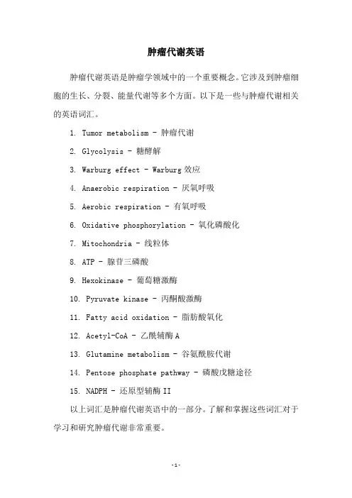
肿瘤代谢英语
肿瘤代谢英语是肿瘤学领域中的一个重要概念。
它涉及到肿瘤细胞的生长、分裂、能量代谢等多个方面。
以下是一些与肿瘤代谢相关的英语词汇。
1. Tumor metabolism - 肿瘤代谢
2. Glycolysis - 糖酵解
3. Warburg effect - Warburg效应
4. Anaerobic respiration - 厌氧呼吸
5. Aerobic respiration - 有氧呼吸
6. Oxidative phosphorylation - 氧化磷酸化
7. Mitochondria - 线粒体
8. ATP - 腺苷三磷酸
9. Hexokinase - 葡萄糖激酶
10. Pyruvate kinase - 丙酮酸激酶
11. Fatty acid oxidation - 脂肪酸氧化
12. Acetyl-CoA - 乙酰辅酶A
13. Glutamine metabolism - 谷氨酰胺代谢
14. Pentose phosphate pathway - 磷酸戊糖途径
15. NADPH - 还原型辅酶II
以上词汇是肿瘤代谢英语中的一部分。
了解和掌握这些词汇对于学习和研究肿瘤代谢非常重要。
- 1 -。
肿瘤细胞破坏基质的物质包括:
1. 蛋白酶:肿瘤细胞产生的蛋白酶可以降解基质中的胶原蛋白、纤维连接蛋白等结构蛋白,从而破坏基质的完整性。
2. 纤溶酶原激活物(uPA):肿瘤细胞可以分泌uPA,它能够激活纤溶酶原,进而引发纤溶酶的活化,从而降解基质中的纤维蛋白。
3. 金属蛋白酶:肿瘤细胞产生的金属蛋白酶可以降解基质中的胶原蛋白、纤维连接蛋白等结构蛋白,从而破坏基质的完整性。
4. 细胞外基质金属蛋白酶抑制剂(TIMP):正常细胞会产生细胞外基质金属蛋白酶抑制剂来抑制金属蛋白酶的活性,但肿瘤细胞可以减少或失去对TIMP的敏感性,从而增加金属蛋白酶的活性,破坏基质。
5. 细胞外囊泡:肿瘤细胞可以释放细胞外囊泡,其中含有多种酶类和蛋白质,可以降解基质中的组织蛋白,从而破坏基质的完整性。

细胞生物学需要掌握的名词概念上次老师说我们考试的时候不能写的一样,你们可以找一下我后面标的页码,自己整理归纳,根据自己的理解来背。
1、lipid rafts model脂筏模型:该模型认为在甘油磷脂维生物膜的主体上,胆固醇、鞘磷脂等富集区域形成相对有序的脂相,如同漂浮在脂双层上的“脂筏”一样载着某些特定生物学功能的各种膜蛋白。
P55在生物膜上胆固醇富集而形成有序脂相,如同脂筏一样载着各种蛋白.脂筏是质膜上富含胆固醇和鞘磷脂的微结构域。
大小约70nm 左右,是一种动态结构,位于质膜的外小页。
2、p53 protein,P53蛋白:313页p53蛋白能调节细胞周期和避免细胞癌变发生。
3、Hayflick limitation Hayflick界限:细胞停止分裂是由细胞自身因素决定的,与环境条件无关,正常细胞具有有限分裂次数,而癌细胞能够在体外无限增殖。
P356细胞,至少是培养的二倍体细胞,不是不死的,而是有一定的寿命;它们的增殖能力不是无限的,而是有一定的界限,这就是Hayflick界线。
4、cell line细胞系:原代培养的细胞一般传至10代左右就不易传下去,细胞生长出现停滞,大部分细胞衰老死亡,但有极少数细胞可能渡过“危机”而传下去。
这些存活的细胞一般又可顺利地传40-50代次,并且仍保持原来染色体的二倍数量及接触抑制的行为。
P435、Nuclear localization signal (NLS);核定位信号:亲核蛋白一般都含有特殊的氨基酸序列,这些内含的特殊短肽保证了整个蛋白质能够通过核孔复合体被转运到细胞核内,这段具有“定向”“定位”作用的序列被命名为核定位信号。
P2326、programmed cell death (PCD)细胞程序性死亡:无论是单细胞生物还是多细胞生物,细胞死亡往往受细胞内由遗传机制决定的“死亡程序”控制,要求特定基因表达,是“主动”而非“被动”的过程。
P3417、biomembrane生物膜:真核生物内部存在由膜围绕构建的各种细胞器。
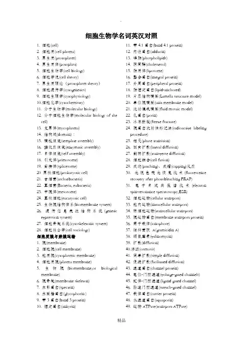
细胞生物学名词英汉对照1. 细胞(cell)2. 细胞质(cell plasma)3. 原生质(protoplasm)4. 原生质体(potoplast)5. 细胞生物学(cell biology)6. 细胞学说(cell theory)7. 原生质理论(protoplasm theory)8. 细胞遗传学(cytogenetics)9. 细胞生理学(cytophysiology)10.细胞化学(cytochemistry)11. 分子生物学(molecular biology)12. 分子细胞生物学(molecular biology of the cell)13. 支原体(mycoplasma)14. 结构域(domain)∶15. 模板组装(template assembly)16. 酶效应组装(enzymatic assembly)17. 自体组装(self assembly)18. 引发体(primosome)19. 剪接体(splicesome)20 原核细胞(prokaryotic cell)21. 古细菌(archaebacteria)22. 真细菌(Bacteria, eubacteria)23. 中膜体(mesosome)24. 真核细胞(eucaryotic cell)25. 生物膜结构体系(biomembrane system)26. 遗传信息表达结构系统(genetic expression system)27. 细胞骨架系统(cytoskeletonic system)28. 细胞社会学(cell sociology)细胞质膜与跨膜运输1. 膜(membrane)2. 细胞膜(cell membrane)3. 胞质膜(cytoplasmic membrane)4. 细胞质膜(plasma membrane)5. 生物膜(biomembrane,or biological membrane)6. 膜骨架(membrane skeleton)7. 血影蛋白(spectrin)8. 血型糖蛋白(glycophorin )9. 带3蛋白(band 3 protein)10. 锚定蛋白(ankyrin) 11. 带4.1蛋白(band 4.1 protein)12. 内收蛋白(adducin)13. 磷脂(phospholipids)14. 胆固醇(cholesterol)15. 脂质体(liposome)16. 整合蛋白(integral protein)17. 外周蛋白(peripheral protein)18. 脂锚定蛋白(lipid-anchored)19. 片层结构模型(Lamella structure model)20. 单位膜模型(unit membrane model)21. 流动镶嵌模型(fluid mosaic model)22. 孔蛋白(porin)23. 冰冻断裂(freeze fracture)24. 膜蛋白放射性标记法(radioactive labeling procedure)25. 相变(phase transition)26. 侧向扩散(lateral diffusion)27. 翻转扩散(transverse diffusion)28. 细胞融合(cell fusion)29. 成斑(patching)、成帽(capping)反应30. 光脱色荧光恢复技术(fluorescence recovery after photobleaching FRAP)31. 电子自旋共振谱技术(electron spin-resonance spectroscopy,ESR)32. 细胞运输(cellular transport)33. 胞内运输(intracellular transport)34. 转细胞运输(transcellular transport)35. 膜运输蛋白(membrane transport protein)36. 离子载体(ionophore)37. 短杆菌肽A(gramicidin A)38. 缬氨霉素(valinomycin)39. 扩散(diffusion)40.渗透(osmosis)41. 简单扩散(simple diffusion)42. 促进扩散(facilitated diffusion)43. 通道蛋白(channel protein)44. 电位-门控通道(voltage-gated channels)45. 配体-门控通道(ligand gated channel)46. 胁迫门控通道(stretch-gated channel)47. 载体蛋白(carrier protein)48. 水通道蛋白(aquaporin)49. 运输ATPase(transport ATPase)50. 协同运输(cotransport)51. 磷酸化运输(phosphorylating transport)细胞通讯1. 细胞通讯(cell communication)2. 信号传导(cell signalling)3. 信号转导(signal transduction)4. 信号分子(signaling molecules)5. 激素(hormone)6. 内分泌信号(endocrine signaling)。
每⽇专业名词通俗解释2022年03⽉01⽇HSCR⽂章的⼀些科普progenitor和precursor的区别The main difference between progenitor and precursor cells is that progenitor cells are mainly multipotent cells that can differentiate into many types of cells, whereas precursor cells are unipotent cells that can only differentiate into a particular type of cells.糖酵解glycolysis糖酵解(英语:glycolysis,⼜称糖解)是把葡萄糖(C6H12O6)转化成丙酮酸(CH3COCOO− + H+)的代谢途径。
在这个过程中所释放的⾃由能被⽤于形成⾼能量化合物ATP和NADH。
糖酵解作⽤及其各种变化形式发⽣在⼏乎所有的⽣物中,⽆论是有氧和厌氧。
糖酵解的⼴泛发⽣显⽰它是最古⽼的已知的代谢途径之⼀。
糖酵解作⽤是所有⽣物细胞糖代谢过程的第⼀步。
糖酵解作⽤是⼀共有10个步骤酶促反应的确定序列。
在该过程中,⼀分⼦葡萄糖会经过⼗步酶促反应转变成两分⼦丙酮酸。
糖酵解作⽤发⽣在⼤多数⽣物体中的细胞的胞质溶胶。
脂肪酸分解Fatty acid catabolism脂肪酸的氧化作⽤發⽣在粒線體(mitochondria)內,脂肪酸必須先和ATP反應,轉變為活化的中間產物,才能與其他酵素作更進⼀步的代謝,⾧鏈的Fatty acyl-CoA不能穿過粒線體內膜進⼊粒線體基質,需藉⾁酸素(carnitine)運送機制,脂肪酸氧化(Fatty acid oxidation)。
氧化磷酸化(英语:oxidative phosphorylation,缩写作 OXPHOS)是细胞的⼀种代谢途径,该过程在真核⽣物的线粒体内膜或原核⽣物的细胞膜上发⽣,使⽤其中的酶及氧化各类营养素所释放的能量来合成三磷酸腺苷(ATP)。
细胞生物学英文注释及名词解释work Information Technology Company.2020YEAR细胞生物学英文注释及名词解释1.Cell biology:细胞生物学以“完整细胞的生命活动”为着眼点,从分子、亚细胞、细胞和细胞社会的不同水平,用动态的和系统的观点来探索和阐述生命这一基本单位的特性。
2.RNA interference:通过促使特定基因的miRNA降解来高效、特异地阻断体内特定基因表达,这种现象称为RNA干扰。
3.Cell membrane:细胞膜是包围在细胞质表面的一层薄膜,又称质膜(plasma membrane)。
4.Lipid rafts:由于鞘脂的脂肪酸尾比较长,因此这一区域比膜的其他部分厚,更有秩序且较少流动,被称为“脂筏”。
5.Endomembrane system:人们把细胞内在结构、功能以及发生上相互密切关联的其他所有膜性结构细胞器统称为内膜系统。
6.Endoplasmic reticulum(ER):在细胞质的内质区分布着一些由小管、小泡相互连接吻合形成的网状结构,称为内质网。
7.Molecular chaperone:热激蛋白虽然能够通过对其各自作用对象的识别、结合来协助它们的折叠组装和转运,但其本身却不参与最终作用产物的形成,也不会改变其自身的基本分子生物学特性,由此被称之为分子伴侣。
8.Signal peptid:指导蛋白多肽链在糙面内质网上进行合成的决定因素,是被合成肽链N 端的一段特殊氨基酸序列,即信号肽。
9.Nuclear import signal:核输入信号是指凡是在细胞质中合成的核蛋白质,其肽链中均含有由7个氨基酸组成的特异性信号序列,负责分拣并指导蛋白质从细胞质通过核孔复合体输入到细胞核内。
又称为核定位信号(nuclear localization signal,NLS)10.Golgi complex (高尔基复合体)11.Lysosome (溶酶体)12.Peroxisome (过氧化物酶体)13.Cytoskeleton:细胞骨架是指真核细胞中与保持细胞形态结构和细胞运动有关的纤维网络,包括微管、微丝、中间丝。
Cell Stem CellArticlePax6Is a Human NeuroectodermCell Fate DeterminantXiaoqing Zhang,1Cindy T.Huang,1Jing Chen,2Matthew T.Pankratz,1Jiajie Xi,2Jin Li,2Ying Yang,3Timothy Vaute,1 Xue-Jun Li,1Melvin Ayala,1Gennadiy I.Bondarenko,4Zhong-Wei Du,1Ying Jin,3Thaddeus G.Golos,4,5and Su-Chun Zhang1,6,*1Waisman Center and the WiCell Institute,Madison,WI53705,USA2Department of Anatomy and Embryology,Fudan University Shanghai Medical School,Institute of Stem Cells and Regenerative Medicine, Fudan University,Shanghai,200032,China3Shanghai Institute of Biological Sciences,Chinese Academy of Science,Shanghai,200031,China4Wisconsin National Primate Research Center5Department of Comparative Biosciences,School of Veterinary Medicine,and Obstetrics and Gynecology,School of Medicineand Public HealthUniversity of Wisconsin-Madison,Madison,WI53715,USA6Department of Anatomy and Department of Neurology,School of Medicine and Public Health,University of Wisconsin,Madison,WI53705,USA*Correspondence:zhang@DOI10.1016/j.stem.2010.04.017SUMMARYThe transcriptional regulation of neuroectoderm(NE) specification is unknown.Here we show that Pax6 is uniformly expressed in early NE cells of human fetuses and those differentiated from human embry-onic stem cells(hESCs).This is in contrast to the later expression of Pax6in restricted mouse brain regions. Knockdown of Pax6blocks NE specification from hESCs.Overexpression of either Pax6a or Pax6b, but not Pax66PD,triggers hESC differentiation. However,only Pax6a converts hESCs to NE.In contrast,neither loss nor gain of function of Pax6 affects mouse NE specification.Both Pax6a and Pax6b bind to pluripotent gene promoters but only Pax6a binds to NE genes during human NE specifica-tion.Thesefindings indicate that Pax6is a transcrip-tional determinant of the human NE and suggest that Pax6a and Pax6b coordinate with each other in determining the transition from pluripotency to the NE fate in human by differentially targeting pluripo-tent and NE genes.INTRODUCTIONIn mammals,the stepwise cell fate transition during early embry-onic development is orchestrated by sequential activation/inac-tivation of lineage-determining transcription factors(Yamanaka et al.,2006).Oct4,Sox2,and Nanog are required for maintaining pluripotency of the inner cell mass(ICM)or the epiblast in a blas-tocyst embryo(Avilion et al.,2003;Chambers et al.,2003;Mitsui et al.,2003;Nichols et al.,1998).Differentiation of the ICM to extraembryonic tissues is governed by Cdx2and Gata6,tran-scription factors that repress pluripotency while inducing genes of the trophectoderm and extraembryonic endoderm,respec-tively(Jedrusik et al.,2008;Koutsourakis et al.,1999;Niwa et al.,2005).After the formation of extraembryonic tissues,the pluripotent epiblasts are converted to three germ layers during gastrulation,but how these processes are regulated remains unknown.One of the best-studied processes during gastrulation,neuro-ectoderm(NE)specification,is at the center of developmental biology.Studies in lower vertebrates,including frogs and chicks, indicate that many transcription factors are involved in NE spec-ification,including zincfinger proteins,Sox family,Otx family, and helix-loop-helix transcription factors(Mizuseki et al.,1998; Nakata et al.,1997;Rex et al.,1997;Sheng et al.,2003).To date,it is unclear which transcription factor is responsible for the conversion from pluripotent cells to NE in mammals.The most promising factor is Sox1,because its expression pattern parallels NE formation in mouse(Bylund et al.,2003;Pevny et al.,1998).However,Sox1-knockout mice do not exhibit severe brain deficits,probably because of compensation by other Sox members(Nishiguchi et al.,1998).Similarly,the tran-scriptional determinant for human NE specification is unknown. The failure in identifying mammalian transcriptional determi-nants underlying NE specification is at least partly due to the lack of model systems that permit easy genetic manipulation and direct observation of developmental processes.Embryonic stem cells(ESCs),derived from the ICM or epiblast,differentiate to cells/tissues of the three germ layers according to develop-mental principles(Murry and Keller,2008;Stern,2005;Zhang, 2006).When human ESCs(hESCs)are differentiated toward the neural fate under a chemically defined medium in the absence of growth factors,NE cells appear around day6–8and form neural tube-like rosettes at day14with corresponding gene expression patterns(Li et al.,2005;Pankratz et al.,2007;Zhang et al.,2001; Zhang and Zhang,2010).This differentiation process resembles in vivo development of the neural plate and neural tube,and it therefore represents a useful tool for studying the molecular underpinnings of human NE specification(Zhang,2006). During hESC neural differentiation,the initial NE cells do not express Sox1,the earliest marker of NE in mouse embryos or90Cell Stem Cell7,90–100,July2,2010ª2010Elsevier Inc.in NE differentiated from mouse ESCs(mESCs)(Li et al.,2005; Pankratz et al.,2007;Pevny et al.,1998;Suter et al.,2008; Ying et al.,2003).Instead,Pax6,a paired box(Pax)transcription factor expressed in region-specific neural progenitors after neural tube closure in mouse(Schmahl et al.,1993;Walther and Gruss,1991),is uniformly expressed in hESC-derived NE (Li et al.,2005;Pankratz et al.,2007).These observations raise an intriguing possibility that Pax6may play a novel role in human NE specification.Three isoforms of Pax6have been identified. The canonical Pax6a harbors two DNA binding domains,the paired domain(PD)and homeodomain(HD),and a proline-serine-threonine(PST)-rich transactivation domain.Pax6b is a spliced variant of Pax6,which is produced by insertion of14 amino acids(exon5a)into the PD,thus conferring different DNA binding specificity(Epstein et al.,1994;Kozmik et al., 1997;Walther and Gruss,1991).The third isoform of Pax6 (Pax66PD)lacks the paired domain.Both Pax6a and Pax6b are expressed in the brain,whereas Pax66PD is identified only in eye and olfactory bulb(Kim and Lauderdale,2006). In rodents,Pax6is essential for the development of several organ systems,including eye,pancreas,and cerebrum(Chi and Epstein,2002).In the present study,by genetic manipulation of ESCs,we discovered that Pax6is necessary and sufficient for NE specification from human but not mouse ESCs.We also found that cell lineage specification of ESCs not only requires repression of pluripotent genes but also depends on induction of the target lineage genes.RESULTSPax6Is Uniformly Expressed in Early Human,but Not Mouse,NEDuring mouse development,Pax6isfirst detected in neural progenitors of the developing forebrain at E8.5–E9.5,1day after the formation of Sox1-expressing NE cells within the neural plate/tube(Bylund et al.,2003;Pevny et al.,1998;Walther and Gruss,1991).However,NE cells differentiated from various hESC lines(H1,H9,H13,HSF1,HSF6)and induced pluripotent stem cells(iPSCs)under different conditions uniformly express Pax6while Sox1are still negative(Gerrard et al.,2005;Hu et al.,2010;Li et al.,2005;Pankratz et al.,2007;Wu et al., 2010;Yao et al.,2006).Importantly,the Pax6-expressing NE cells can be readily patterned to region-specific,Sox1-express-ing neural progenitors,which will give rise to various neuronal subtypes,including dorsal and ventral forebrain,midbrain,spinal cord,and retinal cells(Li et al.,2005,2009;Meyer et al.,2009;Pankratz et al.,2007;Yan et al.,2005;Zhang et al.,2001). This suggests that the early Pax6-expressing human NE cells represent a primitive state.We thus hypothesized that Pax6 may play a unique role in NE specification besides regional patterning in human.Western blotting analysis revealed that Pax6was detectable6days after hESC differentiation,whereas Sox1started to be detected around day14(Figure1A).This was confirmed by immunostaining,showing that Pax6,but not Sox1, was expressed in NE cells at day8of differentiation from the H1 and H9hESC lines as well as a human iPSC line(Figure S1A available online).In contrast,Pax6was not detected until2–3 days after Sox1expression during mouse ESC neural differenti-ation(Figure1B),consistent with previous reports(Bylund et al.,2003;Suter et al.,2008).It is also noteworthy that both Pax6a and Pax6b,but not Pax66PD,were expressed in early human NE cells,as confirmed by an antibody recognizing the C terminus of Pax6(Figures S1B–S1D;Kim and Lauderdale,2006). Validation analysis in human fetal tissues(Figure S1E)revealed that at E18(Carnegie stage8–9),when the neural plate begins to form,Pax6,but not Sox1,was detected in the single-layered NE cells that were also Sox2positive(Figure1C).This expression pattern was retained at E21(Carnegie stage10),in which the neural plate becomes pseudo-multiple layered.By the time that forebrain and midbrain have already been clearly demar-cated at E26(Carnegie stage11–12),Pax6was now restricted to the forebrain and part of the spinal cord but absent inthe Figure1.Expression of Neural Transcription Factors in Fetuses and along ESC Differentiation(A and B)Western blotting shows temporal expression of Pax6and Sox1along human and mouse ESC differentiation.Arrowheads,Pax6a(lower)and Pax6b (upper).(C)Pax6and Sox2,but not Sox1,are expressed in the neural plate of day18 and day21human fetuses,and Sox1is detected in the brain and neural tube of day26human fetus.(D)Sox1and Sox2are expressed throughout the mouse neural plate and neural tube from day8to10.5whereas Pax6is absent in day8embryos but present in the forebrain and neural tube at day10.5.Scale bars represent100m m(C)and50m m(D).Brightfield images are provided in Figure S1.Cell Stem CellPax6Is the Human Neuroectoderm DeterminantCell Stem Cell7,90–100,July2,2010ª2010Elsevier Inc.91midbrain whereas both Sox1and Sox2were expressed in all NE cells(Figure1C).Our previous study showed that NE cells differ-entiated from rhesus monkey ESCs also exhibited early Pax6 expression(Pankratz et al.,2007).Consistent with the in vitro observations,NE cells of rhesus monkey fetuses uniformly expressed Pax6,but not Sox1(Figure S1F).In contrast to primates,Sox1and Sox2were highly expressed in the mouse neural plate at E8whereas Pax6was not expressed(Figure1D). At E10.5,Pax6was expressed in the dorsal forebrain and spinal cord,but not in the midbrain,whereas Sox1and Sox2were ubiquitously expressed in all NE cells(Figure1D).Thus,Pax6 is expressed by early human but not mouse NE cells,suggesting a potential distinct role of Pax6in human NE specification.Pax6Is Required for NE Specification from hESCsWe then built ESC lines that constitutively express RNAi for Pax6 (targeting the homeodomain sequence and thus all three iso-forms)or luciferase(Luc,as a control)through lentiviral infection (Figure S2A),and the knockdown efficacy was confirmed by western blotting(Figure2A)and RT-PCR(Figures7A and7B). After10days of neural differentiation under our chemically defined conditions,hESC-derived NE cells with Luc RNAi pre-sented typical columnar NE morphology and organized into rosettes(Figure2C;Pankratz et al.,2007;Zhang et al.,2001). Noticeably,differentiating hESCs with Pax6RNAi remained as round aggregates formed by round cells but not migrating columnar cells(Figure2C).Consistent results were obtained with different lines(with or without GFP;Figure S2A)and different batches of differentiation,indicating that the knockdown pheno-type was not due to asynchronized differentiation or different viral integration.The lack of columnar NE cells after Pax6knockdown indicates failure of NE differentiation.Microarray analyses,by means of mRNA pooled from different transgenic lines,showed that about 500genes were up-or downregulated more than5-fold in the Luc RNAi control line after6days of differentiation(Figure2D). Consistent with our previous report(Pankratz et al.,2007),the downregulated genes were related to ESC/epiblast(e.g.,Oct4, Nanog,and Myc)and the upregulated genes(Lhx2,Six3,Six6, Lmo3,Meis2,N-cadherin,FGF8,FGF9,Delta like1homolog, and Wnt5b)were associated with the early NE(Tables S1and S2).In contrast,fewer genes were up-or downregulated in the Pax6knockdown cells no matter what threshold(fold change) was set(Figure2D).The50most up-and downregulated genes during differentiation of the control ESCs were less changed in the Pax6knockdown lines(Figure2E),which were confirmedby qRT-PCR(Figure7B).Thus,cells with Pax6knockdown largely retained pluripotent gene expression and had much less NE gene expression.Cell cycle analyses revealed no differ-ential cell death or proliferation after Pax6knockdown(Figures S6A–S6C).Therefore,Pax6knockdown prevents hESCs from differentiation,thus trapping them in the pluripotent state. After another1–2weeks of differentiation,NE cells from the Luc RNAi group readily formed NE aggregates and generated b III-tubulin-positive neurons.In contrast,cells with Pax6knock-down under the same conditions rarely formed NE spheres and they failed to differentiate into neurons in adherent culture (Figures2B and2C).These data also suggest that cells derived from Pax6RNAi lines are not properly developed to the NE stage.To exclude the possibility that the requirement of Pax6in NE specification was due to our differentiation protocol,we adopted a new neural differentiation protocol through dual SMAD signaling inhibition(Chambers et al.,2009).Again,knock-down of Pax6severely blocked pluripotent gene downregulation and NE gene upregulation even with the addition of BMP inhibi-tors(Figure S2B).To further exclude the possibility of cell culture artifact,undifferentiated hESCs were injected subcutaneously into severe combined immunodeficient(SCID)mice to produce teratomas,an in vivo system allowing ESC to differentiate into multilineages including neural tissues.Teratoma generation efficiency and size were comparable in both control and Pax6 knockdown groups.NE rosettes,revealed by hematoxylinand Figure 2.Knockdown of Pax6Inhibits Human NE Specification In Vitro(A)Western blotting confirms knockdown of Pax6in hESCs along neural differ-entiation.(B and C)Neural differentiation of hESCs with Pax6knockdown shows a loss of migrating columnar NE cells and impairment in subsequent formation of NE aggregates and generation of b III-tubulin-positive neurons.Scale bars repre-sent50m m.Values are mean±SEM.**p<0.01versus Luc RNAi control. (D)Microarray analyses show that fewer genes are up-or downregulated when the Pax6-RNAi hESCs were differentiated toward the neural fate.(E)Analysis of the50most up-and downregulated genes along differentiation of the control ESCs showed that these genes were less changed in the Pax6 knockdown lines.Cell Stem Cell Pax6Is the Human Neuroectoderm Determinant92Cell Stem Cell7,90–100,July2,2010ª2010Elsevier Inc.eosin(H&E)staining and confirmed by immunostaining for Sox1 and Sox2,were frequently observed in teratomas formed by hESCs with Luc RNAi but rarely in the Pax6RNAi group(Figures 3A and3B).Nevertheless,mesoderm(cartilage)and endoderm (gut epithelium)derivatives were observed in both Luc and Pax6knockdown tumors(Figure3A).Western blotting analyses of individual teratomas validated that the levels of neural transcription factors Sox1and Sox2drastically decreased in the Pax6knockdown tumors,whereas the endodermal marker, alpha-fetoprotein(AFP),and epidermal marker,cytokeratin, were expressed at similar levels in both groups(Figure3C). These data indicate that the requirement of Pax6for human NE specification is not a culture artifact and Pax6is probably a potential downstream factor of extracellular neural inducers during human NE specification.Pax6Is Not Required for Mouse NE SpecificationThe opposite temporal expression pattern of Pax6and Sox1in human versus mouse suggests a differential role of Pax6in NE specification in these two species.To test this hypothesis,we infected the D3and Sox1/GFP reporter(Ying et al.,2003)mESCs with Pax6or Luc RNAi lentiviruses(the RNAi targeting sequence is identical between human and mouse)and confirmed the knockdown efficiency by western blotting(Figure S2E).Differen-tiation to Sox1-expressing mouse NE cells,indicated by GFP, was readily observable at day6and reached a peak at day9–10,consistent with western blotting analyses(Figure1B). However,knockdown of Pax6did not affect the Sox1level as evaluated byfluorescent microscopy or FACS,suggesting that Pax6is not necessary for mouse NE specification(Figures S2Cand S2D).Western blotting with the naive mESCs(D3line) confirmed that neither Pax6nor Luc RNAi altered the expression of Sox1(Figure S2E).The Pax6RNAi-expressing mouse NE cells further differentiated to neurons with similar efficiency as the Luc RNAi control(Figure S2F).The side-by-side comparison of Pax6 RNAi effects on human versus mouse ESC neural differentiation strongly suggests that Pax6is a crucial transcription factor for NE specification in human,but not mouse.Overexpression of Pax6in hESCs Downregulates Pluripotent Gene ExpressionWe next expressed Pax6a and Pax6b(with GFP fusion to the C terminus)in hESCs under the elongation factor(EF)1a promoter through lentiviral infection(Figure S1B).GFP expression was visible30–40hr after viral infection in both GFP-and Pax6-GFP-overexpressing cells with the highest GFP expression at day4–5.Three days after infection,forced expression of GFP alone had no effect on Oct4or Nanog expression,whereas overexpression of either Pax6a-GFP or Pax6b-GFP resulted in loss of Oct4and Nanog expression even under the culture condi-tions that favored ESC maintenance(Figures4A and4B). Overexpression of Pax66PD,however,did not affect Oct4or Nanog expression(Figure S3),indicating the requirement of the paired domain in downregulating pluripotent genes.Further experiments with Pax6mutants indicated that deletion of the N-terminal PAI domain or the PST transactivation domain,but not the HD of Pax6,abrogated the effect of Pax6in repressing Oct4and Nanog(Figure S3).Therefore,except for the HD,all of the major parts of the Pax6molecule,including the paired domain and the PST domain,are required for the effect of Pax6on hESC differentiation.Overexpression of Pax6a but Not Pax6b Directs hESCs to NEAlthough both Pax6a and Pax6b downregulated pluripotent genes,it was not known whether the two Pax6isoforms acted similarly on NE specification.By monitoring the hESC cultures daily,we discovered that unlike the GFP control cells,the initially scattered Pax6a-GFP cells gradually aggregated in the hESC colonies(Figure4C).Similar aggregation was observed in Pax6a6HD mutant(Figure S3).8days after lentiviral infection, Pax6a-positive cells exhibited an elongated columnar morphol-ogy and formed rosettes(Figure4D),indicative of their neural identity.Interestingly,we found that Pax6b-GFP-expressing cells migrated to the edge of the hESC colonies and eventually they became largeflat cells,giving a membranous appearance outside of the hESC colonies(Figures4C and4D).Byfluorescent microscopy,we noticed kidney-like or horseshoe-shape large nuclei with two or more lobes in most Pax6b-GFP-positive cells (Figure4D).The migration property,cell morphology,and multi-ploid nuclei suggest that the Pax6b-expressing cells have adop-ted a trophoblast-like fate.Although forced expression of Pax6a downregulated Oct4and Nanog quickly,expression of another pluripotent factor,Sox2 (also a NE transcription factor),was retained(Figure5A).The Pax6a-overexpressing cells also expressed fatty acid binding protein7(Fabp7)and N-cadherin(Figures5B and5C),which are specifically expressed in NE cells.It should be notedthat Figure 3.Knockdown of Pax6Inhibits Human Neural Induction In Vivo(A and B)Pax6RNAi hESC-generated teratomas possess far fewer neural rosettes,confirmed by Sox1and Sox2staining,as compared to the luciferase RNAi lines.Values are mean±SEM.**p<0.01(t test)versus Luc RNAi.Scale bar represents100m m.(C)Teratomas from Pax6RNAi lines express much lower amount of Sox1and Sox2proteins(28.3%±12.2%for Sox1and18.4%±5.8%for Sox2)but a similar level of a-fetoprotein and cytokeratin as compared to the luciferase RNAi lines.Cell Stem CellPax6Is the Human Neuroectoderm DeterminantCell Stem Cell7,90–100,July2,2010ª2010Elsevier Inc.93N-cadherin was distributed evenly on the membrane of the Pax6a-expressing cells.We have previously shown that the primitive NE cells express N-cadherin evenly on the cell membrane whereas regional neural progenitors that express Sox1and are polarized express N-cadherin on the lumen side (Pankratz et al.,2007).Hence,the specific expression pattern of N-cadherin in Pax6a-overexpressing cells indicates their primitive NE state,which coincides with our finding that most Pax6a-positive cells were negative for Sox1(Figure 5D).Occa-sionally,Sox1was found in the Pax6a-positive cells.Interest-ingly,the Sox1-expressing cells always had lower Pax6a expression (Figure 5D).In contrast to Pax6a,Pax6b-overex-pressing cells showed no expression of any neural marker tested,confirming their nonneural identity.Furthermore,both Pax6a and Pax6b cells lacked expression of Brachyury and AFP,mesodermal and endodermal markers,respectively,or Gata6,an extraembryonic endodermal maker (data not shown).Thus,although both Pax6a and Pax6b triggered hESC differen-tiation through downregulation of pluripotent genes,only Pax6a directed the cells to a neural fate.In contrast to the results seen with hESCs,overexpression of either Pax6a or Pax6b in mESCs neither changed the ESC morphology nor induced the formation of neural rosettes.Over-expression of Pax6a or Pax6b in mESCs did not decrease Oct4expression and the mESCs could be passaged continuouslyasFigure 4.Overexpression of Pax6a or Pax6b Results in Differentiation to NE and Tro-phectoderm,Respectively(A and B)Overexpression of Pax6a or Pax6b in hESCs for 3days causes Oct4and Nanog down-regulation.(C)5days after lentiviral infection,Pax6+/Oct4Àcells start to aggregate.Pax6b cells are present primarily outside of the hESC colony.(D)8days after infection,Pax6a +cells form columnar NE and organize into rosettes whereas Pax6b +cells stay outside of the hESC colony and possess kidney-or horseshoe-shape nuclei.Insets show the different nuclear morphology of Pax6a +and Pax6b +cells.Scale bars represent 50m m.normal ESCs (Figures S4A and S4B).Therefore,the prominent ESC-differenti-ation and neural-inducing effects of Pax6are unique to human ESCs.Pax6a but Not Pax6b Induces NE Gene ExpressionExpression of either Pax6a or Pax6b dif-ferentiates hESCs rapidly and this pre-vented us from establishing stable trans-genic lines for biochemical studies.We therefore built inducible Pax6a,Pax6a-GFP,Pax6b-GFP,and GFP clonal hESC lines by using a lentivirus-based inducible system (Xia et al.,2008).As shown by the inducible GFP line,doxycycline treatment or induction of GFP expression did notalter the morphology and growth of hESCs.In contrast,induction of Pax6a-GFP expression in hESCs for 3–4days trigged neural rosette formation in the ESC colony (Movies S1and S2).We again found that Pax6b-GFP-overexpressing cells tended to localize in the periphery of the colony and they possessed the same kidney-like or horseshoe-shape nuclei as seen previously (data not shown).These results confirmed the observations made with constitutive Pax6-expressing cells that Pax6a,but not Pax6b,promotes NE specification.To examine the dynamics of Pax6effects,we performed qRT-PCR analyses after Pax6was induced for 1,3,or 5days in ESC culture conditions.Consistent with microarray data (Figure 2E;Table S1),neural differentiation of normal hESCs was accompa-nied by upregulation of neural transcription factors including Lhx2,Six3,Six6,Lmo3,and Meis2as well as neural-related signaling molecules,such as Fabp7,Lix1,Dlk1,Dach1,and N-cadherin at days 6and 10(Figure 6A).Induction of GFP expression did not alter the gene expression pattern in hESCs (Figure 6B).Pax6a or Pax6a-GFP expression greatly induced those neural genes within 1–3days,but not genes of extraem-bryonic lineages,mesoderm,endoderm,or epidermal tissues (Figure 6B).These results suggest that Pax6a induces neural gene expression and the fusion of GFP to Pax6does not interfere with its function.In animal studies,Pax6is important for eye and pancreas development and brain patterning.RT-PCR analysisCell Stem CellPax6Is the Human Neuroectoderm Determinant94Cell Stem Cell 7,90–100,July 2,2010ª2010Elsevier Inc.indicated that retinal (Crx ,Chx10,and RPE65),mesoendodermal (Brachyury ),and pancreatic (Sox17,Hnf1b ,and Pdx1)genes or regional patterning genes (FoxG1,En1,Hoxb4,and Nkx2.1)were not induced by Pax6a (Figures S5A–S5C),further sup-porting the NE specification effect of Pax6a.In contrast,overex-pression of Pax6b-GFP did not induce NE gene expression or characteristic genes from other germ layers except Cdx2(Figure 6B),a key factor for trophectoderm development.In this case,Cdx2was not increased until 5days after induction of Pax6b.It is noteworthy that the NE-inducing effect of Pax6a is quick and robust.Even in the presence of Activin A and Bio (a GSK3b inhibitor),a condition that favors mesoendoderm differentiation (Kroon et al.,2008),Pax6a overexpression induced neural rosette formation within hESC colonies (Figure S5D)with concomitant elevated expression of NE genes and repressed mesoendoder-mal transcripts (Figures S5E and S5F).These data suggest that Pax6is an intrinsic regulator of human NE specification.Pax6a and Pax6b Coordinate with Each Other to Specify the NE FateBecause both Pax6a and Pax6b were expressed during hESC NE differentiation (Figures 1A and 7A)but overexpression of Pax6a alone was sufficient to convert hESCs to NE,we asked whether Pax6b was needed for NE specification.We selected one RNAi sequence targeting exon5a that can specifically knock down Pax6b (Figure 7A;Figure S2A).qRT-PCR showed that similar to knock down of both isoforms,specific knockdownofFigure 5.Overexpression of Pax6a Induces Neural Genes(A)Overexpression of Pax6a,but not Pax6b,in hESCs maintains Sox2expression.(B and C)Pax6a-positive cells express pan neural markers,Fabp7,and N-cadherin.(D)Most Pax6a-overexpressing cells do not express Sox1.However,some cells with a lower Pax6a level (weaker GFP)are positive for Sox1(arrows).Scale bars represent 50m m.Pax6b reduced pluripotent gene downre-gulation and neural gene upregulation during normal NE differentiation,although at a modest level (Figure 7B).These re-sults suggest that Pax6b is also required for human NE specification.Because overexpression of Pax6b cannot induce neural genes,this result suggests that the way Pax6b functions in human NE specification is through coordinating with Pax6a in downregulation of pluripo-tent genes,which is a prerequisite for subsequent upregulation of neural genes.In addition,the neural blocking effect was reproduced with two Pax6RNAi constructs,ensuring that the phenotype was due to knock down of Pax6,but not off-target effects.We then asked whether Pax6can regulate lineage genes directly.Pax6a-GFP,Pax6b-GFP,and GFP lines were induced with doxycycline for 1and 3days,and chromatin immunoprecip-itation (ChIP)analysis was performed to examine the binding of Pax6to promoters of lineage-specific genes.GFP protein did not show any binding to the pluripotent genes or neural genes (data not shown).Both Pax6a and Pax6b were found to localize to the Oct4and Nanog promoters (Figure 7C).Pax6bound to the Nanog promoter 1day after Pax6was induced,earlier than it bound to the Oct4promoter.This is consistent with the observa-tion that Nanog was downregulated earlier than Oct4in normally differentiated cells (Figure 7B).As expected,only Pax6a bound to the promoters of neural genes that were upregulated after Pax6a expression,mostly at day 3(Figure 7C).In summary,both Pax6a and Pax6b bound to the promoters of pluripotent genes,corresponding to the downregulation of Oct4and Nanog .Pax6a,but not Pax6b,occupied the promoters of neural genes,coinciding with the NE fate mediated by Pax6a.DISCUSSIONSince the groundbreaking work by Spemann and Mangold,signaling pathways that lead to NE induction,including BMP inhibition and FGF activation,are now well established (Levineand Brivanlou,2007;Mun˜oz-Sanjua ´n and Brivanlou,2002;Stern,2005,2006).However,transcriptional networks that control NE specification are not well defined.Our present study provides evidence that Pax6is both necessary and sufficient for NECell Stem CellPax6Is the Human Neuroectoderm DeterminantCell Stem Cell 7,90–100,July 2,2010ª2010Elsevier Inc.95。
Amphipathic molecule/兼性分子0109 Anchoring junction/锚定连接150 Adhesion belt/黏合带501 adhesion plaque/粘合斑154 Active transport/主动运输123 Autophagy/自体吞噬502 Autophgosome/自噬体202 Autolysis/自溶作用205 Acrosome/顶体207 Apoptosis/细胞凋亡410 Biological membrane/生物膜108 Ca2+ pump/钙泵0118 caryoty pe/核型310 Cell membrane/细胞膜0101 Cell surface/细胞表面0102 Cell coat/细胞外被0103 Cell junction/细胞连接148 Cell recognition/细胞识别141 Cell cycle/细胞周期401 Cell cycle engine/细胞周期引擎503 Cell cycle checkpoint/细胞周期检控点504 Cell differentiation/细胞分化0414 cell determination/细胞决定413 Cell communication/细胞通讯140 cellular respiration/细胞呼吸228 Cellular aging/细胞衰老0433 Cellular death/细胞死亡0434 Centromere/着丝粒0505 Centromere domain/着丝粒结构域506 Chromatin/染色质507 Chromosome/染色体0508 Chromatids/染色单体307 Centriole cycle/中心粒周期406 Communication junction/通讯连接152 Carrier protein/载体蛋白0114 Channel protein/通道蛋白0115 Channel diffusion/通道扩散127 Clathrin/网格蛋白0133 Coated pit/有被小窝510 Coated vesicle/有被小泡511 Cytoskeleton/细胞骨架231 Contact inhibition/接触抑制145 Cristae/(线粒体)嵴222 central body;centrosome/中心体241 Desmosomes/桥粒151 Dedifferentiation/去分化415 Euchromatin/常染色质0305 Endosome/内体512 Endomembrane system/内膜系统0201 Endoplasmic reticulum;ER/内质网513 Exportin;Export protein/输出蛋白0514 Endocytosis/胞吞/内吞/入胞作用0129 Exocytosis/胞吐作用515Elementary particle/基粒223embryonic induction/胚胎诱导417Fluid mosaic model/液态镶嵌模型0112Facilitated diffusion/易化扩散0126free diffusion/自由扩散125free radical/自由基435Free ribosomes/游离核糖体319Fixed ribosome/附着核糖体320Golgi apparatus/complex/高尔基复合体0219G protein/G蛋白155Gap junction/间隙连接153Glycosy lation/(蛋白质)糖基化216Gene differential expression/基因差次表达0416Heterophagy/异体吞噬516Heterochromatin/异染色质0306housekeeping gene/持家基因0420hemidesmosomes/半桥粒159human leucocyte antigen ,HLA/人白细胞抗原147Intermediate filaments;IF/中间纤维239Integral protein/膜内在蛋白0104Intermediate junction/中间连接ly sosome/溶酶体518mosaic protein/镶嵌蛋白519ionic pump/离子泵116ionic channel/离子闸门通道122informational molecule/信号分子karyotype;cary otype/核型310karyotype analysis/核型分析0521kinetochore动粒308Luxury gene/奢侈基因0418lipid rafts model/脂筏模型0522leader sequence/peptide/导肽0523liposome/脂质体0524ligand gated channel/配体闸门通道120ligand /配体135microfilaments;MF/微丝0236microtubule;MT/微管0232mitosis/有丝分裂402meiosis/减数分裂411mitotic apparatus/有丝分裂器409membrane receptor/膜受体139mitochondrion/线粒体221mtDNA/线粒体DNA 0227microsome/微粒体211microtubule-associated protein;MAP/微管相关蛋白质0233MTOC/微管组织中心235membrane flow/膜流0220membrane transport protein/膜转运蛋白113membrane structure/膜相结构157myosin/肌球蛋白238nucleoid/类核体210nuclear envelope/membrane/核膜525nuclear pore complex/核孔复合体0302nuclear lamina/核纤层0303nucleoskeleton/核骨架0526nucleosome/核小体0304nucleolus/核仁0527Nuclear localization signal;NLS/核定位序列0528Na+K+pump/钠钾泵0117nucleolar organizing region,NOR/核仁组织区0311NP/核质指数;核质比301oxidative phosphorylation/氧化磷酸化230peripheral protein/膜周边蛋白质0105phase transition/相变110passive transport/被动运输124peroxisome/过氧化物酶体529primary messenger/第一信使143primary lysosome/初级溶酶体208primary constriction/主缢痕530phagocytosis/吞噬作用131pinocytosis/胞饮作用130polyribosome/多聚核糖体0217proteoglycan /蛋白聚糖107polarization/极化138ribosome/核糖体531respiratory chain/呼吸链226restriction point/R点;限制点0403residualbody/残余小体203Russell’s body/罗氏小体212receptor mediated endocytosis/受体介导的胞吞作用0132Resting Potential,RP/静息电位137signal transduction/信号传导532signaling molecules/信号分子142second messenger/第二信使143signal recognition particle;SRP/信号识别颗粒0215signal peptide/信号肽0214secondary lysosome/次级溶酶体209structural protein/结构蛋白质0533solenoid/螺线管534supersolenoid/超螺线管535simple diffusion/简单扩散125synaptonemal complex;SC/联会复合体0412spindle/纺锤体0240semiautonomous organelle/半自主性细胞器224sarcoplasmic reticulum/肌质网218tetrad/四分体0538Telomeres/端粒309transmembrane protein/跨膜蛋白0106tight junction/紧密连接149transport by vesicle foration /膜泡运输0128transcytosis/穿胞吞吐作用136telolysosome/终末溶酶体0203transmembrane signal transduetion/膜受体介导的跨膜信号转导536tricarboxylic acid cycle/三羧酸循环229unit membrane/单位膜0111uniport/单运输0537voltage-gated-channel/电压闸门通道1216119质子泵6134出胞作用6144细胞膜抗原6146 AB0血型抗原6156细胞体积守恒定律6158非膜相结构6160受体的激活6204粒溶作用6206异噬作用6213脱粒6225 F1因子6234微管蛋白流6237肌动蛋白微丝6242中心粒小轮6312染色体病6313翻译6314遗传密码6315密码子6317同义密码子6318核糖体循环6321密码子兼并性6322同功受体6404 G0期细胞6405终端分化细胞6406中心粒周期6407细胞分裂周期基因6419奢侈蛋白6421管家蛋白6422干细胞6423畸胎瘤6424全能性6425多能细胞pleuripotent stem cell;PSC6426单能细胞6427免疫细胞6428裸细胞6429双重标志细胞6430嵌合体6431癌基因6432抑癌基因6436细胞坏死性死亡6437阻遏物repressor6438癌细胞0开头的四位编号是可能的重点,所以提到最前面,保留原三位编号顺序,1至4的三位编号为单元分布,5开头的是补充编号,6开头的四位编号是无英译非重点0101细胞膜又称细胞质膜是位于细胞最外层,围绕整个细胞质的一层薄膜,主要由脂类和蛋白质构成。
LETTERSDerivation of pluripotent epiblast stem cells from mammalian embryosI.Gabrielle M.Brons 1,Lucy E.Smithers 2,Matthew W.B.Trotter 2,Peter Rugg-Gunn 1{,Bowen Sun 1,Susana M.Chuva de Sousa Lopes 3,Sarah K.Howlett 4,Amanda Clarkson 5,Lars Ahrlund-Richter 6,Roger A.Pedersen 1&Ludovic Vallier 1Although the first mouse embryonic stem (ES)cell lines were derived 25years ago 1,2using feeder-layer-based blastocyst cul-tures,subsequent efforts to extend the approach to other mam-mals,including both laboratory and domestic species,have been relatively unsuccessful.The most notable exceptions were the derivation of non-human primate ES cell lines 3followed shortly thereafter by their derivation of human ES cells 4.Despite the apparent common origin and the similar pluripotency of mouse and human embryonic stem cells,recent studies have revealed that they use different signalling pathways to maintain their pluripo-tent status.Mouse ES cells depend on leukaemia inhibitory factor and bone morphogenetic protein,whereas their human counter-parts rely on activin (INHBA)/nodal (NODAL)and fibroblast growth factor (FGF).Here we show that pluripotent stem cells can be derived from the late epiblast layer of post-implantation mouse and rat embryos using chemically defined,activin-containing culture medium that is sufficient for long-term main-tenance of human embryonic stem cells.Our results demonstrate that activin/Nodal signalling has an evolutionarily conserved role in the derivation and the maintenance of pluripotency in these novel stem cells.Epiblast stem cells provide a valuable experi-mental system for determining whether distinctions between mouse and human embryonic stem cells reflect species differences or diverse temporal origins.We initially determined that prolonged culture of human ES cells in chemically defined medium 5(CDM)containing activin A and FGF2(CDM/AF)maintained their fundamental characteristics (See Supplementary Data).We then tested similar conditions for derivation of pluripotent cells from pre-and post-implantation rodent embryos.Isolated inner cell masses (ICM)grown in CDM/AF never gave rise to pluripotent cell lines,but underwent rapid differentiation (Table 1and Supplementary Fig.2).In contrast,whenlate epiblast layers were dissected from pre-gastrulation stages (5.75days post coitum (d.p.c.)/embryonic day (E)5.75)for a B63CBA F 1genetic background or 6.5d.p.c.for a NOD genetic background)and cultured for 24h in CDM/AF,they formed colonies of compact cells with high nucleo–cytoplasmic ratios (Fig.1c),a characteristic trait of pluripotent stem cells.Cells on the periphery started to differentiate the following day,producing a broad ring of stromal cells surround-ing smaller aggregates of compact cells,which grew to form larger colonies of compact cells 4–5days later (Fig.1a–j).Such colonies were picked,fragmented into smaller clumps using collagenase and mechanical dissociation,and similarly passaged at 5-day intervals.Immunostaining after 20passages showed that these colonies of compact cells expressed the pluripotency markers Oct-4(Pou5f1),Nanog and SSEA-1(Fut4).They were designated epiblast stem cells (EpiSCs)on the basis of their origin from pure late epiblast cell layers.The high rate of derivation (83%for B63CBA F 1background and 93%for the NOD genetic background;Table 1)was striking for strains considered ‘non-permissive’for mouse ES cell derivation 6.Expression of Oct-4,Nanog and SSEA-1approached 95%and was maintained for more than 40passages (Fig.1k;Supplementary Fig.3),suggesting that EpiSCs represent a near-homogenous population of pluripotent cells.EpiSCs grew as flat,compact colonies and were thus morphologi-cally distinct from mouse ES cells,which form rounded colonies (Fig.1k).Unlike mouse ES cells,passaging of EpiSCs using trypsin or other single-cell dissociation methods induced widespread cell death.No EpiSC lines could be derived in the presence of LIF and/or BMP4(Supplementary Fig.4a),the two factors required for mouse ES cell derivation and self-renewal.On the other hand,the activin receptor inhibitor SB431542induced rapid differentiation of EpiSCs (Supple-mentary Fig.3and Supplementary Fig.4b),showing that EpiSC plur-ipotency depends strictly on activin/Nodal signalling.Finally,mouse1Department of Surgery and Cambridge Institute for Medical Research,Addenbrooke’s Hospital,University of Cambridge,Cambridge CB20XY,UK.2CR-UK Viral Oncology Group,Wolfson Institute for Biomedical Research,UCL,Cruciform Building Gower Street,London WC1E 6BT,UK.3Wellcome Trust/Cancer Research UK Gurdon Institute of Cancer and Developmental Biology and Department of Physiology,University of Cambridge,Tennis Court Road,Cambridge CB2,1QR,UK.4Juvenile Diabetes Research Foundation/Wellcome Trust Diabetes and Inflammation Laboratory,University of Cambridge,Cambridge CB20XY,UK.5Medical Genetics Department Cambridge University Hospital NHS Foundation Trust,Kefford House Maris Lane Cambridge CB22FF,UK.6Dept of Laboratory Medicine Clinical Research Centre,Karolinska University Hospital Karolinska Institutet 14157Stockholm,Sweden.{Present address:Hospital for Sick Children,Toronto Medical Discovery Tower 101College Street,Toronto,Ontario M5G 1L7,TMDT,Canada.Table 1|Efficiency of derivation of EpiSCs from mouse and rat embryosNumber of epiblastsd.p.c.TemperatureMatrixGrowth factors added to CDMLines derivedRate (%)Mouse RatMouse RatB 63CBA NODWistarSprague-DawleyB 63CBANODWistarSprague-Dawley323.537u C Fibro Activin 1FGF 20065.7537u C Fibro Activin 1FGF 258396.537u C Fibro Activin 1FGF 2910056.537u C Fibro Activin 1FGF 21noggin 480147.7538u C FCS Activin 428.5207.7537u C Fibro Activin15Vol 448|12July 2007|doi:10.1038/nature05950191EpiSCs could be derived in CDM supplemented with activin alone (data not shown),indicating that FGF was not strictly required during this process.However,FGF improved the overall quality of the cul-tures,suggesting that it reinforces the efficiency of activin signalling,as in human ES cells 7.Taken together,these observations show that,despite their expression of standard markers of pluripotency,mouse EpiSCs differ significantly from mouse ES cells in their growth factor dependence,their colony morphology,and in being averse to pas-saging as single cells.Importantly,EpiSCs were also derived from rat embryos (which have not previously yielded ES cells through conven-tional derivation methods)at pre-gastrulation stages (E7.5–7.75)(Fig.1a–j,Fig.1k and Supplementary Information),suggesting that activin/Nodal signalling may have a broader role than that of LIF and BMP4in maintenance of pluripotency in embryonic stem cells.We characterized EpiSCs and their differentiated progeny using both quantitative (Q-PCR)and qualitative (PCR,immunostaining)methods.(Because of the apparent similarity of mouse and rat EpiSCs,we focused this further characterization on mouse EpiSCs,except where noted).EpiSCs expressed higher levels of Oct-4and Nanog and similar levels of Sox2as compared with mouse ES cells (Fig.2a).Conversely,EpiSCs did not express Rex1(Zfp42),a specific marker of the ICM 8that is silenced in early epiblast cells just after implantation but is expressed in mouse ES cells (Fig.2a).EpiSC transcript levels for Gbx2,a marker of mouse ICM and ES cells,was one fourth that of mouse ES cells (Supplementary Fig.5a).Onthe other hand,transcripts coding for FGF5and Nodal,two genes specifically expressed in the late epiblast layer 8after implantation,were clearly detected in EpiSCs,in contrast to their low levels in mouse ES cells (Fig.2a).Interestingly,early primitive ectoderm-like (EPL)cells that were established by growing mouse ES cells in HepG2-conditioned medium in the absence of LIF 9also express FGF5,but not Rex1.However,EPL cells revert to ES cells in the presence of LIF—which is not possible with EpiSCs (data not shown)—and EPL cells are unable to differentiate in vitro into extra-embryonic tissues,which can be generated from EpiSCs grown in the presence of BMP4(see below),thus excluding the possibility that EpiSCs are EPL cells.Curiously,alkaline phosphatase activity,which generally marks pluripotent cells (including human ES cells and mouse ES cells),was present in the late epiblast layer itself,but disappeared abruptly on culture and was undetectable in EpiSCs (Fig.2b;Supplementary Fig.5b).The lack of alkaline phosphatase activity also distinguishes EpiSCs from germ cells because the activity persists in primordial germ cells 10,11in the gastrulating embryo 12and in their in vitro derivatives,the embryonic germ cells.In addition,EpiSCs did not express germ cell markers such Blimp1and Stella (Supplementary Information),thereby confirming that EpiSCs are not derivatives of primordial germ cells.To define further the molecular properties of EpiSCs,we performed a global analysis of their expression profile using gene expression microarrays.Expression profiles of EpiSCs were compared to thoseMouse embryos5.75 dpcDissected mouseepiblastRat epiblast colonyday 1Rat epiblast colonyday 2Rat EpiSCs p7Rat embryos at 7.5 dpc Dissected rat epiblastMouse epiblast colonyday 1Mouse epiblast colonyday 2Mouse EpiSCsp32a b c d eji h g f kMouse EpiSCsRat EpiSCsmESCsSSEA1Nanog Oct-4Figure 1|Derivation of pluripotent epiblast stem cells (EpiSCs)from the late epiblast layer of embryos at post-implantation stages.a –j ,Successive stages in EpiSC derivation from mouse (top)and rat (bottom)embryos.a ,f ,Mouse and rat embryos at pre-gastrula stages (respectively,5.75d.p.c and 7.75d.p.c).b ,g ,Pure epiblast layer obtained after removal of the extra-embryonic tissues.c ,h ,Epiblast layer grown for 24h in CDM supplemented with activin (and FGF2for mouse embryos).d ,i ,Epiblast outgrowth after 48h of culture.Arrows indicate compact colonies of undifferentiated cells surrounded by stromal cells.e ,j ,EpiSC colony after prolonged culture (32passages or 9months for mouse EpiSCs;7passages or 2months for rat EpiSCs).Scale bar,100m m.k ,Expression of pluripotency markers in mouse ES cells,in mouse EpiSCs and in rat EpiSCs.Oct-4,Nanog and SSEA-1expression were analysed by immunofluorescence in the mouse ES cell (mESC)line R1of 129strain at passage (p)35,in mouse EpiSCs of the NOD genetic background at p20,and in rat EpiSCs of the Wistar strain at p15.Scale bar,100m m.Nuclei are shown by Hoechst staining.LETTERSNATURE |Vol 448|12July 2007192of ICMs from mouse blastocysts,late epiblast cells from post-implantation mouse embryos,and mouse ES cells using multidimen-sional scaling(Fig.2c).This showed that EpiSCs had fewer differences with dissected late epiblast than with ICM or mouse ES cells,suggest-ing that EpiSCs are transcriptionally similar to their embryonic tissue source.Despite sharing expression of core transcription factor circuitry with mouse ES cells,the EpiSCs established by culturing late epiblast layers in CDM/AF differ from mouse ES cells in other key attributes,suggesting that EpiSCs are a novel type of embryonic stem cell that closely resembles the late epiblast layer of the intact post-implantation mouse embryo from which they have been derived.To assess the extent of pluripotency of EpiSCs,we used a com-bination of in vivo and in vitro assessments involving ectopic transplantation,chimaera production,and embryoid body forma-tion to examine their differentiative potential.EpiSC colonies injected into the testis capsule of immunodeficient mice were capable of forming teratomas containing a wide variety of tissues,including muscle,cartilage,neuronal rosettes,liver and gut(Fig.3a–d),thereby reaffirming the multi-lineage pluripotency of EpiSCs.Next,their RelativegeneexpressionOct-4Pou5f10.0MDS1MDS20.51.0R1 p14aCGR8 p25B6.10a p38NOD21 p261.52.02.5Nanog Sox2Rex1Zfp42FGF5NodalEpiSCs hESCsEpiblast colonybcFigure2|Embryonic identity of EpiSCs.a,Expression of pluripotencymarkers specific for pre-and post-implantation embryonic stages in mouse EScells and in mouse EpiSCs.Q-PCR analyses were performed to detect the genesdenoted in two mouse ES cell lines(R1p14and CGR8p25),and in2mouseEpiSC lines(B6.10a p38,NOD21p26).Expression for each denoted gene wasnormalized with B2m(beta-2microglobulin)and the resulting comparativelevels,calculated from cycle thresholds,are indicated.Similar results wereobtained in three independent experiments and error bars indicate theirstandard deviation.b,Absence of alkaline phosphatase activity in EpiSCs.Enzymatic activity for alkaline phosphatase was analysed in mouse epiblastcultured for12h after dissection and mouse EpiSCs of B63CBA F1geneticbackground(left and middle panels).Human(h)ESCs were used as positivecontrols(right panel).Scale bar,100m m.c,Comparison of expression profilesof ICM from embryos at the blastocyst stage,late epiblast from embryos atpost-implantation stages,mouse ES cells and mouse EpiSCs.Microarrayanalysis of the transcriptional profile of ICM(mICM),epiblast(mEpi),mouseEScells(mESCs)andmouseEpiSCs(EpiSC)were compared using an MDSplot(see Supplementary Information for full details of analysis).a bcedβIII tubulin(Tubb3)Sox1Nestin(Nes)Brachyury(T)Sox17MesoendodermNeuroectodermFigure3|EpiSCs are capable of differentiating into the three primary germlayers in vitro and in vivo.a,Teratomas from mouse EpiSCs.a–d,Differentiated derivatives of all three embryonic germ layers.Areasenclosed in boxes in a are enlarged in panels b(neuronal ganglion),c(striated muscle)and d(gut epithelium with Goblet cells).Scale bar,100m m.e,Expression of mesendoderm and neuroectoderm markers indifferentiated derivatives of EpiSCs.EpiSCs(NOD21p20)were grown inculture conditions inducing neuroectoderm or mesendodermdifferentiation.Expression of brachyury,Sox17,b III tubulin,Sox1andnestin was then analysed by immunostaining.Scale bar,50m m. NATURE|Vol448|12July2007LETTERS193capacity for integration into pre-implantation-stage mouse embryos was examined by injecting EpiSCs into mouse blastocysts or by aggregating mouse embryos at the8cells or morula stage with EpiSC clumps(see Supplementary Information).Only two chimaeras were obtained out of385injected blastocysts,and germline transmis-sion was not observed.Together these results suggest that,unlike the case for mouse ES cells,the pre-implantation embryo does not rep-resent a compatible environment for EpiSCs,possibly owing to a developmental asynchrony that limits the ability of EpiSCs to colonise the host embryos.Alternatively,sparse chimaerism could be explained by a limited capacity of EpiSCs for development into early cell lineages.Accordingly,to examine their differentiative capacity in greater detail,we determined whether EpiSCs could differentiate into a wide variety of cell types in vitro.Differentiation was achieved either by growing EpiSC colonies as embryoid bodies(Supplementary Information)or by differentiating them as monolayers into meso-derm,endoderm and ectoderm cells using a protocol devised for human ES cells(L.V.,Touboul T.,Chng,Z.,Millan,E.,Trotter, M.,Weber,A.&R.A.P.,unpublished observations).Generation of derivatives of the three germ layers was confirmed by the expression of early and late markers(Supplementary Fig.7a)including brachy-ury(mesoderm),Sox17(endoderm),Sox1,nestin and b2III tubulin (neuroectoderm)(Fig.3e).As a further assessment of their in vitro differentiative capacity,EpiSCs were exposed to BMP4,which has been shown to induce differentiation of human ES cells into prim-itive endoderm and trophectoderm13.EpiSCs grown in CDM sup-plemented with BMP4in the absence of activin signalling rapidly differentiated into cells expressing markers of primitive endoderm (Sox7,Gata4,Gata6)and trophectoderm(Cdx2,Hand1,Eomes, H19,a CG/Cga)(Supplementary Fig.7b and7c).Moreover,brachy-ury,Sox17and Mixl1were not detected in these conditions(Sup-plementary Fig.7b),suggesting that BMP4is insufficient to drive differentiation of EpiSCs into mesendoderm.Finally,similar results were obtained with clonal sublines of mouse EpiSCs(Supplementary Information),demonstrating that single EpiSC cells were capable of recapitulating the entire range of differ-entiative outcomes and thereby ruling out the possibility of a mixed cell population with diverse capabilities.All together,these results demonstrate that EpiSCs were capable of differentiating into deriva-tives of all three primary germ layers and into extra-embryonic tissues in vitro.Such broad differentiative capacity reaffirms the pluripotent status of EpiSCs,thus leading to the conclusion that limited EpiSC contribution in chimaeras is attributable to develop-mental asynchrony.Taken together,these results demonstrate that pluripotent stem cells can be cultured from widely separated mam-malian species using chemically defined culture medium supple-mented with activin.On this basis,we conclude that the activin/ Nodal pathway has a central role in pluripotency of a new type of embryonic stem cells derived from mammalian embryos,specifically representing the late epiblast cell population just before gastrulation. EpiSCs differ from mouse ES cells not only in their embryonic tissue of origin but also in the signalling pathway maintaining their pluripotent status(activin/Nodal versus LIF).This dependence of EpiSC pluripotency on activin/Nodal signalling also highlights their resemblance to late epiblast in vivo.Indeed,recent publications have established that embryos mutant for Nodal lack the expression of the pluripotency markers Oct-4and Nanog,instead ectopically expres-sing markers of anterior neuroectoderm14,15.Our results extend these recent findings by showing that activin/Nodal signalling not only is necessary for blocking neuroectoderm specification in vivo,but also that it is sufficient for maintenance of pluripotency in stem cells cultured from the late epiblast.The dependency of mouse ES cells on LIF signalling has focused attention on the early epiblast(E4.5or equivalent)as their likely tissue of origin16.This reflects the narrow temporal role of LIF/GP130signalling,which is required for mouse ICM viability only during delayed implantation.Moreover,mouse embryos that are homozygous mutants for Nodal17or its type I and II receptors18,19,progress well beyond the blastocyst stage,arresting only at pre-gastrula stages,consistent with the hypothesis that LIF and activin/Nodal signalling immortalize stem cell populations at distinct developmental stages,namely,early and late epiblast,respectively. Intriguingly,EpiSCs share with human ES cells not only a depend-ence on activin/Nodal signalling but also other key differences from mouse ES cells that have previously been attributed to species diver-gence(flattened colony morphology,inefficient clonal propagation and a limited capacity for colonising pre-implantation embryos20). Moreover,despite having similar core transcription factor circuitry, human ES cells and mouse ES cells have substantially different target genes for Oct-4and Nanog21,22.Also,the epigenetic stability of human ES cells seems to be greater than mouse ES cells,as shown here and previously by their extent of monoallelic imprinted gene expression23,24.Finally,EpiSCs(like human ES cells13)have been shown to differentiate into trophectoderm in the presence of BMP4,whereas mouse ES cells have little or no capacity for contri-bution to either primitive endoderm or trophectoderm lineages in chimaeric embryos25and differentiate into trophectoderm only when their Oct-4gene is mutated by homologous recombination26.In summary,EpiSCs are temporally defined,embryonic stem cells that add a strategically important experimental system for under-standing the development of pluripotency.It has not escaped our attention that the unique properties of human ES cells,now seen to be shared with EpiSCs,could reflect a similar late epiblast origin. Understanding the relationships between cell-surface-mediated growth factor activities,core transcription factor circuitry and the epigenetic modifications accompanying pluripotency in EpiSCs could contribute significant insights to this issue.METHODSFor derivation of EpiSCs,the late epiblast layer was dissected intact from pre-gastrula stage mouse[E5.75,(B6XCBA)F1mated inter se;E6.5,NO D mated inter se]or rat[E7.5-7.75Wistar or Sprague Dawley]embryos after15-20min treatment at4u C in Cell Dissociation Buffer(Gibco).Dissection was accomp-lished in Calcium and Magnesium free Flushing and Handling Medium using glass needles to completely separate the epiblast layer from the extraembryonic tissues.Epiblast layers were then placed in chemically defined medium(CDM)5, supplemented with Activin A(20ng/ml,RandD systems)and FGF2(12ng/ml, RandD systems).Every4-5days,cells were harvested using5mg/ml collagenase IV(Invitrogen)then plated into plates(Costar)pre-coated either with fetal bovine serum(FBS)or with15m g/ml of human Fibronectin(Chemicon)for 20min at37C and then washed twice in PBS.(Additional methods can be found in Supplementary Information).Full Methods and any associated references are available in the online version of the paper at /nature.Received15January;accepted24May2007.Published online27June2007.1.Evans,M.J.&Kaufman,M.H.Establishment in culture of pluripotential cells frommouse embryos.Nature292,154–156(1981).2.Martin,G.R.Isolation of a pluripotent cell line from early mouse embryos culturedin medium conditioned by teratocarcinoma stem cells.Proc.Natl A 78,7634–7638(1981).3.Thomson,J.A.et al.Isolation of a primate embryonic stem cell line.Proc.NatlA92,7844–7848(1995).4.Thomson,J.A.et al.Embryonic stem cell lines derived from human blastocysts.Science282,1145–1147(1998).5.Johansson,B.M.&Wiles,M.V.Evidence for involvement of activin A and bonemorphogenetic protein4in mammalian mesoderm and hematopoieticdevelopment.Mol.Cell.Biol.15,141–151(1995).6.Brook,F.A.et al.The derivation of highly germline-competent embryonic stemcells containing NOD-derived genome.Diabetes52,205–208(2003).7.Vallier,L.,Alexander,M.&Pedersen,R.A.Activin/Nodal and FGF pathwayscooperate to maintain pluripotency of human embryonic stem cells.J.Cell Sci.118, 4495–4509(2005).8.Pelton,T.A.,Sharma,S.,Schulz,T.C.,Rathjen,J.&Rathjen,P.D.Transientpluripotent cell populations during primitive ectoderm formation:correlation of in vivo and in vitro pluripotent cell development.J.Cell Sci.115,329–339(2002).LETTERS NATURE|Vol448|12July2007 1949.Rathjen,J.et al.Formation of a primitive ectoderm like cell population,EPL cells,from ES cells in response to biologically derived factors.J.Cell Sci.112,601–612 (1999).10.Resnick,J.L.,Bixler,L.S.,Cheng,L.&Donovan,P.J.Long-term proliferation ofmouse primordial germ cells in culture.Nature359,550–551(1992).11.Matsui,Y.,Zsebo,K.&Hogan,B.L.Derivation of pluripotential embryonic stemcells from murine primordial germ cells in culture.Cell70,841–847(1992). 12.De Felici,M.&McLaren,A.Isolation of mouse primordial germ cells.Exp.Cell Res.142,476–482(1982).13.Xu,R.H.et al.BMP4initiates human embryonic stem cell differentiation totrophoblast.Nature Biotechnol.20,1261–1264(2002).14.Camus,A.,Perea-Gomez,A.,Moreau,A.&Collignon,J.Absence of Nodalsignaling promotes precocious neural differentiation in the mouse embryo.Dev.Biol.295,743–755(2006).15.Mesnard,D.,Guzman-Ayala,M.&Constam,D.B.Nodal specifies embryonicvisceral endoderm and sustains pluripotent cells in the epiblast before overt axial patterning.Development133,2497–2505(2006).16.Nichols,J.,Chambers,I.,Taga,T.&Smith,A.Physiological rationale forresponsiveness of mouse embryonic stem cells to gp130cytokines.Development 128,2333–2339(2001).17.Conlon,F.L.et al.A primary requirement for nodal in the formation andmaintenance of the primitive streak in the mouse.Development120,1919–1928 (1994).18.Song,J.et al.The type II activin receptors are essential for egg cylinder growth,gastrulation,and rostral head development in mice.Dev.Biol.213,157–169(1999).19.Gu,Z.et al.The type I serine/threonine kinase receptor ActRIA(ALK2)is requiredfor gastrulation of the mouse embryo.Development126,2551–2561(1999). 20.James,D.,Noggle,S.A.,Swigut,T.&Brivanlou,A.H.Contribution of humanembryonic stem cells to mouse blastocysts.Dev.Biol.295,90–102(2006). 21.Boyer,L.A.et al.Core transcriptional regulatory circuitry in human embryonicstem cells.Cell122,947–956(2005).22.Loh,Y.H.et al.The Oct4and Nanog transcription network regulates pluripotencyin mouse embryonic stem cells.Nature Genet.38,431–440(2006).23.Rugg-Gunn,P.J.,Ferguson-Smith,A.C.&Pedersen,R.A.Epigenetic status ofhuman embryonic stem cells.Nature Genet.37,585–587(2005).24.Sun,B.W.et al.Temporal and parental-specific expression of imprinted genes in anewly derived Chinese human embryonic stem cell line and embryoid bodies.Hum.Mol.Genet.15,65–75(2006).25.Beddington,R.S.&Robertson,E.J.An assessment of the developmental potentialof embryonic stem cells in the midgestation mouse embryo.Development105, 733–737(1989).26.Niwa,H.,Miyazaki,J.&Smith,A.G.Quantitative expression of Oct-3/4definesdifferentiation,dedifferentiation or self-renewal of ES cells.Nature Genet.24, 372–376(2000).Supplementary Information is linked to the online version of the paper at /nature.Acknowledgements We thank A.McLaren for her support.We thank A.Smith for CGR8cells,A.Nagy for R1cells,P.Andrew for the SSEA-1antibody,L.Wicker for access to NOD mice,and S.Thiru for advice in the teratoma analysis.This work was supported by an MRC International Appointments Initiative grant(R.A.P),MRC/ Juvenile Diabetes Research Foundation Centre funding(R.A.P.,I.G.M.B.),Remedi (I.G.M.B.),the Wellcome Trust Functional Genomics Initiative on Stem Cells(L.E.S, M.W.B.T.),the Addenbrookes National Institute for Health Research Biomedical Research Centre,and a Diabetes UK Career Development fellowship(L.V.).We dedicate this paper to the memory of our colleague Isabelle Bouhon.Author Contributions L.V.conceived the experiment and did the molecular analysis;I.G.M.B.derived and cultured the EpiSCs;R.A.P.performed the epiblast dissections and together with I.G.M.B.carried out the aggregation,chimaera and clonal assays;L.E.S.and M.T.obtained the microarray data;P.R-G.performed the epigenetic analysis;B.S.performed the blastocyst immunosurgery and ICM cultures;S.M.C.d.S.L.did the Stella–GFP dissection and Blimp1staining;S.K.H. provided PCR analysis of chimaeras;A.C.did karyotyping of human ES cell lines; L.A-R.carried out the teratoma work;L.V.,R.A.P.and I.G.M.B.analysed the data and co-wrote the paper.Author Information Reprints and permissions information is available at /reprints.The authors declare no competing financial interests. Correspondence and requests for materials should be addressed to L.V.(lv225@).NATURE|Vol448|12July2007LETTERS195doi:10.1038/nature05950METHODSEpiSC and human ES cell culture in feeder-free and serum-free conditions. For feeder-and serum-free culture,human ES cells(H9and H1;WiCell, Madison),hSF-6(UCSF,San Francisco)and EpiSCs were grown in chemically defined medium(CDM)5,supplemented with activin(10ng ml21for human ES cells;20ng ml21for mouse and rat EpiSCs;RandD systems)and FGF2 (12ng ml21,RandD systems for human ESCs and mouse EpiSCs).The com-position of CDM was50%IMDM(Gibco)plus50%F12NUT-MIX(Gibco), supplemented with7m g ml21of insulin(Roche),15m g ml21of transferrin (Roche),450m M of monothioglycerol(Sigma)and5mg ml21bovine serum albumin fraction V(Europabioproducts).Every4–5days,cells were harvested using5mg ml21collagenase IV(Invitrogen)or Accutase(BioWest)and then plated into plates(Costar)pre-coated either with fetal bovine serum(FBS)or with15m g ml21of human Fibronectin(Chemicon)for20min at37u C and then washed twice in PBS.EpiSCs and human ES cells were regularly frozen in a medium containing90%Serum Replacer(Invitrogen)and10%DMSO(Sigma). ICM immuno-surgery and epiblast dissection.For derivation at the blastocyst stage,ICMs were isolated from3.5d.p.c.blastocysts obtained from B63CBA F1 mice mated inter se using immuno-surgery and then placed in CDM/AF on fibronectin.For derivation at post-implantation stages,the late epiblast layer was dissected intact from pre-gastrula stage mouse(E5.75,B63CBA F1mated inter se;E6.5,NO D mated inter se)or rat(E7.5–7.75Wistar or Sprague Dawley) embryos after15–20min treatment at4u C in Cell Dissociation Buffer(Gibco). Dissection was accomplished in calcium-and magnesium-free Flushing and Handling Medium using glass needles to completely separate the epiblast layer from the extra-embryonic tissues.Epiblast layers were then placed in CDM supplemented with growth factors(activin(20ng ml21)and FGF2(12ng ml21)) for culture as above.Subcloning of EpiSCs.EpiSC colonies were dissociated into single cells after 5min of treatment in0.05%Trypsin-EDTA(Gibco)at37u C.Individual cells were then seeded in three different culture conditions.Subcloning in CDM/AF on fibronectin never gave any EpiSC colonies,confirming that clonal propaga-tion efficiency of EpiSCs in chemically defined medium is very low.Two other approaches based on feeder cells were then used to generate EpiSC sublines.The first approach consisted of seeding individual NOD background EpiSCs on green fluorescent protein(GFP)-expressing EpiSC colonies that had been irradiated to block their ing this approach,the clonal efficiency of EpiSCs was 5%;three non-fluorescent colonies were picked and amplified to study their property of differentiation.Another method of subcloning consisted of seeding individual NOD background EpiSCs on a layer of irradiated mouse embryonic fibroblasts in CDM/AF.The clonal efficiency of EpiSCs in these culture condi-tions was,15%with single cells seeded either by limiting dilution or using glass pipettes;11EpiSC sublines derived on mouse feeders were picked and amplified in CDM/AF to study their capacity to differentiate into the three germ layers and into extra-embryonic tissues.。