Cell,Vol.109,153–156,April 19,2002,Copyright 2002by Cell Press
Minireview
Structure and Function
of the Eukaryotic Ribosome:The Next Frontier
All three RNA polymerases are required:RNA polymer-ase I (Pol 1)makes the 28S,18S,and 5.8S rRNAs,Pol II produces the messenger RNAs encoding ribosomal proteins and Pol III synthesizes the remaining 5S rRNA.These building blocks come together in the nucleolus Jennifer A.Doudna 1,3and Virginia L.Rath 2,31
Department of Molecular Biophysics and Biochemistry and
Howard Hughes Medical Institute Yale University
as preribosomal particles,cross the nucleoplasm,exit New Haven,Connecticut 06520through nuclear pores,and mature into functional ribo-2
Department of Molecular and Cell Biology somes in the cytoplasm,requiring coordination in sev-University of California,Berkeley eral cellular compartments.This assembly pathway is Berkeley,California 94720
likely to be closely coupled to ribosome function,pro-ducing ribosomes at the rates and cellular locations required for various activities.For example,the ribo-As the catalytic and regulatory centers of protein syn-some participates directly in polypeptide transport thesis in cells,ribosomes are central to many aspects across membranes and the correct insertion of polytypic of cell and structural biology.Recent work highlights membrane proteins into specific bilayers.Furthermore,the unique properties and complexity of eukaryotic eukaryotic ribosomes are highly regulated by an array ribosomes and their component rRNAs and proteins.of initiation and elongation factors,many of which differ from those associated with prokaryotic ribosomes.Here,Ribosomal RNAs,the most abundant cellular RNA spe-we focus on recent structural work on the eukaryotic cies,have evolved as the catalytic,organizational,and ribosome,highlighting both the exceptional progress in regulatory hub of protein biosynthesis in all cells.These this field and the exciting questions that remain.RNAs form the bulk of the ribosome,a large RNA-protein Structural Studies of the Eukaryotic Ribosome
particle whose two subunits together decode messen-The eukaryotic ribosome contains an additional rRNA ger RNAs and synthesize the corresponding polypep-molecule and 20–30more proteins compared to pro-tides.The structure and function of ribosomes have karyotic ribosomes,yet similarities between many com-been studied extensively over the course of the last half-ponents suggested that the ribosome “core”is con-century,culminating in the recent publication of crystal served in all organisms.This fundamental conservation,structures of the prokaryotic 50S and 30S ribosomal as well as some fascinating differences,have been re-subunits and the intact 70S ribosome.These crystallo-vealed recently in a reconstruction of the yeast 80S
graphic results revealed unequivocally that the ribo-ribosome at 15A
?resolution by cryo-electron micros-some is a ribozyme,confirming a starring role for ribo-copy (Spahn et al.,2001a).Cryo-electron microscopy somal RNA as the catalyst of peptide bond formation.(cryo-EM)is an attractive approach to the study of ribo-In addition to spurring enormous progress in under-some structures because the requirements for sample standing the translational mechanism and the mode of amounts and purity are considerably lower than typically antibiotic inhibitors of ribosome function (Ramakrish-necessary for obtaining crystals.Furthermore,confor-nan,2002),they have stimulated new interest in ex-mational dynamics of ribosomes and their complexes tending our understanding to the more complicated eu-are readily visualized.
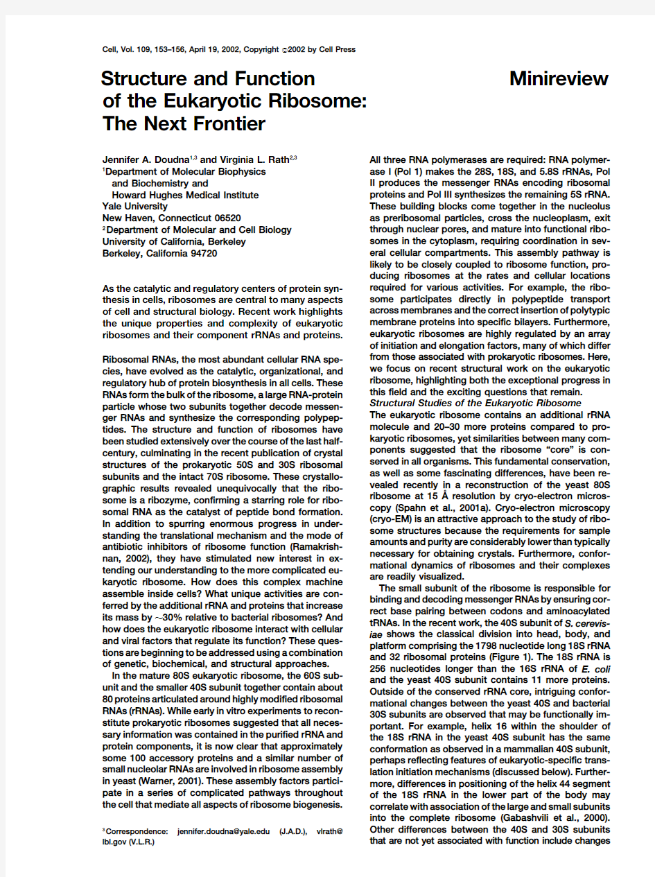
karyotic ribosome.How does this complex machine The small subunit of the ribosome is responsible for assemble inside cells?What unique activities are con-binding and decoding messenger RNAs by ensuring cor-ferred by the additional rRNA and proteins that increase rect base pairing between codons and aminoacylated its mass by ?30%relative to bacterial ribosomes?And tRNAs.In the recent work,the 40S subunit of S.cerevis-how does the eukaryotic ribosome interact with cellular iae shows the classical division into head,body,and and viral factors that regulate its function?These ques-platform comprising the 1798nucleotide long 18S rRNA tions are beginning to be addressed using a combination and 32ribosomal proteins (Figure 1).The 18S rRNA is of genetic,biochemical,and structural approaches.256nucleotides longer than the 16S rRNA of E.coli In the mature 80S eukaryotic ribosome,the 60S sub-and the yeast 40S subunit contains 11more proteins.unit and the smaller 40S subunit together contain about Outside of the conserved rRNA core,intriguing confor-80proteins articulated around highly modified ribosomal mational changes between the yeast 40S and bacterial RNAs (rRNAs).While early in vitro experiments to recon-30S subunits are observed that may be functionally im-stitute prokaryotic ribosomes suggested that all neces-portant.For example,helix 16within the shoulder of sary information was contained in the purified rRNA and the 18S rRNA in the yeast 40S subunit has the same protein components,it is now clear that approximately conformation as observed in a mammalian 40S subunit,some 100accessory proteins and a similar number of perhaps reflecting features of eukaryotic-specific trans-small nucleolar RNAs are involved in ribosome assembly lation initiation mechanisms (discussed below).Further-in yeast (Warner,2001).These assembly factors partici-more,differences in positioning of the helix 44segment pate in a series of complicated pathways throughout of the 18S rRNA in the lower part of the body may the cell that mediate all aspects of ribosome biogenesis.
correlate with association of the large and small subunits into the complete ribosome (Gabashvili et al.,2000).Other differences between the 40S and 30S subunits 3
Correspondence:jennifer.doudna@https://www.doczj.com/doc/9c8483376.html, (J.A.D.),vlrath@
https://www.doczj.com/doc/9c8483376.html, (V.L.R.)
that are not yet associated with function include changes
Cell
154
ments in position were required to fit these segments
into the EM map,revealing large differences in the posi-
tions of the rRNA segments that contact L1and L11.
For example,helices43and44shift by some15A?and
helix78moves by30A?.Movements of the part of the
ribosome bearing L1and L11have also been observed
in the yeast ribosome on binding to the translocation
factor EF2(Gomez-Lorenzo et al.,2000).These two re-
gions are important in tRNA binding and release,and
their mobility may be required to accomplish both tasks.
One of the more intriguing aspects of the yeast80S
ribosome images is the presence of expansion seg-
ments in the large subunit rRNA,visible as extra density
in the separated RNA map that is not accounted for by
the X-ray model of the archaeal large ribosomal subunit
23S rRNA.These expansion segments can be found in
all domains of the5.8S/25S rRNA,and are concentrated
on approximately opposite sides of the60S subunit.
Many participate in tertiary and quaternary contacts,
including two additional bridges to the40S subunit not
seen in bacteria.Ribosomal proteins not found in https://www.doczj.com/doc/9c8483376.html,parison of EM Density for rRNA from E.coli and
rial or archaeal large subunits were also identified,using S.cerevisiae Ribosomes
homology modeling,but some twelve regions of unmod-Upper panels,rRNA density computationally identified within the
eled protein density are present on the solvent side of small ribosomal subunit of E.coli(left)or S.cerevisiae(right),respec-
the large subunit.Interestingly,much of this density tively.The head(h),shoulder(sh),platform(pt),spur(sp),body(b),
and helix44(h44)are indicated;in orange,expansion segments in forms multiple contacts to other parts of the ribosome, the S.cerevisiae rRNA.Lower panels,rRNA density computationally implicating these additional yeast proteins in stabilizing identified within the large ribosomal subunit of E.coli(left)or the extra rRNA or interacting with eukaryotic-specific S.cerevisiae(right),respectively.The central protuberance(CP),translation factors or regulators.
stalk base(SB),sarcin-ricin loop(SRL)and several helices are indi-
Interaction between the large and the small subunit cated;in purple,expansion segments in the S.cerevisiae rRNA.
of the ribosome is a fundamental property of translation. Note that in the E.coli density,parts of the L1-bearing protuberance
and the stalk base are missing,while in S.cerevisiae,these features The small subunit binds mRNA and the anticodon por-are visible in the map.tion of the tRNA and is responsible for translational fidel-
ity by ensuring base pairing between the codon and
anticodon during the decoding process.The large sub-in the shape and position of the beak(see Figure1),a
unit binds the acceptor ends of the tRNAs and catalyzes result of differences in the length of a region known as
peptide bond formation between the nascent polypep-helix33and the presence of additional protein density.
tide chain and the incoming aminoacylated tRNA.Both Segments of the rRNA that contain additional se-
subunits are involved in translocating the mRNA by one quences,or insertions,in the yeast ribosome can be
trinucleotide codon each cycle.Previous EM work on identified in some cases based on the EM density and
the E.coli70S ribosome combined with the X-ray crys-sequence comparisons.A series of these expansion tallographic map of the T.thermophilus70S ribosome segments(ES)(ES12,ES6,ES3,see Figure1)that con-identified7bridges between the two subunits,all of nect the platform of the40S with the lower part of the which occur in the yeast ribosome.In addition,four new body form a structure of unknown function not found in bridges,which may be eukaryotic specific,can be identi-the prokaryotic ribosome.fied from the present work.Most of the subunit interac-The large ribosomal subunit contains the peptidyl tions involve direct RNA-RNA contacts,consistent with transferase active site responsible for catalyzing peptide the idea that an ancestral form of the ribosome might bond formation during protein synthesis.In yeast,this have been composed entirely of RNA.
60S subunit includes25S rRNA(3392nucleotides),5.8S A glimpse of the mechanism of tRNA recognition by rRNA(158nucleotides),5S rRNA(121nucleotides),and a eukaryotic ribosome comes from the tRNA observed 45proteins.This makes the yeast5.8S/25S rRNA646in the peptidyl or P site of the yeast80S structure.Based nucleotides longer than the corresponding bacterial(E.on the docked models of the rRNAs,ribosomal proteins coli)rRNA and505nucleotides longer than the archaeal and the tRNA,Spahn et al.propose that the P site codon (H.marismortui)rRNA.The yeast large subunit has12may interact with helix44of the18S rRNA,and the tRNA and14proteins more,respectively,than its bacterial or with helices24,30,31,and43of18S rRNA as well archaeal counterparts.Despite these additional compo-as ribosomal protein S16.These interactions in yeast nents,its core is structurally similar to that of the H.appear similar to those in bacteria,supporting the idea marismortui23S rRNA.Segments of the rRNA that con-that the most fundamental,and ancient,activities of the tact ribosomal proteins L1and L11were taken from ribosome are the same in all kingdoms of life(Carter et the crystal structures of the bacterial T.thermophilus al.,2000;Yusupov et al.,2001).
(Yusupov et al.,2001)and T.maritima(Wimberly et al.,Targeting Ribosomes to Membranes
1999)rRNAs,respectively,because these regions are In addition to synthesizing proteins,ribosomes are re-disordered in the H.marismortui coordinates.Adjust-
sponsible for directing polypeptides to their correct cel-
Minireview
155
tation rather than the discrete pore previously observed
for an empty ribosome channel complex in detergent
(Beckmann et al.,1997).This suggests that the signal
sequence itself doesn’t alter the channel shape or fill
the gap.
The four attachment sites observed at the outer sur-
face of the polypeptide exit tunnel in the60S subunit
and on the Sec61p complex apparently include both
ribosomal protein and rRNA components,a result which
contradicts an experiment showing that the isolated
large subunit rRNA alone bound to the channel with
high affinity(Prinz et al.,2000).It may be that ribosomal
proteins do not contribute energetically to channel
docking,though this has not yet been investigated.In
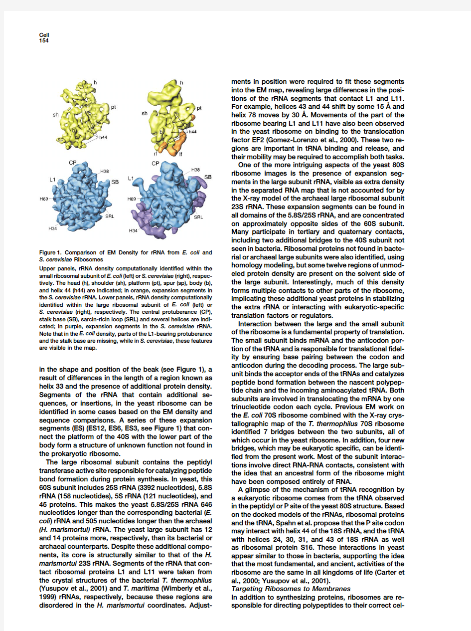
any case,the EM structure provides a set of predicted Figure2.Yeast80S Ribosome Bound to the Sec61Protein Complex
contacts that can now be explored genetically and bio-Density corresponding to Sec61,red;P-site-bound peptidyl tRNA,
chemically,an important step toward understanding ri-green;40S proteins,aqua;small subunit(18S)rRNA,yellow;60S
bosome-channel communication.
proteins,orange;large subunit(25S/5.8S/5S)rRNAs,blue.
Interestingly,rRNA may play an important role in regu-
lating access to the emerging polypeptide during protein
https://www.doczj.com/doc/9c8483376.html,parison of the translating complex with lular location.Many of the approximately30%of pro-
previous cryo-EM studies of yeast ribosomes reveals a teins that are inserted into or transported across cellular
significant rRNA conformational change that occurs membranes contain an N-terminal signal sequence that
near the polypeptide exit site.The location of a large is recognized during translation by the highly conserved
segment of density identified as the main helix of expan-signal recognition particle(SRP).As the signal sequence
sion segment27(ES27)in25S rRNA,one of the rRNA emerges from the ribosome,SRP binds and targets this
insertions characteristic of80S ribosomes,adopts one ribosome-nascent chain complex(RNC)to the ER mem-
of two preferred positions.In one conformation,the helix brane.Hand-off of the nascent chain from the SRP to
is found close to the polypeptide exit site on the ribo-the translocation channel,a heterotrimeric membrane
some,while in the other it rotates90degrees away from protein called,in yeast,the Sec61p complex,creates a
the exit tunnel and binds near the arm of ribosomal leak-proof seal that enables cotranslational export of
protein L1.Reconstructions of different samples reveal the polypeptide(Figure2).
both conformations,and in some cases,evidence for Direct association of the ribosome with the Sec61p
partial occupancy of both positions in the same EM complex in the absence of SRP or its receptor has been
map.However,in the complex with Sec61p,ES27is demonstrated in several ways,and this interaction has
found only near L1,implying that Sec61p binding stabi-been visualized at a new level of detail using cryo-EM
lizes ES27to a locus where it cannot interfere with na-(Beckmann et al.,2001).Preparation of an appropriate
scent chain translocation.This highly dynamic rRNA sample required the clever design of an mRNA encoding
structure may control access of nonribosomal factors a yeast membrane protein,dipeptidylaminopeptidase
such as chaperones,modifying enzymes,or the Sec61p B(DAP2),known to be translocated cotranslationally.
complex to the tunnel exit site and thereby to the emerg-Yeast ribosomes were programmed in a cell-free trans-ing nascent chain.
lation system with a truncated form of this mRNA con-Viral Recruitment of Ribosomes
taining an N-terminal affinity tag and the first120amino The unique features of the eukaryotic ribosome,located acids of DAP2including the signal sequence.Following primarily on its outer solvent-exposed surfaces,are translation termination by the addition of cyclohexa-prime targets for interaction with regulatory factors. mide,active translocating ribosomes were assembled These surfaces can also be exploited by viruses for using immunoprecipitated RNCs and a purified sample ribosome recruitment during host cell infection.Many of the heterotrimeric Sec61in a membrane free system.viruses circumvent cellular controls on5?-end-depen-A control sample contained Sec61p complexed with dent translation initiation by utilizing internal ribosome ribosomes isolated from a translation reaction with no entry site(IRES)RNAs located within viral mRNAs up-added mRNA.stream of coding sequences.In hepatitis C virus(HCV), Both samples revealed the expected overall appear-the IRES binds the40S subunit in the absence of any ance of the80S ribosome,with additional density for other factors to form a high-affinity complex that pre-the Sec61p complex visible at the exit site of the large cedes translation initiation.A20A?resolution cryo-EM ribosomal subunit tunnel.In both the translating and reconstruction of the mammalian40S subunit com-inactive ribosomes,a gap of at least15A?between the plexed with functional and disabled forms of the HCV channel surface and the ribosome leaves the channel IRES provides clues to the mechanisms of ribosome unsealed.This gap,observed in similar mammalian and recruitment and translation initiation(Spahn et al., yeast ribosomal complexes at lower resolution,leaves2001b).
open the question of channel function without concomi-The overall structure of the mammalian40S subunit tant membrane leakage.The overall shape of the Sec61p compares well with the40S portion of the yeast80S complex in the presence or absence of mRNA is the ribosome EM map described above.The IRES RNA same,appearing as a compact disc with a central inden-
binds on the solvent side of the40S subunit and involves
Cell
156
Gabashvili,I.S.,Agrawal,R.K.,Spahn,C.M.,Grassucci,R.A.,Frank, recognition of structural elements unique to eukaryotic
J.,and Penczek,P.(2000).Cell100,537–549.
ribosomes.Since the HCV IRES is functional only with
Gomez-Lorenzo,M.G.,Spahn,C.M.T.,Agrawal,R.K.,Grassucci, mammalian ribosomes,its mode of recognition may in
R.A.,Penczek,P.,Chakraburtty,K.,Ballesta,J.P.G.,Lavandera,J.L., fact be highly specific for particular rRNA,proteins,or
Garcia-Bustos,J.F.,and Frank,J.(2000).EMBO J.19,2710–2718. structures.An?100nucleotide region at the5?end of
Prinz,A.,Behrens,C.,Rapoport,T.A.,Hartmann,E.,and Kalies,K.U. the IRES,domain II,is particularly interesting because
(2000).EMBO J.19,1900–1906.
its location on the ribosome partially overlaps with the
Ramakrishnan,V.(2002).Cell108,557–572.
exit(E)site that houses the deacylated tRNA prior to its
Spahn,C.M.,Beckmann,R.,Eswar,N.,Penczek,P.A.,Sali,A.,Blo-release after peptide bond formation.Although domain
bel,G.,and Frank,J.(2001a).Cell107,373–386.
II alone does not bind the40S subunit or contribute to
Spahn,C.M.,Kieft,J.S.,Grassucci,R.A.,Penczek,P.A.,Zhou,K., IRES affinity for the40S subunit,translation initiation is Doudna,J.A.,and Frank,J.(2001b).Science291,1959–1962.
impaired without it.Domain II also appears responsible Warner,J.R.(2001).Cell107,133–136.
for a pronounced40S subunit conformational change
Wimberly,B.T.,Guymon,R.,McCutcheon,J.P.,White,S.W.,and induced by IRES binding that may correlate with IRES Ramakrishnan,V.(1999).Cell97,491–502.
translational efficiency.The40S subunit head position Yusupov,M.M.,Yusupova,G.Z.,Baucom,A.,Lieberman,K.,Ear-relative to the body is altered near the beak,clamping nest,T.N.,Cate,J.H.,and Noller,H.F.(2001).Science292,883–896. down on the mRNA on one side while simultaneously
opening up the helix18–34region to allow the transla-
tional start site in the mRNA to insinuate into the entry
channel.This conformational change,not observed in
a complex between the40S subunit and an IRES RNA
lacking domain II,suggests a key role for domain II in
the correct positioning of viral mRNA.The IRES may
directly place the message in the decoding center of the
ribosome or stabilize the40S subunit in a conformation
necessary for mRNA binding in the absence of canonical
initiation factors.Many intriguing questions remain:
what is the functional relevance of the conformational
change observed with the HCV IRES?Do other IRES
RNAs bind and influence ribosome conformation in a
similar way?Is this conformation a common feature of
ribosomes primed for translation by cellular translation
initiation factors?Future structural and biochemical ex-
periments will be required to address these and other
aspects of the complicated process of translation initi-
ation.
Conclusion
As some of the most ancient cellular components,ribo-
somal RNAs have been crafted over evolutionary eons
for efficient catalysis and regulation of all aspects of
protein synthesis.Much attention has lately been fo-
cused on the conserved“core”of the ribosome,which
appears to be structurally similar in all kingdoms of life.
But it may well be that investigation of its less conserved
rRNA and protein elements will uncover properties and
activities of the eukaryotic ribosome that are unique in
higher organisms.It also seems likely that understand-
ing eukaryotic ribosomal RNA processing,chemical
modification,folding,structure,and activities will illumi-
nate fundamental aspects of cellular RNA metabolism
in general.The eukaryotic ribosome represents the next
frontier in cell and structural biology,whose mysteries
now seem much more tractable,and more interesting,
the more we know.
Selected Reading
Beckmann,R.,Bubeck,D.,Grassucci,R.A.,Penczek,P.,Verschoor,
A.,Blobel,G.,and Frank,J.(1997).Science278,2123–2126.
Beckmann,R.,Spahn,C.M.,Eswar,N.,Helmers,J.,Penczek,P.A.,
Sali,A.,Frank,J.,and Blobel,G.(2001).Cell107,361–372.
Carter,A.P.,Clemons,W.M.,Jr.,Brodersen,D.E.,Morgan-Warren,
R.J.,Wimberly,B.T.,and Ramakrishnan,V.(2000).Nature407,
340–348.