Drug Metabolism Reviews , 2009; 41(1): 1–7
Re vie w A Rt icl e
Biological degradation of aflatoxins
Qinghua Wu 1, Alena Jezkova 2, Zonghui Yuan 1, Lucie Pavlikova 2, Vlastimil Dohnal 2,3,4, and Kamil Kuca 3,4,5
1
National Reference Laboratory of Veterinary Drug Residues (HZAU)/MAO Key Laboratory of Food Safety Evaluation, Huazhong Agricultural University, Wuhan, People’s Republic of China, 2Department of Food Technology, Faculty of Agronomy, Mendel University of Agriculture and Forestry in Brno, Brno, Czech Republic, 3Department of Chemistry, Faculty of Sciences, J.E. Purkinje University, Usti nad Labem, Czech Republic, 4Department of Toxicology, Faculty of Military Health Sciences, University of Defence, Hradec Kralove, Czech Republic, and 5Centre of Advanced studies, Faculty of Military Health Sciences, University of Defence, Hradec Kralove, Czech Republic
Address for Correspondence : Kamil Kuca, Department of Toxicology and Centre of Advanced Studies, Faculty of Military Health Sciences, University of Defence, Hradec Kralove, Czech Republic; E-mail: kucakam@pmfhk.cz (Received 09 September 2008; accepted 17 October 2008)
Introduction
Aflatoxins are a group of highly toxic secondary
m etabolites produced mainly by Aspergillus species fungi. A. flavus produces only aflatoxins B, while A. para-siticus produces both aflatoxins B and G. Aflatoxins can be found mainly in cereals, oilseeds, tree nuts, spices, and milk. Among 18 different types of aflatoxins, such as B 1, B 2, G 1, G 2, P , Q, M 1, M 2, B 2a , etc., were identified. The most commonly occurring ones in fungi cultures are aflatoxins B 1, B 2, G 1, and G 2, then aflatoxins M 1 and M 2 in milk.
Naturally occurring aflatoxins and aflatoxin B 1 (AFB 1) are classified by the International Agency for Research on Cancer (IARC) as group 1 carcinogens (“carcinogenic
to humans”) (IARC, 2002). The AFB 1 metabolite, 8,9-epoxide, forms DNA adducts primary with N 7 of guanine (Cullen and Newberne, 1994). The toxic effect on living organisms is reviewed in a World Health Organization report (WHO, 1998). The toxicity of aflatoxin decreases in order B 1, G 1, B 2, and G 2. When AFB 1 and aflatoxin B 2 (AFB 2) are consumed, they are metabolized to aflatoxin M 1 (AFM 1), respective M 2 (AFM 2), and distributed in tis-sues and biological fluids and milk (Zarba et al., 1992).Biodegradation of aflatoxins, using microorganisms or enzymes, is one of the well-known strategies for the man-agement of aflatoxins in foods and feeds. The methods of biodegradation are being actively studied and can be a highly promising choice, since it is efficient, specific, and
ISSN 0360-2532 print/ISSN 1097-9883 online ? 2009 Informa UK Ltd DOI: 10.1080/03602530802563850
Abstract
Aflatoxins are cancerogenic compounds produced predominantly by certain strains of the Aspergillus genus. The ideal solution for minimization of health risk that aflatoxins pose is the prevention of foods and feeds contamination. Unfortunately, these contaminants can never be completely removed, and on that account, many studies have been carried out to explore an effective process of their detoxification to a threshold level. Biological decontamination seems to be attractive because it works under mild, environ-mentally friendly conditions. This review is focused on the biological detoxification of aflatoxins, especially aflatoxin B 1, by microorganisms. There are briefly mentioned aflatoxin metabolic pathways in the human and animal body. Microorganisms such as soil or water bacteria, fungi, and protozoa and specific enzymes isolated from microbial systems can degrade aflatoxin group members with varied efficiency to less- or nontoxic products. Some aflatoxin-producing fungi from Aspergillus species have the capability to degrade their own synthesized mycotoxins. Yeasts and lactic acid bacteria work as biological adsorbents that pre-vent aflatoxin’s transfer to the intestinal tract of humans and animals. Aflatoxin B 1 absorbed into the organ-ism could be metabolized by significantly different pathways. They lead to the production of the relatively nontoxic compounds, on the one hand, or to highly toxic active forms on the other hand.Keywords: Aflatoxin; metabolism; biodegradation; decontamination; bacteria
https://www.doczj.com/doc/8315754275.html,/dmr
D r u g M e t a b o l i s m R e v i e w s D o w n l o a d e d f r o m i n f o r m a h e a l t h c a r e .c o m b y C A S C h i n e s e A c a d e m y o f S c i e n c e s o n 01/24/11F o r p e r s o n a l u s e o n l y .
2 Qinghua Wu et al.
environmentally friendly to reduce or eliminate the pos-sible contaminations of aflatoxins in foods and feeds.Soil bacteria
Many bacteria in soil are able to degrade aflatoxins. Flavobacterium aurantiacum NRRL B-184, a kind of bac-teria from soils and water, showed a very high capability of detoxifing a flatoxins i n f eeds a nd f oods (Ciegler e t a l., 1966a). The aflatoxin-contaminated substance and F . aurantiacum NRRL B-184 were mixed together and incubated at 28°C for 12 hours, all of the aflatoxin G was removed, as well as a part of aflatoxin B, which was diminished. Ducking assays implied detoxification of aflatoxin solutions by F . aurantia-cum NRRL B-184 was complete, with no new toxic prod-ucts being formed. Lillehoj showed that this bacterium can remove AFM 1 from milk in 1971 (Shapira, 2004). Later, it was observed that the radioactively labeled 14C-AFB 1 is partially metabolized and partially adsorbed to F . aurantiacum cells (Line et al., 1994). D’Souza and Brackett (1998, 2000, 2001) have been monitoring the effects of cations and several chemical compounds on AFB 1 degradation by these bacte-ria. For example, Cu 2+, Mn 2+, and Zn 2+ lower the reduction capacity of F . aurantiacum . This confirms the influence of the enzymes in the degradation process. More applications of F . aurantiacum for decontamination of food/feed were discussed in a review (Bata and Lásztity, 1999).Other microorganisms were also tested for their p ossible ability to degradate aflatoxins. The strain Nocardia aster-oides reduces AFB 1 by biotransformation to another fluo-rescent product (Arai et al., 1967), and Corynebacterium rubrum is able to detoxify aflatoxin as well (Shapira, 2004).In another work, Mycobacterium fluoranthenivorans sp. nov. DSM44556T isolated from soils of a former coal gas plant, which was polluted with polycyclic aromatic hydrocarbons, was found to be capable of degrading AFB 1 as a single carbon source (Hormisch et al., 2004). The AFB 1 concentration was reduced to amounts of 70–80% of the initial concentration within 36 hours, and no AFB 1 was detectable after 72 hours. In addition, the cell-free extracts of M. fluoranthenivorans sp. nov. DSM44556T degraded AFB 1 more efficiently (Teniola et al., 2005). More than 90% of the initial amount of AFB 1 was degraded at 30°C within 4 hours, and no AFB 1 was detected after 8 hours.
Teniola et al. (2005) investigated Rhodococcus eryth-ropolis isolated from polycyclic aromatic hydrocarbon (PAH) soils for AFB 1 degradation activity. Dramatic reduction of AFB 1 was observed during incubation in the presence of R. erythropolis cells. Then, 17% residual AFB 1 was left after 48 hours and only 3–6% was detecta-ble after 72 hours. In addition, this work team also found AFB 1 was effectively degraded by extracellular extracts from R. erythropolis (only 32% residual AFB 1 was detect-able after 72 hours) (Alberts et al., 2006). Results indi-cated AFB 1 was most likely metabolized to d egradation
p
roducts with chemical properties different from that of AFB 1, because the equipments they utilized could not reveal the formation of any breakdown products.
The high degradation rate and wide temperature range for degradation by both R. erythropolis and M. fluoranthenivorans sp. nov. DSM44556T indicate a potential and promising application for the degradation of AFB 1 in the foods and feeds process.Fungi
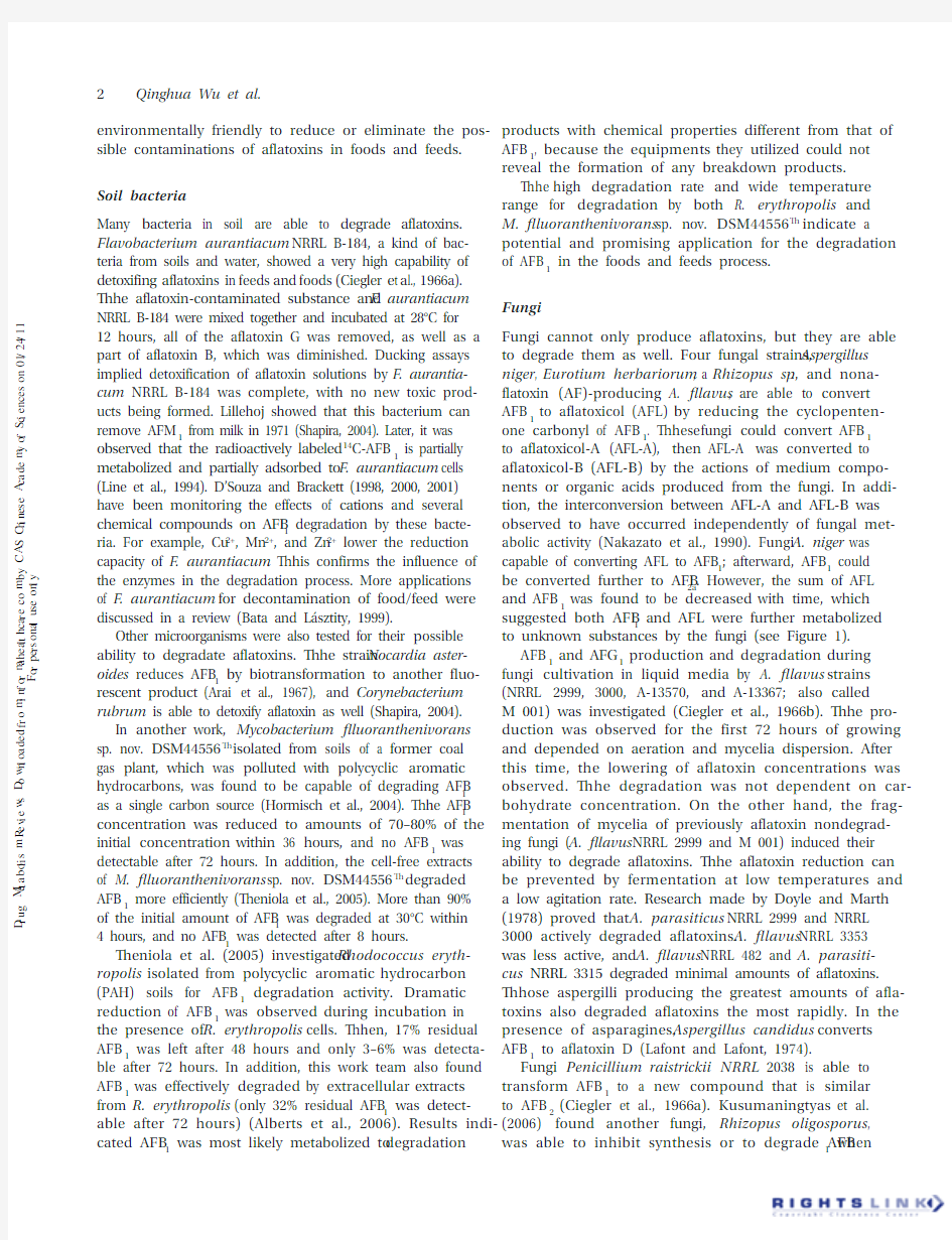
Fungi cannot only produce aflatoxins, but they are able to degrade them as well. Four fungal strains, Aspergillus niger , Eurotium herbariorum , a Rhizopus sp ., and nona-flatoxin (AF)-producing A. flavus , are able to convert AFB 1 to aflatoxicol (AFL) by reducing the cyclopenten-one carbonyl of AFB 1. These fungi could convert AFB 1 to aflatoxicol-A (AFL-A), then AFL-A was converted to aflatoxicol-B (AFL-B) by the actions of medium compo-nents or organic acids produced from the fungi. In addi-tion, the interconversion between AFL-A and AFL-B was observed to have occurred independently of fungal met-abolic activity (Nakazato et al., 1990). Fungi A. niger was capable of converting AFL to AFB 1; afterward, AFB 1 could be converted further to AFB 2a . However, the sum of AFL and AFB 1 was found to be decreased with time, which suggested both AFB 1 and AFL were further metabolized to unknown substances by the fungi (see Figure 1).
AFB 1 and AFG 1 production and degradation during fungi cultivation in liquid media by A. flavus strains (NRRL 2999, 3000, A-13570, and A-13367; also called M 001) was investigated (Ciegler et al., 1966b). The pro-duction was observed for the first 72 hours of growing and depended on aeration and mycelia dispersion. After this time, the lowering of aflatoxin concentrations was observed. The degradation was not dependent on car-bohydrate concentration. On the other hand, the frag-mentation of mycelia of previously aflatoxin nondegrad-ing fungi (A. flavus NRRL 2999 and M 001) induced their ability to degrade aflatoxins. The aflatoxin reduction can be prevented by fermentation at low temperatures and a low agitation rate. Research made by Doyle and Marth (1978) proved that A. parasiticus NRRL 2999 and NRRL 3000 actively degraded aflatoxins. A. flavus NRRL 3353 was less active, and A. flavus NRRL 482 and A. parasiti-cus NRRL 3315 degraded minimal amounts of aflatoxins. Those aspergilli producing the greatest amounts of afla-toxins also degraded aflatoxins the most rapidly. In the presence of asparagines, Aspergillus candidus converts AFB 1 to aflatoxin D (Lafont and Lafont, 1974).
Fungi Penicillium raistrickii NRRL 2038 is able to transform AFB 1 to a new compound that is similar to AFB 2 (Ciegler et al., 1966a). Kusumaningtyas et al. (2006) found another fungi, Rhizopus oligosporus , was able to inhibit synthesis or to degrade AFB 1 when
D r u g M e t a b o l i s m R e v i e w s D o w n l o a d e d f r o m i n f o r m a h e a l t h c a r e .c o m b y C A S C h i n e s e A c a d e m y o f S c i e n c e s o n 01/24/11F o r p e r s o n a l u s e o n l y .
Biological degradation of aflatoxins 3
cultured together with AFB 1-producing fungi A. flavus . R. oligosporus had the best degrading ability on Day 5. Fungi R. arrhizus , R. stolonifer NRRL 1477, and A. arrhi-zus NRRL 2585 transform AFB 1 to aflatoxicol (AFL), trivially named as aflatoxin R o (AFR o ) (Cole et al., 1972). Incubation time longer than 3 days resulted in isomeri-zation to AFR o . A short review about other aflatoxin (AFG 1) degradation by Rhizopus species was published by the same work team (Cole and Kirksez, 1971).
Metabolization of AFB 1 to AFR o was also reported for D. dendroides NRRL 2575, Mucor alterans NRRL 3358, M. griseocyanus NRRL 3359, Absidia repens NRRL 1336, and Helminthosporium sativum NRRL 3356 (Detroy and Hesseltine, 1969). Longer times of incubation led to the formation of new, unidentified blue-fluorescent product.Protozoa
Cells of the protozoon Tetrahymena pyriformis W have the ability to degradate pure AFB 1 to another bright-blue fluorescent product and decrease the AFB 1 concentra-tion to 25% in 30 hours. The course of detoxification of AFG 1 by this strain was different. During 10 hours, the concentration decreased to 80% and was constant for the next 20 hours (Teunisssion and Robertson, 1967). One year later, it was concluded by Robertson et al.
(1970) that T. pyriformis W reduced the carbonyl in the cyclopentane ring of AFB 1 to a hydroxyl group (Figure 2). The reduced aflatoxin appears to be identical to AFR o .Digestive tract microorganisms
Rumen bacteria have been reported to degradate afla-toxins. Kiessling et al. (1984) investigated the metabo-lism of AFB 1 by intact rumen fluid, rumen protozoa, and rumen bacteria in vitro . They found that AFB 1 was not degraded by rumen microorganisms. A slight decrease in the amount of AFB 1 occurred within 30 minutes; how-ever, after this time, no further reduction was evident.In another work, Westlake et al. (1987) studied four strains of Butyrivibrio fibrisolvens (CE46, CE51, CE52, and CE56), one of the predominant rumen bacteria. Results showed that there was no significant degradation of AFB 1. The researchers proposed that microbial activity was solely responsible for aflatoxin’s degradation within the rumen. The efficiency of the rumen metabolism was demonstrated by the fact that only 2–5% of AFB 1 fed to cows reached the intestine (Engel and Hagemeister, 1978; Karlovsky, 1999).
AFB 1-contaminated corn mixed with poultry litter was incubated, and mycotoxin was destroyed by feces microorganisms in several weeks (Jones et al., 1996).
aflatoxicol B (AFL-B)aflatoxicol A (AFL-A)
AFB AFB
2a
AFB 2
Figure 1. Metabolic pathways of aflatoxin B 1 by fungi.
OCH 3
O
O
O
O
OH
aflatoxin B 1
aflatoxin R o (aflatoxicol)
Figure 2. Degradation of AFB 1 by protozoa.
D r u g M e t a b o l i s m R e v i e w s D o w n l o a d e d f r o m i n f o r m a h e a l t h c a r e .c o m b y C A S C h i n e s e A c a d e m y o f S c i e n c e s o n 01/24/11F o r p e r s o n a l u s e o n l y .
4 Qinghua Wu et al.Yeasts and lactic acid bacteria
The mechanism of reducing aflatoxins by yeasts and lactic acid bacteria is due to their adhesion to cell-wall components. Blanco et al. (1993), Jesperson et al. (1994), and Wiseman and Marth (1983) in (Westby et al., 1997) show no effect of lactic acid bacteria. However, many other researchers report very efficient aflatoxin
r eductions associated with fermentation.Yeasts Saccharomyces cerevisiae have capability to bind AFB 1. Further investigation showed that compo-nents of yeast cell wall, called oligomannanes after their chemical modification, esterification, were able to bind up to 95% of AFB 1 (Devegowda et al., 1996). In addition, they are more efficient than commonly used adsorbent HSCAS (hydrated sodium calcium aluminium silicate) (Mahesh and Devegowda, 1996). The addition of 0.05% of esterified glucomannans to the basal diet resulted to improved performance in broilers (Kumprecht et al., 1977; Aravind et al., 2003).
Significant decreasing of AFB 1 concentration dur-ing brewing process in the work of Chu et al. (1975) is probably caused by sorption of mycotoxin on S. cer-evisiae yeasts. This hypothesis is supported by other researchers (Shetty et al, 2007; Celyk et al., 2003). A 19% reduction of spiked AFB 1 during dough fermenta-tion in breadmaking was reported (El-Banna and Scott, 1983).
The addition of yeast cell walls into feed can lead to a decrease of aflatoxin toxic effect in broilers (Stanley et al., 1993; Santin et al., 2003).
AFB 1 was detoxified into AFB 2a during yogurt (Megalla and Hafez, 1982) and dairy product (Megalla and Mohran, 1984) fermentation.
Peltonen et al. (2001) investigated the ability of 12 Lactobacillus , 5 Bifidobacterium , and 3 Lactococcus bacteria strains to bind AFB 1. These bacteria are com-monly used in the food industry as a starter culture in the production of fermented milk and meat products, such as yogurt, cheese, salami, etc. During the last few years, the selected strains of bifidobacteria and lacto-bacilli are rising in use in combination with prebiotics as probiotic cultures applied in renewing of intestinal microflora.
El-Nezami et al. (1998) studied the ability of selected Lactobacillus strains to remove AFB 1 from the cultiva-tion media. The probiotic strains L. rhamnosus GG and L. rhamnosus strain LC-705 showed significant abil-ity to remove toxin from media (around 80% of added amount). The binding of AFB 1 to L. rhamnosus GG decreases its subsequent adhesion capability to Caco-2 cells, which means that these bacteria may reduce the accumulation of aflatoxins in the intestine via increased excretion of an aflatoxin-bacteria complex (Kankaankp?? et al., 2000).
Enzymes
Specific enzymes that are capable of degrading aflatoxins have been purified from microbial systems. The detoxifi-cation by specific enzymes avoids the drawback of using the microorganism, which may, in addition to its deg-radation activity, change flavor or impair the nutritional value and acceptability of the product (Shapira, 2004).A novel aflatoxin degradation enzyme had been
i solated and purified from Pleurotus ostreatus by Motomura et al. (2003). AFB 1 was treated with culture supernatants from 19 mushroom strains. The super-natant from P . ostreatus showed aflatoxin-degradation activity, whereas other strains had no or only weak activ-ity. In addition, the novel enzyme showed the best afla-toxin-degradation activity at 25°C with a pH of 4.0–5.0. Fluorescence measurements suggested that the specific enzyme cleaved the lactone ring of aflatoxin, although the degradation products of aflatoxin were not investi-gated clearly.
In another work (Liu et al., 2001), an enzyme named aflatoxin-detoxifizyme (ADTZ), which exhibited detoxi-fication activity on AFB 1, was isolated and purified from Armillariella tabescens (E-20). AFB 1 seemed to be deg-radated into difuran ring-opening AFB 1, which was less toxic than AFB 1, and the optimum activity for the enzyme was at 35°C with a pH of 6.8. This work team had already isolated a multienzyme that was able to degradate AFB 1 from A. tabescens in the earlier time (Liu et al., 1998). In addition, t hey g ave a p roposed p athway o f t he d egradation of AFB 1 by the multienzyme: AFB 1 was first transformed to AFB 1-epoxide, followed by hydrolysis of the epoxide to give the dihydrodiol. Then, the difuran ring would open in the subsequent hydrolysis step (Figure 3).Metabolism of aflatoxins in animals and humans Generally, there is a great diversity in the metabolism of aflatoxins among different animal species or even, in some cases, individual animals. There are four meta-bolic pathways of AFB 1: O-dealkylation to AFP 1, ketore-duction to AFL, epoxidation to AFB 1-8,9-epoxide (highly toxic, mutagenic, and carcinogenic), and hydroxylation to AFM 1 (highly toxic), AFP 1, AFQ 1, or AFB 2a (all relatively nontoxic). Thus, the main reactions in aflatoxins metab-olism are hydroxylation, oxidation, and demethylation.Most of the metabolites of AFB 1 are able to be trans-formed to further metabolites. AFQ 1 is able to be trans-formed to AFH 1 in liver. For AFB 1-8,9-epoxide, there exists three further metabolic pathways in animals and humans: hydrolysis to form AFB 1-8,9-dihydrodiol, conjugation with to AFB 1-8,9-dihydro-8-(N 7-guanyl)-3-hydroxy (AFB 1-N 7-Gua), and conjugation with soluble nucleophilic molecules, such as glutathione (Yiannikouris and Jouany, 2002).
D r u g M e t a b o l i s m R e v i e w s D o w n l o a d e d f r o m i n f o r m a h e a l t h c a r e .c o m b y C A S C h i n e s e A c a d e m y o f S c i e n c e s o n 01/24/11F o r p e r s o n a l u s e o n l y .
Biological degradation of aflatoxins5 multienzyme
AFB
1AFB
1
-epoxide
AFB
1
-dihydrodiol
O O
O
O O
HO
HO
O O
O
OCH
3
OCH
3
O O
O
Figure 3.Proposed pathway of degradation of AFB
1
by Armillariella tabescens.
Figure 4.Metabolic pathways of aflatoxin B
1
in animals (adapted from Yiannikouris and Jouany (2002)). D
r
u
g
M
e
t
a
b
o
l
i
s
m
R
e
v
i
e
w
s
D
o
w
n
l
o
a
d
e
d
f
r
o
m
i
n
f
o
r
m
a
h
e
a
l
t
h
c
a
r
e
.
c
o
m
b
y
C
A
S
C
h
i
n
e
s
e
A
c
a
d
e
m
y
o
f
S
c
i
e
n
c
e
s
o
n
1
/
2
4
/
1
1
F
o
r
p
e
r
s
o
n
a
l
u
s
e
o
n
l
y
.
6 Qinghua Wu et al.
Various forms of P-450 serve different biotransforma-tion capacities, depending on the animal species. AFB 1 is transformed to AFM 1, AFQ 1, and AFB 1-8,9-epoxide by mixed-function mono-oxygenase in rat liver. In addition, Yoshizawa et al. (1982) found AFM 1 was strictly medi-ated by cytochrome P-448, while AFQ 1 was catalyzed by both P-450 and P-448 in rat liver. Cytochrome P-450-dependent polysubstrate mono-oxygenase system, especially CYP3A4 and CYP1A2, are the major isoforms involved in human liver. However, in distinct contrast, lipoxygenase (LOX) and prostaglandin H synthase (PHS) were found to be the main enzymes of the biotransfor-mation of AFB 1 in human lung, whereas P-450 played a relatively minor role (Donnelly et al., 1996).
Numerous researchers have investigated the metabo-lism of aflatoxins in vitro and in vivo . In an investigation by Roebuck and Wogan (1977), AFL was found to be the main metabolite of AFB 1 in duck liver, whereas AFB 1 was able to be converted to AFQ 1 and AFP 1 by monkey and human liver. In addition, AFB 1 was able to be converted to AFM 1 and AFM 2a in all species (duck, rat, mouse, and monkey) except humans. In vitro metabolism of AFB 2 by animal and human liver was also studied by Roebuck et al. (1978). Duck liver had a much higher level of activ-ity than the tissues from other species. The significant pathway in duck liver was 2,3-desaturation to form AFB 1. AFL was also found in duck liver, as well as AFB 1, AFM 1, and AFM 2. However, AFB 1 was only transformed to AFQ 2 and AFP 2 by rat, mouse, and human liver. Different with ducks, a peptide (or amino acid) conjugate of AFB 2a and a glucuronide conjugate of AFM 1 were the main metabo-lites of AFB 1 in chicken liver in vivo (Chipley et al., 1974).In another work (Salhab and Edwards, 1977), in vitro metabolism of AFL by liver preparations from animals and humans was investigated. AFL was metabolized to AFB 1, AFQ 1, AFH 1, AFP 1, AFM 1, and AFM 2a by liver S12 fractions from monkey, dog, rat, mouse, and human. Human and hamster preparations were most active in the metabolic pathway from AFL to AFB 1, whereas rabbit and trout had the best ability to convert AFB 1 to AFL. However, whether AFH 1 was produced directly form AFL or via the AFB 1 and AFQ 1 intermediates was not identified (metabolic path-ways in animals can be found in Figure 4).
Conclusion
There have been many studies of aflatoxin degradation carried out in laboratory conditions, but no biological system exists to be used in the full commercial sphere currently. Interesting results have been obtained by Flavobacterium aurantiacum application. This soil bacte-rium is supposed to be removing aflatoxins B, G, and M 1 from substrate. The use of Rhodococcus erythropolis and Mycobacterium fluoranthenivorans sp. nov. DSM44556T
seems to be a promising opportunity for the degrada-tion of AFB 1 in the foods and feeds process. Studies sug-gest that certain fungi, including Aspergillus or Rhizopus sp., convert aflatoxin B 1 to less toxic aflatoxicol, aflatoxin B 2, or B 2a . Enzymes isolated from Pleurotus ostreatus and Armillariella tabescens showed aflatoxin-degradationac-tivity as well. Saccharomyces cerevisiae and Lactobacillus spp. can reduce aflatoxin amount due to binding of toxin on its cell walls. Isolated rumen bacteria do not signifi-cantly reduce aflatoxin amount, as they seem to be able to metabolize toxins in bovine ruminal fluid.
The fate of aflatoxin B 1 differs in human and animal organisms and among other species as well. There are four metabolic pathways of AFB 1: O-dealkylation, ketoreduction, epoxidation, and hydroxylation. These reactions lead to the creation of highly toxic aflatoxin B 1-8,9-epoxide and aflatoxin M 1 or relatively nontoxic forms, such as AFP 1, AFQ 1, or AFB 2a .
Acknowledgment
This work was supported by a grant from the Ministry of Industry and Trade of the Czech Republic (Grant no. 2A-1TP1/009).
Declaration of interest : The authors report no conflicts of interest.
References
Alberts, J. F ., Engelbrecht, Y., Steyn, P . S., Holzapfel, W. H., van Zyl, W. H.
2006. Biological degradation of aflatoxin B 1 by Rhodococcus eryth-ropolis cultures. Int. J. Food. Microbiol. 109:121–126.
Arai, T., Tatsuya, I., Koyama, Y. 1967. Antimicrobial activity of aflatox-ins. J. Bacteriol. 93:59–64.
Aravind, K. L., Patil, V . S., Devegowda, G., Umakantha, B.,
Ganpule, S. P . 2003. Efficacy of esterified glucomannan to coun-teract mycotoxicosis in naturally contaminated feed on perform-ance and serum biochemical and hematological parameters in broilers. Poult . Sci. 82:571–576.
Bata, A., Lásztity, R. 1999. Detoxification of mycotoxin-contaminated
food and feed by microorganisms. Trends Food Sci. Technol. 10:223–228.
Blanco, J . L ., C arion, B . A ., L iria, N ., D iaz, S ., G arcia, M . E ., D ominguez, L .,
Suarez, G. 1993. Behaviour of afalatoxins during manufacture and storage of yoghurt. Milchwissenschaft 48:385–389.
Celyk, K., Denly, M., Savas, T. 2003. Reduction of toxic effects of afla-toxin by using baker yeast (Saccharomyces cerevisiae ) in growing broiler chicken diets. Rev. Brasil. de Zootecnia. 32:615–619.
Chipley, J. R., Mabee, M. S., Applegate, K. L., Dreyfuss, M. S. 1974.
Further characterization of tissue distribution and metabolism of [14C] aflatoxin B 1 in chickens. Appl. Microbiol. 28:1027–1029.Chu, F . S., Chang, C. C., Ashoor, S. H., Prentice, N. 1975. Stability of afla-toxin B 1 and ochratoxin A in brewing. Appl. Microbiol. 29:313–316.Ciegler, A., Lillehoj, B., Peterson, R. E., Hall, H. (1966a). Microbial
detoxification of aflatoxins. Appl. Microbiol. 14:934–939.
Ciegler, A., Peterson, R. E., Lagoda, A. A., Hall, H. H. (1966b). Microbial
detoxification of aflatoxin. Appl. Microbiol. 14:826–833.
Cole, R. J., Kirksey, J. W., Blankenship, B. R. 1972. Conversion of afla-toxin B 1 to isomeric hydroxy compounds by Rhizopus subspe-cies. J. Agric. Food Chem. 20:1100–1102.
D r u g M e t a b o l i s m R e v i e w s D o w n l o a d e d f r o m i n f o r m a h e a l t h c a r e .c o m b y C A S C h i n e s e A c a d e m y o f S c i e n c e s o n 01/24/11F o r p e r s o n a l u s e o n l y .
Biological degradation of aflatoxins 7
Cole, R. J., Kirksez, J. W. 1971. Aflatoxin G1 metabolism by Rhizopus
species. J. Agric. Food Chem. 19:222–223.
Cullen, J. M., Newberne, P . M. 1994. Human health, veterinary, and
agricultural significance (pp 1–26). San Diego, California, USA: Academic Press.
Detroy, R. W., Hesseltine, C. W. 1969. Transformation of aflatoxin B 1
by steroid-hydroxylating fungi. Can. J. Microbiol. 15:495–500.Devegowda, G., Aravind, B. I. R., Morton, M. G. 1996. Saccharomyces
cerevisiae and mannanoligosaccharides to counteract aflatoxi-cosis in broilers. Proc. Austr. Poult. Sci. Symp. 8:103–106.
Doyle, M. P ., Marth, E. H. 1978. Aflatoxin is degraded by mycelia from
toxigenic and nontoxigenic strains of aspergilli grown on differ-ent substrates. Mycopathologia 63:145–153.
D’Souza, D. H., Brackett, R. E. 1998. The role of trace metal ions in
aflatoxin B 1 degradation by Flavobacterium aurantiacum . J. Food Prot. 61:1666–1669.
D’Souza, D. H., Brackett, R. E. 2000. The influence of divalent cati-ons and chelators on aflatoxin B 1 degradation by Flavobacterium aurantiacum . J. Food Prot. 63:102–105.
D’Souza, D. H., Brackett, R. E. 2001. Aflatoxin B 1 degradation by
Flavobacterium aurantiacum in the presence of reducing con-ditions and seryl and sulfhydryl group inhibitors. J. Food Prot. 64:268–271.
Donnelly, P . J., Stewart, R. K., Ali, S. L., Conlan, A. A., Reid, K. R.,
Petsikas, D., Massey, T. E. 1996. Biotransformation of aflatoxin B 1 in human lung. Carcinogenesis 17:2487–2494.
El-Banna, A. A., Scott, P . M. 1983. Fate of mycotoxins during process-ing of foodstuffs. I. Aflatoxin B 1 during making of Egyptian bread. J. Food Prot. 46:301–304.
El-Nezami, H., Kankaanp??, P ., Salminen, S., Ahokas, J. 1998. Ability
of dairy strains of lactic acid bacteria to bind a common food carcinogen, aflatoxin B 1. Food Chem. Toxicol. 36:321–326.
Engel, V . G., Hagemeister, H. 1978. On the behaviour of aflatoxin
B1 in the alimentary tract of dairy cows . Milchwissenschaft 33:21–23.
Hormisch, D., Brost, I., Kohring, G. W., Giffhorn, F .,
Kroppenstedt, R. M., Stackebrandt, E., F?rber, P ., Holzapfel, W. H. 2004. Mycobacterium fluoranthenivorans sp. nov., a fluoranthene and aflatoxin B 1 degrading bacterium from contaminated soil of a former coal gas plant. Syst. Appl. Microbiol. 27:653–660.
International Agency for Research on Cancer (IARC)2002. Summaries
and evaluations: aflatoxins . Lyon: IARC Press, 82:171.
Jesperson, L., Halm, M., Kpodo, K., Jakobsen, M. 1994. Significance
of yeasts and moulds occuring in maize dough fermentation for “kenkey”. I nt. J. Food Mircrobiol. 24:239–248.
Jones, F . T., Wineland, M. J., Parson, J. T., Hagler, Jr., W. M. 1996. Poult.
Sci. 75:52–58.
Kankaankp??, P ., Tuomola, E., El-Nezami, H., Ahokas, J.,
Salminen, S. P . 2000. Binding of aflatoxin B 1 alters the adhesion properties of Lactobacillus rhamnosus strain GG in a Caco-2 model. J. Food Prot. 63:412–414.
Karlovsky, P . 1999. Biological detoxification of fungal toxins and its
use in plant breeding, feed, and food production. FeedFood Prod. 7:1–23.
Kiessling, K. H., Pettersson, H., Sandholm, K., Olsen, M. 1984.
Metabolism of aflatoxin, ochratoxin, zearalenone, and three tri-chothecenes by intact rumen fluid, rumen protozoa, and rumen bacteria. Appl. Environ. Microbiol. 47:1070–1073.
Kumprecht, I., Zobac, P ., Siske, V ., Sefton, A. E. 1997. Effects of dietary
mannanoligosaccharide level on live weight and feed efficiency of broilers. Proceedings of 18th Annual Meeting of the Southern Poultry Science Society , 147.
Kusumaningtyas, E., Widiastuti, R., Maryam, R. 2006. Reduction of
aflatoxin B 1 in chicken feed by using Saccharomyces cerevisiae, Rhizopus oligosporus , and their combination. Mycopathologia 162:307–311.
Lafont, P ., Lafont, J. 1974. The Metabolism of aflatoxin B1 by Aspergillus
candidus . Ann. Microbiol. 125:451–457.
Line, E. J., Brackett, R. E., Wilkinson, R. E. 1994. Evidence for deg-radation of aflatoxin B 1 by Flavobacterium aurantiacum . J. Food Protect. 57:788–791.
Liu, D. L., Yao, D. S., Liang, Y. Q., Zhou, T. H., Song, Y. P ., Zhao, L.,
Ma, L. 2001. Production, purification, and characterization of an
intracellular aflatoxin-detoxifizyme from Armillariella tabescens (E-20). Food. Chem. Toxicol. 39:461–466.
Liu, D. L., Yao, D. S., Liang, R., Ma, L., Cheng, W. Q., Gu, L. Q.
1998. Detoxification of aflatoxin B 1 by enzymes isolated from Armillariella tabescens . Food. Chem. Toxicol. 36:563–574.
Mahesh, B. K., Devegowda, G. 1996. Ability of aflatoxin binders to
bind aflatoxin in contaminated poultry feeds—an in vitro study. Proceedings of the 20th World’s Poultry Congress, 296.
Megalla, S. E., Hafez, A. H. 1982. Detoxification of aflatoxin B 1 by
a cidogenous yoghurt. Mycopathologia 77:89–91.Megalla, S. E., Mohran, M. A. 1984. Fate of aflatoxin B-1 in fermented
dairy products. Mycopathologia 88:27–29.
Motomura, M., Toyomasu, T., Mizuno, K., Shinozawa, T. 2003.
Purification and characterization of an aflatoxin degradation enzyme from Pleurotus ostreatus . Microbiol. Res. 158:237–242.Nakazato, M., Morozumi, S., Saito, K., Fujinuma, K., Nishima, T.,
Kasai, N. 1990. Interconversion of aflatoxin B 1 and aflatoxicol by several fungi. Appl. Environ . Microbiol. 56:1465–1470.
Peltonen, K., El-Nezami, H., Haskard, C., Ahokas, J., Salminen, S.
2001. Aflatoxin B 1 Binding by dairy strains of lactic acid bacteria and bifidobacteria. Dairy Sci. 84:2152–2156.
Robertson, A. J., Teunisson, D. J., Boudreaux, G. J. 1970. Isolation
and structure of a biologically reduced aflatoxin B 1. J. Agr. Food Chem. 18:1090–1091.
Roebuck, B. D., Wogan, G. N. 1977. Species comparison of in vitro
metabolism of aflatoxin B 1. Cancer Res. 37:1649–1656.
Roebuck, B. D., Siegel, W. G., Wogan, G. N. 1978. In vitro metabolism of
aflatoxin B2 by animal and human liver. Cancer Res. 38:999–1002.Salhab, A. S., Edwards, G. S. 1977. Comparative in vitro metabolism
of aflatoxicol by liver preparations from animals and humans. Cancer Res. 37:1016–1021.
Santin, E., Paulilo, A. C., Maiorka, A., Nakaghi, L. S. O., Macan, M.,
de Silva, A. V . F ., Alessi, C. A. 2003. Evaluation of the efficiency of Saccharomyces cerevisiae cell wall to ameliorate the toxic effects of aflatoxin in broilers. Int. J. Poult. Sci. 2:241–244.
Shapira, R. 2004. Detection and control. In: Magan, N., & Olsen, M.
(Eds.),Mycotoxins in Food (p 207). Bocca Raton, Florida, USA: CRC Press.
Shetty, P . H., Hald, B., Jespersen, R. 2007. Surface binding of aflatoxin
B 1 by Saccharomyces cerevisiae strains with potential decon-taminating abilities in indigenous fermented foods. Int. J. Food Microbiol. 113:41–46.
Stanley, V . G., Ojo, R., Woldensenbet, S., Hutchinson, D. H. 1993. The
use of Saccharomyces cerevisiae to suppress the effects of aflatox-ins in broiler chicks. Poult. Sci. 72:1867–1872.
Teniola, O. D., Addo, P . A., Brost, I. M., F?rber, P ., Jany, K. D.,
Alberts, J. F ., van Zyl, W. H., Steyn, P . S., Holzapfel, W. H. 2005. Degradation of aflatoxin B 1 by cell-free extracts of Rhodococcus erythropolis and Mycobacterium fluoranthenivorans sp. nov. DSM44556T. Int. J. Food Microbiol. 105:111–117.
Teunisssion, D. J., Robertson, A. J. 1967. Degradation of pure aflatox-ins by Tetrahymena pyriformis . Appl. Microbiol. 15:1099–1103.Westby, A., Reilly, A., Bainbridge, Z. 1997. Review of the effect of fer-mentation on naturally occurring toxins. Food Control 8:329–339.Westlake, K., Mackie, R. I., Dutton, M. F . 1987. Effects of several
mycotoxins on specific growth rate of Butyrivibrio fibrisol-vens and toxin degradation in vitro . Appl. Environ. Microbiol. 53:613–614.
Wiseman, D. W., Marth, E. E. 1983. Behaviour of aflatoxin M1 in
yogurt, buttermilk and kefir. J. Food Prot. 46:115–118.
World Health Organization. 1998. Safety evaluation of certain food
additives and contaminants. Avaliable at: https://www.doczj.com/doc/8315754275.html,/documents/jecfa/jecmono/v040je16.htm
Yiannikouris, A., Jouany, J. P . 2002. Mycotoxins in feeds and their fate
in animals: a review. Anim. Res. 51:81–99.
Yoshizawa, H., Uchimaru, R., Kamataki, T., Kato, R., Ueno, Y. 1982.
Metabolism and activation of aflatoxin B 1 by reconstituted cyto-chrome P-450 system of rat liver. Cancer Res. 42:1120–1124.
Zarba, A., Wild, C. P ., Hall, A. J., Montesano, R., Hudson, G. J.,
Groopman, J. D. 1992. Aflatoxin M 1 in human breast milk from The Gambia, West Africa, quantified by combined mono-clonal antibody immunoaffinity chromatography and HPLC. Carcinogenesis 13:891–894.
D r u g M e t a b o l i s m R e v i e w s D o w n l o a d e d f r o m i n f o r m a h e a l t h c a r e .c o m b y C A S C h i n e s e A c a d e m y o f S c i e n c e s o n 01/24/11F o r p e r s o n a l u s e o n l y .
化学品安全技术说明书 第一部分:化学品及企业标志 化学品中文名称:黄曲霉毒素 化学品俗名或商品名:黄曲霉毒素B1(CAS号1162-65-8); 黄曲霉毒素B2(CAS号7220-81-7); 化学品英文名称:Aflatoxin 企业应急电话(国家或地区代码)(区号)(电话号码): 传真号码(国家或地区代码)(区号)(电话号码): 国家应急电话: 分子式:B1:C17H12O6;B2:C17H14O6 第二部分:成分/组成信息 √纯品混合物有害物成分浓度CAS NO 黄曲霉毒素--1402-68-2 第三部分:危险品概述 危险性类别:剧毒 侵入途径:吸入、食入、经皮吸收。 健康危害:黄曲霉素可引起肝细胞变性、坏死,损害肝脏,故它是一类肝毒素。哺乳动物的细胞培养液中含有微量黄曲霉毒素时,便可使细胞致死,所以它又是一类细胞毒素。环境危害:黄曲霉毒素对植物、微生物、两栖动物、鸟类、甲壳动物、软体动物、昆虫等
都有毒害作用。 燃爆危险:无资料。 第四部分:急救措施 皮肤接触:立即脱去污染的衣着,用大量流动清水冲洗至少15分钟。必要时用6%次氯酸钠擦洗,就医。 眼睛接触:立即提起眼睑,用大量流动清水或生理盐水彻底冲洗至少15分钟。就医。 吸入:迅速脱离现场至空气新鲜处。保持呼吸道通畅。就医。 食入:停止食入,立即送医院救治。 第五部分:消防措施 危险特征:无资料 有害燃烧产物:无资料 灭火方法:一切可以灭火的器材,如雾状水、干粉灭火器等。 第六部分:泄漏应急处理 应急行动:隔离泄漏污染区,限制出入。建议应急处理人员戴防尘面具(全面罩),穿防酸碱工作服。不要直接接触泄漏物。小量泄漏:用洁净的铲子收集于干燥、洁净、有盖的容器中,送至特定区域内处理。大量泄漏:收集回收或运至废物处理场所特定区域内处置。 第七部分:操作处置与储存 操作处置注意事项:密闭操作,注意通风。操作尽可能机械化、自动化。操作人员必须经过专门培训,严格遵守操作规程。建议操作人员佩戴自吸过滤式防尘口罩,戴化学安全防护眼镜,戴橡胶耐酸碱手套。远离易燃、可燃物。避免产生粉尘。配备泄漏应急处理设备。
常见霉菌毒素的种类及危害分析 霉菌毒素是一些霉菌在基质上生长繁殖过程中产生的有毒次级代谢产物。霉菌产毒仅限于少数产毒霉菌的部分菌株。不同的霉菌可产生同一种霉菌毒素,而一种霉菌可产生几种霉菌毒素。 霉菌根据生长条件划分为田间霉菌和仓储霉菌两种。田间霉菌是指镰孢菌属、青霉菌属和麦角菌属等野外菌株,这类霉菌通常是谷物在生长过程中就已感染。仓储霉菌主要是指饲料或原料在储存过程中产生的霉菌,以曲霉菌属为主。 黄曲霉毒素 黄曲霉毒素主要是曲霉菌产生的,其他曲菌、放线菌、镰孢霉菌和青霉菌也能产生黄曲霉毒素。所有动物均对黄曲霉毒素敏感,不过不同动物的敏感性差异较大。在家畜中以仔猪最为敏感。低浓度的黄曲霉毒素污染导致采食量下降、饲料转化率降低和引起机体的免疫抑制。母猪饲喂黄曲霉毒素污染严重的饲料,毒素会通过母乳传播而造成仔猪生长迟缓甚至死亡。此外,黄曲霉毒素还会干扰肝脏的解毒功能以及损害免疫系统。 赭曲霉毒素 赭曲霉毒素是由赭曲霉菌等所产生的一种毒素,分为A、B两种类型。赭曲霉毒素A的毒性较大,主要侵害猪的肾脏和肝脏。赭曲霉毒素可以造成猪的精神沉郁,食欲减退,体重下降,消化功能紊乱,肠炎,甚至腹泻,脱水多尿,伴随蛋白尿和糖尿。妊娠母畜子宫黏膜出血,往往发生流产。中毒后的病理变化以肾脏为主,可见肾脏肥大,呈灰白色,表面凹凸不平,有小泡,肾实质坏死,肾皮质间隙细胞纤维化;近曲小管功能退化,肾小管通透性变差,浓缩能力下降。 呕吐毒素 呕吐毒素属于单端孢霉烯族化合物,主要由禾谷镰刀菌、尖孢镰刀菌、串珠镰刀菌等镰刀菌产生。其危害主要是造成猪只的呕吐,同时降低采食量。呕吐毒素也属于一种很强的免疫抑制剂,它在猪体内可以抑制蛋白质的合成,对快速生长的组织(如皮肤和黏膜)和免疫器官均可产生影响,降低猪群的抵抗力。 玉米赤霉烯酮 玉米赤霉烯酮(F2毒素)由禾谷镰孢霉菌产生,是具有类似雌激素作用的霉菌毒素,临床症状因感染剂量和年龄不同而异。玉米赤霉烯酮对猪影响最大的部位是生殖系统。较低的浓度会诱发女性化现象,较高浓度会干扰排卵、受孕、植入及胚胎的发育。可造成后备母猪或小母猪出现假发情和阴道脱垂或脱肛。该毒素会造成怀孕母猪的流产和死胎、初生仔猪出现八字腿及外阴部肿胀。 T-2毒素
黄曲霉毒素的危害 摘要:黄曲霉毒素是由寄生曲霉和产毒的黄曲霉产生的一种真菌毒素,有很强 的致癌性。许多粮油食品、饲料等都容易被黄曲霉毒素污染,从而危害人类健康和畜牧生产。因此研究黄曲霉毒素的危害,非常必要。 黄曲霉毒素是黄曲霉真菌的代谢化合产物,发生范围分布世界各地,通过感染食物和饲料,致使家禽、家畜及人类发生黄曲霉中毒及各种并发疾病,是目前人们认识最多,研究最广的一种真菌毒素。 什么是黄曲霉毒素?黄曲霉毒素是黄曲霉真菌的次级代谢产物,是一种严重危害人体和动物健康的有毒致癌物质。根据其分子结构与感染方式的不同,黄曲霉毒素分为G1,G2,B1,B2,其化学分 子结构式分别为B1(C17H12O6),B2(C17H14O6),G1(C17H12O7),G2(C17H14O7)后来又在奶中分离出M1和M2 ,G和B的命名分别来自毒素在紫外光下发出的绿色荧光Green和蓝色荧光Blue,M则是由于它最早发生于奶中mike。 黄曲霉毒素对粮油食品的危害;在天然污染的食品中,以黄曲霉素B1最常见,而且毒性也最强,是真菌毒素中致癌力最强的一种。一般在热带和亚热带地区,食品中黄曲霉素的检出率比较高。联合国粮农组织估计,全世界谷物供应的25%受霉菌毒素污染,其中,每年至少有2% 的农产品因黄曲霉素污染而报废,世界上已有大约100 个家对食品中黄曲霉素的含量做了严格限量要求。我国花生及制品、食用
油、油料饼粕及饲料和玉米、大米等农产品及食品的黄曲霉素污染比较严重,其中,以花生和玉米的污染最为严重,成为一些地区肝癌发病率高的主要原因。 黄曲霉毒素对动物性食品的危害:已证实,几乎所有谷物、饲草和各种食品(包括畜产品)都可作为黄曲霉产生、生长基质,都有可能在被黄曲霉菌和寄生曲霉菌污染后产生黄曲霉毒素。但动物源性食品不同于植物源性产品的特征是,活体动物一般不会发生黄曲霉菌或寄生曲霉菌的自然繁殖,因此不会像植物源性产品那样,在植物生长、获环节受到土壤、病害、气候等因素的影响产生毒素。但在动物源产品加工、包装、储存等后期,“冷链”传递的失控或包装材料的污染却可能导致黄曲霉毒素的产生。饲料中黄曲霉毒素是动物源性食品污染的主要原因。由于动物源性产品,特别像牛奶这样的产品,被黄曲霉毒素污染后没有明显的感官特征(不能像花生等产品对霉变粒进行挑选),使得人们对黄曲霉毒素的控制变得更加困难。动物源性产品的黄曲霉毒素主要由直接污染或动物食用被污染的饲料2 种途径引起,因此其控制措施就应针对原因进行研究。其中对于动物源性产品在加工、储藏、包装等环节的控制,主要应遵循食品卫生的操作规范,对特殊动物产品生产工艺进行监测,调整最佳工艺条件等。动物源性产品中黄曲霉毒素污染的控制重点应是饲料的控制,此外还应深入研究奶类产品中黄曲霉毒素去除方法。 黄曲霉毒素对人体的危害:黄曲霉毒素对食品的危害,归根还是对人身体的危害。人们食用被黄曲霉毒素污染的粮食和食品,牲畜使用被
1.1.4 黄曲霉毒素的解毒措施 黄曲霉毒素危害严重,分析每一批家禽饲料中霉菌毒素的含量是不可能的,当慢性霉菌毒素中毒发生时,除了轻微的生产性能下降外,没有任何明确的临床症状。在发展中国家中,每年黄曲霉毒素都会给饲料业及畜牧业带来巨大经济损失。在过去很长的一段时间里,为尽量减少霉菌毒素的危害所做的努力已经取得长足的进步,避免饲料中黄曲霉毒素的含量超过规定允许的浓度是预防和治疗毒素中毒的方法之一。为减少黄曲霉毒素及其代谢物残留在动物性食品中以及减轻毒性反应,适当的方法包括物理分离,化学方法和毒素粘合剂是必须要使用的[42],如沸石化合物,活性炭,珍珠岩,膨润土,硅藻土等已经被使用。 1.1.4.1 物理去毒方法 传统用于霉菌毒素的物理去毒方法主要有混合稀释法、水洗法、热处理、脱壳、磨粉、放射、萃取、吸附等。混合稀释法是将简单地将霉变饲料与未霉变饲料进行混合,以降低饲料中霉菌毒素的浓度,此法成本低,工作量大,不能从根本上解决饲料中毒素的问题。水洗法可显著减少毒素,但只用于湿磨或发酵的前处理,否则干燥成本太高。霉菌素素在谷物表面含量高,脱壳可有效降低霉菌毒素水平,但工作量大。黄曲霉毒素耐热,一些试验表明热处理似乎能降低一些毒素水平,然而实际效果存疑。磨粉处理只能改变毒素的分布,不能减少毒素总量。放射能杀真菌孢子,但不减少毒素含量。有机溶剂可提取花生油、棉籽油中的黄曲霉毒素,几乎可将油中所有的黄曲霉毒素除去,但成本高,应用不广[43]。 在当前的实际生产中,往饲料中添加可以吸附霉菌毒素的物质是一种常用的方法,利用吸附剂来降低机体对毒素的吸收率,减少毒素对机体的毒害作用,众所周知的吸附剂主要有水合硅铝酸钙钠盐、蒙脱石、膨润土、粘土、沸石和活性炭等。汪前红、齐德生等[44,45]报道含有矿物、酵母等的复合吸附剂以及蒙脱石均能降低AFB1对动物的负面作用。在含黄曲霉毒素日粮中添加复合吸附剂或蒙脱石,均能在一定程度上能缓解AFB1对动物的毒性作用,降低AFB1中毒的死淘率,一定程度上恢复动物的生产性能,提高动物产品的质量。物理吸附法虽在一定条件下能吸附霉菌毒素,但其吸附毒素的特异性与广谱性的统一还是一个世界性问题。 1.1.4.2 化学去毒方法
黄曲霉毒素的研究进展 孙丽08食检(1)班20080405210 摘要:黄曲霉毒素(AFT)是一类有毒致癌化合物,污染粮食及其制品,给人类健康造成严重威胁。黄曲霉毒素(AFT)是迄今发现的毒性最强的一类生物毒素,有AFB1、AFB2、AFG1、AFG2等多种形式。本文介绍了黄曲霉毒素的基本结构,致病机理,危害,和预防措施,并且对目前用于黄曲霉毒素的检测方法进行了综述。在对薄层层析法、高效液相色谱法、微柱法及酶联免疫吸附法等检测方法综合分析的基础上,评述了上述方法的优劣和适用条件。 关键词:黄曲霉毒素;致病机理;危害;检测方法;预防措施;进展 黄曲霉毒素(AFT )主要是由黄曲霉和寄生曲霉等真菌产生的一类有毒次生代谢物。在世界不同地区都发现了这些真菌大量存在于供人类食用的食品中,黄曲霉毒素污染已导致严重的食品安全问题[1,2]。自20世纪60年代以来,有关黄曲霉毒素的危害被大量报道,以致黄曲霉毒素已成为最受人们关注的一种真菌毒素[3]。阐明黄曲霉毒素的治病机理,快速,准确地检测食品中黄曲霉度的含量,并制定相应的标准,采取适当的预防措施来降低其危害已成为我们研究的重点。 一.黄曲霉毒素的概述 (1)黄曲霉毒素的发现 上世纪60年代,在英国发生的十万只火鸡突发性死亡事件被确认与从巴西进口的花生粕有关,进一步的调研证明,这些花生粕被一种来自真菌的有毒物质污染,这些研究工作最终使人们发现了黄曲霉产生的有毒代谢物质:黄曲霉毒素。黄曲霉毒素是黄曲霉和寄生曲霉的代谢产物,特曲霉也能产生黄曲霉毒素,但产量较少[4]。产生的黄曲霉毒素主要有B1,B2,G1,G2以及另外两种代谢产物M1,M2.其中M1和M2是从牛奶中分离出来的,B1,B2,G1,G2 ,M1 ,M2在分子结构上十分接近。 (2)黄曲霉毒素化学结构和理化性质 黄曲霉毒素是一组化学结构类似的化合物,目前已分离鉴定出12种,包括B1,B2,G1,G2,M1,M2,P1,Q,H1,GM,B2a和毒醇[5]。黄曲霉毒素的的基本结构为二呋喃环和香豆素,B1是二氢呋喃氧杂萘邻酮的衍生物,即含有一个双呋喃环和一个氧杂萘邻酮(香豆素),前者为基本毒性结构,后者与其致癌性有关[6]。M1是黄曲霉毒素B1在体内经过羟化而衍生成的代谢产物。黄曲霉毒素的主要分子形式含B1,B2,G1,G2,M1,M2等,其中M1和M2主要存在于牛奶中,B1为毒性及致癌性最强的物质。 黄曲霉毒素难溶于水、己烷、乙醚和石油醚,易溶于甲醇、乙醇、氯仿和二甲基甲酰胺等有机溶剂,分子量为312-346,熔点为200-300℃。黄曲霉毒素耐高温,通常加热处理对其破坏很小,只有在熔点温度下才发生分解。黄曲霉毒素遇碱能迅速分解,但此反应可逆,即在酸性条件下又复原。一般来说,温度30℃、相对湿度80%、谷物水份在14%以上(花生的水份在9%以上)最适合黄曲霉繁殖和生长。在24-34℃之间,黄曲霉菌产毒量最高[7]。几乎所有谷物、饲草和各种食品(包括畜产品)都可作为黄曲霉基质[8]。 (3)黄曲霉毒素的分布 黄曲霉毒素存在于土壤、动植物、各种坚果,特别是花生和核桃中。在大豆、稻谷、玉米、通心粉、调味品、牛奶、奶制品、食用油等制品中也经常发现黄曲霉毒素。一般在热带和亚热带地区食品中黄曲霉毒素的检出率比较高。在我国,产生黄曲霉毒素的产毒菌种主
1、1、4 黄曲霉毒素得解毒措施 黄曲霉毒素危害严重,分析每一批家禽饲料中霉菌毒素得含量就是不可能得,当慢性霉菌毒素中毒发生时,除了轻微得生产性能下降外,没有任何明确得临床症状。在发展中国家中,每年黄曲霉毒素都会给饲料业及畜牧业带来巨大经济损失。在过去很长得一段时间里,为尽量减少霉菌毒素得危害所做得努力已经取得长足得进步,避免饲料中黄曲霉毒素得含量超过规定允许得浓度就是预防与治疗毒素中毒得方法之一。为减少黄曲霉毒素及其代谢物残留在动物性食品中以及减轻毒性反应,适当得方法包括物理分离,化学方法与毒素粘合剂就是必须要使用得[42],如沸石化合物,活性炭,珍珠岩,膨润土,硅藻土等已经被使用。 1、1、4、1 物理去毒方法 传统用于霉菌毒素得物理去毒方法主要有混合稀释法、水洗法、热处理、脱壳、磨粉、放射、萃取、吸附等。混合稀释法就是将简单地将霉变饲料与未霉变饲料进行混合,以降低饲料中霉菌毒素得浓度,此法成本低,工作量大,不能从根本上解决饲料中毒素得问题。水洗法可显著减少毒素,但只用于湿磨或发酵得前处理,否则干燥成本太高。霉菌素素在谷物表面含量高,脱壳可有效降低霉菌毒素水平,但工作量大。黄曲霉毒素耐热,一些试验表明热处理似乎能降低一些毒素水平,然而实际效果存疑。磨粉处理只能改变毒素得分布,不能减少毒素总量。放射能杀真菌孢子,但不减少毒素含量。有机溶剂可提取花生油、棉籽油中得黄曲霉毒素,几乎可将油中所有得黄曲霉毒素除去,但成本高,应用不广[43]。 在当前得实际生产中,往饲料中添加可以吸附霉菌毒素得物质就是一种常用得方法,利用吸附剂来降低机体对毒素得吸收率,减少毒素对机体得毒害作用,众所周知得吸附剂主要有水合硅铝酸钙钠盐、蒙脱石、膨润土、粘土、沸石与活性炭等。汪前红、齐德生等[44,45]报道含有矿物、酵母等得复合吸附剂以及蒙脱石均能降低AFB1对动物得负面作用。在含黄曲霉毒素日粮中添加复合吸附剂或蒙脱石,均能在一定程度上能缓解AFB1对动物得毒性作用,降低AFB1中毒得死淘率,一定程度上恢复动物得生产性能,提高动物产品得质量。物理吸附法虽在一定条件下能吸附霉菌毒素,但其吸附毒素得特异性与广谱性得统一还就是一个世界性问题。 1、1、4、2 化学去毒方法
黄曲霉毒素的危害及预防措施 https://www.doczj.com/doc/8315754275.html, 2004-9-24 中国畜牧网 摘要本文主要阐述了黄曲霉毒素的理化特性、对动物和人的危害、在畜产品中的残留以及预防和去毒措施。 关键词黄曲霉毒素危害措施 黄曲霉毒素(Aflatoxin,简写AF)主要是黄曲霉菌和寄生曲霉菌的代谢产物。在温暖潮湿气候地区的粮食和饲料,凡被黄曲霉菌和寄生曲霉菌污染都可能存在黄曲霉毒素。黄曲霉毒素最易污染花生、玉米、棉籽、禽蛋、肉、奶及奶制品,其次是小麦、高粱和甘薯,大豆粕被黄曲霉毒素污染的程度轻些。我国粮食和饲料被黄曲毒素污染率很高,给饲料企业和养殖业主带来了很大损失,人们食用含有黄曲霉毒素的食物危害到人体健康。 1 黄曲霉毒素的理化特性 目前已确定黄曲霉毒素结构的有AFB1、AFB2、AFM1等18种,它们的基本结构中都含有二呋喃环和氧杂萘邻酮(又名香豆素),前者为其毒性结构,后者可能与其致癌有关。黄曲霉毒素难溶于水、己烷、乙醚和石油醚,易溶于甲醇、乙醇、氯仿和二甲基甲酰胺等有机溶剂。分子量为312-346,熔点为200-300℃,黄曲霉毒素耐高温,通常加热处理对其破坏很小,只有在熔点温度下才发生分解。黄曲霉毒素遇碱能迅速分解,但此反应可逆,即在酸性条件下又复原。一般来说,温度30℃、相对湿度80%、谷物水份在14%以上(花生的水份在9%以上)最适合黄曲霉繁殖和生长。在24-34℃之间,黄曲霉菌产毒量最高。几乎所有谷物、饲草和各种食品(包括畜产品)都可作为黄曲霉基质。 2 黄曲霉毒素的危害 2.1黄曲霉毒素对动物的危害 黄曲霉毒素的毒性很大,是目前已发现霉菌中毒性最大的一种。目前发现的18种黄曲霉菌毒素中,AFB1毒性最强,AFM1、AFG1次之,AFB2、AFG2、AFM2毒性较弱。AFB1的毒性是砒霜的68倍,诱发肝癌的能力比二甲基亚硝胺大75倍。其毒性因动物的种类、年龄、性别和体况以及营养状况的不同有差异,年幼动物、雄性动物较敏感。 黄曲霉毒素具有诱导突变、抑制免疫和致癌的作用。黄曲霉毒素作用的靶器官主要是肝脏,动物中毒以全身性出血、消化机能障碍和神经系统紊乱为特征。急性中毒表现为食欲废绝,运动失调,排泄停止,肝炎,黄疸,肝脏充血、出血、肿大、变性和坏死,并伴有严重的血管和中枢神经损伤,动物中毒后几小时至数天内死亡。慢性中毒者早期症状表现为食欲不佳,体重减轻,生产性能降低,胴体和蛋壳品质下降,后期出现黄疸,脂肪肝、肝损伤及抑制动物免疫机能和致癌作用。 猪 猪对霉菌毒素敏感,特别是哺乳或哺乳仔猪。一般来讲,当水平相对较低时,霉菌毒素降低饲料采食量、生产性能和免疫功能。20-200ppb的黄曲霉毒素B1可引起饲料采食量和生产性能下降,但可通过提高特殊日粮养分如赖氨酸或蛋氨酸水平来抵消;严重黄曲霉毒素中毒(1000-5000ppb),可发生急性影响,包括对呼吸的影响。据报道,饲料中黄曲霉毒素含量为2.0mg/kg时,可使猪体重由对照组的33.7kg减少到29.7kg。黄曲霉毒素通过胎盘屏障转移到胎儿,引起胎儿畸形,导致产仔数减少、产弱仔、死胎和木乃伊。急性中毒的个别母畜会发生流产。公猪黄曲霉毒素中毒则表现性欲下降。 家禽
第7期(总第484期) 2019年7月 农产品加工 Farm Products Processing No.7 Jul. 文章编号:1671-9646(2019)07b-0086-04 食品中黄曲霉毒素检测技术的研究进展 史春悦 (天津市南开区教育后勤服务中心,天津300190) 摘要:黄曲霉毒素主要是由黄曲霉、寄生曲霉产生的次级代谢产物,在自然界中尤其是湿热地区分布广泛,污染范围广,具有强致癌和致突变性。目前,用于检测黄曲霉毒素含量的技术主要包括大型仪器分析技术和免疫分析技术。 对当前黄曲霉毒素检测技术的研究进展进行简要综述,旨在为食品中黄曲霉毒素的检测分析提供参考。 关键词:食品;黄曲霉毒素;黄曲霉;寄生曲霉;仪器分析;免疫分析;研究进展 中图分类号:TS201.1文献标志码:A doi:10.16693/https://www.doczj.com/doc/8315754275.html,ki.1671-9646(X).2019.07.059 Research Progress in the Detection of Aflatoxins in Foods SHI Chunyue (Tianjin Nankai District Education Logistics Service Center,Tianjin300190,China)Abstract:Aflatoxins are mainly secondary metabolites produced by Aspergillus flavus and Aspergillus parasiticus.It is widely distributed in nature,especially in hot and humid areas,with a wide range of pollution,and is highly carcinogenic and mu-tagenic.At present,the detection technology for aflatoxins mainly includes large-scale instrumental analysis technology and immunoassay technology.In this paper,a brief review of the current research progress in the detection of aflatoxins was pro-vided to provide reference for the detection and analysis of aflatoxins in food. Key words:food;aflatoxins;Aspergillus flavus;Aspergillus parasiticus;instrumental analysis;immunoassay;research progress 黄曲霉毒素(Aflatoxins,AFs)主要是由黄曲霉(Aspergillus flavus)、寄生曲霉(Aspergillus paraciti-cus)产生的次级代谢产物叭这类真菌在自然界中,尤其是在高温高湿的热带和亚热带地区分布广泛、污染范围广,谷物、豆类、坚果、油脂、调味品、乳制品等易受其污染叫 已知的黄曲霉毒素有20多种,在自然条件下产生的黄曲霉毒素主要包括AFB1,AFB2,AFG1和AFG2[3]O根据其在紫外照射下产生的荧光颜色不同,主要分为黄曲霉毒素B(Blue)与黄曲霉毒素G (Green)。B族黄曲霉毒素包括黄曲霉毒素B1(AFBJ 和黄曲霉毒素B2(AFB J,在紫外照射下呈现蓝色荧光,主要由黄曲霉菌和寄生曲霉菌产生;G族黄曲霉毒素包括黄曲霉毒素G1(AFG)和黄曲霉毒素G2 (AFG),在紫外照射下呈现绿色荧光,主要由黄曲霉菌产生45]。AFM1由AFB1通过体内代谢产生,泌乳动物食用受AFB1污染的食物或饲料后由乳汁或尿液进行分泌冋。 黄曲霉毒素具有强致癌性和毒性,世界卫生组织的癌症研究机构已将其列为I类致癌物。黄曲霉毒素结构类似物的基本结构均有1个氧杂萘邻酮(香豆素)和1个双咲喃环,致癌性主要与香豆素结构有关,毒性主要与二咲喃环结构有关,其中以AFB1最为常见且毒性最强。我国GB/T2761-2017国家标准规定了食品中真菌毒素的限量,分别规定了谷物、豆类、坚果及其制品等产品中AFB1的限量,限量为 0.5~20.0弘g/kg,规定乳及乳制品中AFM1的限量为0.5“g/kg。我国GB/T5009.22—2016国家标准规定 了食品中黄曲霉毒素B族和G族的测定方法,主要包括同位素稀释液相色谱-串联质谱法、高效液相色谱-柱前衍生法、高效液相色谱-柱后衍生法、酶联免疫吸附筛查法、薄层色谱法。目前,用于检测黄曲霉毒素含量的技术基于原理不同,大体上可以分为基于色谱分离、荧光检测或质谱检测原理的大型仪器检测技术和基于免疫学原理的快速检测技术叫 1仪器分析法 用于黄曲霉毒素检测的仪器法分析法主要包括高效液相色谱法(High Performance Liquid Chro-matography,HPLC)、超高效液相色谱法(Ultra HP- 收稿日期:2019-03-30 作者简介:史春悦(1990—),女,硕士,助理工程师,研究方向为食品安全,
【药品名称】通用名称:头孢克肟干混悬剂 【性状】 本品为白色或类白色颗粒,气芳香、味甜。 【适应症】对链球菌属(肠球菌除外),肺炎球菌,淋球菌,卡伦布兰汉球菌,大肠杆菌,克雷白杆菌属,沙雷菌属,变形杆菌属,流感杆菌中头孢克肟敏感菌引起的以下感染有效:慢性支气管炎急性发作,急性支气管炎并发细菌感染,支气管扩张合并感染,肺炎; 肾盂肾炎,膀胱炎,淋球菌性尿道炎; 急性胆道系统细菌性感染(胆囊炎,胆管炎); 猩红热; 中耳炎,鼻窦炎。 【规格】50mg 【用法用量】服用时加水20ml冲服。 成人及体重30公斤以上儿童: 口服,每次50-100mg每日二次;成人重症感染者可加至每次200mg每日二次。 儿童:口服,用量按成人减半。或按每公斤1.5-3.0mg计算给药量每日二次。或遵医嘱。 【不良反应】 在总病例12,879例中,发现包括临床检查值异常在内共294例(2.58%)的不良反应。这些不良反应包括腹泻等消化道症状112例(0.87%),皮疹等皮肤症状29例(0.23%),另外,临床检查值异常包括gpt升高78例(0.61%),got升高58例(0.45%),嗜酸细胞增多26例(0.20%)等。 1.严重不良反应: (1)休克:由于引起休克(<0.1%)的可能性,应密切观察,如有出现不适感、口内异常感、哮喘、眩晕、便意、耳鸣、出汗等现象,应停止给药,采取适当处置。 (2)过敏样症状:有出现过敏样症状(包括呼吸困难、全身潮红、血管性水肿、荨麻疹等)(<0.1%)的可能性,应密切观察,如有异常发生时停止给药,采取适当处置。 (3)皮肤病变:有发生皮肤粘膜眼症候群(stevens-johnson症候群,<0.1%),中毒性表皮坏死症(lyell症候群,<0.1%)的可能性,应密切观察,如有发生发热、头痛、关节痛、皮肤或粘膜红斑、水泡、皮肤紧张感、灼热感、疼痛等症状,应停止给药,采取适当处置。 (4)血液障碍:有发生粒细胞缺乏症(<0.1%,早期症状:发热、咽喉疼、头疼、倦怠感等),溶血性贫血(<0.1%,早期症状:发热、血红蛋白尿、贫血等症状),血小板减少(< 0.1%,早期症状:点状出血、紫斑等)的可能性,且有其它头孢类抗生素造成全血细胞减少的报告,因此应密切观察,例如进行定期检查等,有异常发生时应停止给药,采取适当处置。 (5)肾功能障碍:由于引起急性肾功能不全等严重肾功能障碍(<0.1%=的可能性,因此应密切观察,例如进行定期检查等,如有异常发生时,应停止给药,采取适当处置。 (6)结肠炎:可能引起伴有血便的严重大肠炎例如伪膜性结肠炎等(<0.1%)。如有腹痛、反复腹泻出现时,应立即停止给药,采取适当处置。
誅饲料与添加剂 1概述 黄曲霉素(AFT)为黄曲霉和寄生曲霉产毒菌株的代谢产物。此外,在热带地区,温特曲霉和软毛青霉也能产生少量的黄曲霉毒素。 1.1结构 AFT是二呋喃环和香豆素(氧杂萘邻酮)的衍生物,目前已明确分子结构的约有17种。饲料在自然条件下污染的AFT 主要有AFTB 1 、AFTB2、AFTG1、AFTG2,其中以AFTB1最多,AFTG1其次,AFTB2和AFTG2很少。 1.2理化性质 AFT溶于多种极性有机溶剂,如氯仿、甲醇、乙醇、丙酮、乙二甲基酰胺,难溶于水(在水中最大溶解度为10毫克/升),不溶于石油醚、乙醚和己烷,这是提取和溶解依据。 在365纳米波长的紫外灯下,B族黄曲霉毒素呈蓝色荧光,G族呈黄绿色荧光。 AFT对光、热、酸较稳定,但对强酸、强碱和氧化剂不稳定。AFT能耐高温,一般的蒸煮不易破坏,只有加热到280~300℃时才裂解,高压灭菌2小时,毒力降低25%~33%,4小时降低50%。对紫外光也相对稳定,但在强紫外光照射下可破坏。在酸性和中性介质中稳定,但pH值<3时分解。对碱不稳定,当pH值9~10时,其内酯环开裂生成几乎无毒的盐,荧光也随之消失,但此反应是可逆的,在酸性条件下又复原。 对氧化剂也不稳定,很多氧化剂如次氯酸钠、氯、过氧化氢、臭氧和高硼酸钠等均可使毒素破坏,荧光消失,且氧化剂浓度越高,分解越快。其中,5%次氯酸钠常作为实验室里AFT 的消除剂。 1.3毒性 AFT属剧毒物质,是目前发现最强的化学致癌物质,在世界卫生组织确定重点研究的毒物中被列为首位。不同黄曲霉毒素之间毒性差异很大,根据雏鸭的口服半数致死量可知,AFTB1的毒性最大,半数致死量为0.24~0.56毫克/千克;AFTG1次之,半数致死量为0.78~1.20毫克/千克;AFTB2的半 数致死量为1.68毫克/千克;AFTG 2 的半数致死量为3.45毫克/千克。 由于自然界黄曲霉产毒菌株产生的毒素以AFTB 1 的比例为高,加之其毒性和致癌性最大。因此在检验饲料中黄曲霉毒素 含量和对其进行卫生评价时,一般只以AFTB 1 作为分析指标。 几乎所有的动物对AFT都很敏感,但不同的品种、性别、营养状况对AFT的敏感性不同。一般地说幼年动物比成年动物敏感,雄性动物比雌性动物敏感。不同动物中,雏鸭、仔猪、火鸡为最敏感,绵羊对黄曲霉毒素有较高的耐受性。高蛋白饲料可降低动物对AFT的敏感性。 急性AFT中毒主要表现为出血、贫血、黄疸、血清谷丙转 氨酶(GPT)升高。剖解主要病理变化为广泛性出血、中毒性肝炎。组织学检查可见肝细胞变性坏死,部分肝小叶增生,胆小管增粗。 慢性AFT中毒主要表现为贫血、消瘦。剖解可见肝萎缩硬化和胸腹腔积液。组织学检查可见肝结缔组织增生,病程长的可见肝癌结节。 2一般分析方法 黄曲霉毒素的分析方法很多,有微柱法(与柱层析相似,常用作筛选),高效液相色谱法(HPLC),气相色谱法(GC)和薄层色谱法(TLC)。薄层色谱法分析黄曲霉毒素一般要经过毒素的提取和纯化,定量测定、理化鉴定。 2.1AFT的提取和纯化 黄曲霉毒素提取常用的有机溶剂有甲醇、氯仿、丙酮。根据样品的性质不同,通常在这几种有机溶剂中加入一定比例的水。如花生中黄曲霉毒素的分析方法是用甲醇-水(55∶45)提取的,而棉籽中黄曲霉毒素的分析方法是用丙酮-水(85∶15)提取的。 常用的提取方法是振荡提取法和匀浆提取法。提取液用萃取法纯化或在提取过程中用液固提取法纯化,纯化后的提取液浓缩至干,再用苯-乙腈(98∶2)溶解定容,供薄层色谱分析。 2.2定量分析 AFT分析常用的展开剂有,氯仿-丙酮(92∶8),苯-甲醛-乙酸(90∶5∶5),乙醚-甲醇-水(96∶3∶1)。将上述样液和标准液点于薄层板上,用适合的有机溶剂展开,然后于365纳米的紫外灯下观察。然后用下述方法定量,将提取液稀释,直至薄层板刚好能看到兰紫色荧光。通过荧光的黄曲霉毒素的最低量(一般情况下是0.0004微克)推知提取液中黄曲霉毒素的浓度,或将不同量标准黄曲霉毒素和提取液在同一薄层板上展开。再通过肉眼比较或薄层扫描仪扫描,求出待测样品的含量。 有时将点好样的薄层板先用无水乙醚预展,目的是消除一些杂质的干扰,预展后,黄曲霉毒素应在原点不动。 2.3理化鉴定 为了区别其可能产生荧光并与黄曲霉毒素Rf值相似的物质,必须对黄曲霉毒素进行鉴定。常用鉴定方法有以下2种。 光谱分析法。将薄层板上的荧光斑点(或带)剥离下来,用氯仿-甲醇洗脱,洗脱液蒸发至干,再用氯彷溶解,测定其紫外吸收光谱,并和标准液比较。 衍生物法。黄曲霉毒素能与三氟乙酸反应形成黄曲霉毒素的衍生物,通过标准黄曲霉毒素形成的衍生物与待测样品形成的衍生物的Rf值的比较,即可对待测样品进行鉴定。 黄曲霉毒素及其分析方法 宋彬彬(哈尔滨市饲料科学研究所150018) 輬輮訝 养殖技术顾问2011.7
黄曲霉毒素 一, 概述 1993年黄曲霉毒素被世界卫生组织(WHO)的癌症研究机构划定为1类致癌物,是一种毒性极强的剧毒物质.黄曲霉毒素的危害性在于对人及动物肝脏组织有破坏作用,严重时,可导致肝癌甚至死亡.在天然污染的食品中以黄曲霉毒素B1最为多见,其毒性和致癌性也最强. 1,发现:上世纪60年代,在英国发生的十万只火鸡突发性死亡事件被确认与从巴西进口的花生粕有关.进一步的调研证明,这些花生粕被一种来自真菌的有毒物质污染,这些研究工作最终使人们发现了黄曲霉(Aspergillus.flavus)产生的有毒代谢物质,黄曲霉毒素(Aflatoxins).是黄曲霉和寄生曲霉的代谢产物,特曲霉也能产生黄曲霉毒素,但产量较少.产生的黄曲霉毒素主要有B1,B2 ,G1 ,G2 以及另外两种代谢产物M1 ,M2.其中M1 和M2是从牛奶中分离出来的.B1,B2 ,G1 ,G2 ,M1 和M2的在分子结构上十分接近. 2,化学结构:黄曲霉毒素(Aflatoxins),是一组化学结构类似的化合物,目前已分离鉴定出12种,包括B1,B2,G1,G2,M1,M2,P1,Q,H1,GM,B2a和毒醇.黄曲霉毒素的的基本结构为二呋喃环和香豆素,B1是二氢呋喃氧杂萘邻酮的衍生物.即含有一个双呋喃环和一个氧杂萘邻酮(香豆素).前者为基本毒性结构,后者与致癌有关.M1是黄曲霉毒素B1在体内经过羟化而衍生成的代谢产物.黄曲霉毒素的主要分子型式含B1,B2,G1,G2 ,M1,M2等.其中M1和M2 主要存在于牛奶中.B1为毒性及致癌性最强的物质. 《黄曲霉毒素B1,B2,G1,G2,M1,M2化学结构式》 黄曲霉毒素B1 黄曲霉毒素B2 黄曲霉毒素G1 黄曲霉毒素G2 黄曲霉毒素M1 黄曲霉毒素M2 3,理化特性:在紫外线下,黄曲霉毒素B1,B2发蓝色荧光,黄曲霉毒素G1,G2发绿色荧光.黄曲霉毒素的相对分子量为312-346.难溶于,易溶于油,甲醇,丙酮和氯仿等有机溶剂,但不溶于石油醚,己烷和乙醚中.一般在中性溶液中较稳定,但在强酸性溶液中稍有分解,在pH9-10的强酸溶液中分解迅速.其纯品为无色结晶,耐高温,黄曲霉毒素B1的分解温度为268℃紫外线对低浓度黄曲霉毒素有一定的破坏性. 二, 分布 黄曲霉毒素存在于土壤,动植物,各种坚果,特别是花生和核桃中.在大豆,稻谷,玉米,通心粉,调
黄曲霉毒素不可不知的秘密 在外聚餐时,餐桌上常会有赠送的餐前小吃——花生米,不过,如果你在金灿灿的花生米中无意夹到一颗变黑的,这时可千万不要往嘴里送,因为它们有可能已经被黄曲霉毒素污染而可能会对人体产生一定的危害。那什么是黄曲霉毒素呢? 1、黄曲霉毒素 黄曲霉毒素是一种由黄曲霉和寄生曲霉等真菌经过聚酮途径产生的次生代谢产物,是一组结构类似的化合物总称。迄今为止,已发现的黄曲霉毒素至少包含有黄曲霉毒素B1、B2、G1、G2、M1、M2、P1、Q1、H1、GM、B2a、G2a及毒醇等20种左右结构相似化合物。 通常黄曲霉毒素存在于土壤、动植物、各种坚果特别是花生和核桃中。在大豆、稻谷、玉米、通心粉、调味品、牛奶及奶制品、食用油等制品中也可能会发现黄曲霉毒素。 2、黄曲霉毒素在食物中的限量标准 根据我国国家标准GB2761-2011《食品中真菌毒素限量》的规定,我国食品中黄曲霉毒素B1允许量标准为:玉米及其制品、花生及其制品中不得超过20μg/kg;稻谷、糙米、大米和其他植物油脂中不得超过10μg/kg;小麦、大麦、其他谷物、发酵豆制品和其他熟制坚果及籽类中不得超过5μg/kg;部分调味品(如酱油、醋、酿造酱)中不得超过5μg/kg;婴幼儿配方食品中不得超过0.5μg/kg。乳及乳制品和特殊膳食用食品中黄曲霉毒素M1限量不得超过0.5μg/kg。 3、黄曲霉毒素的检测方法 (1)薄层色谱法(TLC法) TLC法是测定黄曲霉毒素的经典方法,是我国测定食品及饲料中黄曲霉毒素B1的标准方法之一(GB/T5009.23-2006)。适用于各种食品中黄曲霉毒素B1、B2、G1、G2的测定。(2)免疫分析法 免疫化学分析法是以抗原抗体的免疫化学反应为基础进行抗原抗体含量测定的方法。用于黄曲霉毒素测定的免疫分析法主要有免疫亲和层析净化—荧光光度法、免疫亲和层析净化—高效液相色谱法、酶联免疫吸附法等。 ▲免疫亲和层析净化—荧光光度法和免疫亲和层析净化—高效液相色谱法免疫亲和层析技术的方法特异性好、灵敏度高,是目前我国现行国家标准推荐的方法之一(GB/T18979-2003),适用于玉米、花生及其制品(花生酱、花生仁、花生米)、大米、小麦、植物油脂、酱油、食醋等食品中黄曲霉毒素的测定。 ▲酶联免疫吸附法(ELISA) ELISA法是应用抗原抗体特异性反应和酶的高效催化作用来测定黄曲霉毒素含量的免疫分析方法,其相对应的标准是GB/T5009.22-2003(第二法),适用于粮食、花生及其制品、薯类、豆类、发酵食品及酒类等各种食品中黄曲霉毒素B1的测定。 4、黄曲霉毒素的预防与控制措施 (1)挑选霉粒法 因黄曲霉毒素主要集中在霉坏、破损、皱皮、变色和虫蛀等的粮粒中,这些带毒颗粒比健康颗粒轻,外表也较易辨认,可用机械或人工掏除。 (2)植物油加碱去毒法
生物降解花生粕中黄曲霉毒素B_1的研究 科技动态 生物降解花生粕中黄曲霉毒素B1的研究 王玉陈现伟山东临沂大学生命科学学院 摘要为探讨生物降解黄曲霉毒素的作用,试验采用在花生粕中接种黄曲霉菌,并在30 ℃环境中培养7 d,使其产生黄曲霉毒素,然后分别采用高温高压法、添加1 %乳酸菌和1 %酵母菌3种方法处理试样,并测定其黄曲霉毒素B1(AFB11,但仍有72.3 %的AFB1存在;经乳酸菌30 ℃厌氧培养72 h,可使99.4 %的AFB1得到破坏;经酵母菌30 ℃培养72 h 可使83.4 %的AFB1得到降解,且乳酸菌的处理效果显著优于酵母菌的处理效果。 关键词乳酸菌酵母菌花生粕黄曲霉毒素B1 中图分类号: S 816.32 文献标志码: B 文章编号:1002 - 2813(2012)01 - 0080 - 02黄曲霉毒素(AFT) 是一类能够致癌、致畸和致突变的真菌毒素,被世界卫生组织列为一级致癌物,主要包括AFB1、AFB2、AFG1、AFG2、AFM1、AFM2、AFB2a、AFG2a、AFBM2a和AFGM2a 等,其中以黄曲霉毒素B1(AFB1)毒性最强, 危害最大。AFT 污染是全球性的问题,在发展中国家尤为严重。花生粕在生产、储存、运输及加工过程中极易受黄曲霉菌的污染。受霉菌污染的花生粕一方面使其饲用价值降低,甚至失去商品价值,造成巨大的经济损失。另一方面由黄曲霉菌产生的毒素易引起人和动物中毒。目前对AFT去毒方法除了传统的物理吸附和化学处理方法外,生物学去毒方法以其安全、有效和环保的特点倍受关注,具有较好的发展和应用前景。 1.2 试验仪器 霉菌培养箱(MJ-160B-Ⅱ型);远红外快速烘干箱(YHG500-BS-Ⅱ);AFT测定仪(EAB1-95型);植物粉碎机(FZ102型)。1.3 试验方法1.3.1 试验分组与处理 将花生粕粉碎后放入烘干箱中130 ℃灭酶,烘干,然后加水,使水分含量为20 %,接种黄曲霉菌,并混合均匀,平均分为A、B、C和D 4份,其中A作为空白组。放入霉菌培养箱中30 ℃培养7 d,检测AFB1的含量。将经培养后B组花生粕再连续培养72 h后,进行高温和高压处理(121 ℃,102.9 kPa,30 min);C组花生粕添加1 %乳酸菌,混匀,并进行厌氧密封;D组添加1 %酵母菌,混匀。将A、B、C和D放入霉菌培养箱中30 ℃培养72 h,检测AFB1含量。每组检测样品数量为3。1.3.2 AFB1检测 AFB1的检测使用AFB1 酶联免疫定量测试盒进行测定,具体方法按照试剂盒测定程序进行,每处理取3个平行样进行AFB1含量检测。1.4 试验数据统计与分析 采用SPSS13.0统计分析软件对试验数据进行方差分析(ANOVA),LSD法组间比较。P<0.05为差异显著,P<0.01为差异极显著;所有试验结
头孢克肟分散片说明书 以下内容仅供参考,请以药品包装盒中的说明书为准。 头孢克肟分散片 Cefixime Dispersible Tablets 【药品名称】 通用名称:头孢克肟分散片 中文名称:头孢克肟分散片 商品名称:头孢克肟分散片 【成份】 本品主要成份为头孢克肟。 【适应症】 本品适用于对头孢克肟敏感的链球菌属 (肠球菌除外),肺炎球菌、淋球菌、卡他布兰汉球菌、大肠杆菌、克雷伯杆菌属、沙雷菌属、变形杆菌属及流感杆菌等引起的下列细菌感染性疾病: .支气管炎、支气管扩张症 (感染时),慢性呼吸系统感染疾病的继发感染,肺炎; . 肾盂肾炎、膀胱炎、淋球菌性尿道炎;
. . 胆囊炎、胆管炎; . . 猩红热; . . 中耳炎、副鼻窦炎。 . 【规格】 0.1 g(以 C16H15N5O7S2计) 【用法用量】 . 成人和体重 30 公斤以上的儿童:本品可直接口服,或将本品加入适量温开水中待分散溶解后(可以搅拌)服用,每次50~100 mg(效价)(每次半片~1 片),一日二次。此外,可以根据年龄、体重、症状进行适当增减,对重症患者,可每次口服 200 mg(效价)(每次口服 2 片),一日二次。 . .
小儿,本品可直接口服,或将本品加入适量温开水中待分散溶解后(可以搅拌)服用,每次 1.5~3 mg(效价)/kg(体重),一日二次。此外,可以根据症状进行适当增减,对于重症患者,每次可口服 6 mg(效价)/kg(体重),一日二次。 . 儿童一日二次,每次按下列剂量口服: . 6 个月至 1 岁每次 1/7~1/5 片;1 岁至 2 岁每次 1/5~ 1/4 片;2 岁至 4 岁每次 1/4~1/3 片;4 岁至 6 岁每次1/3~2/5 片;6 岁至 9 岁每次 2/5~1/2 片;9 岁至 14 岁每次 1/2~2/3 片;14 岁至 18 岁每次 2/3~1 片。 . 【不良反应】 临床研究资料表明,本品主要不良反应为包括腹泻等消化道反应(0.87%)、皮疹等皮肤症状(0.23%)、临床检查值异常(包括 GPT 升高(0.61%)、GOT 升高(0.45%)、嗜酸细胞增多(0.20%))等,具体如下: . 严重不良反应: .
黄曲霉毒素是一种强烈的致癌物质,能使人体或动物的免疫功能丧失,诱导畸形、癌症的发生。黄曲霉毒素是毒性极强的化合物,AFB1的急性毒性为成年人半至死量(LD50)10.0。黄曲霉毒素急性中毒症状主要表现为呕吐、厌食、发热、黄疸和腹水等肝炎症状。而黄曲霉毒素的“三致”(致突变、致癌、致畸性)危害性,更引起人们的关注。黄曲霉毒素是目前所知致癌性最强的化合物,对鱼类、禽类、家畜和灵长目类动物的实验肿瘤诱导作用极大,并且能同时诱导多种癌症。黄曲霉毒素对人的致癌性虽然缺乏直接证据,但大量的流行病学调查均证实,黄曲霉毒素的高摄入量和人类肝癌的发病率密切相关。因此,世界各国对食品中的黄曲霉毒素的含量做出了严格的规定。 黄霉菌是微生物世界的一个大家族,黄曲霉菌是这个大家族的一员。黄曲霉菌本身是无毒的,但在其繁殖代谢的过程中可分泌出有毒的物质黄曲霉毒素。黄曲毒素是一种剧毒物质,它损害动物的肝脏,引起肝细胞坏死、肝纤维化、肝硬化等病变。黄曲霉毒素是目前发现的最强的致癌物质之一。 1 主要可诱发肝痛,还能诱发胃癌、肾癌、直肠癌及乳腺、卵巢、小肠等部位的肿瘤黄曲霉毒素对人体健康威胁很大。目前已确定其化学结构,黄曲霉毒素B1、B2、C1、G2等17种,其中趴毒性最大。食物中的花生、花生油、玉米、大米、棉籽等最容易污染上黄曲霉寿素,小麦,大麦也常被污染,豆类一般污染较轻,工业化生产的发酵制品如面酱。咸肉、火腿、香肠等肉类食品,亦能受到黄曲霉菌的污染。我国卫生标准规定,花生、花生油、玉米中,黄曲霉毒素含量不超过20微克/公斤;大米、食用油不得超过10微克/公斤;其它粮食、豆类、发酵食品不得超过5微克/公斤;婴儿食品中不得有黄曲毒素。 2 受黄曲霉菌污染的粮及食品不能食用 轻度污染的粮及其他食品,可以用一些简单的方法将毒素破坏掉或除去。日常生活中可以用以下方法去毒: 2.1 剔除霉变粮粒因毒素主要集中在霉变的粮粒中,凡表面长有黄绿色霉菌,或破损皱缩、变色、变质的花生米和玉米,都有可能污染黄曲霉毒素。在食用前应仔细挑选,剔除霉变粒。2.2 提高加工精度稻谷污染黄曲霉菌后,米中的毒素主要集中在米糠层,如果在稻谷加工时,将糙米碾得精一点,尽量除去米糠层,可降低大米中毒素的含量。玉米中的黄曲霉毒素有 54~72%集中在皮层和胚中,如在加工时提取胚,可除去大部分毒素。用含黄曲霉毒素的玉米制成玉米淀粉,毒素的含量仅为原有含量的1%。 2.3 水洗去毒将污染上黄曲霉菌的大米用清水反复搓洗五六次,一直洗到水清时再煮饭,可除去大部分毒素。 2.4 加热去毒蒸煮、爆炒或油炸可减少一部分黄曲霉毒素。轻度污染的花生米,爆炒可使黄曲霉毒素B1、C1含量分别减少65%和62%;若用油炸,可使黄曲霉毒素Bl、 Cl含量分别减少69%和67%。大米煮成米饭,一般能破坏20%的黄曲霉毒素。用高压锅煮米饭,去毒效果比普通锅煮饭好。 2.5 植物油中的去毒在含黄曲霉毒素的植物油中,加入活性白陶土或活性炭等吸附剂,搅拌后静置沉淀,取上层清油,毒素含量大为降低。如在含毒花生油中加入1.5%的白陶土,可使含毒量从每公斤100微克降至10微克以下。用含黄曲霉毒较低的植物油烹调食物时,先将油倒入锅内烧至冒微烟(约120℃时),可除去油中90%以上的毒素。如果菜肴中加葱、姜、蒜等辛香料,对除去黄曲霉毒素效果更为理想。 2.6 山苍子去毒用中药山苍子或山苍子胶丸均可,每百公斤粮食用十四五粒胶丸。用法是先把胶丸剖开,放在小瓶中,用透气纱布扎住瓶口,然后连瓶埋人米缸中,盖上缸盖,让芳香油自然挥发,熏蒸粮食,即可消除黄曲霉毒素。将山苍子野果或干果直接埋人粮食中,亦可收到良好的效果。3 黄曲霉素的去除方法 黄曲霉毒素的污染是一个全球性的难题。黄曲霉在食品的加工、生产、贮存、运输等各个