Gastric cancer—molecular and clinical dimensions
- 格式:pdf
- 大小:842.33 KB
- 文档页数:13
胃癌前病变的核心病机及方药作者:魏华民赵引段佳易来源:《世界中医药》2020年第22期摘要胃癌前病变的逆转对预防胃癌发生有重要意义。
但由于缺乏典型癥状、体征,导致临床中认识多样、治疗方法混杂。
对该病核心病机的深入认识是发挥中医药优势,有效预防胃癌的关键。
本文从胃癌前病变的宏观现象、微观现象、分子机制和临床药理4个方面搜索证据,探讨了胃癌前病变的核心病机及方药,初步探索了该病的核心病机确立原则和方法。
关键词胃癌前病变;核心病机;半夏泻心汤;中医学Abstract Reversion of gastric precancerous lesion is important for the prevention of gastric cancer.There are different understanding and treatments about it,due to lacking of typical symptoms and physical signs.Deep understanding of its core pathogenesis is the key to exert superiority of traditional Chinese medicine and prevent gastric cancer.Evidences are collected from 4 aspects including macro phenomenon,microphenomenon,molecular mechanism and clinical pharmacology core pathogenesis and prescription of gastric precancerous lesion,initially establishing the principles and methods of the pathogenesis of the disease.Keywords Gastric precancerous lesion; Core pathogenesis; Banxia Xiexin Decoction;TCM 中图分类号:R273 文献标识码:A doi:10.3969/j.issn.1673-7202.2020.22.029疾病是在多种因素的共同作用下具有一定演变规律的现象,由于事物是在内在核心矛盾作用下发展演变的,所以能够涵盖最基本最核心的病理变化的病机可称为核心病机,它是疾病发生、发展、演变的内在核心矛盾,具有时间和空间的多维性,能够反映疾病的病因、病位、病性及病势的变化[1]。
胃癌组织 Hp 检测、SOX2蛋白表达变化及意义李多;赵文月;齐维娟;高会斌;白文元【摘要】目的:探讨幽门螺旋杆菌(Hp)感染和性别决定相关基因簇2(SOX2)在胃癌发生、发展中的作用。
方法选择慢性萎缩性胃炎患者40例(慢性萎缩性胃炎组)、萎缩性胃炎伴肠化生或不典型增生患者40例(不典型增生组)、胃癌患者50例(胃癌组),采用13 C呼气试验和吉姆萨染色法检测各组Hp感染情况,采用免疫组化法检测各组SOX2蛋白表达情况,分析Hp感染及SOX2蛋白表达与胃癌患者临床病理参数的关系。
结果胃癌组、不典型增生组Hp阳性例数明显高于慢性萎缩性胃炎组(P均<0.05),SOX2阳性表达例数明显低于慢性萎缩性胃炎组(P均<0.05),且胃癌组SOX2阳性表达例数低于不典型增生组(P<0.05),胃癌组、不典型增生组Hp阳性例数比较P>0.05。
Hp阳性与胃癌患者肿瘤浸润深度有关(P<0.05),与患者性别、年龄、肿瘤直径、临床分期、组织分化程度均无关(P均>0.05);SOX2阳性表达与胃癌临床分期、浸润深度、组织分化程度有关(P均<0.05),与患者性别、年龄、肿瘤直径无关(P均>0.05)。
胃癌组织Hp感染与SOX2阳性表达呈负相关(r=-0.811,P<0.01)。
结论 Hp感染和SOX2阳性表达增加均与胃癌的发生、发展有关。
【期刊名称】《山东医药》【年(卷),期】2016(056)028【总页数】3页(P55-57)【关键词】胃癌;幽门螺旋杆菌;性别决定相关基因簇2【作者】李多;赵文月;齐维娟;高会斌;白文元【作者单位】河北北方学院附属第一医院,河北张家口075000;河北北方学院附属第一医院,河北张家口075000;河北北方学院附属第一医院,河北张家口075000;河北北方学院附属第一医院,河北张家口075000;河北医科大学附属第二医院【正文语种】中文【中图分类】R735.2文献报道,胃癌发病原因与幽门螺旋杆菌(Hp)感染、遗传、环境、饮食等有关,其发病过程为正常胃黏膜→慢性萎缩性胃炎→胃黏膜不典型增生→胃癌[2,3]。
[28] Ferchaud-Roucher V, Zair Y, Aguesse A, et al. Omega 3improves both apob100-containing lipoprotein turnover and their sphingolipid profile in hypertriglyceridemia[J]. The Journal of Clinical Endocrinology & Metabolism, 2020, 105(10): 3152-3164.[29] Arca M, Borghi C, Pontremoli R, et al. Hypertriglyceridemiaand omega-3 fatty acids: Their often overlooked role in cardiovascular disease prevention[J]. Nutrition, Metabolism and Cardiovascular Diseases, 2018, 28(3): 197-205.[2023-10-01收稿]胃肠道肿瘤包括胃腺癌、结肠腺癌、直肠腺癌等。
其中,结直肠癌是全球第三大常见癌症,也是癌症相关死亡的第四大原因[1-2],而胃癌是全球第五大常见癌症,同时也是癌症死亡的第三大原因[3]。
近年来,我国胃肠道肿瘤的发病率逐年升高[4]。
胃肠道肿瘤的发病大多与遗传、不良的饮食习惯以及幽门螺杆菌感染有关[5]。
最新研究表明,吸烟也是胃肠道肿瘤发生的危险因素之一[6]。
目前,手术仍是胃肠道肿瘤的首选治疗方法[7]。
然而,高昂的费用和术后疼痛给胃肠道肿瘤患者带来了巨大的负担。
此外,胃肠道肿瘤患者复发也较常见,因此,探索其他潜在的治疗方案尤为必要。
半胱氨酸是人体20种氨基酸中含硫氨基酸之一,是蛋白质功能(调节、催化或结合)位点内高度保守的残基,因其独特的化学性质赋予其特殊功能,如与高亲和力金属结合、形成二硫键的能力[8]。
DOI :10.13193/j.issn.1673-7717.2021.05.040中药治疗慢性萎缩性胃炎研究进展吉跃进,沈洪,朱磊(南京中医药大学附属医院,江苏南京210029)摘要:慢性萎缩性胃炎是临床常见的慢性炎症性疾病,也是胃癌最常见的癌前疾病。
“炎癌转化”的分子机制是当前肿瘤基础研究的热点,如何延缓或逆转“炎癌转化”这一途径已成为预防胃癌发生发展的重要思路。
对于慢性萎缩性胃炎,西医尚缺乏有效的治疗手段,而中医药在治疗慢性萎缩性胃炎方面一直有着独特的优势。
对2015—2019年在中国知网公开发表的关于中药治疗慢性萎缩性胃炎的临床试验进行检索总结,并对近5年相关的机制研究进行综述,从临床试验和机制研究两方面来探讨中药治疗慢性萎缩性胃炎的近期研究进展,以期为后续的临床和基础研究提供思路。
关键词:中药;慢性萎缩性胃炎;临床试验;机制研究;研究进展中图分类号:R259.733.2文献标志码:A文章编号:1673-7717(2021)05-0166-06Research Progress on Traditional Chinese Medicine in Treatment of Chronic Atrophic GastritisJI Yuejin ,SHEN Hong ,ZHU Lei(Affiliated Hospital of Nanjing University of Chinese Medicine ,Nanjing 210029,Jiangsu ,China )Abstract :Chronic atrophic gastritis (CAG )is not only a common chronic inflammatory disease in clinic ,but also the most common precancerous disease of gastric cancer (GC ).The molecular mechanism of inflammation -cancer transformation is the focus of current tumor basic research.How to delay or reverse the pathway of inflammation -cancer transformation has become an important approach to prevent the occurrence and development of GC.Western medicine is still lack of effective therapies ,and traditional Chinese medicine (TCM )has a unique advantage in the treatment of CAG.In this article ,the clinical trials of TCM in CAG published in CNKI from 2015to 2019were searched and summarized ,and the related mechanism studies within the past five years were reviewed.The paper discussed the recent research progress of TCM in the treatment of CAG from two aspects of clinical trials and mechanism researches ,in order to provide ideas for prospective clinical and basic researches.Keywords :traditional Chinese medicine ;chronic atrophic gastritis ;clinical trials ;mechanism researches ;research progress基金项目:国家重点研发计划项目(2017YFC1700104);国家中医药行业科研专项(201407001)作者简介:吉跃进(1991-),男,江苏连云港人,博士研究生,研究方向:中医内科脾胃病。
分子标志物在胃癌发病机制及转化医学研究中的应用朱正纲;严超;张俊;刘文韬;燕敏;沈炜炜;程时丹;傅国辉;刘炳亚;于颖彦;瞿颖;吴云林;顾琴龙;李建芳;李琛【摘要】With high-throughput omics techniques, the gastric cancer-related molecular markers were screened, and the biological functions of candidate gastric cancer molecular markers were studied. The results indicated that IPO-38, gastrin, IL-33, and FAM5C and MYLK methylation in circulation might serve as markers for early diagnosis of gastric cancer. It was also revealed that FRZB, TXNL2, PHF10, MPS-1 and PTP1B were highly expressed in tissues of gastric cancer, which was associated with the proliferation, differentiation, apoptosis and invasion of tumor cells. The high expression of AE1 in gastric cells might keep pi6 in cytoplasm, and refrain it from entering nuclei, which served as proto-oncogene. IRX1 and miR-126 were weakly expressed in tissues of gastric cancer, which served as anti-oneogene. The research lays a good foundation for the clinical translational research of screening of candidate gastric cancer markers, development of diagnostics, molecular targets for therapeutics and prognosis prediction. The research findings have won the second prize of National Science Progress in 2008 and first prize of Shanghai Science Progress in 2012.%该项目以分子标志物为切入点,以高通量组学技术为基础,筛选出胃癌相关的分子标志物,对胃癌候选分子标志物进行生物学功能研究.结果发现,IPO-38、胃泌素、IL-33以及循环血中的FAM5C和MYLK高甲基化等胃癌血清诊断学标志物可作为胃癌早期诊断的标志物.研究还发现,FRZB、TXNL2、PHF10、MPS-1、PTP1B在胃癌组织中均呈高表达,与肿瘤细胞的增殖、分化、凋亡和侵袭等有关;胃癌细胞中AE1高表达可以将p16扣押在胞质,阻止其入核而起到原癌基因的作用;IRX1和miR-126在胃癌组织中均呈低表达,在胃癌中的作用类似抑癌基因.该研究为筛选胃癌候选标志物、开发诊断、分子靶点和预后判断等临床转化研究奠定了良好的基础,并荣获2008年国家科技进步二等奖和2012年上海市科技进步一等奖.【期刊名称】《上海交通大学学报(医学版)》【年(卷),期】2012(032)009【总页数】13页(P1185-1197)【关键词】胃癌;分子标志物;诊断;治疗;预后;转化研究【作者】朱正纲;严超;张俊;刘文韬;燕敏;沈炜炜;程时丹;傅国辉;刘炳亚;于颖彦;瞿颖;吴云林;顾琴龙;李建芳;李琛【作者单位】上海交通大学医学院附属瑞金医院外科上海消化外科研究所上海市胃肿瘤重点实验室,上海200025;上海交通大学医学院附属瑞金医院外科上海消化外科研究所上海市胃肿瘤重点实验室,上海200025;上海交通大学医学院附属瑞金医院外科上海消化外科研究所上海市胃肿瘤重点实验室,上海200025;上海交通大学医学院附属瑞金医院外科上海消化外科研究所上海市胃肿瘤重点实验室,上海200025;上海交通大学医学院附属瑞金医院外科上海消化外科研究所上海市胃肿瘤重点实验室,上海200025;上海交通大学基础医学院病理学教研室,上海200025;上海交通大学医学院附属瑞金医院外科上海消化外科研究所上海市胃肿瘤重点实验室,上海200025;上海交通大学基础医学院病理学教研室,上海200025;上海交通大学医学院附属瑞金医院外科上海消化外科研究所上海市胃肿瘤重点实验室,上海200025;上海交通大学医学院附属瑞金医院外科上海消化外科研究所上海市胃肿瘤重点实验室,上海200025;上海交通大学医学院附属瑞金医院外科上海消化外科研究所上海市胃肿瘤重点实验室,上海200025;上海交通大学医学院附属瑞金医院外科上海消化外科研究所上海市胃肿瘤重点实验室,上海200025;上海交通大学医学院附属瑞金医院外科上海消化外科研究所上海市胃肿瘤重点实验室,上海200025;上海交通大学医学院附属瑞金医院外科上海消化外科研究所上海市胃肿瘤重点实验室,上海200025;上海交通大学医学院附属瑞金医院外科上海消化外科研究所上海市胃肿瘤重点实验室,上海200025【正文语种】中文【中图分类】R735.2胃癌仍居我国高发恶性肿瘤之列,总体疗效迄今仍未臻满意。
胃癌组织中KAI1/CD82、p27和p53的表达水平及其临床意义崔荣花,李建旺,元建华,张曙波,毛山山,陈琼慧,李兴,张玮芳,谢宗宙中南大学湘雅医学院附属海口医院肿瘤科,海南海口570208【摘要】目的研究胃癌组织中KAI1/CD82、p27、p53蛋白的表达水平及其临床意义。
方法收集中南大学湘雅医学院附属海口医院胃肠外科2020年6月至2021年6月期间接受胃癌切除术治疗的43例胃癌患者标本,将标本分为43例胃癌组、43例配对的癌旁组(距癌2cm)和43例配对的正常组(距癌5cm 以上),运用免疫印迹(Western blot)方法检测并比较三组组织中的KAI1/CD82、p27、p53蛋白表达水平,分析胃癌组织中KAI1/CD82、p27、p53的表达水平与临床病理参数的关系,采用Pearson 相关分析法分析其与胃癌的临床病理特征的关系。
结果胃癌组中的KAI1/CD82、p27、p53蛋白表达水平明显低于癌旁组和正常组,且癌旁组明显低于正常组,差异均有统计学意义(P <0.05);KAI1/CD82、p27、p53蛋白的阳性表达率与肿瘤组织的分化程度、临床分期和淋巴结是否转移有关(P <0.05),而与年龄、性别无关(P >0.05);经Pearson 相关分析结果显示,胃癌组织中KAI1/CD82与p27蛋白和p53蛋白的表达均呈正相关(r =0.586、0.426,P <0.05)。
结论胃癌组织中的KAI1/CD82、p27、p53蛋白表达水平明显降低,联合检测KAI1/CD82、p27、p53可能成为胃癌诊断及预后评估的新的分子标记物。
【关键词】】胃癌;KAI1/CD82;p27;p53;免疫印迹;诊断;预后【中图分类号】R735.2【文献标识码】A【文章编号】1003—6350(2024)07—0978—04Expression of KAI1/CD82,p27,and p53in gastric cancer tissue and their clinical significance.CUI Rong-hua,LI Jian-wang,YUAN Jian-hua,ZHANG Shu-bo,MAO Shan-shan,CHEN Qiong-hui,LI Xing,ZHANG Wei-fang,XIE Zong-zhou.Department of Oncology,the Affiliated Haikou Hospital of Xiangya Medical College,Central South University,Haikou 570208,Hainan,CHINA【Abstract 】ObjectiveTo investigate expression levels of KAI1/CD82,p27,and p53protein in gastric cancertissues and assess their clinical significance.MethodsSpecimens were collected from 43gastric cancer patients whounderwent gastrectomy at the Department of Gastrointestinal Surgery,the Affiliated Haikou Hospital of Xiangya Medi-cal College,Central South University,from June 2020to June 2021.The specimens were divided into three groups:43cases of gastric cancer group (gastric cancer tissue),43cases of pericancerous group (paired pericancerous tissue,2cm from the cancer),and 43cases of normal group (paired normal tissue,more than 5cm from the cancer).The expression levels of KAI1/CD82,p27,and p53proteins in the three groups of tissues were detected and compared using the Western blot.And the relationship between the expression levels of KAI1/CD82,p27,and p53in gastric cancer tissue and clinical pathological parameters was analyzed.The relationship between their expressions and the clinical pathological charac-teristics of gastric cancer was assessed using Pearson correlation analysis.ResultsExpression levels of KAI1/CD82,p27,and p53proteins were significantly lower in gastric cancer group compared to pericancerous group and normal group.Positive expression of KAI1/CD82,p27,and p53was associated with tumor differentiation,clinical staging,and lymph node metastasis (P <0.05),but not with age or gender (P >0.05).Pearson correlation analysis showed posi-tive correlations among KAI1/CD82,p27,and p53protein expressions in gastric cancer tissues (r =0.586,0.426,P <0.05).ConclusionKAI1/CD82,p27,and p53protein expressions are significantly reduced in gastric cancer -bined detection of these proteins could serve as novel molecular markers for diagnosing and prognosticating gastric cancer.【Key words 】Gastric cancer;KAI1/CD82;p27;p53;Immunoblotting;Diagnosis;Prognosis·论著·doi:10.3969/j.issn.1003-6350.2024.07.014基金项目:海南省卫生健康行业科研项目(编号:21A200338、22A200306)。
胃癌免疫治疗研究进展吕元皓;季鸣;陈晓光【摘要】2015年,我国有近50万人死于胃癌,临床治疗药物和治疗方法的短缺是高死亡率的原因之一。
肿瘤免疫治疗是近十年来最热的研究领域,其在血液肿瘤上取得的成功使得人们对“治愈”肿瘤有了信心。
近年来,胃癌的免疫治疗研究取得了一些成果,但临床预后仍然亟待改善。
本文列举了几种胃癌研究领域常见的免疫治疗药物及方法的临床研究进展,从而探讨胃癌免疫治疗的发展前景。
%500 thousands people died from gastric cancer in China over 2015,shortage of clinical medicines and therapeutic methods is one of the reasons for the high mortality.Tumor immunotherapy is the hottest area of research over the past decade,whose success in hematological malignancies makes people raised their co nfidence to “cure” cancer.In recent years,gastric cancer immunotherapy research has made some achievements,while clinical outcomes still need improvement.In this paper, the progress of clinical research of common immunotherapeutic drugs and methods in several fields of gastric cancer research are reviewed to discuss the development prospects of immunotherapy for gastric cancer .【期刊名称】《中国生化药物杂志》【年(卷),期】2016(036)011【总页数】3页(P197-199)【关键词】胃癌;免疫治疗;肿瘤疫苗;抗体;CTLA-4;PD-1;CIK【作者】吕元皓;季鸣;陈晓光【作者单位】中国医学科学院北京协和医学院药物研究所,北京 100050;中国医学科学院北京协和医学院药物研究所,北京 100050;中国医学科学院北京协和医学院药物研究所,北京 100050【正文语种】中文【中图分类】R96胃癌是我国第二大高发肿瘤,仅次于肺癌,由于发病隐匿,多数胃癌诊断时即为晚期,使治疗难度大大增加,目前临床上治疗胃癌的有效药物较少,导致胃癌在我国的死亡率居高不下[1]。
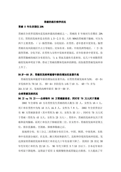
胃癌的流行病学状况胃癌 5 年生存期仅20%胃癌在全世界范围内是发病率最高的癌症之一。
胃癌的 5 年相对生存期在 20%左右,男性的发病率是女性的 1.5~2.5 倍。
大约 90%的胃癌属于腺癌,可分为两个主要类型:( 1)肠型胃癌,分化较好,在男性、老年患者中更常见。
肠型胃癌在高风险地区中占主导地位,比如东亚、东欧、中南美洲等地区。
( 2)弥漫型胃癌,分化不好,在男性与女性中发病率更接近,在年轻患者中更常见。
弥漫型胃腺癌的地区分布更均匀,与 A 型血及遗传关系密切。
近几十年来肠型胃癌的发病率明显下降,带动了胃癌的整体发病率的降低,而弥漫型胃癌发病率却上升了。
50岁~80 岁,胃癌的发病率随着年龄的增加而显著升高胃癌的发病率随着年龄的增加而显著升高,以男性胃癌发病率为例, 45~54 岁发病率为 70/10 万, 55~64 岁阶段为 145.7/10 万, 65~74 岁为264.3/10 万。
发病的高峰年龄在 50岁~80 岁。
全球胃癌发病状况90 万 vs 70 万——全球每年 90 万胃癌新患者,同时有 70 万人死于胃癌2002 年全球每 10 万名男性发生胃癌的的人数为 22 人,女性为 10.4 人;死亡率在男性中为每 10 万人 16.3 人,女性为 7.9 人。
2002 年全世界估计有 90 万胃癌新患者(其中男性为 60 万,女性为 33 万),同时有 70 万人死于胃癌(男性为 45 万人,女性为 25 万人)。
男性中,胃癌的发病率仅次于胃癌和前列腺癌,而死亡率仅次于肺癌居第二位。
在女性中,胃癌的发病率居第五位,排在乳腺癌、宫颈癌、肺癌和肠癌之后。
按地理分布,有三分之二的胃癌分布在日本、中国、韩国、中南美洲、东欧和中东的部分地区,在北美、澳大利亚和新西兰、北欧和印度的发病率较低。
发达国家的胃癌的发病率和死亡率在近几十年有显著下降了,美国在 20 世纪 50 年代年死亡率约为 22/10 万, 90 年代下降至 3.7/10 万以下。
Ki67、LRP在胃癌中的表达及其临床意义开题报告【摘要】胃癌是全球常见的肿瘤之一,其病因复杂,且具有高度异质性。
本文旨在探究Ki67、LRP在胃癌中的表达及其临床意义。
通过对近年来相关文献的综述和分析,发现Ki67、LRP在胃癌中的表达水平显著升高,并且与胃癌的恶性程度密切相关。
Ki67在胃癌的分子病理诊断中起到了重要的作用,可以预测患者的生存率及其预后。
同时,LRP是一个与多种肿瘤相关的膜糖蛋白,在胃癌中也起到了重要的作用,其表达水平与胃癌细胞的侵袭和转移密切相关。
本文的研究发现,Ki67、LRP在胃癌中的表达水平升高,可以作为预测胃癌预后和治疗效果的生物标志物。
为进一步明确胃癌分子机制,提高病例诊断及治疗水平提供了新的研究方向,有望为胃癌的早期诊断和预防提供依据。
【关键词】胃癌,Ki67,LRP,表达,生物标志物【Abstract】Gastric cancer is one of the common tumors worldwide, which is complex in etiology and highly heterogeneous. This paper aims to explore the expression and clinical significance of Ki67 and LRP in gastric cancer. Through a review and analysis of relevant literature in recent years, it was found that the expression level of Ki67 and LRP in gastric cancer was significantly increased, which was closely related to the malignancy of gastric cancer. Ki67 plays an important role in the molecular pathological diagnosis of gastric cancer, which can predict the survival rate and prognosis of patients. Meanwhile, LRP is a membrane glycoprotein associated with multiple tumors, which also plays an important role in gastric cancer. Its level of expression is closely related to the invasion and metastasis of gastric cancer cells.The research found that the increased expression of Ki67 and LRP in gastric cancer can be used as a biomarker to predict the prognosisand therapeutic effect of gastric cancer. To further clarify the molecular mechanism of gastric cancer, this provides a new research direction to improve the diagnosis and treatment level of cases, and is expected toprovide a basis for the early diagnosis and prevention of gastric cancer.【Keywords】Gastric cancer, Ki67, LRP, expression, biomarker。
木仓医学考研复试SCI长难句肝胆胃肠外科第一章-肝细胞恶性肿瘤Hepatocellular carcinoma(HCC)is the third leading cause of cancer--related deaths worldwide,and hepatitis B virus(HBV)infection is one of its leading causes.During the past several years,next-generation sequencing studies using bulk tumor samples have revealed considerable intratumor molecular and genetic heterogeneity in HCC.Such intratumor heterogeneity poses a great challenge for tumor characterization and therapeutic management of HCC patients.As is well known,tumor initiation and evolution are mediated by sequential genetic alterations in single cells.Single-cell sequencing has the potential to provide new insights into cancer bio-logical diversity that were difficult to resolve in genomic data from bulk tumor samples.在全球范围内导致癌症相关死亡的原因中,肝细胞癌(HCC)位列第三,而乙型肝炎病毒感染是其重要病因之一。
World Chin J Digestol 2004 February;12(2):262-265世界华人消化杂志 ISSN 1009-3079 CN 14-1260/R2004年版权归世界胃肠病学杂志社PO Box 2345 Beijing 100023, China Fax: +86-10-85381893Email:****************** • 胃癌 GASTRIC CANCER •胃癌前病变p21ras肝脏疾病及提高溃疡愈合质量的研究.广州中医药大学科学研究与发展总体规划重点及广东省中医药管理局资助课题, No. GH0016 300011项目负责人: 胡玲, 510405, 广东省广州市机场路12号大院, 广州中医药大学脾胃研究所. pqhl@21cn. com收稿日期: 2003-08-07 接受日期: 2003-09-24Relationship between expression of p21ras , c-erbB-2 and p53 and TCM syn-drome in gastric precancerous lesionsLing Hu, Shao-Xian LaoLing Hu, Shao-Xian Lao, Piwei Institute, Guangzhou University of Tradi-tional Chinese Medicine, Guangzhou 510405, Guangdong Province, China Supported by the Funds of Guangzhou University of TCM and Guangdong Provincial Administration of TCM, No.GH0016, 300011Correspondence to: Dr. Ling Hu, Piwei Institute, Guangzhou University of Traditional Chinese Medicine, Guangzhou, 510405, Guangdong Province,******************Received: 2003-08-07 Accepted: 2003-09-24AbstractAIM: To explore the relationship between p21ras, c-erbB-2, p53 protein expression and TCM syndrome in gastric precancerous lesions (GPL).METHODS: Forty cases with endosmotically and patho-logically confirmed GPL were studied, including 24 cases of moderate dysplasia of gastric mucosa, 9 cases of severe dysplasia, 7 cases of incomplete colonic intestinal metaplasia. By the differential diagnosis of TCM, pi-wei deficiency of Qi and Yin associated stagnation of Qi, stomach-heat, and blood stasis were 10,12 and 18 cases respectively.Expression of p21ras, c-erbB-2 and p53 proteins was de-tected by the LSAB immunohistochemical method.RESULTS: Overexpression of p21ras , c-erbB-2 and p53 pro-teins was found in GPL, and gradually increased with the progress of lesions; but among gastric mucosa of the mederate, severe dysphasia and incomplete colonic intesti-nal metaplasia, there are no differences in the expression of the genes (P >0.05). Among the differential associated symptoms and signs, the expression of p21ras and p53oncogene proteins was the blood stasis > stomach-heat and Qi stagnation (P <0.01), and the expression of c-erbB-2 oncogene protein was the blood stasis and stom-ach-heat >Qi stagnation(P <0.05).CONCLUSION: Overexpression of p21ras , c-erbB-2 and p53proteins is found in GPL. The expression is related with the differentially associated symptoms and signs.Hu L, Lao SX. Relationship between expression of p21ras , c-erbB-2 and p53 and TCM syndrome in gastric precancerous lesions. Shijie Huaren Xiaohua Zazhi 2004;12(2):262-265摘要目的: 观察胃癌癌前病变p21ras其中中度异型增生24例兼胃热者12例c-erbB-2c-erbB-2且其表达随着病变的进展而升高p21ras气滞者(P <0.01)胃热者大于兼气滞者(P <0.05).结论: p21ras其表达与不同中医兼证有关 GPL)指胃黏膜中重度异型增生和/或不完全性结肠化生p53等癌基因异常表达与GPL的发生密切相关[1-14]. 中药治疗不仅能使GPL患者的临床症状得到缓解还是中药对各相关癌基因蛋白的调控作用均存在着一定的差异. 中医认为GPL是以脾胃气阴两虚为本热毒为标虚实夹杂的综合征候群织中存在着一定的差异是否提示GPL组织中相关癌基因蛋白的表达可能具有一定的证候特异性?为此c-erbB-2采用四点法若为肠上皮化生胃黏膜尚需进一步采用HID-ABpH2. 5-PAS法作黏液组织化学染色以确认属于不完全性结肠化生者; 然后再结合临床的中医辨证肾功能明显异常中药新药临床研究指导原则第一辑. 1993:88拟定食后加重; 舌质胖嫩加次症2项即可诊断. 兼气滞症: 胃脘胀满不适; 胀痛连胁; 嗳气频作; 反酸或嘈杂; 脉弦. 兼血瘀症: 久痛不已; 痛有定处;刺痛; 舌质暗或有瘀斑或瘀点. 兼胃热症: 口干口苦; 大便干结; 舌苔黄. 具备以上兼症中任何两项即为兼有该兼症. 随机双盲法收集GPL病例共40例重度9例女8例; 中医辨证兼气滞症10例5-10 a者13例LSAB). 鼠抗人p21ras单克隆抗体鼠抗人p53蛋白(DO-7)及LSAB试剂盒均为丹麦DAKO产品; DAB 为福建迈新公司产品. 用PBS替代一抗作阴性对照p21ras及c-erbB-2位于胞质c-erbB-2表达主要定位于胞质(图1但少量也表达在胞质.p21ras但中重度异型增生及不完全性结肠化生之间未见显著性差异见表1.2.2 不同兼症中癌基因的表达 本组GPL病例中不同中医兼症p21ras从脾胃气阴两虚兼气滞各癌基因的表达逐渐增加表2 胃癌癌前病变不同兼症p21ras, c-erbB-2及p53表达情况(%)中医兼症 n p21ras c-erbB-2 p53气滞1020.020.010.0胃热12 33.3350.0a 16.67血瘀1872.2b 77.78a61.1daP <0.05, χ2 : 4.85 vs 气滞; bP <0.01, χ2: 8. 02 vs 气滞胃热.3 讨论p21ras是c-Ha-ras癌基因的蛋白产物而是在异型增生阶段即出现其在一定水平持续过度表达与肿瘤的启动有关[28-29]. c-erbB-2癌基因定位于染色体17q21胃黏膜异型增生中c-erbB-2癌基因蛋白的表达随着病变的进展呈上升趋势编码393个氨基酸残基的胞核磷蛋白而且可发生于癌变前的不同阶段c-erbB-2现代医学在针对GPL的治疗却未见明显的突破痞满胃脘痛痛或不痛八五已较一致地认为GPL是以脾胃气阴两虚为本或血瘀的本虚标实之证; 因虚挟邪中药治疗不仅能使GPL患者的临床症状缓解还是中药对之的调控作用均存在着一定的差异. GPL中这种基因表达的差异是否与不同兼症有关?各兼症之间的转化基于此本组研究病例中并随着病变的进展而表达逐渐增加上述各癌基因蛋白的表达均以兼血瘀者最高提示p21rasGPL的三种兼症中兼血瘀者最重GPL组织中p21rasGPL不同中医兼症与胃黏膜组织中上述相关癌基因的表达有一定的相关性; 推测GPL兼症间的转化一方面可为揭示GPL证型的客观化开辟了一条新的途径; 同时为深入研究其病理实质及相关药物的作用机制又提供了必要的前提条件.4 参考文献1费素娟, 陈玉林, 林志发, 陈淑敏, 刘广珍. 胃癌及癌前病变ras P21,P53的表达意义. 世界华人消化杂志 2001;9:465-4662陈小良, 钟国均, 唐录英, 李建忠, 邹仕恩. 胃癌的发生发展与P21表达的关系. 世界华人消化杂志 2000;8:5043陆和平, 郑银宝. 肠化生cerbB-2蛋白在胃肿瘤中的表达及意义. 世界华人消化杂志 2002;10:1050-105113伍银桥, 王孟薇, 吴本俨, 尤纬缔, 祝庆孚. 胃癌前病变演变过程中凋亡相关蛋白和PCNA的表达意义. 世界华人消化杂志 2003;11:1219-122214潘传敬, 刘宽宇. 胃癌增生凋亡与调节基因的表达. 世界华人消化杂志 2003;11:526-53015李庆明, 余谦, 闵存云. P53突变与VEGF在大鼠胃癌中的表达及中药胃康宁的防治作用. 世界华人消化杂志 2003;11:997-100016张旭晨, 高瑞丰, 李炳庆, 马连生, 梅立新, 吴玉珍, 刘凤芹, 廖振林. 胃细胞逆转丸治疗胃癌前期病变的临床与实验研究. 新消化病学杂志 1997;5:216-21817危北海, 张占海, 杨丽彩, 刘晋生, 赵敏, 赵荣莱, 李乾构. 胃安素治疗慢性萎缩性胃炎的临床与实验研究. 华人消化杂志 1998;6:114-117264 ISSN 1009-3079 CN 14-1260/R 世界华人消化杂志 2004年2月15日 第12卷 第2期18周学文. 中医药治疗胃癌癌前病变的临床研究现状与展望. 世界华人消化杂志 1999;7:277-27919胡玲, 劳绍贤, 周福生. 中医药治疗胃癌癌前病变的研究策略. 世界华人消化杂志 2000;8(特刊 8):320陆为民, 单兆伟, 沈洪, 吴静, 朱云华, 朱长乐. 胃舒胶囊治疗慢性萎缩性胃炎癌前病变的临床研究. 世界华人消化杂志 1999;7:822-82321许建青. 胃细胞逆转丸治疗胃癌癌前病变927例的逆转作用. 世界华人消化杂志 1999;7:63322胡玲, 劳绍贤. 胃癌癌前病变P21, p53蛋白表达及胃炎消的调节作用. 华人消化杂志 1998;6:873-87423胡玲, 劳绍贤. 胃癌癌前病变相关基因表达及胃炎消治疗机制的初步探讨. 中国中西医结合脾胃杂志 1999;7:91-9224黄志新, 常东, 劳绍贤, 胡玲, 唐纯志, 陈更新, 匡忠生. 胃炎消对胃癌癌前病变的作用机制探讨. 广州中医药大学学报 2003;20:102-10525李春婷, 王爱云, 单兆伟, 孙志广. 仁术健胃颗粒治疗胃癌前期病变88例临床观察. 中医杂志 2002;43:910-91126胡玲, 劳绍贤, 周福生, 唐纯志. 胃炎消对胃癌癌前病变相关靶基因蛋白表达的调控作用. 浙江中西医结合杂志 2001;11:334-33627唐纯志, 劳绍贤, 胡玲, 匡忠生. 胃炎消治疗胃癌前病变对细胞凋亡及相关基因表达的影响. 中国中西医结合脾胃杂志 2000;8:263-26428Hao Y, Zhang J, Lu Y, Yi C, Qian W, Cui J .The role of ras gene mutation in gastric cancer and precancerous lesions. J Tongji Med Univ 1998;18:141-14429惠延平, 黄高升, 王文亮, 王映梅, 朱晓慧, 马福成. 胃癌及其癌前病变中ras, C-erbB-2, p53癌基因产物的表达. 第四军医大学学报 2001;22:220-22330米建强, 杨石强, 沈铭昌. 胃癌及癌前病变组织中c-erbB-2癌基因产物的表达. 新消化病学杂志 1997;5:152-15331李晓清, 郝丽萍, 张小丽, 龚飞跃, 郭惠学, 伍尤泉. 胃黏膜异型增生C-erbB-2基因表达及其癌变率的研究. 胃肠病学和肝病学杂志 2002;11:269-27132Testino G, Gada D, De Iaco F, Cornaggia M. P53 and Ki-67expression in epithelial gastric dysplasia and in gastric cancer.Panminerva Med 2002;44:369-37133Feng CW, Wang LD, Jiao LH, Liu B, Zheng S, Xie XJ. Expression of p53, inducible nitric oxide synthase and vascular endothelial growth factor in gastric precancerous and cancerous lesions:correlation with clinical features. BMC Cancer 2002;2:834Ming SC. Cellular and molecular pathology of gastric carci-noma and precursor lesions: A critical review. Gastric Cancer 1998;1:31-5035Wang J, Chi DS, Kalin GB, Sosinski C, Miller LE, Burja I,Thomas E. Helicobacter pylori infection and oncogene expres-sions in gastric carcinoma and its precursor lesions. Dig Dis Sci 2002;47:107-11336李晓清, 郝丽萍, 张小丽, 龚飞跃, 郭惠学, 伍尤皇. 胃黏膜不典型增生P53基因表达及其癌变率的研究. 世界华人消化杂志 2002;10:1216-121737劳绍贤, 许鑫梅, 周福生, 余绍源, 贾建国, 郑裕璇, 黄志新, 郭遂成, 卞兆祥. 胃炎消治疗胃癌癌前期病变疗效分析. 中药新药与临床药理 1997;8:72-75胡玲, 等. 胃癌前病变p21ras, c-erbB-2, p53表达与中医证候的关系 265ISSN 1009-3079 CN 14-1260/R 2004年版权归世界胃肠病学杂志社• 消息•中国科技信息研究所信息分析研究中心期刊检索报告: 2002年度世界华人消化杂志 影响因子1.926, WJG影响因子2.579本刊讯 中国科技信息研究所信息分析研究中心期刊检索报告: 2002年度世界华人消化杂志总被引频次4 151他引总引比0.45被引半衰期2.99基金和资助论文比例0.27. 2002年度World Journal of Gastroenterology(WJG) 总被引频次1633他引总引比0.08被引半衰期2.43国际论文比0.14. (世界胃肠病学杂志社 2003-12-27)。
胃癌组织中CXCR3表达与微血管形成的关系及其临床意义李凯;刘杰【摘要】目的探讨胃癌组织中CXCR3的表达和微血管形成的关系并分析其临床意义.方法采用免疫组化法检测169例胃癌组织中CXCR3蛋白表达和肿瘤间质微血管密度(microvascular density,MVD),分析其相关性及其与胃癌临床病理参数的关系.结果 CXCR3表达在分化程度高(P =0.025)、浸润程度浅(P=0.005)、TNM 分期低(P =0.002)及无淋巴结转移(P=0.001)的胃癌患者中更高;MVD在胃癌分化程度低者中更高(P =0.001),与浸润深度、TNM分期及淋巴结转移无关(P>0.05).胃癌组织中CXCR3表达与MVD呈负相关(r=-0.151,P=0.049).Kaplan-Meier分析发现高表达CXCR3的胃癌患者(P=0.015)和低MVD患者(P =0.047)的生存时间延长.结论胃癌中CXCR3的表达与MVD呈负相关,且与胃癌的浸润、转移相关.【期刊名称】《临床与实验病理学杂志》【年(卷),期】2016(032)010【总页数】4页(P1097-1100)【关键词】胃肿瘤;CXCR3;CD34;微血管密度【作者】李凯;刘杰【作者单位】湖北医药学院附属东风总医院病理科,十堰442000;湖北医药学院附属人民医院重症医学科,十堰442000【正文语种】中文【中图分类】R735.2胃癌是全球范围内常见恶性肿瘤之一[1]。
目前胃癌最主要的治疗方式是外科手术治疗,但对于进展期胃癌患者即使应用了胃癌扩大根治术,5年生存率仍较低[2]。
近年来,手术切除术、新辅助化疗及个体化分子治疗等综合治疗取得很大进步,但术后转移率、复发率和病死率未有明显改善[3,4]。
趋化因子受体CXCR3在肿瘤发生、发展中起着复杂的作用,但CXCR3在不同肿瘤中表现出双向的调节作用,即抗/抑肿瘤作用或促肿瘤作用。
如在乳腺癌[5]、黑色素瘤[6]和结直肠癌[7]中CXCR3高表达者易发生淋巴结转移,预后较差;而在慢性淋巴细胞白血病[8]、前列腺癌[9]和肾透明细胞癌[10]中CXCR3高表达者预后较好。
阿帕替尼单药及合用替吉奥治疗晚期胃癌疗效观察纪华清;桂宏亮;束宽山;李兴保;王辉【摘要】目的探讨阿帕替尼单药及合用替吉奥治疗晚期胃癌的临床疗效.方法选取2015年7月至2017年12月铜陵市人民医院收治的40例晚期胃癌患者作为研究对象,采用随机数表法分为观察组和对照组各20例,对照组患者给予阿帕替尼单药治疗,观察组患者在对照组基础上合用替吉奥治疗,每3周为一个疗程.连续用药2个疗程后比较两组患者的临床治疗效果及毒副反应情况,采用卡氏行为状态(KPS)评分评价两组患者治疗后的生活质量水平.结果观察组患者治疗后的疾病控制率为90.0%,明显高于对照组的60.0%,差异有统计学意义(P<0.05),但其总缓解率仅为25.0%,略高于对照组的10.0%,差异无统计学意义(P>0.05);观察组和对照组患者治疗后手足综合征、白细胞减少发生率(30.0%vs 25.0%、75.00%vs 70.0%)比较,观察组较对照组略有提高,但差异均无统计学意义(P>0.05);观察组患者的生活质量改善及稳定者分别为25.0%和55.0%,高于对照组的15.0%和30.0%,但差异均无统计学意义(P>0.05),总有效率为80.0%,明显高于对照组的45.5%,差异具有统计学意义(P<0.05).结论与阿帕替尼单一用药比较,联合替吉奥治疗晚期胃癌短期疗效较为突出,其能在一定程度上改善患者的生活质量,用药期间药物毒副反应虽有所增加,但在患者可耐受范围内.【期刊名称】《海南医学》【年(卷),期】2019(030)005【总页数】3页(P619-621)【关键词】阿帕替尼;替吉奥;联合用药;晚期胃癌;不良反应;生活质量【作者】纪华清;桂宏亮;束宽山;李兴保;王辉【作者单位】铜陵市人民医院肿瘤内科,安徽铜陵 244000;铜陵市人民医院肿瘤内科,安徽铜陵 244000;铜陵市人民医院胃肠外科,安徽铜陵 244000;铜陵市人民医院肿瘤内科,安徽铜陵 244000;铜陵市人民医院肿瘤内科,安徽铜陵 244000【正文语种】中文【中图分类】R735.2近年来我国消化系统恶性肿瘤疾病的发病率呈现逐年上升趋势,其中最具代表性的是胃癌,该疾病起源于胃黏膜上皮的恶性肿瘤,不仅在我国各种恶性肿瘤中发病率居首位,而且其发病还具有明显的地域性差别,特别是在我国的西北与东部沿海地区胃癌发病率比南方地区明显要高[1]。
Department of Gastrointestinal Medical Oncology (R. Wadhwa, S. Song, Y. Yao, J. A. Ajani ),Department of Systems Biology (J.‑S. Lee ), Department ofEpidemiology (Q. Wei ), University of Texas MD Anderson Cancer Center, 1515 Holcombe Boulevard (FC10.3022), Houston, TX 77030, USA.Correspondence to: J. A. Ajanijajani@Gastric cancer—molecular and clinical dimensionsRoopma Wadhwa, Shumei Song, Ju-Seog Lee, Yixin Yao, Qingyi Wei and Jaffer A. AjaniAbstract | Gastric cancer imposes a considerable health burden around the globe despite its declining incidence. The disease is often diagnosed in advanced stages and is associated with a poor prognosis for patients. An in-depth understanding of the molecular underpinnings of gastric cancer has lagged behind many other cancers of similar incidence and morbidity, owing to our limited knowledge of germline susceptibility traits for risk and somatic drivers of progression (to identify novel therapeutic targets). A few germline (PLCE1) and somatic (ERBB2, ERBB3, PTEN, PI3K/AKT/mTOR, FGF , TP53, CDH1 and MET ) alterations are emerging and some are being pursued clinically. Novel somatic gene targets (ARID1A, FAT4, MLL and KMT2C ) have also been identified and are of interest. Variations in the therapeutic approaches dependent on geographical region are evident for localized gastric cancer—differences that are driven by preferences for the adjuvant strategies and the extent of surgery coupled with philosophical divides. However, greater uniformity in approach has been noted in the metastatic cancer setting, an incurable condition. Having realized only modest successes, momentum is building for carrying out more phase III comparative trials, with some using biomarker-based patient selection strategies. Overall, rapid progress in biotechnology is improving our molecular understanding and can help with new drug discovery. The future prospects are excellent for defining biomarker-based subsets of patients and application of specific therapeutics. However, many challenges remain to be tackled. Here, we review representative molecular and clinical dimensions of gastric cancer.Wadhwa, R. et al. Nat. Rev. Clin. Oncol. advance online publication 24 September 2013; doi:10.1038/nrclinonc.2013.170IntroductionGlobally, the incidence of gastric cancer ranks as the fourth most frequent cancer in men and fifth in women, but its death rate is comparable to lung cancer—the most common cancer worldwide.1 Histologically, gastric cancer can be mostly diffuse or intestinal type. Diffuse gastric cancer (DGC) represents poorly differentiated cancer cells interspersed with stromal cells. By contrast, intestinal gastric cancer (IGC) comprises cancer cells that form gland-like tubular structures with limited stromal components.2In 2008, approximately 989,600 (8% of all cancers) new cases of gastric cancer worldwide and 738,000 (10% of all cancer deaths) gastric-cancer-specific deaths were reported. Of these, 70% of deaths occurred in develop-ing nations—with China reporting approximately 40%—owing to the high incidence of gastric cancer in these regions,1 with Asia, Eastern Europe and South America being most affected. Despite these seemingly worrying numbers, the incidence of gastric cancer globally has been declining since World War II;3 several factors that have contributed to the decline include improved living standards 3–6 and early detection strategies, which have reduced the death rate in Japan.7 Helicobacter pylori infection as a risk factor is of importance for preven-tive strat e gies by developing strategic elimination ofthe bacteria in high-risk areas.8 Furthermore, major molecular biology advances have identified small subsets of gastric cancers defined by biomarkers. Of these, the over e xpression of HER2 protein and amplification of its gene ERBB2 have led to new treatment approaches in gastric cancer, such as trastuzumab. At present, addi-tional biomarkers (for example, c-MET, PI3K/Akt/mTOR and HER3) are currently being explored. Greater understanding of the molecular biology and immune biology of gastric cancer is still lacking, but likely to be highly rewarding.This Review focuses on the aspects of the emerging molecular biology of gastric cancer that might lead to improved therapeutics. We discuss the complexity of genomic alterations that drive the disease and show how these present potential opportunities for exploit-ation. The details of molecular signalling are largely not covered if their translational value is unclear. We discuss the role of tumour-initiating cells and the role of many oncogenes in conferring secondary resistance to therapy. In addition, we emphasize the progress—or lack thereof—made in the clinical arena by outlining recent phase III trial results. We aim to highlight advances in molecular and clinical research that reflect the current understanding of gastric cancer, rather than provide an encyclopaedic reference. Details of preventive strategies, impact of new classifications and nuances of surgery and radiotherapy are beyond the scope of this Review.Competing interestsThe authors declare no competing interests.REVIEWSHelicobacter pylori as a risk factorThe risk factors for gastric cancer include old age, smoking, alcohol consumption, above normal body weight, high salt and fat consumption, low vegetable and fruit consumption, low economic status, perni-cious anaemia, other chronic gastric diseases and H. pylori infection.6,9 Of these risk factors, H. pylori is the most fascinating (Figure 1) and best studied. The bacterium is associated with a range of effects, from chronic inflamma t ion to epigenetic modulations that can promote t r ansformation, invasion and metastasis of host cells.H. pylori infection increases the risk of gastric cancer threefold to sixfold,10 and is associated with distal gastric cancer and IGC.11 Chronic active gastritis is an integral part of bacteria-related gastric cancer:11,12 the organism attaches to gastric epithelial cells, leading to inflammation and an increase in the reactive oxygen and nitrogen species, which causes tissue damage.13,14 Furthermore, H. pylori also induces the expression of Toll-like receptors (TLRs), which leads to proliferation of the gastric epithelium.15 The oncogene CagA (now known as S100A8) is produced by carcinogenic H. pylori strains;12,16,17 the gene is included in the cag patho g enicity island and is translocated by the bacterium into the host epithelial cytosol.16,18 Once in the host cell, CagA is phosphory l ated by Src and c-Abl kinases19,20 to its active form to exert a range of effects,such as forming a complex with the SRC homology 2-domain (SH2)-containing tyrosine phosphatase SHP-2 (encoded by PTPN11), resulting in cytoskeletal reorganization that can induce malignant transformation.21 CagA activates the ERK/MAP kinase cascade, resulting in ETS domain-containing protein Elk-1 (Elk-1) phosphorylation and increased proto-oncogene c-Fos (c-Fos) transcription.22 In addition, CagA promotes invasion through activation of the hepatocyte growth factor receptor c-MET, which leads to an oncogenic response.23 CagA also induces E-cadherin-mediated impairment of cell adhesion junctions leading to cytoplasmic and nuclear accumu-lation of β-catenin.24 The protein CagA can bind Crk adaptor (p38) proteins (Crk-II, Crk-I and Crk-L)25 and kinase PAR1,26 eliciting loss of cell polarity, which is an oncogenic event. Furthermore, CagA stimulates the inflammatory pathway by inducing the secretion of cytokines IL-8, IL-1 and TNF-α27–29 through NF-κB in the epi t helium.30 Proinflammatory IL-1 gene cluster polymorphisms (in IL1B, encoding IL-1β, and in the loci for IL1RN and its receptor antagonist) increase the risk of noncardia gastric cancer (that is, cancer that affects the lower stomach).31,32 CagA also upregulates cyclo-oxygenase 2 (COX-2),27,33 which in turn induces the expression of oncogenic prostaglandins.34,35Aside from its CagA-mediated effects, H. pylori reduces the expression of the proapoptotic FAS-associated factor 1 (encoded by FAF1).36 The bacteri u m also mediates increases in another oncoprotein, a q uaporin-3 (encoded by AQP3),37alters DNA methyl-ation of E-cadherin (encoded by CDH1), which is an oncogenic event,38,39 and promotes methylation of the tumour suppressor TFF240 and RUNX3,41 as well as likely promoting the methylation of six other tumour suppres-sors (FLNC, HAND1, THBD, ARPC1B, HRASLS and LOX).42 Finally, H. pylori can lead to aberrant expres-sion of the nucleic acid editing enzyme activation-induced cytidine deaminase (AID), which can promote carcino g enesis through gene editing.43 These elucidated pathways and mechanisms show that H. pylori is carcino-genic, albeit in susceptible individuals. These improved and detailed understandings of H.pylori-induced host events will lead to plausible targets for drug development to reduce the risk of gastric cancer.and its several virulence factors, such as CagA, interact with gastric epithelial cells to induce chronic inflammation, mucosal damage and multiple alterations in gene expression and genetic and epigenetic changes, eventually leading to gastric carcinogenesis. Abbreviations: COX-2, cyclooxygenase-2; CpG island, areas of cytosine and guanine repeats; LPS, lipopolysaccharide; RNS, reactive nitrogen species; ROS, reactive oxygen species; VacA, vacuolating cytoxin A.The genomic profile of gastric cancerThe regions that have a high prevalence of H. pylori—for example, South America, Asia, Africa and Eastern Europe—have few cases of gastric cancer, which implies that genetic susceptibility might be a critical factor to consider.44 Indeed, several studies of single nucleotide polymorphisms (SNPs) and genome-wide association studies (GWAS) have revealed some plausible genes implicated in gastric cancer.Rare germline mutations (<0.001% of the general popu lation)in CDH1 have been implicated in familial cases of gastric cancer.45,46 Intuitively, SNPs can facilitate gastric cancer such that one adverse allele contributes weakly, but multiple adverse alleles can considerably increase the risk.47 Prior investigations of SNPs have focused on genes involved in mucosal protectionagainst H. pylori infection (for example, IL1B, IL1RN and TNFA), carcino g en metabolism (for example, CYP2E1 and GSTM1), deoxynucleotide synthesis (for example, MTHFR and TYMS), DNA repair (for example, MTHFR and XRCC1) and tumour suppressors (for example, TP53 and CDH1). However, these studies have had limited yield and none can be used clinically because SNP studies require customized approaches (with a priori assumptions that alterations in certain functional SNPs would increase susceptibility to gastric cancer). In spite of the correlations between certain SNPs and gastric cancer, prospective validation requiring large, population-based studies have been lacking as they are labour-intensive and resource-intensive.GWAS have been used to scan the whole genome to identify SNPs that are implicated in the disease (Table 1). Such studies have identified genes not previously known to be involved in gastric cancer—for example, PLCE1 (encoding pancreas-enriched phospholipase C) has an oncogenic role in skin and intestinal cancers,48 but is now thought to have a role in gastric cancer. A Japanese research group documented that SNPs in PSCA (encod-ing prostate stem-cell antigen) was associated with diffuse gastric cancer.49,50 The researchers genotyped 188 cases and 752 controls for 85,576 SNPs and then valid data were obtained from 749 cases and 750 controls for 2,753 SNPs. Although the intronic rs2976392 SNP in PSCA was identi f ied as the risk allele and the SNP was in disequilibrium with rs2294008 (located in exon 1), the function of PSCA in gastric tissue is unclear, although its possible tumour-suppressing function in the gastric epi-thelium has been suggested.51 A second study included 1,077 oesophageal cancer (which bears many similari-ties to gastric cardia cancer in terms of environmental risk factors) cases and 1,733 controls and identified 18 ‘hits’ that were validated in 2,766 cases of gastric cardia cancer; PLCE1 rs2274223 and C20orf54 rs1304295 SNPs were shown to be associated with gastric cancer risk.48 Another study of 2,240 gastric cancer cases and 3,302 controls identified the PLCE1 rs2274223 SNP as a risk allele for gastric cardia cancer.52 Indeed, PLCE1 SNPs have been shown to be associated with risk of gastric cancer53,54 and poor prognosis in Chinese patients,55 but not white patients.56 The fourth GWAS included 1,006 cases and 2,273 controls in China and validatedthe genomic data in 3,288 cases and 3,069 controls; SNPrs13361707 (located between PTGER4 and PRKAA1)and the ZBTB20 rs9841504 SNP were associated withincreased risk of gastric cancer.57Cancer stem cells and aberrant pathwaysGastric carcinogenesis is complex and not fully character-ized.58 Although IGC develops after systematic progres-sion from the preneoplastic stages, DGC is thought toarise de novo as the result of downregulation (mutation orpromoter methylation) of CDH1,59,60 thereby permitting tumorigenesis and progression. Nevertheless, accumu-lated genetic alterations—mutations, amplifications,insertions, deletions and recombinations—contribute togastric cancer,61,62 with more alterations accumulating asthe cancer progresses (Table 2). Furthermore, increasingevidence points to the existence of gastric cancer stemcells (CSCs) that initiate malignancy by self-renewal and differentiation. Although the origin of human gastricCSCs is still unclear, the mesenchymal stem cells in thebone marrow have been suggested to have a role.63,64Despite the efforts to genotype IGC and DGC (and iden-tify novel subtypes),65–76 clinically relevant and robustmolecular subtypes have yet to emerge; in one study, IGCand DGC were found not to be genotypically distinct, butdid respond differently on the basis of their histopatho-logical characteristics.73 However, these are preliminaryresults. Further detailed analyses of the implicated cellsand pathways might point to viable targets that canbe exploited in gastric cancer; indeed, some of theses t rategies have been studied in other cancer types.CSCs are known to undergo epithelial– m esenchymaltransition (EMT), activate oncogenic pathways77 andactivate embryogenic signalling pathways,78 whichare essential for the self-renewal and maintenance oftumours. EMT leads to the CSC-like phenotypes.79 CSCsare generally resistant to stress caused by reactive oxygenspecies, which in turn makes them relatively resistant toradiotherapy and chemotherapy. Additionally, CSCs haveself-renewal capacities. These cells depend on the Wnt,Notch and Hedgehog pathways for maintenance.80 Fourpleiotropic transcriptional factors (Snail, Slug, Twist andREVIEWSZeb1/2) orchestrate the EMT and related processes,81 whereas c-MET and TGF-β signalling can be critical for transformation to EMT.82 c-MET activation can induce the reprogramming transcription factors known to support embryonic stem cells and induce differentiated cells to form the pluripotent stem (iPS) cells.83TGF-β can be pro-oncogenic by inducing matrix deposition, immunosuppression and EMT.84–86 TGF-β signalling drives EMT and CSC self-renewal by target-ing microRNAs (miRNAs),81 upregulating Snail family members and repressing E-cadherin.87 Downstream of c-MET and the TGF-β receptor, PI3K/Akt/mTOR signal l ing conveys prosurvival messages that lead to CSC expansion and maintenance.88 Consequently, there has been interest in targeting mTOR with metformin to inhibit CSCs in breast cancer (and possibly other cancers).89 Ras, along with Hedgehog, helps to maintain CSCs:90,91 Ras alters cellular metabolism to provide an advantage to CSCs to gather biomass to support cellular division. Ras also facilitates tolerance to DNA damage during cell cycling by inhibiting the activity of tumour suppressors. Hedgehog components facilitate rapid cell division in CSCs, as is their function in develop i ng embryos. Furthermore, TGF-β is overexpressed in DGC92 and stimulates collagen synthesis and subsequent fibro-sis. TGF-β can be antiapoptotic through trans a ctivation of EGFR.93 The bone morphogenetic proteins (BMPs), which are members of the TGF-β superfamily, activate PI3K/Akt.94 Given the considerable interest in the inhibi-tion of angiogenesis, VEGF and VEGFR overexpression, which is common in IGC through activation of NF-κB by H. pylori, is a potential target.95,96G astric CSCs express CD133, CD44, aldehyde dehydro g enase 1 (ALDH1) and ATP-binding cassette sub-family G member 2 (ABCG2); CD44 and ALDH1 are associated with therapy resistance and might be valid therapeutic targets.97 CD44 imparts stem-cell-like proper-ties by promoting synthesis of intracellular reduced gluta-thione.98 Among the other plausible targets in gastric cancer, HER family members have been of interest.99 The oncogenic properties of HER are conferred through the RAS/MEK/MAPK and PI3K/Akt/mTOR pathways.100,101 Overexpression of HER2 caused by ERBB2 amplifica-tion is more prevalent in IGC than DGC.102,103 Thus, confirm i ng the HER2 status in patients with IGC is more important than in patients with DGC; in DGC HER over-expression is rare and might not be driving the cancer. HER2 interacts with EGFR (also known as HER1), HER3104 and insulin-like growth factor 1 (IGF-1R),105 which are amplified, overexpressed106,107 or acquire acti-vating mutations in cancer.108–110ERBB3 mutations have been reported in 10% of gastric cancers.111 Constitutive activation of c-MET can trigger prolifer-ation and antiapoptotic signals.112 Amplification or over-expression of c-MET, rather than mutation of its gene, can activate receptor tyrosine kinase function.113,114 c-MET overexpression or amplification is common in DGC as well as IGC.115–117 Amplified c-MET crosstalk can acti-vate EGFR, HER2 and HER3 to establish a s i gnalling network leading to constitutive PI3K/Akt signal-ling.118–120 This information enables the understanding of the mechanisms of primary and second a ry resistance when the c-MET pathway is inhibited and can lead to ration a l therapeutic strategies in the future, including dual pathway inhibition. Gastric cancers with c-MET overexpression coexpress EGFR, HER3 or both,121 which is clinically relevant for strategies looking to inhibit both targets concurrently.117,120,121 Indeed, the PI3K/Akt/mTOR pathway is frequently altered as a result of amplification or overexpression (PIK3CA, AKT1), activating muta-tions (PIK3CA)59,122 or loss of PTEN.123 Overexpression of phosphory l ated mTOR can occur in DGC;59 HER3 and FGFR amplification in DGC is another mechanism for PI3K/Akt activation.124,125Although HER, c-MET, PI3K/Akt/mTOR, VEGFR and VEGF have been targeted in gastric and other cancer types, TGF-β and E-cadherin are not targetable because loss of function of a gene is difficult to restore in patients (although it can be achieved in cell lines). Several novel targets worth mentioning include chromatin modifiers, such as ARID1A, MLL3, MLL and FAT4 (cell adhesion), which are of increasing interest and importance.75,126 Frequent mutations in the genes mentioned above have been observed in 47% of gastric cancers and can poten-tially be therapeutic targets. As we uncover the complex-ity of RNA regulation of intracellular processes, more targets are likely to emerge, for example, targeting the mutations in the spliceosome machinery.MicroRNAsmiRNAs have a role in tumorigenesis, tumour pro-gression and metastasis, and will—in all likelihood—c o ntinue to gain importance in diagnostics, monitoring during therapy and as therapeutic targets (Figure 2).127 Oncogenic microRNAs (oncomiRs) are overexpressed and inhibit tumour suppressors, leading to cell prolifer-ation, invasion and reduced apoptosis. For example, over-expression of miR-296-5p in gastric cancer cells increased cell prolifer a tion and inhibition of apoptosis via the repres-sion of tumour suppressor CDX1.128 Furthermore, over-expression of miR-301a directly targets tumour suppressor RUNX3,129 whereas miR-17-5p/20a targets tumour protein p53-inducible nuclear protein 1 (TP53INP1).130 miR-18a levels were correlated with those of survivin (also known as baculoviral inhibitor of apoptosis repeat-containing 5; BIRC5), Bcl-xL (B-cell lymphoma-extra large) and c-Myc, all three being downstream transcriptional targets of STAT3. STAT3-induced transcription can be negatively regulated by PIAS3. Thus, miR-18a acts as an oncomiR by negatively regulating PIAS3.131 miRNA-372 is onco-genic as it targets TNFAIP1 and modulates NF-κB signal-ling in gastric cancer cells.132IRX1, a newly identified tumour suppressor gene, is inacti v ated by miR-544.133 miR-10b is highly expressed in IGC and is associated with the depth of invasion, lymph-node involvement and metastatic progression.134Tumour suppressor microRNAs (tsmiRs), by contrast,are downregulated miRNAs that facilitate the activity of target oncogenes. miR-195 and miR-378 have been shown to be downregulated in gastric cancer (in vivo and in vitro); their target oncogenes are CDK6 and VEGF.135 The oncogene target of miR-133b is FGFR1, which is often amplified in DGC.136 The target of miR-29c is Mcl‑1; acti-vation of this miRNA by celecoxib represses Mcl‑1 and promotes apoptosis of gastric cancer cells.137 miR-34a is down r egulated in gastric cancer cells; its oncogene target is BIRC5.138 miR-145 suppresses protein C-ets-1 (ETS1), also known as p54, by binding to its three-prime untranslated region (3'-UTR) and reducing the onco-genic processes.139 Let-7i is frequently down r egulated in most tumours and is prognostic of lymphatic invasion, nodal metastasis and poor pathological tumour response in patients with gastric cancer.140 Furthermore, ZFX has a role the maintenance of CSCs and is the target of miR-144 in gastric cancer.141 miR-101 targets the 3'-UTR of PTGS2 (also known as COX2) mRNA and its down-regulation in gastric cancer correlates with over e xpression of COX-2 and cell prolifer a tion.142 Finally, miRNA-146a inhibits NF-κB by targeting CARD10 and COPS8 in gastric cancer.143All miRNAs are stable in serum, plasma, gastric juice and other bodily fluids, which makes them ideal bio-markers for the disease.144 Indeed, miR-21 and miR-106a were shown to be overexpressed in the gastric juice of patients compared with normal controls.145 Additionally, levels of miR-421 in the gastric juice of patients was higher than in controls (P <0.001) and it resulted in the earlier diagnosis of gastric cancer than by serum carcino-embryonic antigen.146 Similarly, plasma miR-106b, miR-20a and miR-221 levels were elevated in patientscompared with healthy controls (P <0.05).147 In anotherstudy, plasma levels of miRNA-199a-3p were associ-ated with tumour invasion, lymph-node involvementandmetasta ses.148 Circulating miR-17-5p and miR-20a(miR-17-5p/20a) have also been detected in patientswith gastric cancer and both of these miRNAs corre-lated with overall survival and relapse-free survival.149Finally, the miR-200c expression level in the blood inpatients with gastric cancer were significantly higherthan in the blood of normal controls (P =0.018).150 Clearly,research on several miRNAs holds promise and needs tobe fully developed.Mechanisms of resistanceTreatment resistance is inevitable; the major reasonsfor treatment failure in patients are the occurrence ofprimary and secondary resistance. For example, HER2is the only validated biomarker in gastric cancer, andHER2-overexpressing tumours regularly become resist-ant to targeted agents, such as trastuzumab. Accordingly, understanding the underlying mechanisms of resistanceis important. In HER2-overexpressing tumours, thepredominant mechanism of resistance is compensa-tory signalling by other cell surface receptors.151 HER2-overexpressing cells—when inhibited—reprogrammeother oncogenes, including IGF1R, MET, GDF15 (encod-ing growth differentiation factor 15) and other membersof the ERBB family.152 The IGF1R-mediated resistanceto HER2 targeting involves the PI3K pathway, leading toenhanced degradation of p27Kip1,105,153,154 whereas activatedFigure 2 | microRNA targets and functions in gastric cancer. Some oncomiRs are overexpressed in tumours and inhibit tumour suppressors, leading to cell proliferation, invasion and reduced apoptosis. By contrast, tsmiRs normally target oncogenes that are downregulated in tumours and facilitate the activity of their target oncogenes. Abbreviations: EMT, epithelial–mesenchymal transition; miR, microRNA; oncomiRs, oncogenic microRNAs; tsmiRs, tumour-suppressor microRNAs.REVIEWSc-MET mediates resistance in gastric cancer cells155,156 by restoring shared downstream signalling in the MAPK and Akt pathways.155 Increased levels of EGFR and HER3 ligands can overcome HER2 inhibition.88,124,152 Constitutively activated p95HER2, a truncated HER2 receptor, is the most intriguing mechanism of resistance in response to the blockade of the extracellular domain of HER2.157 This method of conferring resistance to trastu-zumab is mediated by the regulatory DNA that engages a specific protease gene to truncate the receptor. Thus, resist-ance is not caused by acquiring a mutation in the ERBB2 gene itself, rather another gene modifies the receptor and renders the truncated receptor constitutively active. Membrane mucins, such as mucin-4, interact with HER2 in HER2-overexpressing breast cancer cells, resulting in epitope masking that blocks trastuzumab binding.158 Other factors that confer resistance to trastuzumab include focal adhesion kinase (FAK), Src and altera-tions in cell-cycle regulators.159 Constitutive activation of PI3K caused by activating PIK3CA mutations,61 reduced PTEN expression160 or deregulated signalling can induce resistance to HER2 inhibition.61,161,162 Finally, STAT3 activation can mediate resistance as a result of produc-tion of IL-6.163 These data suggest that cancer cells have many redundant mechanisms to overcome resistance to therapy. Accordingly, we have considerable work ahead of us, including exploring antibody–drug c o njugates and immune modulation therapies.Clinical considerationsMuch clinical development is underway in the setting of gastric cancer, which includes many of the targets dis-cussed herein. Many of the recently completed phase III trials should have an impact on patient care and future research directions (Figure 3). Notably, any discussion regarding clinical developments and the future of gastric cancer care must be made with consideration of the regional differ e nces, both in terms of epidemiology and standard practice. The epidemiology and predominant location of primary gastric cancer varies geographically164 because of variations in genetic susceptibility, predomi-nance of certain histological phenotypes (for example,FGFR2 amplification (9%), VEGF/VEGFR overexpression (36–40%), EGFR amplification and overexpression (27–44%), HER-2 amplification and overexpression (7–34%), c-MET amplification (10–15%), kRAS mutation (2–20%), Raf mutation (0–3%),PI3K mutation (4–36%), phospho-Akt expression (29–86%), phospho-mTOR expression (60–88%), PTCH1 overexpression (16%), SMO overexpression (12%) and HER3 mutations (10%, not shown). *No clinical trials of these agents have yet been reported in gastric cancer. ‡No known numbers or percentages for these genes and pathways. Abbreviations: EGFR, epidermal growth factor receptor; FGFR, fibroblast growth factor receptor; GLI, glioma-associated oncogene family zinc finger 1; HDAC, histone deacetylase; HER, human epidermal growth factor receptor; HGF, hepatocyte growth factor;Hh, Hedgehog; IGFR, insulin-like growth factor receptor; MMP, matrix metalloproteinase; mTOR, mammalian targetof rapamycin; PDGFR, platelet-derived growth factor receptor; Ptch-1, protein patched homolog 1; Smo, smoothened; VEGF, vascular endothelial growth factor; VEGFR, vascular endothelial growth factor receptor.。