
How CHT ? Ceramic Hydroxyapatite Works
Paul Ng, Jie He, and Andrew Cohen, Bio-Rad Laboratories, Inc., Hercules, CA 94547 USA
CHT ceramic hydroxyapatite is a versatile chromatography support used for separation of biological molecules as diverse as polyclonal and monoclonal antibodies, antibody fragments, enzymes, nucleic acids, and membrane proteins. The interactions that occur between CHT and the molecules it binds are complex; this article aims to clarify the most significant features of these interactions.
CHT, Ca 10(PO 4)6(OH)2, is a mixed-mode support with
functional groups consisting of pairs of positively charged crystal calcium ions (C-sites) and clusters of six negatively charged oxygen atoms associated with triplets of crystal phosphates (P-sites) (Figure 1). The C-sites, P-sites, and hydroxyl groups are distributed in a fixed pattern on the CHT crystal structure, as presented in classic studies by Kawasaki (1978a, 1978b) and Kawasaki et al. (1985).
In theory, CHT can retain solutes by weak anion exchange or calcium metal affinity with C-sites, by cation exchange with P-sites, and by hydrogen bonding with hydroxyl groups (Gorbunoff 1984a, 1984b, Gorbunoff and Timasheff 1984). Experimental evidence, however, suggests that most proteins bind CHT by a combination of metal affinity and phosphoryl cation exchange (Figure 2), with little contribution by hydrogen bonding. The affinity interaction of protein carboxyl clusters with CHT C-sites represents a classic metal chelating mechanism in which protein carboxyl groups approximate the carboxyl configuration of chelating agents such as EDTA.
chromatography
tech note 5709
Fig. 2. Interaction of proteins with CHT. A , interaction of carboxyl groups and CHT. Note the metal affinity interaction between CHT C-sites and carboxyl groups, and the repulsion of carboxyl groups from P-sites. B , interaction of amino groups with CHT. Note the phosphoryl cation exchange interaction between CHT P-sites and amino groups, and the repulsion of amino groups from C-sites.
A
Fig. 1. Crystal structure of CHT ceramic hydroxyapatite, Ca 10(PO
4)6(OH)2. Each molecule consists of five positively charged calcium pairs (C-sites); two phosphate triplets (P-sites), each with six negatively charged oxygen atoms; and two hydroxyl residues.
Stronger than electrostatic interactions, these metal affinity interactions withstand the presence of even saturated sodium chloride since chloride ions do not form a complex with
Ca2+. This indicates that any anion exchange between CHT C-sites and protein carboxyl groups does not contribute significantly to protein binding (Gorbunoff 1984a, 1984b, Gorbunoff and Timasheff 1984, Gagnon 1998). Further evidence of this comes from the demonstration that acidic proteins are retained more weakly with ascending pH (Ogawa and Hiraide 1995). In addition, binding experiments with urea, which disrupts hydrogen bonds (Tanford 1968, Timasheff and Fasman 1969), indicate that the contribution of hydrogen bonding is likewise negligible.
The contributions of metal affinity and phosphoryl cation exchange are distinctive for every protein and can be investigated by eluting with various neutral salt and phosphate concentrations. Elution of proteins bound by metal affinity interactions requires phosphate, which outcompetes CHT-protein metal interactions with its own strong affinity for calcium. In contrast, elution of proteins bound by cation exchange requires either neutral salts, such as sodium chloride, or buffering salts, such as phosphates. Therefore, elution of proteins by phosphate-mediated buffer can result in distinct separation, depending upon the mechanism by which the protein is bound.
Some proteins, such as lysozyme, bind CHT exclusively by cation exchange between their amino groups and CHT P-sites (Figure 2B), while others, such as bovine serum albumin (BSA), bind almost exclusively by metal affinity interactions (Figure
2A). Rich in carboxyl groups and with a strong affinity for CHT C-sites, BSA elutes in a linear phosphate gradient at pH 6.5
in 110 mM sodium phosphate (Gagnon 1998). However, when
the same linear phosphate gradient is run in the presence of 1.0 M sodium chloride, the phosphate concentration required for elution of BSA drops only to 100 mM (Gagnon et al. 2005a), indicating that ion exchange is a minor contributor to the binding energy while calcium affinity dominates retention.
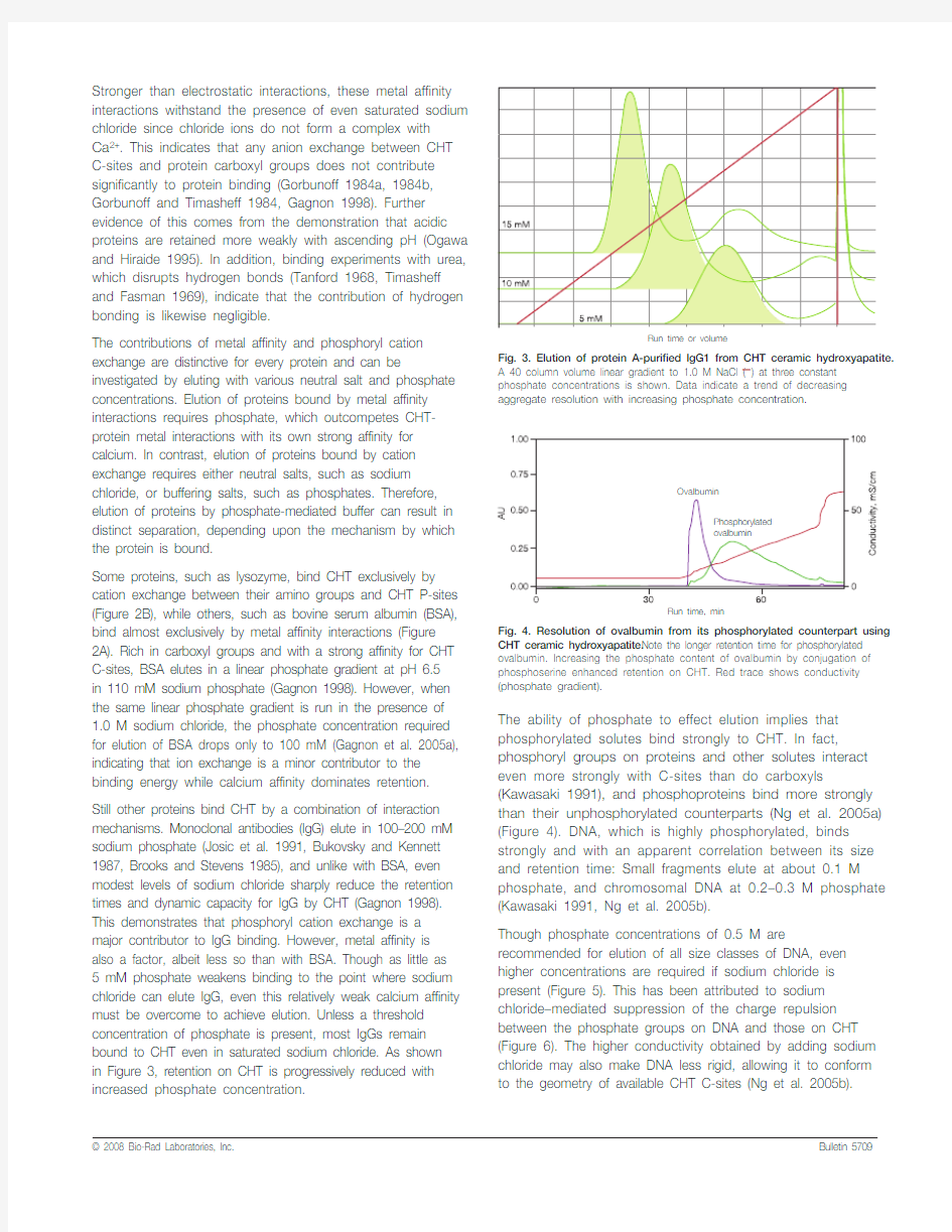
Still other proteins bind CHT by a combination of interaction mechanisms. Monoclonal antibodies (IgG) elute in 100–200 mM sodium phosphate (Josic et al. 1991, Bukovsky and Kennett 1987, Brooks and Stevens 1985), and unlike with BSA, even modest levels of sodium chloride sharply reduce the retention times and dynamic capacity for IgG by CHT (Gagnon 1998). This demonstrates that phosphoryl cation exchange is a major contributor to IgG binding. However, metal affinity is also a factor, albeit less so than with BSA. Though as little as 5 mM phosphate weakens binding to the point where sodium chloride can elute IgG, even this relatively weak calcium affinity must be overcome to achieve elution. Unless a threshold concentration of phosphate is present, most IgGs remain bound to CHT even in saturated sodium chloride. As shown
in Figure 3, retention on CHT is progressively reduced with increased phosphate concentration.The ability of phosphate to effect elution implies that phosphorylated solutes bind strongly to CHT. In fact, phosphoryl groups on proteins and other solutes interact even more strongly with C-sites than do carboxyls (Kawasaki 1991), and phosphoproteins bind more strongly than their unphosphorylated counterparts (Ng et al. 2005a) (Figure 4). DNA, which is highly phosphorylated, binds strongly and with an apparent correlation between its size and retention time: Small fragments elute at about 0.1 M phosphate, and chromosomal DNA at 0.2–0.3 M phosphate (Kawasaki 1991, Ng et al. 2005b).
Though phosphate concentrations of 0.5 M are recommended for elution of all size classes of DNA, even higher concentrations are required if sodium chloride is present (Figure 5). This has been attributed to sodium chloride–mediated suppression of the charge repulsion between the phosphate groups on DNA and those on CHT (Figure 6). The higher conductivity obtained by adding sodium chloride may also make DNA less rigid, allowing it to conform to the geometry of available CHT C-sites (Ng et al. 2005b).
CHT ceramic hydroxyapatite. Note the longer retention time for phosphorylated ovalbumin. Increasing the phosphate content of ovalbumin by conjugation of phosphoserine enhanced retention on CHT. Red trace shows conductivity (phosphate gradient).
Fig. 3. Elution of protein A-purified lgG1 from CHT ceramic hydroxyapatite.
A 40 column volume linear gradient to 1.0 M NaCl (—) at three constant phosphate concentrations is shown. Data indicate a trend of decreasing aggregate resolution with increasing phosphate concentration.
Run time or volume
Fig. 6. Interaction of DNA with CHT ceramic hydroxyapatite. Note the electrostatic repulsion between CHT P-sites and the phosphate backbone of DNA, and the interaction of that backbone with the C-sites and hydroxyl groups. (Symbols same as in Figure 2.)
Fig. 5. Elution of sheared salmon sperm DNA from CHT ceramic hydroxyapatite. Data compare the behavior of DNA on CHT as a function of sodium chloride concentration. Increased NaCl concentration improved DNA retention.
Run time or volume
Endotoxins, which are also phosphorylated, may require up to 1.0 M phosphate for complete removal; subpopulations can elute over the entire range of 0–0.5 M potassium
phosphate, but reductions in retention are apparent when phosphate gradient elution is carried out at high sodium chloride concentrations, indicating that binding involves a cation exchange component (Gagnon et al. 2005b).The mechanism by which proteins interact with CHT is multifaceted. Its unique resolution property makes it a powerful tool for process developers. The ability of CHT to purify a variety of proteins — including monoclonal antibodies, which are leading licensed products or therapeutic candidates in many drug companies —
strengthens its versatility. It is anticipated that CHT will enjoy increasing attention in the years to come.For more information, request bulletin 2156 or visit https://www.doczj.com/doc/8914902865.html,/process/.
References
Brooks T and Stevens A, Preparative HPLC purification of IgG and IgM monoclonal antibodies, Am Lab 17, 54–64 (1985)
Bukovsky J and Kennett R, Simple and rapid purification of monoclonal antibodies from cell culture supernatants and ascites fluids by hydroxylapatite chromatography on analytical and preparative scales, Hybridoma 6, 219–228 (1987)
Gagnon P, An enigma unmasked: how hydroxyapatite works and how to make it work for you, Validated Biosystems (https://www.doczj.com/doc/8914902865.html,) (1998)
Gagnon P et al., Platform purification of IgG with CHT ceramic hydroxyapatite, BioProcess International Asia-Pacific Conference, 7–9 November 2005, Singapore (IBC USA Conferences, Inc., https://www.doczj.com/doc/8914902865.html,) (2005a)Gagnon P et al., Retention behavior of endotoxin, DNA and Protein A on CHT ceramic hydroxyapatite and CFT ceramic fluorapatite, BioProcess International World Conference, 19–21 September 2005, Boston, MA (IBC USA Conferences, Inc., https://www.doczj.com/doc/8914902865.html,) (2005b)
Gorbunoff M, The interaction of proteins with hydroxyapatite. I. Role of protein charge and structure, Anal Biochem 136, 425–432 (1984a)
Gorbunoff M, The interaction of proteins with hydroxyapatite. II. Role of acidic and basic groups, Anal Biochem 136, 433–439 (1984b)
Gorbunoff M and Timasheff S, The interaction of proteins with hydroxyapatite. III. Mechanism, Anal Biochem 136, 440–445 (1984)
Josic D et al., Purification of monoclonal antibodies by hydroxylapatite HPLC and size exclusion HPLC, Biol Chem Hoppe Seyler 372, 149–156 (1991)Kawasaki T, Theory of chromatography of rigid molecules on hydroxyapatite columns with small loads. IV. Estimation of the adsorption energy of nucleoside polyphosphates, J Chromatogr 151, 95–112 (1978a)
Kawasaki T, Theory of chromatography on hydroxyapatite columns with small loads, J Chromatogr 157, 7–42 (1978b)
Kawasaki T, Hydroxyapatite as a liquid chromatographic packing, J Chro-matogr A 544, 147–184 (1991)
Kawasaki T et al., Hydroxyapatite high-performance liquid chromatography: column performance for proteins, Eur J Biochem 152, 361–371 (1985)Ng P et al., Application of ceramic hydroxyapatite for proteomic analysis, ISPPP 2005, 4–6 November 2005, St Petersburg, FL (Barr Enterprises, https://www.doczj.com/doc/8914902865.html,) (2005a)
Ng P et al., Differential retention of DNA on ceramic hydroxyapatite and ceram-ic fluoroapatite, Prep 2005, 2–6 May 2005, Philadelphia, PA (Barr Enterprises, https://www.doczj.com/doc/8914902865.html,) (2005b)
Ogawa T and Hiraide T, Effect of pH on gradient elution of proteins on two types of Macro-Prep ? ceramic hydroxyapatite, Prep '95: Industrial Separation Science Conference, 13 February 1995, East Rutherford, NJ (1995)Tanford C, Protein denaturation, Adv Protein Chem 23, 121–282 (1968)Timasheff S and Fasman G, Structure and stability of biological macromolecules, Marcel Dekker, New York, NY (1969)
Information in this tech note was current as of the date of writing (2006) and not necessarily the date this version (rev A, 2008) was published.
Ordering Information
Catalog # Description*
CHT Ceramic Hydroxyapatite, Type I
158-2000 20 μm particle size, 10 g
157-0020 20 μm particle size, 100 g
157-0021 20 μm particle size, 1 kg
157-0025 20 μm particle size, 5 kg
158-4000 40 μm particle size, 10 g
157-0040 40 μm particle size, 100 g
157-0041 40 μm particle size, 1 kg
157-0045 40 μm particle size, 5 kg
158-8000 80 μm particle size, 10 g
157-0080 80 μm particle size, 100 g
157-0081 80 μm particle size, 1 kg
157-0085 80 μm particle size, 5 kg
732-4322 Bio-Scale? Mini CHT-I cartridge, 40 μm, 1 x 5 ml 732-4324 Bio-Scale Mini CHT-I cartridge, 40 μm, 5 x 5 ml * Larger quantities available on request.Catalog # Description*
CHT Ceramic Hydroxyapatite, Type II
158-2200 20 μm particle size, 10 g
157-2000 20 μm particle size, 100 g
157-2100 20 μm particle size, 1 kg
157-2500 20 μm particle size, 5 kg
158-4200 40 μm particle size, 10 g
157-4000 40 μm particle size, 100 g
157-4100 40 μm particle size, 1 kg
157-4500 40 μm particle size, 5 kg
158-8200 80 μm particle size, 10 g
157-8000 80 μm particle size, 100 g
157-8100 80 μm particle size, 1 kg
157-8500 80 μm particle size, 5 kg
732-4332 Bio-Scale Mini CHT-II cartridge, 40 μm, 1 x 5 ml 732-4334 Bio-Scale Mini CHT-II cartridge, 40 μm, 5 x 5 ml
Life Science Group Bio-Rad
Laboratories, Inc.
Web site https://www.doczj.com/doc/8914902865.html, USA 800 4BIORAD Australia 61 02 9914 2800 Austria 01 877 89 01 Belgium 09 385 55 11 Brazil 55 21 3237 9400 Canada 905 364 3435 China 86 21 6426 0808 Czech Republic 420 241 430 532 Denmark 44 52 10 00 Finland 09 804 22 00 France 01 47 95 69 65 Germany 089 318 84 0 Greece 30 210 777 4396 Hong Kong 852 2789 3300 Hungary 36 1 455 8800 India 91 124 4029300 Israel 03 963 6050
Italy 39 02 216091 Japan 03 6361 7000 Korea 82 2 3473 4460 Mexico 52 555 488 7670 The Netherlands 0318 540666 New Zealand 0508 805 500 Norway 23 38 41 30 Poland 48 22 331 99 99 Portugal 351 21 472 7700 Russia 7 495 721 14 04 Singapore 65 6415 3188 South Africa 27 861 246 723