Cryptochrome1Overexpression Correlates with Tumor Progression and Poor Prognosis in Patients with Colorectal Cancer
Hongyan Yu1.,Xiangqi Meng1.,Jiangxue Wu1,Changchuan Pan3,Xiaofang Ying1¤,Yi Zhou1,Ranyi Liu1, Wenlin Huang1,2*
1State Key Laboratory of Oncology in South China,Cancer Center,Sun Yat-sen University,Guangzhou,People’s Republic of China,2CAS Key Laboratory of Pathogenic Microbiology and Immunology,Institute of Microbiology,Chinese Academy of Science,Beijing,People’s Republic of China,3Medical Oncology,Sichuan Cancer Hospital and Institute,Second People9s Hospital of Sichuan Province,Cheng Du,People’s Republic of China
Abstract
Background:Clock genes drive about5–15%of genome-wide mRNA expression,and disruption of the circadian clock may deregulate the cell’s normal biological functions.Cryptochrome1is a key regulator of the circadian feedback loop and plays an important role in organisms.The present study was conducted to investigate the expression of Cry1and its prognostic significance in colorectal cancer(CRC).In addition,the function of Cry1in human CRC was investigated in cell culture models.
Methods:Real-time quantitative PCR,Western blot analysis and immunohistochemistry were used to explore Cry1 expression in CRC cell lines and primary CRC clinical specimens.MTT and colony formation assays were used to determine effects on cellular proliferation ability.The animal model was used to explore the Cry1impact on the tumor cellular proliferation ability in vivo.Transwell assays were performed to detect the migration ability of the cell lines.Statistical analyzes were applied to evaluate the diagnostic value and the associations of Cry1expression with clinical parameters.
Results:Cry1expression was up regulated in the majority of the CRC cell lines and168primary CRC clinical specimens at the protein level.Clinical pathological analysis showed that Cry1expression was significantly correlated with lymph node metastasis(p=0.004)and the TNM stage(p=0.003).High Cry1expression was associated with poor overall survival in CRC patients(p=0.010).Experimentally,we found that up-regulation of Cry1promoted the proliferation and migration of HCT116cells,while down-regulation of Cry1inhibited the colony formation and migration of SW480cells.
Conclusions:These results suggest that Cry1likely plays important roles in CRC development and progression andCry1may be a prognostic biomarker and a promising therapeutic target for CRC.
Citation:Yu H,Meng X,Wu J,Pan C,Ying X,et al.(2013)Cryptochrome1Overexpression Correlates with Tumor Progression and Poor Prognosis in Patients with Colorectal Cancer.PLoS ONE8(4):e61679.doi:10.1371/journal.pone.0061679
Editor:Xin-Yuan Guan,The University of Hong Kong,China
Received January10,2013;Accepted March13,2013;Published April23,2013
Copyright:?2013Yu et al.This is an open-access article distributed under the terms of the Creative Commons Attribution License,which permits unrestricted use,distribution,and reproduction in any medium,provided the original author and source are credited.
Funding:This work was supported by grants from the National Basic Research Program of China(973Program,No.2011CB504805,No.2010CB52994),from National Natural Science Foundation of China(30973448)and Guangdong Recruitment Program of Creative Research Group.The funders had no role in study design,data collection and analysis,decision to publish,or preparation of the manuscript.
Competing Interests:The authors have declared that no competing interests exist.
*E-mail:hwenl@https://www.doczj.com/doc/8710512685.html,
¤Current address:Department of Radiotherapy,Hubei Cancer Hospital,Wu han,People’s Republic of China
.These authors contributed equally to this work.
Introduction
Circadian rhythms are daily oscillations in various biologic processes.In recent years,regulation of circadian rhythm has become better understood.The molecular mechanism of circadian oscillations in the suprachiasmatic nuclei(SCN)and peripheral cells is based on the feedback loops of eight core circadian genes[1,2]:Period1(Per1),Period2(Per2),Period3 (Per3),Clock,Bmal1,Casein Kinase I e(CKI e),Cryptochrome1(Cry1) and Cryptochrome2(Cry2).Among the eight genes,the Crys accumulate in the cytoplasm and then enter the nucleus, promoting the formation of stable Per/Cry/CK1e complexes.Once in the nucleus,the Crys break apart the Bmal1/Clock-associated transcriptional complex,resulting in inhibition of Cry and Per transcription and derepression of Bmal1 transcription.The molecular clock of the peripheral tissues coordinates the transcription of the circadian genes.The circadian genes are largely tissue specific and link key tissue functions to the circadian environment,allowing these impor-tant functions to be available at specific times when they are most needed[3,4,5].
In mammals,about5–15%of genome-wide mRNA expression is driven by circadian genes,including key cell cycle regulators (such as cyclin D1),oncogenes,and tumor suppressors,such as c-
myc and WEE-1(a kind kinase that blocks cell division)[6].Disruption of the circadian clock may deregulate normal cellular biological functions and have significant effects on human health,causing conditions such as sleep disorders,gastrointestinal and cardiovascular illnesses,and depression.The circadian clock is also associated with an increased incidence of several epithelial cancers [7,8,9,10,11,12].In mouse models,transplanted tumors grow twice as fast in SCN-lesioned mice as in sham-lesioned animals [13].These findings suggest that a close connection exists between circadian organization and the development of various cancers.Relations between circadian genes and cancer have been demonstrated in recent years.The host circadian clock has been reported to play an important role in the endogenous control of tumor progression [14].Cry1,one member of the Cryptochrome family,has been shown to be essential to the negative arm of the circadian feedback loop [15,16].
Colorectal cancer (CRC)is one of the three leading causes of cancer-related mortality worldwide [17].CRC patients fre-quently develop lymph node metastases during the early stages of the disease.During the advanced stages,the majority of patients also develop liver,lung and peritoneum metastases.The factors involved in CRC metastasis are largely unknown;however,the dysregulation of molecular processes is considered to result in the growth and metastasis of CRC.Consequently,the importance of markers that promote the development of CRC has been emphasized,as they might provide therapeutic targets [18,19].
However,studies assessing the relations of Cry1expression to clinicopathological features and outcomes in colorectal cancer have not been reported.We,therefore,evaluated the expression levels of Cry1in human colorectal cancer tissues and matched non-tumor mucosa and analyzed the clinical significance of Cry1expression inCRC patients.Our data demonstrate that Cry1expression is significantly correlated with the TNM stage and lymph node status.Thus,Cry1may serve as a new diagnosis marker and therapeutic target for CRC therapy.
Materials and Methods Patients and follow-up
One hundred and sixty-eight colorectal cancer patients of all TNM stages at the Cancer Center of Sun Yat-sun University between September 1999and December 2005were included in the study.Ten paired tissues from the 168samples were used for a q-PCR assay.
The study was approved by the Ethics Committee of the Sun Yat-sen University Cancer Center Institutional Board,and written informed consent was obtained from all of the patients.
Clinical data,including age,gender,tumor size,tumor location,preoperative carcinoembryonic antigen (CEA)levels and carbohydrate antigen 19-9(CA199)levels,were collected from unprocessed case reports.
Pathological parameters,such as tumor invasive depth,differ-entiation grade and histological pattern were collected from pathological reports and checked by pathologists.The patients were followed-up once every three months for the first two years,once every six months during the third and fourth years,and once a year after the fifth year postoperatively.
All patients were contacted by phone to check on their health status;the last follow-up date was March 1,2012.The disease-free survival (DFS)and overall survival (OS)times were computed from the operation date to metastasis or recurrence date or the date of death,or the last censor time.
Cell culture
The human CRC cell lines SW480,SW620,HCT116,HT29,the human embryonic kidney cell line GP293and the normal colon epithelium cell line FHC were obtained from the American Type Culture Collection.The THC8307cells were obtained from our own laboratory collection.The CRC cell lines were grown in DMEM supplemented with 10%fetal bovine serum (FBS;Invitrogen,Carlsbad,CA).The FHC cells were grown in DMEM:F12medium containing 0.005mg/ml insulin,10ng/ml
cholera
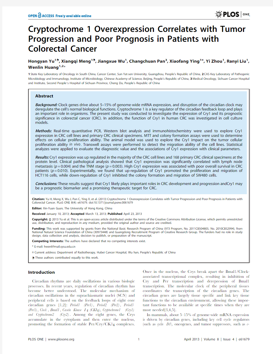
Figure 1.The expression of Cry1protein in colorectal cancer sections.Representative immunohistochemical images of colorectal cancer tissue specimens indicating negative or weakly detectable Cry1staining (A and B );moderate Cry1staining (C );and strong Cry1staining (D )are shown.Magnification is 6200(A,B,C and D ).doi:10.1371/journal.pone.0061679.g001
toxin,100ng/ml hydrocortisone and0.005mg/ml transferrin and supplemented with10%fetal bovine serum,100ng/ml streptomycin and100U/ml penicillin.All of the cell lines were cultured in a humidified chamber with5%CO2and at 37u C.
Quantitative reverse transcription-PCR
Total RNA was extracted using the TRIzol Reagent H(DSBIO, Guangzhou,China)according to the manufacturer’s instructions. Complementary DNA(cDNA)was synthesized from2m g of total RNA using M-MLV reverse transcriptase(Promega,Madison, WI).cDNA was amplified by AmpliTaq H Gold DNA polymerase (Applied Biosystems,Foster,CA)and gene specific primers.The following primers were used for Cry1,59-GAGTATGATTCT-GAGCCCTTTG-39(forward),59-GGTTGTCCACCATT-GAGT-39(reverse).GAPDH was used as an internal control. Western Blot analysis
Total cellular proteins were extracted and separated in SDS-PAGE gels,and Western Blot analysis was performed according to standard procedures.GAPDH was used as a loading control on the same membrane.The primary antibodies that were used included monoclonal anti-Cry1(1:1000,Abcam,USA)and anti-GAPDH (1:2000,Santa Cruz Biotechnology,USA).Proteins were visualized using the ECL procedure(Amersham Biosciences,USA).
Cell transfection
The coding sequence of Cry1was amplified and cloned into the NotI and BamHI site of pcDNA3.1+to generate a pcDNA3.1+-Cry1expressing vector;the resulting construct was confirmed by sequencing.Cry1siRNA(GCAAGAGAAUUUGCUUAAUTT) and control siRNA(UUCUCCGAACGUGUCACGUTT)were purchased from Shanghai Genepharma Co.Ltd.(Shanghai, China).
For transient transfection,SW480cells(66105cells per well) and HCT116cells(56105cells per well)were seeded in6-well plates at60%confluence and transfected with100nM of oligonucleotides or4m g of plasmid,using Lipofectamine2000 ((Invitrogen)according to the manufacturer’s instructions.After 6h of incubation at37u C,the transfection medium was replaced with3ml of complete medium containing10%FBS.Cells were collected for western blot,proliferation and invasion assays at different times.
Retrovirus packing and transduction
Cry1and controlsequences were cloned into the Xho I and Cla I site of the pLNCX2Retrovirus vector.Virus packing was performed in GP293cells.GP293cells were cultured in DMEM with10%FBS in a37u C incubator with5%CO2.Forty-eight hours after transfection,the supernatant was collected by centrifugation at 1000g for10min.The HCT116cells were transduced with the retrovirus containing Cry1or control sequences plasmids.Forty-eight hours after infection,G418(600ug/ml)was added to the media for2weeks to select the stable cells infected with the retrovirus.Western blotting assays were used to detect the expression of Cry1in two stable cell lines as described above. Cell viability assay
The3-(4,5-dimethylthiazole-2-yl)-2,5-biphenyl tetrazolium bromide(MTT)assay was used to detect cell proliferation. HCT116cells were plated in96-well at16103cells/well.The spectrophotometric absorbance of each sample was measured at 490nm.All experiments were repeated three times,and the average results were calculated.
For the colony formation assay,HCT116cells(46103cells per10cm2plate)overexpressing Cry1or control GFP and SW480cells(56103cells per10cm2plate)down-regulated for the expression of Cry1by siRNA or a negative control were seeded in complete medium.The cells were cultured for14days at37u C in5%CO2humidified air.Colony formation and growth were visualized by crystal violet staining.The numbers of colonies containing.50cells were determined,and12fields were counted.
In vitro migration assay
The migration ability of cells was measured in transwell chambers(8m m pore;BD Biosciences,Franklin Lakes,NJ).The
Table1.Clinicopathological findings and correlation with Cry1expression.
Variables No.(%)Cry1low Cry1high P value Total cases16867(39.9)101(60.1)
Age(years)
#65127(75.6)47(37.0)80(63.0)
.6541(24.4)20(48.8)21(51.2)0.181 Gender
Male89(53.0)39(43.8)50(56.2)0.268 Female79(47.0)28(35.4)51(64.6)
Tumor location
Colon85(50.6)34(40.0)51(60.0)0.975 Rectum83(49.4)33(39.8)50(60.2)
Tumor size(cm){
,=592(55.1)33(35.9)59(64.1)0.285
.575(44.9)33(44.0)42(56.0)
Histology
Adenocarcinoma146(86.9)57(39.0)89(61.0)0.567 Mucinous22(13.1)10(45.5)12(54.5)
Tumor invasive depth
T1–T210(6.0)6(60.0)4(40.0)0.180
T3–T4158(94.0)61(38.6)97(61.4)
Lymph node status
N098(58.3)48(49.0)50(51.0)*0.004 N1(n.=1)70(41.7)19(27.1)51(72.9) Preoperative CEA(ng/mL){{
,598(65.8)38(38.8)60(61.2)0.330
.=551(34.2)24(47.1)27(52.9)
AJCC/TNM stage
I–II94(56.0)47(50.0)47(50.0)*0.003 III–IV74(44.0)20(27.0)54(73.0) Preoperative CA199(ng/
mL){{{
,=35118(80.3)51(43.2)67(56.8)0.392
.3529(19.7)10(34.5)19(65.5)
*Statistically significant.Numbers in parentheses indicate the proportion of
tumors with a specific clinical or pathological feature in a given Cry1subtype. {Analysis for this parameter was available for167cases.
{{Analysis for this parameter was available for149cases.
{{{Analysis for this parameter was available for147cases.
doi:10.1371/journal.pone.0061679.t001
bottom chamber was filled with 700m l of DMEM containing 10%FBS.For the migration assay,tumor cells (16105cells in a total volume of 200m l)were placed in the upper chamber and incubated at 37u C in 5%CO 2humidified air.After 24h cultured,non-migrating cells on the upper surface of the membrane were removed,and the cells that migrated to the underside of the polycarbonate membrane were fixed with ethanol and stained with crystal violet for 10min.The number of migrating cells was then determined from 5independent microscopic fields.The mean of triplicate assays for each experimental condition was used for analysis.
Immunohistochemical assay
The expression of Cry1in primary tumors and adjacent noncancerous colorectal mucosa were examined using an immu-nohistochemical assay.The immunohistochemical assay was performed within five days of section preparation.Paraffin sections were cut to a thickness of 4um and mounted on silanized slides.The sections were then dewaxed,rehydrated and blocked with 0.3%hydrogen peroxide.Tissue antigens were retrieved with a microwave oven set at 95u C for 25minutes and cooled to room temperature in 10mmol/sodium citrate buffer (pH 6.0).Each
slice was then washed with phosphate-buffered saline (PBS)and incubated overnight at 4u C with Cry1(1:400,clone ab54649,abcam,Cambridge,UK).Primary antibodies were diluted by background reducing components (S2022,Dako,Glostrup,Den-mark).The secondary antibody was employed with the Envision Detection Kit (Dako).The slides were stained for 2min with diaminobenzidine tetrahydrochloride (DAB)and then counter-stained with hematoxylin.Tissue treated with antibody dilution solution was used as a negative control.All controls yielded satisfactory results.
Evaluation of staining
The specimens were evaluated by two investigators,who were unaware of the clinical outcome and who independently reviewed the stained slides.Each slide was assessed under microscopy at 6200magnification.The expression of Cry1was evaluated using H-scores.The H-scores consisted of an assessment of the intensity of staining and the percentage of the staining area having a given intensity.Only stained malignant cells were assessed.The samples were grouped into the following four categories based on the intensity of nuclear staining:none (0),weak (1),medium (2)and strong (3).
The
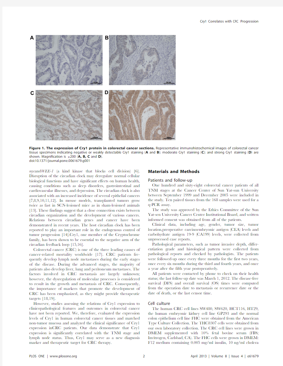
Figure 2.The overexpression of Cry1mRNA and protein in colorectal cancer tissues.(A )A representative image of Cry1staining in colorectal cancer tissues is shown.(B )A representative image of Cry1staining in adjacent noncancerous tissues is shown.(C )Cry1protein expression level was higher in tumor tissues compared to adjacent control tissue as detected by immunoblotting (mean 6SEM;n =109;***,P ,0.001).(D )Average T/N ratios of Cry1mRNA expression in paired colorectal cancer (T)and normalmucosa tissues (N)were quantified by qPCR and normalized to GAPDH.Error bars represent the standard deviation of the mean (SD)calculated from three parallel experiments.Magnification is 6200.doi:10.1371/journal.pone.0061679.g002
indexed sum was obtained by multiplying the intensity grade by the percentage of staining area.
The Cry1expression level was dichotomized according to DFS and OS by a ROC curve.The cut-off value of the positive rate was the maximized sum of the sensitivity and specificity points.
In vivo proliferation assays
Female athymic BABL/c nude mice(4–5weeks old)were purchased from the Medical Animal Center Guangdong Province (Guangdong,China).All animal studies were conducted inaccor-dance with NIH animal useguidelines and the current Chinese regulations and standardson the use of laboratory animals.To determine the proliferation capacity of the cell linesstably high expressing Cry1in vivo,a total of16106cells were injected subcutaneously into the left of nude mice and the negative control group were injected into the right(n=9).The tumor volume was evaluated using the following formula:tumorvolume=4p/ 36(width/2)26(length/2).Four weeks after injection,the animals were sacrificed.
Statistical analysis
All data are presented as mean6SEM unless stated otherwise.A paired t test was used to test for the differences in Cry1expression between matched tumor and benign mucosa.
Table2.Univariate analysis of clinicopathological parameters and molecular expression.
Variables Disease free survival Overall survival
n`5-y(%)P value n`5-y(%)P value Age(years)
#6526/12780.622/12781.7
.6511/4180.00.32610/4174.10.290 Gender
Male18/8982.60.52215/8982.20.512 Female19/7978.017/7977.0
Tumor location
Colon15/8584.10.19712/8584.80.112 Rectum22/8376.620/8374.7
Tumor size(cm){
,=520/9277.40.79019/9277.70.446
.516/7582.212/7583.5
Histology
Adenocarcinoma35/14678.80.11431/14678.20.065 Mucinous2/2290.91/2295.5
Tumor invasive depth
T1–T22/1090.00.7862/1078.80.927
T3–T435/15879.830/15880.7
Lymph node status
N014/9889.413/9885.9*0.022 N1(n.=1)23/7067.9*0.00119/7071.4
Preoperative CEA(ng/mL){{
,513/9886.0*0.00413/9885.50.058
.=518/5170.313/5178.1
AJCC/TNM stage
I–II10/9493.4*,0.0019/9489.7*,0.001 III–IV27/7463.923/7467.2
Preoperative CA199(ng/mL){{{
,=3520/11886.0*0.03017/11884.1*0.033 .3511/2965.59/2969.0
Cry1expression
Low8/6789.1*0.0116/6792.3*0.010 High29/10174.726/10177.6
*Statistically significant.
{Analysis for this parameter was available for167cases.
{{Analysis for this parameter was available for149cases.
{{{Analysis for this parameter was available for147cases.
`The number of positive events/total events.5-y(%),five-year rate.
doi:10.1371/journal.pone.0061679.t002
The statistical significance of the in vitro studies was analyzed using Student’s t -test.Kaplan-Meier and log-rank tests of the equality of the survivors were used to draw the survival curves by high versus low Cry1IHC scores (as defined by the ROC curve).All p values were two-sided,and P ,0.05was the level for statistically significant.All statistical analyses were conducted using SPSS software package,version 16.0(SPSS Inc.,Chicago,IL,USA).
Results
Characteristics of patients and tumors
A total of 168patients who received radical resection between January 1999and December 2005were studied.The character-istics of all the patients are summarized in Table 1.The mean patient age was 54.6years (SD,14.3).The median follow-up duration was 84months (range,4to 146).Eighty-five tumors (50.6%)were located in the colon.Ten out of the 168tumors (6.0%)were T1or T2.Ninety-four tumors were TNM stage I–II (56.0.0%).The median number of harvested lymph nodes was 19(range from 0to 48).Cry1protein expression levels were used for immunohistochemical analysis.Cry1was stained mainly in the cytoplasm and nuclear regions of the cells.High Cry1protein expression was detected in 101samples (60.1%)and weak or negative staining was observed in 67tumor samples (39.9%,Figure 1).
Cry1is overexpressed in colorectal cancer tissues
Cry1mRNA expression in colorectal cancer was investigated using RT-PCR;analyzes of Cry1mRNA were executed on ten matched pairs of colorectal cancer samples and adjacent noncancerous tissue samples.Cry1mRNA was expressed at higher levels in eight of the colorectal cancer tissue samples than in adjacent noncancerous tissues.The differential high expression ranged from 1.1-fold to 13.1-fold (Figure 2D).Consistent with these data,Cry1protein was also up regulated in colorectal cancers compared with the matched control tissue samples (Figure 2A–C).
Association between Cry1expression and clinicopathological variables
The association of Cry1expression and clinicopathological variables was further analyzed.Cry1expression and all of the other clinicopathological parameters were dichotomized into two groups.The results are summarized in Table 1.There was a significant correlation between Cry1expression and both the TNM stage (p =0.003)and lymph node status (p =0.004).
Among the 168colorectal cancer patients,no significant correlation was found between Cry1expression and gender,age,location of primary mass,tumor size,tumor
differentiation
Figure 3.The level of Cry1protein expression affects overall survival and disease-free survival.Kaplan–Meier curves with univariate analysis (log-rank)for colorectal cancer patients with high Cry1expression (n =101)versus low or no Cry1expression for overall survival (n =67)(A )and disease-free survival (n =67)(B )are shown.Higher expression of Cry1positively correlated with the poor patientoutcomes.
doi:10.1371/journal.pone.0061679.g003
Figure 4.Overexpression of Cry1mRNA and protein in colorectal cancer cell lines.Expression of Cry1mRNA and protein in colorectal cancer cell lines (SW480,SW620,HT29,THC8307,and HCT116)and FHC were examined by qPCR (A )and Western blotting (B ).Expression levels were normalized to GAPDH.Error bars represent the standard deviation of the mean (SD)calculated from three parallel experiments,*,P ,0.05.
doi:10.1371/journal.pone.0061679.g004
grade,histological type,Preoperative CA199or CEA level (p .0.05).
Association between Cry1expression and survival
The application of Cry1expression as a prognostic marker for CRC patients was also investigated.At the end of the follow-up period (March 1,2012),32patients had died from colorectal cancer-related diseases.The five-year survival rate of DFS was 74.7%for the Cry1high-expression group.This rate was significantly lower than the survival rate (89.1%)for the low-expression group (p =0.011).Similarly,the five-year survival rate of OS was 77.6%in the Cry1high-expression group and 92.3%in the low-expression group (p =0.010)(Table 2and Figure 3).
Cry1is highly expressed in most of the CRC cell lines
To investigate the potential role of Cry1in the tumorigenesis of colorectal cancer,the expression of Cry1mRNA and protein was determined for five CRC cell lines (SW480,HT29,SW620,THC8307and HCT116)and a normal colon epithelium cell line,FHC.Cry1mRNA expression was at most 10-fold higher in the
colorectal cancer cell lines than in the FHC cells (Figure 4A).Cry1protein was highly expressed in the colorectal cancer cell lines and only weakly expressed in the FHC cells (Figure 4B).
Cry1promotes tumor growth and proliferation
Next,we investigated the effects of Cry1manipulation (through gain-of-function and loss-of-function)on the proliferation of cancer cells.
We exogenously overexpressed Cry1in the HCT116cells,which have a lower endogenous expression.The impact of Cry1on cellular proliferation was then evaluated using MTT and clonogenic assays.The results showed that overexpression of Cry1can promote cellular proliferation in instantaneous transfection HCT116cells (p ,0.001,Fig.5B).In contrast,the control group showed no significant effect on cellular proliferation.The colony formation assays also showed that overexpression of Cry1significantly increased the number of colonies formed after 14days of culture compared with the control cells (p =0.033,Fig.5C,D).The link between Cry1and colorectal cancer cell growth was further established using colony formation assays after Cry1knockdown.As shown in Figure 5E and 5F,knockdown
of
Figure 5.Effects of Cry1on cell growth.(A )Cry1protein levels were upregulated in HCT116cells and downregulated in SW480cells.(B )MTT assays on HCT116cells 24,48,and 72h after transfection with Cry1or GFP control (***,p ,0.001).(C–D )Colony formation assay of HCT116cells transfected with Cry1or GFP control (*,p =0.033).(E–F )Inhibition of SW480cell colony formation capacity by Cry1siRNA relative to control (**,p =0.007).Experiments were repeated at least three times,and representative data are presented;bars ,SD.*,P ,0.05;**,P ,0.01,***,p ,0.001.doi:10.1371/journal.pone.0061679.g005
endogenous Cry1expression by siRNA resulted in a dramatic inhibition of clone formation by SW480cells(p =0.007)
Cry1promotes themigration of colorectal cancer cells
As for the Cry1expression was associated with lymph node status,the effect of Cry1on the migration of colorectal cancer cells was explored using the transwell assay to examine the effects of Cry1overexpression or deletion.In the transwell assays,HCT116cells transfected with Cry1or GFP control plasmids were seeded into the chambers,and their migration potential was determined 24h after transfection.The assays showed that the migration capacity of HCT116cells overexpressing Cry1was increased by 112%compared with the control cells (p =0.019,Fig.6A).
Confirmation that Cry1promoted the migration of colorectal cancer cells was assessed by the transfection of SW480cells with Cry1-siRNA or negative siRNA.After 24h of culture,the migration capacity of the knock-down Cry1SW480cells was reduced by 52.4%compared with the control cells (p ,0.001,Fig.6B).
Cry1promoted CRC growth in vivo
To further investigate the oncogenic properties of Cry1in vivo ,we constructed a retrovirus vector overexpressingCry1and
established two stable cell lines,which were named ctrl-HCT116and Cry1-HCT116(Fig.7A).These two cell lines were injectedsubcutaneously into the flanks of nude mice.Tumor progression was studied over time.At 4weeks post-implantation,the mice were sacrificed,and the tumors were removed.As shownin Fig.7B–C,the volume and weight of the tumors resulting from injection of Cry1-HCT116cells were significantly larger and heavier than those originated from the ctrl-HCT116cells.Takentogether,these observations are consistent with the in vitro results and indicate that Cry1has the ability to promote CRC cell growth in vivo .
Discussion
To our knowledge,this is the first study to evaluate the protein expression levels of Cry1in human colorectal cancer.Clearly,Cry1expression was higher in cancerous tissues and cells than that in normal tissues and cells.We also studied the relationship of the expression levels of Cry1to patient outcomes and clinicopatho-logical features.Our results suggest that overexpression of Cry1gene could be a useful predictor of lymph node metastasis,TNM stage and poor outcomes in patients with colorectal
cancer.
Figure 6.Cry1promotes CRC cells migration.(A )Migration assays of HCT116cells transfected with Cry1or GFP control.(n =3,*,p =0.019)(B )Migration assays of SW480cells transfected with Cry1-siRNA or negative control.(n =3,***,p ,0.001).Representative images are shown above,and the average number of cells per field at the indicated time points are shown below.Data are the mean of three independent experiments.doi:10.1371/journal.pone.0061679.g006
Several previous studies have compared the expression levels of Cry1between cancer tissue and adjacent normal mucosa.One study found that Cry1and other circadian gene (Cry2,Per2and BamlI)mRNA expression levels were similar in colon cancer and adjacent normal mucosa [20].Another study found that mRNA expression levels of Cry2and Per2were down regulated in colorectal cancer.This study also found higher Cry1expression in the tumor mucosa of cancers located in distal colorectal segments.Cry1mRNA levels in CRC tissues were also significantly associated with patient age and sex [21].In human epithelial ovarian cancer,Cry1mRNA expression level was clearly higher in cancerous tissues than in normal tissues [22].Our findings are in partial disagreement with these studies.In our study,Cry1gene protein expression levels were higher in cancerous tissues than in adjacent noncancerous tissue.The same results were found in colorectal tumor cells and normal cells,and overexpression of Cry1was found to promote growth and migration in colorectal tumor cells.However,no significant correlation was found between Cry1expression and gender,age,location of primary mass,tumor size,tumor differentiation grade,histological type,preoperative CA199or CEA level (p .0.05).These results seem to be reasonable for the following reason.Cry1protein expression levels may be inconsistently associated with mRNA expression levels for these environmental factors.However,the proteins play the most important role in the biologic effects,and we detect protein expression of Cry1in CRC tissues.
We also examined the relationships of Cry1expression with clinicopathological features and patient outcomes.The Cry1protein levels were significantly associated with the AJCC/TNM stage (with the highest levels detected in TNM stages III–IV)and lymph node involvement (with the highest levels detected in the case of positive lymph nodes).Circadian genes have been implicated in cell cycle regulation [23].Cry1mutant mice have an elevated level of Wee1in many tissues,including the liver.Wee1kinase blocks cell division by inhibiting the G2-M transition.High Cry1expression may inhibit the ability of Wee1to promote
cell proliferation,thereby providing a survival advantage for CRC [24].Moreover,the Cry1protein is known to complex with adenylyl cyclase,and overexpression of Cry1reduces cAMP production in response to PGE2,isoproterenol,and even the direct adenylyl cyclase activator,forskolin [25].Studies have reported that high concentrations of cAMP can inhibit the migration and metastasis of human prostate cancer cells.PKA is a protein kinase that is related to the cAMP signaling pathway.Studies have suggested that PKA inhibits the activity and function of RhoA [26,27].RhoA is a key member of the Rho family of small GTP-binding proteins and mediates signaling related to cytoskeletal arrangement,migration,proliferation and gene expression [28,29,30].Previous studies have found that invasive growth and metastasis were repressed in a variety of cancer cells (including CRC cells)when RhoA activity was inhibited [26,31].The current findings suggest that a Cry1-cAMP/PKA-RhoA mediated pathway is involved in the migration and metastasis of CRC.
Our immunostaining data agree with these studies.Higher TNM stage and the presence of lymph node metastasis were significantly correlated with higher levels of Cry1expression,suggesting that Cry1can be used as a marker to determine the progression of CRC.Our future studies will be aimed to more fully dissect the molecular mechanism underlying Cry1promotion of colorectal tumor cell growth,migration and the progression of CRC.
Conclusions
In summary,the present study showed that Cry1expression was up regulated in the majority of the CRC clinical tissue specimens at the protein level.Higher expression of Cry1positively correlated with the aggressive phenotype of colorectal cancer and predicted poor patientoutcomes.We also present experimen-tal evidence that overexpression of Cry1in colorectal cancer cell lines promoted cell proliferation and migration.Based on these findings,we conclude that Cry1is functionally important in
the
Figure 7.Cry1promoted CRC growth in vivo .(A )The expression of Cry1was markedly increased in the stable cell line Cry1-HCT116compared with control stable cell line ctrl-HCT116.GAPDH was used as an internal control.(B )Representative images of tumors derived from Cry1-HCT116or ctrl-HCT116,following subcutaneous xenograft transplants in nude mice (C )Overexpression of Cry1promoted colorectal cancer growth.Tumor cells were injected subcutaneously into nude mice.Mice were sacrificed after 4weeks,and the volume of each tumor was measured every 4days.Bars,6SEM;*P ,0.05,**p =0.01.
doi:10.1371/journal.pone.0061679.g007
development and progression of colorectal cancer and that Cry1 may serve as a new target for colorectal cancer therapy. Acknowledgments
We thank Jiemin Chen and Ling Zhou(Sun Yat-SenUniversity, Guangzhou,People’s Republic of China)for their technical assistance.Author Contributions
Conceived and designed the experiments:WLH JXW.Performed the experiments:HYY XQM.Analyzed the data:XFY RYL.Contributed reagents/materials/analysis tools:CCP YZ.Wrote the paper:HYY.
References
1.Lee C,Etchegaray JP,Cagampang FR,Loudon AS,Reppert SM(2001)
Posttranslational mechanisms regulate the mammalian circadian clock.Cell107: 855–867.
2.Reppert SM,Weaver DR(2002)Coordination of circadian timing in mammals.
Nature418:935–941.
3.Chu G,Yoshida K,Narahara S,Uchikawa M,Kawamura M,et al.(2011)
Alterations of circadian clockworks during differentiation and apoptosis of rat ovarian cells.Chronobiol Int28:477–487.
4.Hayashida S,Kuramoto Y,Koyanagi S,Oishi K,Fujiki J,et al.(2010)
Proxisome proliferator-activated receptor-alpha mediates high-fat,diet-en-hanced daily oscillation of plasminogen activator inhibitor-1activity in mice.
Chronobiol Int27:1735–1753.
5.Luo Y,Tian W,Cai L,Wang Y,Zhang J,et al.(2009)Expression profiling
reveals a positive regulation by mPer2on circadian rhythm of cytotoxicity receptors:Ly49C and Nkg2d.Chronobiol Int26:1514–1544.
6.Hunt T,Sassone-Corsi P(2007)Riding tandem:circadian clocks and the cell
cycle.Cell129:461–464.
7.Schibler U(2007)The daily timing of gene expression and physiology in
mammals.Dialogues Clin Neurosci9:257–272.
8.Schernhammer ES,Laden F,Speizer FE,Willett WC,Hunter DJ,et al.(2001)
Rotating night shifts and risk of breast cancer in women participating in the nurses’health study.J Natl Cancer Inst93:1563–1568.
9.Schernhammer ES,Laden F,Speizer FE,Willett WC,Hunter DJ,et al.(2003)
Night-shift work and risk of colorectal cancer in the nurses’health study.J Natl Cancer Inst95:825–828.
10.Viswanathan AN,Hankinson SE,Schernhammer ES(2007)Night shift work
and the risk of endometrial cancer.Cancer Res67:10618–10622.
11.Sack RL,Auckley D,Auger RR,Carskadon MA,Wright KP,Jr.,et al.(2007)
Circadian rhythm sleep disorders:part I,basic principles,shift work and jet lag disorders.An American Academy of Sleep Medicine review.Sleep30:1460–1483.
12.Engelen E,Janssens RC,Yagita K,Smits VA,van der Horst GT,et al.(2013)
Mammalian TIMELESS Is Involved in Period Determination and DNA Damage-Dependent Phase Advancing of the Circadian Clock.PLoS One8: e56623.
13.Filipski E,King VM,Li X,Granda TG,Mormont MC,et al.(2002)Host
circadian clock as a control point in tumor progression.J Natl Cancer Inst94: 690–697.
14.Koyanagi S,Kuramoto Y,Nakagawa H,Aramaki H,Ohdo S,et al.(2003)A
molecular mechanism regulating circadian expression of vascular endothelial growth factor in tumor cells.Cancer Res63:7277–7283.15.Ozturk N,Song SH,Ozgur S,Selby CP,Morrison L,et al.(2007)Structure and
function of animal cryptochromes.Cold Spring Harb Symp Quant Biol72:119–131.
16.Hanoun M,Eisele L,Suzuki M,Greally JM,Huttmann A,et al.(2012)
Epigenetic silencing of the circadian clock gene CRY1is associated with an indolent clinical course in chronic lymphocytic leukemia.PLoS One7:e34347.
17.Cunningham D,Atkin W,Lenz HJ,Lynch HT,Minsky B,et al.(2010)
Colorectal https://www.doczj.com/doc/8710512685.html,ncet375:1030–1047.
18.Singh R,Lillard JW,Jr.,Singh S(2011)Chemokines:key players in cancer
progression and metastasis.Front Biosci(Schol Ed)3:1569–1582.
19.Wang H,Wu J,Meng X,Ying X,Zuo Y,et al.(2011)MicroRNA-342inhibits
colorectal cancer cell proliferation and invasion by directly targeting DNA methyltransferase1.Carcinogenesis32:1033–1042.
20.Oshima T,Takenoshita S,Akaike M,Kunisaki C,Fujii S,et al.(2011)
Expression of circadian genes correlates with liver metastasis and outcomes in colorectal cancer.Oncol Rep25:1439–1446.
21.Mazzoccoli G,Panza A,Valvano MR,Palumbo O,Carella M,et al.(2011)
Clock gene expression levels and relationship with clinical and pathological features in colorectal cancer patients.Chronobiol Int28:841–851.
22.Tokunaga H,Takebayashi Y,Utsunomiya H,Akahira J,Higashimoto M,et al.
(2008)Clinicopathological significance of circadian rhythm-related gene expression levels in patients with epithelial ovarian cancer.Acta Obstet Gynecol Scand87:1060–1070.
23.Matsuo T,Yamaguchi S,Mitsui S,Emi A,Shimoda F,et al.(2003)Control
mechanism of the circadian clock for timing of cell division in vivo.Science302: 255–259.
24.Gauger MA,Sancar A(2005)Cryptochrome,circadian cycle,cell cycle
checkpoints,and cancer.Cancer Res65:6828–6834.
25.Narasimamurthy R,Hatori M,Nayak SK,Liu F,Panda S,et al.(2012)
Circadian clock protein cryptochrome regulates the expression of proinflamma-tory cytokines.Proc Natl Acad Sci U S A109:12662–12667.
26.Chen Y,Wang Y,Yu H,Wang F,Xu W(2005)The cross talk between protein
kinase A-and RhoA-mediated signaling in cancer cells.Exp Biol Med (Maywood)230:731–741.
27.Wang Y,Chen Y,Chen M,Xu W(2006)AKAPs competing peptide HT31
disrupts the inhibitory effect of PKA on RhoA activity.Oncol Rep16:755–761.
28.Kjoller L,Hall A(1999)Signaling to Rho GTPases.Exp Cell Res253:166–179.
29.Sahai E,Marshall CJ(2002)RHO-GTPases and cancer.Nat Rev Cancer2:
133–142.
30.Takai Y,Sasaki T,Matozaki T(2001)Small GTP-binding proteins.Physiol Rev
81:153–208.
31.Narumiya S(1996)The small GTPase Rho:cellular functions and signal
transduction.J Biochem120:215–228.