Lycium barbarum polysaccharide LBPF4-OL may be a new Toll-like
- 格式:pdf
- 大小:1.16 MB
- 文档页数:10
蓝实的有效成分(中英文实用版)英文文档:The bioactive components of Lanzhi (Lycium barbarum) have been the subject of extensive research due to its numerous potential health benefits.The fruit of this plant, often referred to as Chinese wolfberry or goji berry, contains a rich array of vitamins, minerals, and antioxidants that may contribute to its efficacy in promoting health and longevity.One of the key active ingredients in Lanzhi is betalains, which are natural pigments responsible for the red-orange color of the berries.Betalains have been shown to have powerful antioxidant properties, helping to protect the body against oxidative stress and potentially reducing the risk of various chronic diseases.Additionally, Lanzhi contains a variety of vitamins and minerals, including vitamin C, vitamin A, and trace minerals such as zinc and selenium.These nutrients are important for maintaining overall health and supporting various bodily functions.The fruit also contains a unique polysaccharide called Lycium barbarum polysaccharide (LBP), which has been studied for its potential immune-boosting effects.LBP is thought to enhance the activity of immune cells, thereby strengthening the body"s natural defense mechanisms against pathogens.Furthermore, Lanzhi has been traditionally used in Chinese medicine to improve vision and kidney function.While scientific evidence supporting these traditional uses is limited, some studies suggest that the active components in Lanzhi may have a beneficial effect on these aspects of health.In conclusion, the bioactive components of Lanzhi, such as betalains, vitamins, minerals, and Lycium barbarum polysaccharide, contribute to its potential health benefits.Further research is needed to fully understand the efficacy and mechanisms of action of these components.中文文档:蓝实(Lycium barbarum)的有效成分一直是广泛研究的热点,因其具有众多潜在的健康益处。
枸杞多糖对糖尿病大鼠的降血糖作用及血清TNF-α IL-6水平的影响于蕾;崔宏伟【摘要】目的探讨枸杞多糖(LBP)对链脲佐菌素(STZ)致糖尿病大鼠的降血糖作用以及对其血清炎症因子肿瘤坏死因子(TNF-α)、白介素6(IL-6)水平的影响.方法分别给予50只健康雄性Wistar-Imamichi大鼠腹腔注射STZ(40mg/kg)制备糖尿病模型.待模型成功后,分别灌胃给予不同剂量的LBP进行干预治疗.在同等条件下,选取罗格列酮为阳性对照药物,给予大鼠腹腔注射生理盐水作为空白对照组.连续灌胃给药30d.对各组大鼠一般行为学、空腹血糖(FBG)、空腹胰岛素(FNS)、胰岛素敏感指数(ISI)以及胰岛素抵抗(HOMA-IR)、血清TNF-α、IL-6的水平进行测定.结果与正常对照组大鼠体重相比,模型组大鼠的体重显著下降,差异有统计学意义(P<0.05),与模型组大鼠体重相比较,罗格列酮组、LBP组(低、中、高)各组大鼠体重随着实验进程而增加,差异有统计学意义(P<0.05).药物干预糖尿病模型大鼠后,与模型组比较,罗格列酮组、LBP(低、中、高)组大鼠FBG、FNS、血清IL-6、血清TNF-α水平逐渐降低,差异有统计学意义(P<0.05),ISI指数有所提高,而IR指数下降,差异有统计学意义(P<0.05).结论 LBP对STZ致糖尿病大鼠模型具有降低血糖的作用,可能的机制与降低血清炎症因子及改善脂质代谢有关.【期刊名称】《浙江临床医学》【年(卷),期】2015(017)012【总页数】3页(P2062-2064)【关键词】枸杞多糖;糖尿病;大鼠;IL-6;TNF-α【作者】于蕾;崔宏伟【作者单位】010020 内蒙古自治区中医医院药剂科;010050 内蒙古医科大学附属医院临床医学研究中心【正文语种】中文枸杞多糖(lycium barbarum polysaccharide,LBP)是来源于枸杞的干燥成熟果实含有如阿拉伯糖、葡萄糖及木糖等单糖成分,以及多种微量元素和氨基酸[1,2],具有多样的生物活性,如抗氧化、抗衰老、抗肿瘤、降血糖和增强机体免疫力等功效[3~5]。
款冬的功效与作用款冬,又称枸杞,是一种传统的中草药材,已有上千年的历史。
款冬的功效与作用广泛,被人们广泛应用于食疗和药疗等方面。
以下是关于款冬的功效与作用的详细介绍。
1. 提升免疫力款冬中含有丰富的多糖和抗氧化物质,能够增强人体的免疫力,抵抗疾病和外界环境的侵袭。
研究发现,款冬能够增加淋巴细胞的活性,增强巨噬细胞的吞噬能力,激活机体细胞免疫和体液免疫功能。
因此,常服用款冬可以提升免疫力,预防和缓解感冒、淋巴瘤等疾病。
2. 延缓衰老款冬中的多种抗氧化物质,如维生素C、维生素B1、维生素E等,可以中和自由基,减少细胞的氧化损伤,从而延缓衰老过程。
此外,款冬还含有一种特殊的成分LBP(Lycium barbarum polysaccharide),该成分被广泛应用于抗衰老产品中,能够促进皮肤新陈代谢,改善皮肤弹性,减少皱纹和黑斑的产生。
因此,经常食用款冬可以保持皮肤的健康和年轻。
3. 保护视力款冬中富含的胡萝卜素类物质,特别是β-胡萝卜素,是维持和保护视觉健康的重要成分。
β-胡萝卜素在人体内转化为维生素A,对眼睛的视网膜有保护作用,能够减少黄斑变性、青光眼等眼疾的发生。
此外,款冬中还富含的维生素C、维生素E和锌等微量元素,也对眼睛的健康起到积极的作用。
因此,适量摄入款冬可以保护视力,减少眼部疾病的风险。
4. 提高精力与改善睡眠款冬中的多糖和多种氨基酸能够刺激大脑分泌多巴胺和去甲肾上腺素等神经递质,提高精力和抗疲劳能力。
此外,款冬还含有一种叫做γ-氨基丁酸(γ-aminobutyric acid)的物质,它是一种主要的抑制型神经递质,在大脑起到镇静和促进睡眠的作用。
因此,适量摄入款冬可以改善睡眠质量,保持精力充沛。
5. 调节血糖和血压款冬具有降血糖和降血压的作用,对于糖尿病患者和高血压患者来说具有特别的益处。
研究发现,款冬中的多糖能够抑制糖尿病大鼠体内的血糖升高,同时还可以增强胰岛素的分泌,改善胰岛素抵抗。
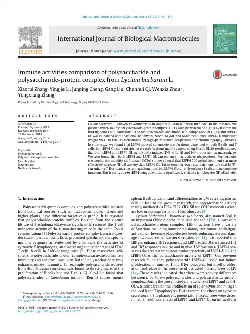
International Journal of Biological Macromolecules 65(2014)441–445Contents lists available at ScienceDirectInternational Journal of BiologicalMacromoleculesj o u r n a l h o m e p a g e :w w w.e l s e v i e r.c o m /l o c a t e /i j b i o m acImmune activities comparison of polysaccharide andpolysaccharide-protein complex from Lycium barbarum L.Xiaorui Zhang,Yingjie Li,Junping Cheng,Gang Liu,Chunhui Qi,Wenxia Zhou ∗,Yongxiang Zhang ∗Beijing Institute of Pharmacology and Toxicology,Beijing 100850,PR Chinaa r t i c l ei n f oArticle history:Received 9January 2013Received in revised form 12December 2013Accepted 7January 2014Available online 11February 2014Keywords:Lycium barbarum L.Polysaccharide-protein complex Lymphocytes MacrophageNuclear factor-Ba b s t r a c tLycium barbarum L.,known as wolfberry,is an important Chinese herbal medicine.In the research,we purified water-soluble polysaccharide-protein complex (LBPF4)and polysaccharide (LBPF4-OL)from the fruiting bodies of L.barbarum L.The monosaccharide and amino acid composition of LBPF4and LBPF4-OL was elucidated with fractional acid hydrolization,GC/MC and NMR techniques.LBPF4-OL molecular weight was 181kDa,as determined by high-performance gel-permeation chromatography (HPGPC).In vitro assay,we found that LBPF4induced splenocyte proliferations depended on both B cells and T cells,but LBPF4-OL induced splenocyte proliferations mainly depended on B cells.ELISA results showed that both LBPF4and LBPF4-OL significantly induced TNF-␣,IL-1and NO production on macrophage.We also found that both LBPF4and LBPF4-OL can enhance macrophage phagocytosis.Furthermore,electrophoretic mobility shift assay (EMSA)studies suggest that LBPF4100g/ml treatment can more effectively increase NF-B activity than LBPF4-OL.Taken together,our results demonstrate that LBPF4can enhance T,B cells and macrophages functions,but LBPF4-OL can only enhance B cells and macrophage functions.This is partly due to LBPF4being able to more significantly enhance lymphocytes NF-B activity.©2014Elsevier B.V.All rights reserved.1.IntroductionPolysaccharide-protein complex and polysaccharides isolated from botanical sources,such as mushrooms,algae,lichens and higher plants,have different target cells profile.It is reported that polysaccharide-protein complex isolated from the culture filtrate of Tricholoma lobayense significantly restored the T-cell mitogenic activity of the tumor-bearing mice in the same Con A concentrations [1].Polysaccharide-protein complex from Scolopen-dra subspinipes mutilans L.Koch promoted specific and nonspecific immune response as evidenced by enhancing the activities of cytotoxic T lymphocytes,and increasing the percentages of CD4+T cells,B cells in S180-bearing mice [2].These researches indi-cated that polysaccharide-protein complex can activate both innate immunity and adaptive immunity.But the polysaccharide mainly enhances innate immunity.For example,polysaccharide isolated from Acanthopanax senticosus was found to directly increase the proliferation of B cells but not T cells [3].Kuo-I Lin found that polysaccharides of Ganoderma lucidum (Reishi)causes mouse∗Corresponding authors.Tel.:+861066931625;fax:+861068211656.E-mail addresses:zhouwx@ ,zhouwx@ ,zhangx r@ (W.Zhou),zhangyx@ (Y.Zhang).splenic B cell activation and differentiation to IgM-secreting plasma cells.In fact,to the present research,the polysaccharide activity mainly mediated via TLR4,TLR2,CR3,SR and CD19molecules which are low or not expression on T lymphocytes [4].Lycium barbarum L.,known as wolfberry,also named Goji,is an important Chinese herbal medicine and tonic [5,6].L.barbarum polysaccharide-protein complex (LBP)fractions have a variety of functions including immunoregulatory,antitumor,antifatigue,antioxidant,lowering blood glucose levels,reducing neuronal dam-age and blood-retinal barrier disruption [7–11].It is reported that LBP can enhance Th1response,and LBP-treated DCs enhanced Th1and Th2responses in vitro and in vivo.LBP fraction 4(LBPF4)pos-sesses the greatest immunostimulatory activity of LBPF1-5[12,13].LPBF4-OL is the polysaccharide moiety of LBPF4.Our previous research found that,polysaccharide LBPF4-OL could not induce proliferation of purified T and B lymphocytes,but B cell prolifer-ation took place in the presence of activated macrophages or LPS [14].These results indicated that there were activity differences between L.barbarum polysaccharides and polysaccharide-protein complex.During the present study,the activity of LBPF4and LBPF4-OL was compared on the proliferation of splenocytes and mitogen induced B and T lymphocytes.Furthermore,the effects on cytokine secretion and the phagocytic potential of macrophages were deter-mined.In addition,effects of LBPF4and LBPF4-OL on intracellular0141-8130/$–see front matter ©2014Elsevier B.V.All rights reserved./10.1016/j.ijbiomac.2014.01.020442X.Zhang et al./International Journal of Biological Macromolecules65(2014)441–445signaling molecules including NF-B and BSAP(B cell specific acti-vator protein)were tested.2.Materials and methods2.1.MaterialsFemale BALB/c mice aged between six and eight weeks were obtained from the Beijing Vitalriver Experimental Animal Center, and were housed in a controlled environment(12h light/12h dark photoperiod,22±2◦C,45–55%relative humidity).Experiments were carried out according to the National Guide for Care and Use of Laboratory Animals,and were approved by the Bioethics Committee of the Beijing Institute of Pharmacology and Toxico-logy.Pronase E was obtained from Bacillus L.and bovine serum albumin from St.Louis,MO,USA.Sodium dodecyl sulfate(SDS), Concanavalin A(Con A)and lipopolysaccharide(LPS)were pur-chased from Sigma–Aldrich(St.Louis,MO);3H-thymidine was obtained from GE Healthcare(Buckinghamshire,UK).2.2.Extraction and purification of LBPF4and LBPF4-OLA total of20kg of L.barbarum was procured from a local drug store in Ningxia province of China.LBP was isolated from dried L.barbarum fruit with water,and the water extract was precip-itated with ethanol.LBP was separated using DEAE-cellulose ion exchange chromatography andfive homogenous fractions were obtained.LBPF4was purified by gelfiltration chromatography using a Sephadex G-100column.LBPF4(80mg)was dissolved in3.5mL0.1N Tris–hydrochloric acid buffer(pH8.0)containing 0.1%calcium chloride.One percent(g/g)Pronase E was added to the buffer and digested for24h at37◦C,followed by a further 48h digestion with Pronase E.Crude LBPF4-OL was subjected to gel permeation chromatography using a Sephadex G-100column (100cm×1.5cm)at aflow rate of0.5mL/min.The absorbance of total sugar was measured at490nm using a Shimadzu UV-1601 spectrophotometer(Kyoto,Japan).Protein content was measured using the Bradford method,with bovine serum albumin as the standard.Elution was monitored by measuring the absorbance at280nm using an SPD-10A UV-VIS detector(Shimadzu,Kyoto, Japan).2.3.Monosaccharide and amino acid composition analysis ofLBPF4and LBPF4-OLThe monosaccharide and amino acid composition were eluci-dated with fractional acid hydrolization,GC/MC and NMR tech-niques.1mg LBPF4,dissolved in0.5mL HCl solution(6.0mol L−1), was hydrolyzed in a closed tube at110◦C for24h,then amino acids were analyzed after the tube was opened and dried by drawing water.The molecular weight of LBPF4-OL was determined using the HPGPC method.Measurements were performed using HPGPC with a Waters515instrumentfitted with GPC software(Millennium), using a Waters2410RI detector.A calibration curve for molecu-lar weight determination was obtained using a series of Dextran T standards.This calibration curve correlated the molecular weights of standards with the HPGPC retention time,and was subsequently used for evaluating the molecular weight of LBPF4-OL.2.4.Splenocyte and lymphocyte proliferationThe spleen cells suspension of Balb/C mice was prepared in ster-ile condition.Spleen cells(5×10⁵)were stimulated with serial concentrations of LBPF4-OL including10,50and100g/mL.Con A (0.5g/mL)and LPS(5g/mL)were positive controls for the pro-liferation of T and B cells,respectively.The level of3H-thymidine incorporated into cells was measured using a-scintillation counter(BECKMAN LS6500).2.5.Mouse peritoneal macrophage preparationPeritoneal cells were harvested four days after intraperi-toneal injection of1mL of a starch solution,and isolated using 10mL PBS containing antibiotics(penicillin10U/mL and strepto-mycin1mg/mL).Peritoneal cells(1×10⁶cells/mL)were harvested, washed and resuspended in RPMI-1640;1.0×10⁶cells per well were plated in24-well tissue culture plates and incubated at37◦C in a5%CO2humidified incubator for2h.Non-adherent cells were removed by thorough washing,leaving monolayer of adherent cells comprising approximately99%macrophages.2.6.Analysis of TNF-˛,IL-1ˇand nitric oxide productionAdherent peritoneal macrophages(∼95%purity)were har-vested through vigorous pipetting and cultured with LBPF4-OL for 24h.The levels of TNF-␣and IL-1in supernatants were measured with TNF-␣and IL-1ELISA kits.The production of NO was indi-rectly assessed by calculating nitrite levels in culture supernatants using Griess reagent.2.7.Chicken erythrocytes phagocytosis assayThe phagocytic potential of adherent macrophages was deter-mined using a chicken erythrocyte phagocytosis assay.Freshly collected chicken erythrocytes were washed three times in PBS (pH7.2)and a2%chicken erythrocytes suspension was prepared in RPMI-1640.The Petri dishes per treatment group was stimulated with1.0mL of the chicken erythrocyte suspension.The free chicken erythrocytes coverslips were washed individually,and residual cells werefixed with1:1methanol-acetone(v/v)and stained using Giemsa staining solution.The numbers of macrophages containing or free from cytoplasmic chicken erythrocytes,and the number of chicken erythrocytes per phagocytic macrophage,were recorded for each coverslip.2.8.EMSACells were treated with LPS(10g/mL),LBPF4(50g/mL), LBPF4-OL(50g/mL)or medium for8h.Cells were harvested and washed with PBS,and a crude nuclear preparation was homog-enized in10mM HEPES pH7.8,1.5mM MgCl2,10mM KCl,and 1mM DTT.Protein concentrations in nuclear extracts were deter-mined using BCA protein assay kits(Pierce Biotech.,Rockford, IL).Equal amounts of supernatant protein(usually15g)were incubated with a32P-labeled double-stranded oligonucleotide.The probe sequences were as follows:NF-B5 -GCC TGG GAA AGT CCC CTC AAC T-3 ,5 -AGT TGA GGG GAC TTT CCC AGG-3 ,BSAP5 -TGT GAC GCA GCG GTG GGT GAC GAC T-3 ,5 -AGT CGT CAC CCA CCG CTG CGT CAC A-3 .DNA-protein complexes were resolved using electrophoresis on a5%glycerine-polyacrylamide gel.2.9.Statistical analysisThe data were treated by ANOVA,p<0.05was selected prior to experiments to reflect statistical significance.The results were presented as the mean±SD.X.Zhang et al./International Journal of Biological Macromolecules65(2014)441–445443 Table1Monosaccharide and amino acid content analysis of LBPF4-OL and LBPF4.Rha,l-rhamnose;Ara,l-arabinose;Gal,d-galactose;Xyl,d-xylose.Polysaccharide Carbohydrate content Amino acid content Monosaccharide molar ratioRha Ara Gal Xyl LBPF485.60% 1.72%0.43 1.50 2.500.23LBPF4-OL C:39.06%,H:5.73%0%0.05 1.33 1.00Fig.1.Gelfiltration chromatogram of LBPF4-OL using a Sephadex G-100column(1.5cm×100cm).3.Results3.1.Carbohydrate and amino acid composition analysis ofLBPF4-OL and LBPF4It was reported that polysaccharide-protein complex activity may from the polysaccharide or the protein portion,but also can be the result of the binding complex[15,16].Results of frac-tional acid hydrolization,GC/MC and NMR tests showed that the LBPF4molecular weight was214.8kDa,and composed with17 amino acids and4kinds of monosaccharide.The molar ratios were that Asp(9.61):Thr(6.77):Ser(12.80):Glu(10.33):Pro(5.15):Gly (12.57):Ala(11.64):Cys(1.52):Val(6.04):Met(1.03):Ile(3.74):Leu (4.64):Tyr(3.06):Phe(3.00):Lys(3.94):His(1.25):Arg(2.92).And the monosaccharide molar ratios were Ara(1.5):Gal(2.5):Rha (0.43):Xyl(0.23).The LBPF4-OL was consisted with3kinds of monosaccharide in molar ratios of Rha(0.05):Ara(1.33):Gal(1.00). The LBPF4carbohydrate and amino acid content were85.6%and 1.72%respectively(Table1).The polysaccharides and protein con-tent of LBPF4-OL was tested(Fig.1),and a single symmetrical absorbance peak was observed on an ultraviolet(UV)spectrum at 490nm,and no significant peak was observed at280nm.The results indicated that the main components of LBPF4-OL were polysaccha-ride substances but not a polysaccharide-protein complex.3.2.Homogeneity and molecular weight analysis of LBPF4-OLA single and symmetrical peak on HPGPC suggested that the polysaccharide is homogeneous(Fig.2).Based on the equation determined by linear regression made by different standard dex-trans,the molecular weight was estimated to be181kDa.The result indicates that LBPF4-OL is a purified homogeneous polysaccharide.3.3.Effect of LBPF4and LBPF4-OL on lymphocyte proliferationWe previously reported that LBPF4-OL cannot induce activa-tion and proliferation of T lymphocytes[14].In the research,we found that both LBPF4and LBPF4-OL induced spleen cell prolif-eration in a dose-dependent manner(Fig.3A).LBPF4(50g/mL) significantly enhanced spleen cell proliferation approximately3.2-fold,while LBPF4-OL enhanced proliferation7.2-fold.There was a significant difference in lymphocyte proliferation induced by LBPF4 and LBPF4-OL.LBPF4(10,50,100g/mL)but not LBPF4-OL(10, 50,100g/mL)can significantly enhanced the Con A induced T lymphocyte proliferation(Fig.3B).However,LPS induced B cell proliferation was enhanced by both LBPF4and LBPF4-OL(10, 50,100g/mL)(Fig.3C).The results indicated that LBPF4induced lymphocyte proliferation predominantly depended on both B and T cells and LBPF4-OL induced lymphocytes proliferation only depended on the B cells.3.4.Effect of LBPF4and LBPF4-OL on macrophage TNF-˛,IL-1ˇand NO productionRegulation of macrophage function by polysaccharides has been reported in diverse ways including promoting macrophage activa-tion,inducing the secretion of TNF-␣,IFN-␣and NO,andenhancingFig. 2.HPGPC analysis of LBPF4-OL using a TSK–Gel G4000SW column (7.5mm×300mm).444X.Zhang et al./International Journal of Biological Macromolecules 65(2014)441–445Fig.3.Effect of LBPF4-OL and LBPF4on mixed splenocytes (A),Con A (1g/ml)stimulated T lymphocytes (B)and LPS (5g/ml)stimulated B lymphocytes (C)proliferation in vitro.Values are the mean ±SD of triplicate experiments (∗P <0.05,∗∗P <0.01compare to medium group,#P <0.05,##P <0.01compare to LBPF4group).Fig.4.Effect of LBPF4(g/mL)and LBPF4-OL (g/mL)on production of TNF-␣(A),IL-1(B)and nitric oxide (C)by macrophages.Values are mean ±SD of triplicate experiments (∗P <0.05,∗∗P <0.01compare to LPS group).phagocytosis and antigen presenting ability [17,18].The stimula-tion of murine peritoneal macrophages with LBPF4and LBPF4-OL resulted in a comparable dose-dependent increase of the produc-tion of TNF-␣and IL-1.In addition,we also found that LBPF4and LBPF4-OL at concentrations of 10,50and 100g/mL increased the secretion of NO to comparable levels.LPS 5g/mL significantly induced TNF-␣,IL-1and NO production (Fig.4).These results demonstrate that that LBPF4-OL is the major immune activator of LBPF4on macrophages.3.5.Effect of LBPF4and LBPF4-OL on peritoneal macrophage phagocytosis activityLBPF4and LBPF4-OL (10g/mL)had no significant effects on the phagocytic activity of resting macrophages in mice,but the macrophage chicken erythrocyte phagocytic activity was signif-icantly increased by low concentrations of LBPF4and LBPF4-OL (Fig.5).Furthermore,there was no difference between LBPF4and LBPF4-OL in terms of inducing the phagocytic activity ofmacrophages.These results indicate that LBPF4and LBPF4-OL can increase the macrophage phagocytosis.3.6.Effect of LBPF4and LBPF4-OL on BSAP and NF-ÄB activityAt present,polysaccharide intracellular signaling mechanism research focuses predominantly on NF-B and MAPK [19,20].In the research,we investigated the levels of BSAP and NF-B in splenocytes using the EMSA method.As we found that in a lower concentration of 10g/ml,both LBPF4-OL and LBPF4cannot stimulating the mixed lymphocytes proliferation (Fig.3A),which indicated that LBPF4and LBPF4-OL (10g/ml)may not enhance the BSAP activity.Furthermore,10g/ml LBPF4cannot induce the TNF-␣and NO productions (Fig.4A and C),which indicated that LBPF4(10g/ml)may not enhance the NF-B activity.And we found that LBPF4and LBPF4-OL (50g/ml)significantly promote BSAP and NF-B activity.So we speculated that 50g/ml but not 10g/ml was a suitable concentration to observe the differences between LBPF4and LBPF4-OL on BSAP and NF-B activity.Especially theLBPF4Fig.5.Effect of LBPF4(g/mL)and LBPF4-OL (g/mL)on peritoneal macrophage phagocytosis index (A)and phagocytosis rate (B).Values are the mean ±SD of triplicate experiments (∗P <0.05,∗∗P <0.01compare to LPS group).X.Zhang et al./International Journal of Biological Macromolecules65(2014)441–445445Fig.6.Effect of LBPF4and LBPF4-OL on BSAP and NF-B expression.Splenocytes were prepared in sterile conditions.The splenocytes were stimulated with LPS (5g/mL),LBPF4(50g/mL)or LBPF4-OL(50g/mL)plexes were resolved using electrophoresis on a5%glycerine–polyacrylamide gel.treatment to be more effectively activated splenocytes NF-B than LBPF4(Fig.6).4.ConclusionA water-soluble polysaccharide-protein complex(LBPF4)and polysaccharide(LBPF4-OL)were isolated the fruiting bodies of L. barbarum L.through a series of DEAE anion exchange cellulose and gel-permeation chromatography.Its physicochemical properties and inducing lymphocytes proliferation activity were investigated. This polysaccharide showed a single and symmetrical peak on HPGPC,indicating its homogeneity,and its molecular weight was determined to be1.81×105Da.The results of the present research clearly demonstrate that LBPF4-OL cannot induce proliferation of Con A-activated spleen cells.We also fund that LBPF4-OL50g/mL was more effective at inducing proliferation of splenocytes and LPS-stimulated B cells than LBPF4100g/mL.This research also demonstrates that LBPF4-OL and LBPF4significantly and comparably induce TNF-␣,IL-1and NO production,and enhance macrophage phagocytosis of neutral red and chicken red blood cells.In EMSA experiments,there were no differences between LBPF4and LBPF4-OL in terms of inducing splenocytes BSAP expression.Moreover,LBPF4can more effectively induced NF-B expression in splenocytes than LBPF4-OL.This may be because LBPF4not only can also promote the expression of NF-B on B lymphocytes,but also can promote the expression of NF-B on T lymphocytes[21].In conclusion,the data demonstrate that L.barbarum polysac-charide LBPF4-OL can only enhance B cell and macrophage functions,but polysaccharide-protein complex LBPF4can enhance both T and B cells and macrophage functions.This due to LBPF4 being able to more significantly enhance lymphocytes NF-B activ-ity than LBPF4-OL.AcknowledgmentsThis work was supported by the National Natural Science foun-dation of China(No.81102451),National Key Technology R&D Program(2012BAI29B07)and the National Science and Technology Major Project(2012ZX09301003-476002-001).References[1]F.Liu,V.E.Ooi,W.K.Liu,S.T.Chang Gen,General Pharmacology27(1996)621–624.[2]H.X.Zhao,Y.Li,Y.Z.Wang,J.Zhang,X.M.Ouyang,R.X.Peng,J.Yang,Food andChemical Toxicology50(2012)2648–2655.[3]S.B.Han,Y.D.Yoon,H.J.Ahn,H.S.Lee,C.W.Lee,W.K.Yoon,S.K.Park,H.M.Kim,International Immunopharmacology3(2003)1301–1312.[4]K.Török,M.Kremlitzka,N.Sándor,E.A.Tóth,Z.Bajtay,A.Erdei,ImmunologyLetters143(2012)131–136.[5]P.Bucheli,K.Vidal,L.Shen,Z.Gu,C.Zhang,ler,J.Wang,Optometry andVision Science88(2011)257–262.[6]N.C.Lin,J.C.Lin,S.H.Chen,C.T.Ho,A.I.Yeh,Journal of Agriculture and FoodChemistry59(2011)10088–10096.[7]S.Y.Li,D.Yang,C.M.Yeung,W.Y.Yu,R.C.Chang,K.F.So,D.Wong,A.C.Lo,PLoSOne6(2011)16380.[8]G.Li,D.W.Sepkovic,H.L.Bradlow,N.T.Telang,G.Y.Wong,Nutrition and Cancer61(2009)408–414.[9]O.Potterat,Planta Medica76(2010)7–19.[10]Y.S.Ho,M.S.Yu,X.F.Yang,K.F.So,W.H.Yuen,R.C.Chang,Journal of AlzheimersDisease19(2010)813–827.[11]X.Shan,J.Zhou,T.Ma,Q.Chai,International Journal of Molecular Sciences12(2011)1081–1088.[12]Z.S.Chen,B.K.Tan,S.H.Chan,International Immunopharmacology8(2008)1663–1671.[13]L.J.Huang,G.Y.Tian,Y.X.Zhang,C.H.Qi,Chemical Journal of Chinese University428(2001)407–411.[14]X.R.Zhang,W.X.Zhou,Y.X.Zhang,C.H.Qi,H.Yan,Z.F.Wang,B.Wang,Journalof Ethnopharmacology136(2011)465–472.[15]Y.Z.Tao,Y.Y.Zhang,L.Zhang,International Journal of Biological Macro-molecules45(2009)109–115.[16]C.H.Yeh,H.C.Chen,J.J.Yang,W.I.Chuang,F.Sheu,Journal of Agricultural andFood Chemistry58(2010)8535–8544.[17]I.Ando,Y.Tsukumo,T.Wakabayashi,S.Akashi,K.Miyake,T.Kataoka,K.Nagai,International Immunopharmacology2(2002)1155–1162.[18]Z.S.Chen,J.H.Lu,N.Srinivasan,B.K.H.Tan,S.H.Chan,Journal of Immunology182(2009)3503–3509.[19]X.He,J.Shu,L.Xu,C.Lu,A.Lu,Molecules17(2012)3155–3164.[20]R.Maeda,T.Ida,H.Ihara,T.Sakamoto,Bioscience Biotechnology Biochemistry76(2012)501–505.[21]C.C.Cheng,S.P.Yang,W.S.Lin,L.J.Ho,i,S.M.Cheng,W.Y.Lin,InternationalImmunopharmacology13(2012)354–361.。
枸杞中枸杞多糖的药理作用及提取方法的研究进展摘要:研究表明,枸杞具有调节机体免疫、抗肿瘤、抗氧化、延缓衰老、降血脂、降血糖、保护遗传系统等药理作用。
枸杞多糖为其主要有效成分。
近几十年对枸杞多糖的研究多集中于其药理作用以及提取、分离、纯化及检测方面[1]。
本文对枸杞的主要有效成分枸杞多糖的药理作用及其提取纯化工艺的研究进展做一综述。
目的:为枸杞多糖的进一步临床应用和研发提供参考。
关键词:枸杞;枸杞多糖;药理作用;提取Summary: The research indicate that the Lycium barbarum has the effect such as immunomodulation, anti-tumor,antioxidation, delaying senescence, lowering blood sugar, blood fat ,protect genetic system and so on. Its main effective component is LBP. In recent several decades, the research on LBP is most concentrate on its pharmacological effects, extraction, isolation and purification,assaying[1]. This article makes a review of the pharmacological activities and extraction industrial art on the main effective component of Lycium barbarum —polysaccharide. It provides the reference for LBP’s further clinical application and further research.Keywords: Lycium barbarum ; Lycium barbarum polysaccharide(LBP) ; pharmacological effects ;extraction枸杞为茄科植物宁夏枸杞Lycium barbarum L的成熟果实;性味甘平,归肝肾经。
枸杞子为枸杞的干燥成熟果实,系茄科(Solanaceae )、茄族(Solanceae Reihb )、枸杞亚族(Lyciinae Wettst )、枸杞属(Lycium L.),最负盛名的是宁夏枸杞(Lycium barbarum L .)。
枸杞子是我国的传统名贵中药材,它在我国传统医学中具有重要的地位,其药用价值备受历代医家的推崇。
枸杞子的药理和保健作用与其中含有的生物活性物质枸杞多糖(Lycium barbarum polysaccharide ,LBP )有很大关系。
根据检索的文献可见,从中医到西医,从国内到国外,枸杞多糖都是近年来研究枸杞药理和保健作用的主要点。
现代科学研究表明,枸杞多糖具有调节免疫、清除自由基、抑制肿瘤、延缓衰老、降血脂、降血糖、降血压和抗疲劳等作用[1,2],笔者综述了枸杞多糖在生物活性方面新的研究进展,以期为枸杞多糖的进一步开发研究提供参考。
1 枸杞多糖的生物活性1.1 抗氧化及抗衰老作用LBP 的抗氧化功能已经在大量的动物实验中被证实。
纯化的枸杞多糖LBP 具有强力清除DPPH 自由基的活性,此外,老年动物口服30dLBP ,结果清楚地表明,LBP 具有抗氧化活性和对皮肤氧化损伤的保护作用[3]。
龚涛等[4]用高(400mg/kg ·d)、中(200mg/kg ·d)、低(100mg/kg ·d)3个剂量的枸杞粗多糖生理盐水溶液对D-半乳糖(100mg/kg ·d)致衰老模型小鼠和正常小鼠灌胃,连续灌胃30d ,测定结果表明,枸杞粗多糖能较显著(P<0. 01)提高小鼠血清、肝脏及脑组织中SOD 活性,降低MDA 含量,对小鼠具有显著的抗氧化、抗衰老作用。
邵鸿娥等[5]在枸杞多糖对小鼠体内抗氧化酶活性及耐力的影响枸杞多糖生物活性的研究进展燕宪涛,路新国(扬州大学旅游烹饪学院,江苏扬州 225127)的实验中也发现,LBP 可使小鼠血中SOD 活性明显升高、MDA 含量明显降低,认为LBP 具有良好的体内抗氧化作用,可以延缓衰老。
淫羊藿苷对白消安致小鼠睾丸功能损伤的作用马丽媛;王佳;郭小宇;刘春莲;焦海燕【期刊名称】《宁夏医科大学学报》【年(卷),期】2024(46)3【摘要】目的探讨淫羊藿苷(icariin,ICA)对白消安致睾丸功能损伤的治疗作用。
方法造模阶段:将小鼠分为对照组(20%DMSO)和模型组(4 mg·kg^(-1)白消安),每组8只。
使用ICA对模型小鼠进行治疗:将小鼠分为对照组(20%DMSO)、模型组(4 mg·kg^(-1)白消安)、ICA治疗组(4 mg·kg^(-1)白消安+120 mg·kg^(-1)ICA)、枸杞多糖(lycium barbarum polysaccharide,LBP)治疗组(4 mg·kg^(-1)白消安+40 mg·kg^(-1)LBP,ICA阳性对照组)共4组,每组11只。
所有实验小鼠均使用6周龄ICR雄性小鼠。
测定小鼠睾丸及附睾质量;用牛鲍式计数板进行精子计数,使用全自动精子分析仪检测精子存活率,应用Diff-Quik快速染色试剂盒检测精子畸形率;HE染色观察睾丸组织并进行Johnsen评分。
采用检测试剂盒检测睾丸组织丙二醛(malondialdehyde,MDA)、谷胱甘肽(glutathione,GSH)和脂质过氧化物(lipid peroxides,LPO)含量。
结果与对照组相比,白消安诱导的模型组小鼠睾丸、附睾质量降低(P均<0.05),睾丸指数降低(P<0.01),小鼠精子数量和精子存活率降低(P均<0.001),精子总畸形率升高(P<0.001),Johnsen评分降低(P<0.001);睾丸组织中MDA含量增加(P<0.01),GSH含量降低(P<0.01),LPO含量升高(P<0.001)。
与模型组相比,ICA治疗组睾丸和附睾质量、睾丸指数、精子存活率和Johnsen评分均升高(P均<0.001),精子数量增加(P<0.01),精子总畸形率降低(P<0.001);睾丸组织中MDA含量降低(P<0.01),GSH含量增加(P<0.001),LPO含量降低(P<0.001)。
手机应用程序,为师生提供便捷的在线点餐和预订服务。
这种系统可以根据个人需求和口味偏好定制餐食,有效减少现场点餐时因缺乏准确估计而导致的过量取餐和食物浪费。
在食堂内部署专门的监督人员,其主要职责是提醒和引导师生在取餐时根据实际需求适量选择,避免盲目取餐和过度浪费。
通过监督和指导,可以有效提高师生对食物浪费问题的重视,从而养成良好的用餐习惯[6]。
②通过举办各种宣传活动,如主题讲座、海报展示、社交媒体推广等,向师生宣传“光盘行动”的理念和重要性。
同时,可以设立奖励机制,鼓励那些积极参与并表现出色的个人或团体,以激励更多人加入减少食物浪费的行动中来。
③建立完善的厨余垃圾处理流程,确保食堂产生的厨余垃圾得到及时、有效的处理。
这些垃圾可以通过分类收集、生物降解等方式转化为有价值的资源,如有机肥料等,从而实现资源的循环利用。
同时可以借此机会教育师生认识到厨余垃圾的价值和正确处理的重要性。
4.2 推广使用环保餐具为了进一步推广环保理念,并在日常生活中实践减少资源浪费的目标,高校要积极倡导并提供可循环餐具,包括不锈钢碗、木质筷子和可清洗的餐盘等,它们不仅结实耐用,而且可以反复使用,避免了频繁更换一次性餐具的麻烦。
通过选择这些可循环餐具,能明显减少塑料垃圾的产生,降低对环境的压力。
为了进一步强化师生的环保意识,还可以鼓励师生们自带餐具。
为了激励大家积极参与,可以推出一系列优惠措施,如为自带餐具的师生提供折扣或积分奖励。
以上措施在一定程度上能够促进资源的有效利用,更能培养师生们节约资源的良好习惯。
在推广使用可循环餐具的过程中,不可忽视卫生和清洁的重要性。
因此,要严格规范餐具的消毒和清洁流程,以确保每一件餐具在再次使用前都经过彻底的清洁和消毒处理。
通过加强卫生管理和严格执行食品安全标准,确保循环使用餐具的清洁卫生,为师生们提供一个安全、健康的用餐环境。
这样不仅能够消除使用可循环餐具带来的健康顾虑,也能为环保餐具的推广提供坚实的支撑和保障。
枸杞的生物学功能及其在动物生产中的应用作者:葛影影何国戈郑经成胡克科别又才来源:《安徽农业科学》2021年第03期摘要枸杞是一种传统的药食兼用型中药,属于茄科枸杞属灌木植物。
枸杞的主要生物活性物质有枸杞多糖、甜菜碱、枸杞色素等,具有抗菌、抗氧化、降血糖、免疫调节等生理功能。
枸杞及其提取物、枸杞粉、枸杞渣等作为功能性饲料添加剂在动物生产中的研究已有报道,应用价值高、开发前景广。
综述了枸杞的主要活性成分及生理功能,枸杞在动物生产中的实际应用,旨在为枸杞的合理开发利用提供参考。
关键词枸杞;活性成分;生理功能;动物生产中图分类号S816.7文献标识码A文章编号0517-6611(2021)03-0025-03doi:10.3969/j.issn.0517-6611.2021.03.006AbstractLycium barbarum is a traditional Chinese medicine and food, and belong to the shrub plants of theSolanaceae and Lycium.The main biological active substances of Lycium barbarum have Lycium barbarum polysaccharide, betaine, Lycium barbarum pigment, etc., which have physiological functions including antibacterial, antioxidant, hypoglycemic and immune regulation.Researches on Lycium barbarum and its extracts, Lycium barbarum powder, Lycium barbarum residue,which can be used as functional feed additives in animal productions have been reported, it has a high application value and broad development prospects.The paper reviewed the main active ingredients and physiological functions of Lycium barbarum, the practical application in animal production,so as to provide a reference for the reasonable development and utilization of Lycium barbarum.Key wordsLycium barbarum;Active ingredient;Physiological function;Animal production枸杞(Lycium barbarum)是一種多年生茄科枸杞属灌木植物,喜冷凉,耐干旱,适应性强,常生于山坡、荒地、丘陵、盐碱地及路边,也可在我国宁夏、青海、甘肃等地区大面积种植。

中国果菜China Fruit &Vegetable综合利用Comprehensive Utilization第41卷,第6期2021年6月果蔬天然活性成分对雄性生殖障碍的改善作用张遥遥,郝佳美,张柏林,任建武*(北京林业大学生物科学与技术学院,北京100083)摘要:雄性不育多表现为雄性生殖系统组织病变及生精功能障碍,其病因多样,诊断具有复杂性和特殊性。
目前,治疗雄性不育症的西药效果还不够理想,而果蔬天然活性成分治疗有一定的优势,能使其得到有效改善。
本文探讨了雄性不育症可能存在的原因,以及现阶段研究发现的对生殖障碍有缓解作用的果蔬天然活性成分,为今后生殖障碍的治疗提供研究方向及思路。
关键词:不育症;氧化应激;抗氧化;天然活性成分中图分类号:R965.1文献标志码:A文章编号:1008-1038(2021)06-0088-06DOI:10.19590/ki.1008-1038.2021.06.016Effects of Natural Active Ingredients from Fruits and Vegetableson Male Reproductive DysfunctionZHANG Yao-yao,HAO Jia-mei,ZHANG Bo-lin,REN Jian-wu *(College of Biological Sciences and Biotechnology,Beijing Forestry University,Beijing 100083,China)Abstract:Male sterility is characterized by pathological changes of male reproductive system andspermatogenesis dysfunction.The etiology of male infertility is diverse,and the diagnosis is complex and specific.At present,the effect of Western medicine in the treatment of male infertility is not ideal,and natural active ingredients have certain advantages,which can effectively improve it.This paper discussed the possible causes of male infertility,and the natural active ingredients found at this stage to alleviate reproductive dusfunction,so as to provide research direction and ideas for the treatment of reproductive disorders in thefuture.Keywords:Infertility;oxidative stress;anti-oxidation;natural active ingredients收稿日期:2021-05-10基金项目:北京市怀柔区科技计划项目“林下黄精资源保存、繁育及多糖提取”(SHFZ2020-4)第一作者简介:张遥遥(1993—),女,在读硕士,研究方向为食品科学工作*通信作者简介:任建武(1967—),男,教授,博士,主要从事药食同源的研究工作雄性不育症是造成哺乳动物生殖问题的重要原因之一。
枸杞多糖的提取及其抗衰老研究进展作者:娄佳景鸿燕白梦婷陈景鲜卢超来源:《现代农业科技》2020年第14期摘要; ; 枸杞是一种传统的药食同源材料,枸杞多糖是枸杞的主要活性成分之一。
本文对近年来枸杞多糖的提取及其抗衰老研究进展进行了综述,以期为枸杞的开发利用提供参考。
关键词; ; 枸杞;枸杞多糖;提取;抗衰老中图分类号; ; R284.2; ; ; ; 文献标识码; ; A文章编号; ;1007-5739(2020)14-0210-01枸杞是茄科枸杞属植物,是我国传统的药食兼用的名贵中药材。
枸杞多糖(Lycium Barbarum Polysaccharide,LBP)是其重要的活性成分,具有抗氧化、清除自由基、抗疲劳、免疫调节等多种药理作用。
具有抗衰老作用的传统中药包括人参、枸杞、山药、牛膝等[1]。
大部分起到抗衰老药理作用的有效成分主要是多糖类、黄酮类等化合物。
目前,有关枸杞多糖抗氧化、清除自由基、调节免疫力功能等作用的研究均取得了一定的进展,为今后产业化利用奠定了基础。
1; ; 枸杞多糖的提取方法1.1; ; 水提法水提法的优点包括提取成本低、安全性高、操作简单,但是耗时较长,提取效率较低。
在水提过程中的料液比、提取温度、浸提时间、浸提次数等因素都会影响枸杞多糖得率。
在一定范围内,随着料液比增加,枸杞多糖提取率呈现先增加后下降的趋势。
枸杞多糖的提取率随着提取温度的升高逐渐增高。
随着浸提时间的增加,枸杞多糖的提取率也呈现先增加后下降的趋势,一般控制在2~7 h之间。
随着提取次数的增加,枸杞多糖提取率逐渐增加,但是增幅明显减小[2]。
提取次数一般控制在1~3次之间,提取次数越多,经济成本也越高。
1.2; ; 酶解提取法酶解提取法的优点是反应快、产量高、反应温度温和、易于操作,但是为了保证酶的活性,对温度和pH值有较高的要求,因而成本相对比较高。
酶解提取法有最适酶解温度,在最适温度之前,随着温度的提高,反应速度加快,枸杞多糖的提取率增加,但是超过最适温度后,酶促反应速度下降,则提取率下降。
营养学报2005年第27卷第1期 79枸杞多糖对高脂血症小鼠血脂及脂质过氧化的影响Effects of Lycium barbarum Polysaccharides on Serum Lipids and LipidPeroxidation in Hyperlipidemic Mice朱彩平,张声华(华中农业大学食品科技学院,武汉 430070)ZHU Cai-ping,ZHANG Sheng-hua(School of Food Science and Technology, Huazhong Agricultural University, Wuhan 430070,China)心脑血管病在我国居死亡原因之首位[1],研究表明,心脑血管病的发生与机体血清中高胆固醇含量有关,而通过药物或膳食干预能有效降低胆固醇水平并降低其发病危险性[2]。
枸杞为一种传统中药材,枸杞多糖(Lycium barbarum polysaccharide,LBP)为其主要生物活性成分,已证明LBP有增强机体免疫力[3,4]和抗肿瘤[5,6]等作用,但对降血脂抗氧化能力的研究尚未见报道。
1 材 料 与 方 法1.1 材料枸杞多糖:枸杞子为宁夏枸杞(Lycium barbarum L.)的干燥果实。
将枸杞子烘干粉碎,用乙醇水溶液于80℃浸提3次,滤渣用蒸馏水于100℃浸提4次,合并浸提液,60℃下真空浓缩,乙醇沉淀,丙酮-石油醚混合液脱色,真空干燥,水溶解,Sevage法脱蛋白,乙醇沉淀,抽滤,冷冻干燥,水溶解,上Sephadex G-75柱,蒸馏水洗脱,收集洗脱液,透析,真空浓缩,冷冻干燥得白色絮状的枸杞多糖[7]。
绞股蓝总甙:由广州白云山中药厂生产的“绞股蓝总甙片”研磨粉碎得到。
总胆固醇(TC)、甘油三酯(TG)、高密度脂蛋白(HDL-C)测定试剂盒以及超氧化物歧化酶(SOD)、丙二醛(MDA)测定试剂盒:南京建成生物工程研究所提供。
枸杞子对眼睛健康的保护作用研究枸杞子对眼睛健康的保护作用摘要:枸杞子是一种传统中药材,常被用于保护眼睛健康。
本文通过对已有研究的综述,探讨了枸杞子对眼睛健康的保护作用。
研究发现,枸杞子富含多种营养成分,包括维生素C、维生素E、锌、铜等,这些成分都对眼睛健康有积极作用。
此外,枸杞子还含有丰富的多糖类物质,具有抗氧化、抗炎和抗衰老的功效,能够保护眼睛的细胞免受损伤。
虽然目前的研究结果还不完善,但综合来看,枸杞子对眼睛健康的保护作用已得到初步证实。
未来的研究还需要进一步确认枸杞子的剂量和使用方式,并探索其与其他药物的联合应用,以更好地发挥其作用。
关键词:枸杞子,眼睛健康,维生素,多糖,抗氧化1. 引言眼睛是人们感知世界的重要器官,保护眼睛健康对于维持良好的视力和生活质量至关重要。
随着人口老龄化和电子屏幕的广泛使用,各类眼部疾病的发病率逐年增加,因此,寻找有效的保护眼睛健康的方法变得尤为重要。
枸杞子是一种已有数千年历史的传统中药材,在中医中被广泛使用。
许多人认为枸杞子对眼睛健康有益,并常常将其作为食物补充剂使用。
然而,关于枸杞子对眼睛健康的作用,目前的研究结果还不完整,缺乏系统综述的研究。
本论文将从枸杞子的营养成分、抗氧化和抗炎作用等多个方面,综述枸杞子对眼睛健康的保护作用,并讨论现有研究的不足之处。
希望通过这篇论文的总结,能够为未来进一步研究和应用提供一定的参考,推动枸杞子在眼睛健康保护领域的应用。
2. 枸杞子的营养成分枸杞子富含多种营养成分,包括维生素C、维生素E、维生素A、锌、铁、铜、硒等。
这些营养成分在保护眼睛健康方面起到至关重要的作用。
2.1 维生素C维生素C是一种强效的抗氧化剂,能够中和自由基对眼睛细胞的损害。
研究发现,维生素C的摄入与晶状体淡化和年龄相关性黄斑变性的发生率呈负相关关系。
2.2 维生素E维生素E是一种脂溶性抗氧化剂,能够保护眼睛的细胞免受氧化损伤。
研究表明,适量的维生素E摄入与老年人黄斑变性的风险降低相关。
食品实验枸杞(Lycium barbarum)是一种多年生茄科枸杞属灌木植物,喜冷凉、耐干旱,适应性强,常生于山坡、荒地、丘陵、盐碱地及路边,在我国宁夏、青海、甘肃等地区大面积种植。
枸杞是药食同源的一种植物,其药用部位主要有果实、果柄、果茎、叶片和根部,在我国已有两千多年的用药历史。
枸杞的主要营养成分及活性物质有枸杞多糖、甜菜碱、类胡萝卜素、枸杞黄酮、蛋白质及17种氨基酸等。
枸杞多糖(Lycium barbarum polysaccharide,LBP)作为一种水溶性多糖,是枸杞中最主要的活性成分之一,具有促进免疫、抗氧化、降血糖血脂、保护和改善生殖功能等作用。
甜菜碱是存在于枸杞果实、叶片、果柄中的主要生物碱之一,可以维持细胞渗透压,是机体良好的渗透调节剂。
枸杞色素是存在于枸杞果实中的呈色物质之一,是枸杞的重要营养活性成分,主要含有类胡萝卜素、叶黄素和其他呈色物质,具有增强免疫力、补虚益精、清热明目的保健功能。
枸杞叶片清甜爽滑,在两广地区可作蔬菜食用。
目前,枸杞可以加工成各种食品、饮料、油料、保健酒、保健品等,如枸杞饼干、枸杞饮品、枸杞保健酒。
近年来,浓缩果蔬汁类饮料成为一种新型的营养健康饮品,它是采用物理方法直接打浆或是浓缩后蒸发掉一部分水分,加水复原后具有果蔬汁类应有特征的饮品。
这种饮品以不添加任何添加剂、原汁原味为主要卖点,可以达到健康、营养的目的,同时也解决了新鲜水果蔬菜存在的难以长时间贮存、易腐败的问题。
由于枸杞营养价值丰富,所以将其打浆后可添加到其他饮品中,以起到相应的作用。
下面探讨了枸杞原浆的制作方法。
一、原材料及设备原材料:枸杞果(青海) ;主要仪器与设备:粉碎机、高压均质机、胶体磨、灌装机、杀菌锅。
二、加工工艺1.工艺流程。
枸杞果→挑选→清洗→打浆→过滤→均质→灌装→杀菌→成品检验。
枸杞原浆制作方法研究2.原料的标准与处理。
枸杞果要求果皮呈鲜红、紫红色或枣红色,具有枸杞应有的滋味、气味,无正常视力可见外来杂质,状态呈类纺锤形略扁稍皱缩。
Lycium barbarum polysaccharide LBPF4-OL may be a new Toll-like receptor 4/MD2-MAPK signaling pathway activator and inducerXiao-rui Zhang a ,Chun-hui Qi a ,Jun-ping Cheng a ,Gang Liu a ,Lin-juan Huang b ,Zhong-fu Wang b ,Wen-xia Zhou a ,⁎,Yong-xiang Zhang a ,⁎aBeijing Institute of Pharmacology and Toxicology,Beijing 100850,Chinab Key Laboratory of Resource and Biotechnology in Western China,Ministry of Education,Shaanxi Provincial Key Laboratory of Biotechnology,Life Science College,Northwest University,Xi'an 710069,Chinaa b s t r a c ta r t i c l e i n f o Article history:Received 24October 2013Received in revised form 12December 2013Accepted 12January 2014Available online 23January 2014Keywords:Lycium barbarum polysaccharides TLR4/MD2complex MacrophagesMitogen-activated protein kinase In flammatory factor LymphocytesRecognition of the utility of the traditional Chinese medicine Lycium barbarum L.has been gradually increasing in Europe and the Americas.Many immunoregulation and antitumor effects of L.barbarum polysaccharides (LBP)have been reported,but its molecular mechanism is not yet clear.In this study,we reported that the activity of the polysaccharide LBPF4-OL,which was puri fied from LBP,is closely associated with the TLR4–MAPK signaling pathway.We found that LBPF4-OL can signi ficantly induce TNF-αand IL-1βproduction in peritoneal macro-phages isolated from wild-type (C3H/HeN)but not TLR4-de ficient mice (C3H/HeJ).We also determined that the proliferation of LBPF4-OL-stimulated lymphocytes from C3H/HeJ mice is signi ficantly weaker than that of lymphocytes from C3H/HeN mice.Furthermore,through a bio-layer interferometry assay,we found that LPS but not LBPF4-OL can directly associate with the TLR4/MD2molecular complex.Flow cytometry analysis indicat-ed that LBPF4-OL markedly upregulates TLR4/MD2expression in both peritoneal macrophages and Raw264.7cells.As its mechanism of action,LBPF4-OL increases the phosphorylation of p38-MAPK and inhibits the phos-phorylation of JNK and ERK1/2,as was observed through Western blot analysis.These data suggest that the L.barbarum polysaccharide LBPF4-OL is a new Toll-like receptor 4/MD2-MAPK signaling pathway activator and inducer.©2014Elsevier B.V.All rights reserved.1.IntroductionLycium barbarum is an important traditional Chinese herbal medicine for the promotion of health and longevity and as a food supplement [1,2].In recent years,the health care functions of L.barbarum are gradu-ally gaining the interest of the US and Europe [3].Modern medicine found that the polysaccharide of L.barbarum (LBP)is the main active in-gredient of L.barbarum .Many bene ficial health effects of LBPs,such as immune regulation,anti-stress,anti-colon cancer,neuroprotective,and anti-diabetic effects,have been reported [4–12].Several studies also found that LBPs exhibit myocardial protection,eye protection,anti-radiation damage,and anti-aging effects [4–12].However,the molecular mechanism of LBPs has not yet been elucidated.LBPs (100mg/mL)can increase the co-expression of I-A/I-E and CD11c and the secretion of IL-12p40in bone marrow-derived dendritic cells (BMDCs),which suggests that LBPs are capable of promoting both the phenotypic and functional maturation of murine BMDCs in vitro [13].LBPs can also up-regulate the DC expression of CD40,CD80,CD86,and MHC class H molecules,down-regulate the uptake of Ag by DCs,enhance the co-stimulatoryactivity of DCs,and induce IL-12p40and p70production.Furthermore,LBPs induce the development of an enhanced Th1response,and LBP-treated DCs exhibited enhanced Th1and Th2responses in vitro and in vivo .Several studies have provided a rationale for the use of LBP under various clinical conditions to enhance host immunity and suggest the use of LBP as a potent adjuvant for the design of DC-based vaccines [14].We previously found that macrophages,rather than T and B cells,are the principal immunostimulatory target cells of the L.barbarum poly-saccharide LBPF4-OL.All of these results suggest that macrophages and dendritic cells are the primary target cells of LBPs [15].At present,TLR4/2is generally believed to be the main receptor of polysaccharides.A Japanese scholar first reported that saf flower poly-saccharides activate the transcription factor NF-kB via Toll-like receptor 4and induce cytokine production [16].More recently,South Korean and Chinese scholars began to report that the Toll-like receptor-mediated activation of B cells and macrophages by polysaccharides isolated from the cell culture of Acanthopanax senticosus and polysaccharides from Dioscorea batatas induce tumor necrosis factor-alpha secretion via Toll-like receptor 4-mediated protein kinase signaling pathways [17,18].Anti-TLR4monoclonal antibody blocking and the use of cells from TLR4-defective mice are two mainstream methods for revealing the molecular mechanism of active polysaccharides,but these methods do not directly indicate whether the polysaccharide is a ligand of TLR4.AInternational Immunopharmacology 19(2014)132–141⁎Corresponding authors at:Beijing Institute of Pharmacology and Toxicology,27TaiPing Avenue,Beijing 100850,China.Tel.:+861066931625;fax:+861068211656.E-mail addresses:zhouwx@ (W.Zhou),zhangyx@ (Y.Zhang).1567-5769/$–see front matter ©2014Elsevier B.V.All rights reserved./10.1016/j.intimp.2014.01.010Contents lists available at ScienceDirectInternational Immunopharmacologyj o u r n a l h om e p a g e :w w w.e l s e v i e r.c o m /l o c a t e /i n t i m plarge number of studies suggest that several polysaccharides can signif-icantly activate the TLR4–MAPK signaling pathway and induce TNF-α, IL-1β,and IFN-α/βproduction.However,it is currently unclear whether the LBP activity is related to the TLR4–MAPK signaling pathway and whether LBP can be directly combined with TLR4.In this study,we in-vestigated the molecular mechanism of the polysaccharide LBPF4-OL, which was extracted from LBP,in peritoneal macrophages and murine RAW264.7macrophage cells.2.Material and methods2.1.AnimalsSix-week-old female BALB/c mice and C3H/HeN mice were pur-chased from Vital River Experimental Animal Center(Beijing,China) and were housed in a controlled environment(12-h light/12-h dark photoperiod,22±2°C,45–55%relative humidity).Female C3H/HeJ mice were obtained from The Jackson Laboratory.All of the mice were used at the age of8to10weeks.All of the experiments were conducted according to the National Guide for the Care and Use of Laboratory An-imals and were approved by the Bioethics Committee of the Beijing In-stitute of Pharmacology and Toxicology.2.2.ReagentsLipopolysaccharide(LPS from Escherichia coli,serotype055:B5)was purchased from Sigma-Aldrich(St.Louis,MO,USA).Ethylenediamine tetraacetic acid disodium salt(EDTA-Na2)was purchased from Sinopharm Chemical Reagent(Shanghai,China).TNF-αand IL-1βELISA kits were purchased from Dakewe Biotech(Beijing,China). Anti-mouse TLR4/MD2,anti-CR3,and anti-TLR2were purchased from BioLegend.Extracellular signal-regulated kinase(ERK),C-Jun N-terminal kinase(JNK),and p38mitogen-activated protein kinase (MAPK)were purchased from Cell Signaling Technology(Beverly,MA, USA).RPMI(Roswell Park Memorial Institute)1640medium was pur-chased from Gibco BRL(Life Technologies,USA).Fetal bovine serum (FBS)was purchased from Hyclone Laboratories,and3H-thymidine was obtained from GE Healthcare(Buckinghamshire,UK).2.3.Preparation of LBPF4-OLLBPF4-OL was isolated from L.barbarum fruit as described previously [6].First,crude LBP was isolated from L.barbarum fruit.Briefly,dried L.barbarum fruit was extracted with water,and the water extract was precipitated with ethanol.The free proteins were removed using the Sevag reagent(CHCl3:n-BuOH=4:1).The crude LBP was obtained through dialysis and lyophilization.Second,the polysaccharide–protein complex LBPF4was isolated from LBP.Briefly,LBP was separated through DEAE-cellulose ion exchange chromatography(successively eluted with water,followed by0.05M,0.1M,0.25M,and0.5M NaHCO3),andfive homogenous fractions,designated LBPF1,LBPF2, LBPF3,LBPF4,and LBPF5,were obtained.LBPF4was then purified main-ly through gelfiltration chromatography using a Sephadex G-100col-umn with0.1M sodium chloride–water solution as the eluent,and LBPF4was obtained.Third,LBPF4-OL was released from LBPF4.Briefly, 80mg of LBPF4was dissolved in3.5mL of0.1N Tris–hydrochloric acid buffer(pH8.0)containing0.1%calcium chloride.Then,1%(g/g) Pronase E was added to the buffer,and the sample was allowed to digest for24h at37°C and then an additional48h with a supplement of0.5% (g/g)Pronase E.Then,100μL of methylbenzene was used to inhibit the growth of the bacterium.LBPF4-OL wasfinally purified using a Sephadex G-100column,and the polysaccharide LBPF4-OL released from LBPF4was obtained.The molecular weight of LBPF4-OL was181 kD,as determined by SDS-PAGE.The protein content was found to be ~0%,as determined through the Bradford method using a protein assay kit according to the manufacturer's protocol.LBPF4-OL was dis-solved in PBS or normal saline andfiltered through a0.22-μmfilter. 2.4.Isolation of peritoneal macrophagesMice were injected i.p.with1mL of3%thioglycolate(Sigma-Aldrich). After three days,the peritoneum was washed with10mL of ice-cold PBS. The cells were incubated on24-well plates with DMEM(Invitrogen Life Technologies)containing10%FCS(HyClone Laboratories),100U/mL penicillin,100U/mL streptomycin,2mM L-glutamine,1mM sodium py-ruvate,and nonessential amino acids for3h at37°C in5%CO2.The nonadherent cells were removed by washing.2.5.ELISAThe resident cells from the peritoneal cavity of mice were collected, and the macrophage population was enriched according to the method described by Shalev et al.[19].The levels of TNF-αand IL-1βin the su-pernatants were measured with precoated TNF-αand IL-1βELISA kits according to the manufacturer's instructions.The absorbance was mea-sured at450nm with a reference wavelength of570nm using a spec-trometer(BMG POLARStar Galaxy v4.0,Germany).2.6.Cell lineRAW264.7is a tumor macrophage cell line derived from BALB/c mice(20)that retains phagocytic and Ag-presenting capabilities (21).RAW264.7cells were obtained from American Type Culture Collection(ATCC)and were raised in full medium at37°C in a5% CO2atmosphere.2.7.Splenocyte proliferation assayC3H/HeN and C3H/HeJ mice were sacrificed.Their spleen was asep-tically removed and minced through a40-μm nylon cell strainer to achieve a single-cell suspension.The red blood cells were depleted with Tris–NH4Cl lysis buffer(0.144M NH4Cl,0.017M Tris–HCl).A total of5×105spleen cells were stimulated with LBPF4-OL at serial con-centrations,including5,10,50,100,and200μg/mL.The spleen cells were cultured in RPMI-1640medium supplemented with10%FBS,pen-icillin(100U/mL),and streptomycin(100μg/mL)at37°C in a5%CO2 humidified incubator for72h and were pulsed with3H-thymidine (1μCi/well)during the last18h of incubation.The cells were harvested on glassfiberfilters using a Filtermate cell harvester(Packard).The amount of3H-thymidine incorporated into the cells was measured using aβ-scintillation counter(Beckman LS6500).The results are expressed as cpm(counters per minute)of the stimulated cells and cpm of the unstimulated cells.2.8.Flow cytometryThe cells were washed with cold PBS containing0.1%NaN3and1% FBS.A total of106cells were incubated with PE-conjugated anti-mouse TLR4/MD2or isotype control IgG(BD Biosciences)for20min at room temperature.The cells were washed in PBS containing0.1% FBS and analyzed byflow cytometry(BD Calibur™).2.9.Western blotThe cells were washed in1×PBS and lysed in lysis buffer[10mM Tris–HCl(pH7.5),10mM NaH2PO4/NaHPO4(pH7.5),130mM NaCl, 1%Triton X-100,10mM NaPPi,1mM phenylmethylsulphonylfluoride, and2μg/mL pepstatin A]for30min on ice.The lysates were then centri-fuged at30,000rpm and4°C for30min.The supernatant was collected, and the protein content was then measured using a Bio-Rad protein assay kit before its analysis.The cytoplasmic or nuclear protein samples133X.Zhang et al./International Immunopharmacology19(2014)132–141were loaded at a concentration of10μg of protein/lane,separated by so-dium dodecyl sulfate-polyacrylamide gel electrophoresis(SDS-PAGE)in a10–15%gel,and transferred to polyvinylidene difluoride membranes (Immun-Blot PVDF membrane,0.2μm;Bio-Rad).The membranes were blocked with5%nonfat powdered milk in1×Tris-buffered saline(TBS) containing0.1%Tween20for1h and then incubated with primary anti-bodies overnight at4°C.The membranes were then treated with horse-radish peroxidase-coupled secondary antibodies for1h at4°C and washed with T-TBS after each antibody binding reaction.The detection of each protein was performed using an enhanced chemiluminescence (ECL)kit(Millipore Co.,USA).2.10.Bio-layer interferometry(BLI)The kinetic estimates were obtained using the ForteBio Octet RED system(ForteBio).The octet analysis was performed at30°C in HBS-EP+0.1mg/mL BSA running buffer.Briefly,the anti-human IgG Fc cap-ture(AHC)sensors from ForteBio were used to capture the TLR4/MD2-Fc onto the kinetic surface of the sensor.TLR4/MD2-Fc was loaded to saturation onto the anti-human Fc biosensors,washed in buffer(30s), and placed for3min in the wells containing LBPF4-OL,LPS,and FBS. The one-shot KD was calculated based on the1:1model,and the aver-age KD was extracted from the binding results obtained forfive different concentrations of the analyses.2.11.Statistical analysisUnless specifically mentioned,all of the data are expressed as the means±S.D.The ANOVA procedure was used to assess the significance of the differences between groups.For each significant treatment effect, Dunnett-type multiple comparisons were used to compare the means of multiple groups.All of the statistical analyses were conducted using the SPSS12.0software package.A value of P b0.05was used as the thresh-old for significance.3.Results3.1.Anti-TLR4/2treatment inhibits LBPF4-OL-induced TNF-a and IL-1βproductionOur previous research study found that LBPF4-OL induces the pro-duction of multiple cytokines,including TNF-α,IL-1ß,IL-2,IL-6,IL-8, IL-10,IL-12,and GM-CSF,in splenocytes in a dose-dependent manner [15].Of all of these cytokines,TNF-αand IL-1βwere the most significant-ly up-regulated.To determine whether the reported polysaccharide-related receptors,including TLR4/2and CR3,are related to the activities of L.barbarum polysaccharides,macrophages were treated with anti-TLR4/2antibody,and the concentrations of TNF-αand IL-1βin the cell culture supernatants were examined.Treatment with LBPF4-OL for24 h stimulated TNF-αand IL-1βproduction compared with the medium control(Fig.1A).Anti-TLR4(0.31,0.62,1.25,and2.5μg/mL)treatment inhibited the LBPF4-OL(100μg/mL)-induced secretion of TNF-αand IL-1βin a concentration-dependent manner.Anti-TLR4(0.62μg)also significantly inhibited the LPS(5μg/mL)-induced secretion of TNF-αand IL-1β(Fig.1B).We further found that anti-TLR2(0.31, 0.62,1.25,and2.5μg/mL)treatment partially inhibited the LBPF4-OL(100μg)-induced TNF-αand IL-1βsecretion,and anti-TLR2(0.62μg/mL)has no apparent effect on the LPS(5μg/mL)-induced TNF-αand IL-1βsecretion(Fig.1C).Neither anti-CD11b nor anti-CD18/anti-CD11b combined with anti-CD18had an effect on the LBPF4-OL-and LPS-induced TNF-αand IL-1βproduction(Fig.1D). These results suggest that the LBPF4-OL-induced TNF-αand IL-1βproduction is TLR4/2-related and not CR3-related.3.2.Anti-TLR4/MD2treatment inhibits LBPF4-OL-induced lymphocyte proliferationThe immunoenhancement activity of LBP is mainly reported to stimulate mouse splenocyte proliferation[15,20,21]and to induce the phenotypic and functional maturation of DCs with strong immu-nogenicity[14].A recent study found that the water-soluble polysac-charide fraction LBP-IV isolated from L.barbarum leaves significantly enhanced the proliferation of splenocytes stimulated by Con A or LPS [22].To investigate whether TLR4and CR3are related to the immu-nological enhancement activity of LBPF4-OL,we further observed the effect of LBPF4-OL on lymphocyte proliferation through antibody pretreatment.BALB/c splenocytes were prepared,and lymphocyte separation medium was used to obtain the lymphocytes.The lym-phocytes were pre-treated with anti-TLR4and anti-CR3antibody for1h in vitro,and the3H-TdR incorporation method was then used to analyze the LBPF4-OL-and LPS-induced lymphocyte prolifer-ation.Fig.2A shows that LBPF4-OL induces lymphocyte proliferation in a concentration-dependent manner,but the stimulation intensity is significantly weaker than that obtained with LPS.Fig.2B and C shows that anti-TLR4can completely inhibit the LBPF4-OL-induced lymphocyte proliferation,but anti-CR3has no effect on the LBPF4-OL-induced lymphocyte proliferation.These results indicate that the LBPF4-OL-induced lymphocyte proliferation is TLR4-dependent and not CR3-dependent.3.3.LBPF4-OL cannot induce TNF-αand IL-1βproduction in peritoneal macrophages from C3H/HeJ miceThe C3H/HeJ mouse is TLR4-defective[23].To further clarify wheth-er TLR4is a LBPF4-OL activity-related site and to determine whether the production of inflammatory cytokines induced by LBPF4-OL is associat-ed with TLR4,the supernatant TNF-αand IL-1βconcentrations of peri-toneal macrophages from C3H/HeJ mice were analyzed.As shown in Fig.3,LBPF4-OL(50,100μg/mL)significantly increased the TNF-αand IL-1βproduction by peritoneal macrophages from C3H/HeN mice but had no effect on the TNF-αand IL-1βproduction by macrophages from C3H/HeJ mice.In addition,we found that the TNF-αand IL-1βpro-duction by peritoneal macrophages from C3H/HeJ mice was obviously weaker than that obtained from C3H/HeN macrophages.The results show that TLR4is in fact an activity-association receptor of LBPF4-OL for the induction of TNF-ɑand IL-1βproduction,but it is unclear whether TLR4is a LBPF4-OL binding site.3.4.LBPF4-OL-stimulated proliferation of lymphocytes from C3H/HeJ mice is weaker than that of lymphocytes from C3H/HeN miceTo determine whether the lymphocyte proliferation induced by LBPF4-OL is associated with TLR4,the in vitro proliferation rates of the lymphocytes from C3H/HeJ and C3H/HeN mice were compared throughFig.1.Anti-TLR4/2treatment inhibits LBPF4-OL-induced TNF-ɑand IL-1βproduction.BALB/c mice were sacrificed,and their spleen cells were prepared.The spleen cells(5×10⁵)were stimulated with LPS(5μg/mL),Dextran(100μg/mL),anti-TLR4(0.31–2.5μg/mL),anti-TLR2(0.31–2.5μg/mL),anti-CR3(0.31–1.25μg/mL),and LBPF4-OL(10–100μg/mL)for24h.(A)LBPF4-OL induced TNF-ɑand IL-1βproduction in peritoneal macrophages in vitro.*P b0.05compared with the medium control.(B)Anti-TLR4treatment inhibited LBPF4-OL (100μg/mL)-induced TNF-ɑand IL-1βproduction.**P b0.01compared with the medium control;##P b0.01compared with LPS;$$P b0.01compared with LBPF4-OL.(C)Anti-TLR2treat-ment inhibits LBPF4-OL(100μg/mL)-induced TNF-ɑand IL-1βproduction.**P b0.01compared with the medium control;##P b0.01compared with LBPF4-OL.(D)Anti-CR3treatment had no effect on LBPF4-OL(100μg/mL)-induced TNF-ɑand IL-1βproduction.**P b0.01compared with the medium control.The production of TNF-ɑand IL-1βwas measured by ELISA. The data represent the means±SD of four replicates.134X.Zhang et al./International Immunopharmacology19(2014)132–141135 X.Zhang et al./International Immunopharmacology19(2014)132–141the 3H-TdR incorporation method.As shown in Fig.4,the effect of LBPF4-OL on the proliferation of C3H/HeN mouse splenocytes is obvi-ously weaker than that on the proliferation of C3H/HeJ mouse splenocytes,and this effect is dependent on the concentration (5to 200μg/mL)of LBPF4-OL.However,the proliferation of lymphocytes from C3H/HeJ mice induced by LBPF4-OL is signi ficantly higher than that observed in the control group.The results show that TLR4is in fact an activity association receptor of LBPF4-OL for the induction of lymphocyte proliferation.The results also indicate that other receptors in addition to TLR4may participate in the LBPF4-OL-induced lympho-cyte proliferation.3.5.LBPF4-OL cannot directly bind to TLR4in vitroMany of the bioactivities of botanical polysaccharides are closely associated with TLR4;however,it has not yet been reported whether these polysaccharides can be directly combined with TLR4.LPS is a glycolipid located in the outer membrane of Gram-negative bacteria and is composed of an amphipathic lipid A component,hydrophilic polysaccharides in the core,and O-antigen.Lipid A represents the conserved molecular pattern of LPS and is the main inducer of the immunological responses to LPS [24].TLR4in association with MD-2is responsible for the physiological recognition of LPS [25].In this study,a new method based on bio-layer interferometry (BLI)was used to determine the binding and dissociation of LBPF4-OL and LPS with TLR4in vitro [26].As shown in Fig.5A,the AHC sensorcombined effectively with the FC fragment of human rat recombi-nant TLR4molecules,and the maximum response value was found to be 0.8nm.Fig.5B shows that LPS directly binds to TLR4and rapidly reached saturation within 10s in a concentration-dependent man-ner.Because we cannot acquire the molecular weight of LPS,it is im-possible to quantify the interactions through equilibrium binding analyses.This result shows that LPS can bind directly to TLR4in vitro .As a comparison,the association of LBPF4-OL with TLR4was tested under the same conditions,and we found that LBPF4-OL at different concentrations (31.25–500μg/mL)cannot bind to TLR4.These results suggest that LBPF4-OL cannot directly associate with TLR4.3.6.LBPF4-OL increases TLR4expression on the membranes of peritoneal macrophages and RAW264.7cellsIt has been reported that one of the molecular mechanisms of the immune-enhancing effect of polysaccharides is the induction of the expression of TLR4on the cytomembrane.Therefore,we further ob-served the effect of LBPF4-OL on the expression of TLR4on peritone-al macrophages and RAW264.7cells by flow cytometry.As indicated in Fig.6A,peritoneal macrophages stimulated with LBPF4-OL or LPS for 24h cannot induce the expression of TLR4on the cytomembrane,and a high expression of TLR4was observed in these cells after stim-ulation for 48h with both LPS and LBPF4-OL.A partial recovery of the TLR4expression was observed in cells stimulated with LBPF4-OLforFig.2.Anti-TLR4/MD2treatment inhibits LBPF4-OL-induced lymphocyte proliferation.BALB/c mice were sacri ficed,and their spleen cells were prepared.The spleen cells (5×10⁵)were pretreated with anti-CR3(0.62μg/mL)and anti-TLR4/MD2(0.62μg/mL)and then stimulated with LPS (5μg/mL)and LBPF4-OL (100μg/mL)for 72h.(A)Anti-TLR4/MD2treatment inhibits the LBPF4-OL-induced proliferation of the spleen cells.*P b 0.05and **P b 0.01compared with the medium control;#P b 0.05and ##P b 0.01compared with LPS treatment.(B)Anti-CR3treatment has no effect on the LBPF4-OL-induced proliferation of spleen cells;*P b 0.05and **P b 0.01compared with the medium control.The cell proliferation was measured by 3H-thymidine incorporation assay.The data represented as mean the means ±SD of four replicates.136X.Zhang et al./International Immunopharmacology 19(2014)132–14172h.In contrast,the LPS-induced TLR4expression was completely recovered after 72h.In addition,we observed the effect of LBPF4-OL on TLR4expression in the macrophage cell line RAW264.7.Incontrast to its effect on peritoneal macrophages,the expression of TLR4on the RAW264.7membrane was signi ficantly increased by both LBPF4-OL and LPS after 24h.Moreover,we found thattheFig.3.LBPF4-OL cannot induce TNF-αand IL-1βproduction by peritoneal macrophages from C3H/HeJ mice.Both C3H/HeN and C3H/HeJ mice were sacri ficed,and their spleen cells were prepared.The spleen cells (5×10⁵)were stimulated with LPS (5μg/mL),galactan (100μg/mL),and LBPF4-OL (10–100μg/mL)for 24h.(A)LBPF4-OL could not induce TNF-ɑproduction by the peritoneal macrophages from C3H/HeJ mice.(B)LBPF4-OL could not induce IL-1βproduction by the peritoneal macrophages from C3H/HeJ mice.*P b 0.01compared with the LPS-stimulated spleen cells from C3H/HeN mice;#P b 0.05and ##P b 0.01compared with the LBPF4-OL-stimulated spleen cells from C3H/HeN mice.The TNF-ɑand IL-1βproduction levels were measured by ELISA.The data represent the means ±SD of four replicates.510501002003H -t h y m i d i n e i n c o p o r a t i o n (c p m )2000400060008000LBPF4-OL(g/ml)Fig.4.The LBPF4-OL-stimulated proliferation of lymphocytes from C3H/HeJ mice is weaker than that of lymphocytes from C3H/HeN mice.Both C3H/HeN and C3H/HeJ mice were sacri ficed,and their spleen cells were prepared.The spleen cells (5×10⁵)were stimulated with LBPF4-OL (5–200μg/mL)for 72h and pulsed with 3H-thymidine (1μCi/well)during the last 18h.The cells were harvested on glass fiber filters using a Filtermate cell harvester.*P b 0.05and **P b 0.01compared with the LBPF4-OL-stimulated spleen cells from C3H/HeN mice.The 3H-thymidine incorporation method was used to determine the cell proliferation.The data represent the means ±SD of four replicates.137X.Zhang et al./International Immunopharmacology 19(2014)132–141expression of TLR4in LBPF4-OL-treated cells was recovered to a nor-mal level,but the TLR4expression in LPS-treated cells was signi fi-cantly lower than that observed in the control group after 48h.These results suggest that LBPF4-OL and LPS trigger different molec-ular mechanisms to induce TLR4expression.The above results also suggest that the immune-enhancing effect of LBPF4-OL is due to TLR4expression.3.7.LBPF4-OL regulates the phosphorylation of MAPKsMitogen-activated protein kinase (MAPK)signal transduction path-ways are ubiquitous and highly evolutionarily conserved mechanisms of eukaryotic cell regulation [27].The multiple MAPK pathways present in all eukaryotic cells enable coordinated and integrated responses to di-verse stimuli.It is de finite that both LPS and the otherbotanicalFig.5.LPS and not LBPF4-OL can bind to TLR4on the octet platform.(A)Loading of label-free mouse TLR4(aa24–334)-Fc to immobilized anti-human IgG Fc capture (AHC)sensors in the octet,which was used to detect the molecular –molecular interactions via bio-layer interferometry.(B)LPS association and dissociation with mouse TLR4(aa24–334)show an increase in the saturation response with increasing concentration.(C)LBPF4-OL does not associate with mouse TLR4(aa24–334).Bio-layer interferometry (BLI)was used to analyze the association and dissociation of polysaccharides with TLR4.The experiment was repeated three times,and representative results are shown.138X.Zhang et al./International Immunopharmacology 19(2014)132–141polysaccharide can effectively evoke the MAPK signal pathway through TLR4.However,the effects of L.barbarum polysaccharides on MAPK sig-nal pathway have not yet been reported.In the present study,the effect of LBPF4-OL on p-38,ERK1/2,and SAPK/JNK phosphorylation was ob-served through the WB method (Fig.7).We found that phospho-p38was markedly increased after stimulation with LBPF4-OL at a concentra-tion of 100μg/mL for 30min,and the level of phospho-p38was recov-ered after 45min.In contrast,phospho-ERK1/2was signi ficantly inhibited early during treatment (within 30min)with LBPF4-OL and returned to the normal level after 45min.LBPF4-OL has no effect on the level of SAPK over a period of 1h.We also found that LBPF4-OL markedly inhibits the phospho-JNK level within 1h.In contrast,we ob-served that the treatment of cells with LPS for 45min signi ficantly en-hanced the levels of phos-p-38,phos-ERK1/2,and phospho-SAPK/JNK.These results indicate that LBPF4-OL can regulate the p-38,ERK1/2,and JNK MAPK signaling pathways in macrophages.4.DiscussionImmunomodulation and antitumor effects are one of the main activ-ities of L.barbarum polysaccharide.Previous studies have shown that the polysaccharide –protein complex from L.barbarum (LBP3p)can sig-ni ficantly inhibit the growth of transplantable sarcoma S180and in-crease macrophage phagocytosis;in addition,the form of antibody secreted by spleen cells,spleen lymphocyte proliferation,CTL activity,and IL-2mRNA expression level were increased,and lipid peroxidation was reduced in S180-bearing mice [28].Moreover,L.barbarum polysac-charide (LBP),which was isolated with boiling water from L.barbarum fruits,can inhibit the proliferation of HeLa cells [29].The present study provides the first demonstration that L.barbarum polysaccharide stimu-lates tumor necrosis factor-alpha (TNF-α)and IL-1βgeneration and promotes lymphocyte proliferation in a TLR4-dependent manner.TNF-α,a pro-in flammatory cytokine that was initially identi fied and named as a result of its anti-tumoral properties,is involved in the path-ogenesis of multiple chronic in flammatory or autoimmune diseases.TNF-αcan act on monocytes and macrophages in an autocrine manner to enhance various functional responses and induce the expression of other immunoregulatory and in flammatory mediators.IL-1βis involved in fever during the induction of the acute-phase protein response.In ad-dition,IL-1βcooperates with IFN-γand IL-12to induce tumor death by NK cells.It has been reported that,after an initial phase of growth,some murine tumor cells transduced with the TNF-αgene either completely regress or demonstrate decreased tumorigenicity.Consistently,Ganoderma atrum polysaccharide (PSG-1)can activate macrophages and increase TNF-α,IL-1β,and nitric oxide production via TLR4-dependent signaling pathways,improve immunity,and inhibit tumor growth [30].All of these results strongly suggest that TLR4plays an im-portant role in the immunomodulation and antitumor effects of L.barbarum polysaccharide LBPF4-OL.In this study,we found that the underlying mechanisms induced by LBPF4-OL and LPS on the TLR4–MAPK signaling pathway are dif-ferent.First,we found that the anti-TLR4,anti-TLR2,and anti-CR3treatments resulted in markedly different effects on the LPS-and LBPF4-OL-induced secretion of in flammatory cytokines.As shown in Fig.1,both anti-TLR4and anti-TLR2treatments signi ficantly inhibited the LBPF4-OL-induced TNF-αand IL-1βsecretion.Howev-er,anti-TLR4and not anti-TLR2inhibited the LPS-induced TNF-αand IL-1βproduction by macrophages.In addition,as shown in Fig.3,we found that LBPF4-OL markedly induced the secretion of TNF-αand IL-1βby C3H/HeN mice peritoneal macrophages but had no effect on the secretion of cytokines by macrophages from TLR4-defective C3H/HeJ mice.On the contrary,LPS signi ficantly induced the secretion of TNF-αand IL-1βBy C3H/HeJ mouse peritoneal macrophages in vitro .These results are consistent with the activity characteristics of LPS.Studies have shown that the LPS –TLR4interaction requires the participation of many other molecules,such as CD14/LBP and HSP70,and this molecular community is known as the LPS receptor family [31–33].In addition,these receptors are clustered together and are involved in LPS-induced transmembrane signal transduction.Studies have also found that TLR4de ficiency cannot fully reduce the activity of LPS.These results indicate that,in addition to TLR4,LBPF4-OL can induce the secretion of in flamma-tory factors by TLR2,and LPS can induce the secretion of in flammatory factors through an unknown receptor.Second,there is a clear difference in the TLR4expression induced by LPS and LBPF4-OL.Vicky Sender found that LPS can signi ficantly induce TLR4expression on the cell mem-brane of alveolar macrophages,and this expression peaks after 30minFig.6.LBPF4-OL increases TLR4expression on the membranes of peritoneal macrophages and RAW264.7cells.Macrophages or Raw264.7cells were cocultured with LBPF4-OL and LPS for 24,36,and 48h,and the PE-conjugated anti-mouse TLR4/MD2complex (0.25μg per million cells in 100μL)was used for the flow cytometric analysis.(A)LBPF4-OL induces TLR4expres-sion in peritoneal macrophages from C3H/HeN mice.(B)LBPF4-OL induces TLR4expression in Raw264.7cells.139X.Zhang et al./International Immunopharmacology 19(2014)132–141。