Immunoproteasomes Preserve Protein Homeostasis upon Interferon-Induced Oxidative Stress
- 格式:pdf
- 大小:1.09 MB
- 文档页数:12
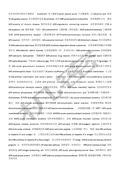
医学术语和名称英文缩略语A:adrenalin 肾上腺素A:aortic second sound 主动脉瓣第二音AA:amino acid 氨基酸AA:aplastic anemia 再生障碍性贫血Ab:antibody 抗体ABE:acute bacterial endocarditis 急性细菌性心内膜炎ACD:anemia of chronic disease 慢性病贫血ACE:angiotensin converting enzyme 血管紧张素转化酶 A cell:accessory cell 辅佐细胞(免疫)ACh:acetylcholine 乙酰胆碱(神经递质)AChE:acetycholinesterase 乙酰胆碱脂酶AChR:acetylcholine receptor 乙酰胆碱受体ACTH:adrenocorticotropic hormone 促肾上腺皮质激素ACV:acyclovir 无环鸟苷(抗病毒药)AD:autosomal dominant 常染色体显性AD:Alzheimer's disease 阿尔茨海默氏病ADA:adenosine deaminase 腺苷脱氨酶ADCC:antibody-dependent cellular cytotoxicity 抗体依赖性细胞介导细胞毒作用抗利尿激素抗酸杆菌Ag:antigen 抗原人工授精syndrome 获得性急性淋巴细胞白硬化症)AML:acute细胞性白血病急性高山病AMS:atypical measles syndrome 非典型麻疹综合征AMY:amylase 淀粉酶AN:analgesic nephritis 止痛药肾炎ANA:antinuclear antibody 抗细胞核抗体ANP:atrial natriuretic peptide 心房利钠肽(即心钠素)Anti-HBc:antibody to hepatitis B core antigen 抗乙型肝炎核心抗体Anti-HBe:antibody to hepatitis B e antigen 抗乙型肝炎e抗体Anti-HBs:antibody to hepatitis B surface antigen 抗乙型肝炎表面抗体(即AuAg)ANUG:acute necrotizing ulcerative gingivitis 急性坏死性溃疡性龈炎AP:alternate pathway 旁路途径(补体活化)APB:atrial premature beat 房性期前收缩APC:antigen-presenting cell 抗原呈递细胞APC:acute pharyngoconjunctival fever 急性咽结合膜热APP:acute-phase protein 急性期蛋白APRT:adenine phosphoribosyltransferase 腺嘌呤磷酸核糖转移酶(嘌呤回收途径)APTT:activated partial thromboplastin time 活化部分凝血活酶时间APUD:amine precursor uptake and decarboxylation 胺前体摄取和脱羧(细胞)AR:aortic regurgitation 主动脉反流AR:autosomal recessive 常染色体隐性ARA-A:adenine arabinoside 阿糖腺苷(抗病毒药)ARA-C:cytosine arabinoside 阿糖胞苷(抗癌药)ARAS:ascending reticular activation system 上行网状激活系统ARC:AIDS-related complex 艾滋病相关复合征ARD:acute respiratory disease 急性呼吸道病ARDS:adult respiratory distress syndrome 成人呼吸窘迫综合征ARF:acute renal failure 急性肾功能衰竭ARF:acute rheumatic fever 急性风湿热AS:aortic stenosis 主动脉瓣狭窄AS:ankylosing spondylitis 强直性脊柱炎Arg:arginine 精氨酸ASA:acetylsalicylic acid 乙酰水杨酸ASD:Alzheimer'smodulator 生物反应调节剂BSA:body surface area 体表面积BSP:bromsulphalein 酚四溴肽磺酸钠,磺溴肽钠(肝功能试验)BT:bleeding time 出血时间BUN:blood urea nitrogen 血尿素氮C:complement 补体CABG:coronary artery bypass grafting 冠状动脉旁路移植术CAD:coronary artery disease 冠状动脉病CAD:computer-aided diagnosis 计算机辅助诊断CAH:congenital adrenal hyperplasia 先天肾上腺增生cAMP:cyclic adenosine monophosphate 环一磷酸腺苷CAPD:continuous ambulatory peritnoeal dialysis 持续性非卧床式腹膜透析CA T:calcium antagonist 钙拮抗剂CA VH:continuous arteriovenous hemofiltration 连续动静脉血液滤过CA VHD:continuous arteriovenous hemodialysis 连续动静脉血液透析CA VHP:continuous arteriovenous hemoperfusion 连续动静脉血液灌流CA VP:continuous arteriovenous plasmapheresis 连续动静脉血浆换出CBC:complete?blood?count?全部血细胞计数CBF:cerebral?blood?flow?脑血流量CCK-PZ:cholecystokinin-pancreozymin?缩胆囊素-促?胰酶素CCNU:cyclohexyl-chloroethyl-nitrosourea?氯乙环己?亚硝脲(抗癌药)CCU:cardiac?care?unit?心脏病监护中心CD:cluster?of?differentiation?免疫细胞表面分化抗?原美国疾病监控?中心C?dyn:dynamic?compliance?动态顺应性CEA:carcinoembryonic?antigen?癌胚抗原CEI:converting?enzyme?inhibitor?转化酶抑制剂CEP:chronic?eosinophilic?pneumonia?慢性嗜酸细胞?增多性肺炎CF:complement?fixation?补体结合CF:chemotactic?factor?趋化因子CF:cystic?fibrosis?囊性纤维化CFU:colony?forming?unit?集落形成单位CGD:chronic?granulomatous?disease??慢性肉芽肿病CGL:chronic?granulocytic?leukemia?慢性粒细胞白血?病CH:compromized?host?免疫力低下寄主C:constant?domain?of?heavy?chain?重链稳定区(免?疫球蛋白)CHD:coronary?heart?disease?冠心病CHF:congestive?heart?failure?充血性心力衰竭CHO:carbohydrate?碳水化合物,糖CI:cardiac?index?心脏指数Ci:curie?居里(放射强度单位)CIC:circulating?immune?complex?循环免疫复合物CIE:countercurrent?immunoelectrophoresis?对流免?疫电泳CIS:carcinoma?in?situ?原位癌CISN:chronic?interstitial?nephritis??慢性间质性?肾炎(免?疫球蛋白肺-胸廓顺? 应性病心输出量儿茶酚转移酶心肺复Raynaud's调反应蛋白集落刺激因子CT:clotting time 闭合容积CV陷半胱氨酸&敌敌畏分化因子DHEA:dehydroepiandrosterone 脱氢表雄甾酮DHT:dihydrotestosterone 二氢睾丸酮DI:diabetes insipidus 尿崩症DIC:disseminated intravascular coagulation 弥漫性血管内凝血DIP:distal interphalangeal 远指间(关节)DIT:diiodotyrosine 二碘酪氨酸DJD:degenerative joint disease 退行性关节病(即骨性关节病)DKA:diabetic ketoacidosis 糖尿病酮症酸中毒DLE:discoid lupus erythromatosus 盘状红斑狼疮DM:diabetes mellitus 糖尿病DM:dermatomyositis 皮肌炎DNA:deoxyribonucleic acid 脱氧核糖核酸DOC:11-deoxycorticosterone 11-脱氧皮质酮Dopa:dihydroxyphenylalanine 二羟苯丙氨酸,多巴DP:diastolic pressure 舒张压DP:discharge precautions 排出物隔离DPG:diphosphoglyceric acid 二磷酸甘油酸DPN:diphosphopyridine nucleotide 二磷酸吡啶核苷酸(同NAD,即辅酶I)DSA:digital substraction angiography 数字减影血管造影DSCG:disodium cromoglycate 色甘酸钠(过敏反应介质阻释药)DSH:deliberate self harm 蓄意自伤DSM:Dignostic nd Sttisticl Mnul of Mentl Disorders 《精神障碍诊断统计手册》DST:dexamethasone suppression test 地塞米松抑制试验DT:delirium tremens 震颤谵妄DTH:delayed-type hypersensitivity 迟发过敏DTIC:dimethyl imidazole carboxamide 氮烯咪胺(抗癌药)DTP:diphtheria tetanus pertussis 白喉-破伤风-百日咳(三联疫苗)DUB:dysfunctional uterine bleeding 功能失调性子宫出血DVT:deep vein thrombosis 深静脉血栓形成D/W:dextrose in water 葡萄糖液E:enzyme 酶E:estrone 雌酮E:estradiol 雌二醇E:estriol 雌三醇EABV:effective arterial blood volume 有效动脉血容量EACA:epsilon-aminocaproic acid 6-氨基己酸(纤溶酶激活剂抑制药)EAC-rosette:erythrocyte-antibody-complement rosette 红细胞-抗体-补体玫瑰花结EAE:experimental allergic encephalomyelitis 实验性变应性脑脊髓炎EBA:epidermolysis bullosa acquisita 获得性大疱性表皮松解症EBP:eosinophilic basic protein 嗜酸细胞碱性蛋白EBV:Epstein-Barr virus 爱泼斯坦-巴尔二氏病毒ECF:extracellular fluid 细胞外液ECF-A:eosinophil chemotactic factor of anaphylaxis 过敏反应嗜酸细胞趋化因子ECG:electrocardiogram 心电图ECHO:echocardiography 超声心动图ECHOvirus:enteric cytopathogenic human orphan virus 人类肠道细胞病变孤儿病毒ECM:external cardiac massage 胸外心脏按压ECM:erythema chronicummigrans?慢性游走性红斑(见?LD)ECT:electroconvulsive?therapy??电惊厥疗法,电抽?搐疗法(即电休克疗法)ECT:emission?computed?tomography?发射计算机断层?成像检心电图肌电图务系统应肾上腺素肠道隔离内窥镜逆期苷酸用力呼气量FFA:free?fatty?acid?游离脂肪酸FFM:fat-free?mass?不含脂肪物质FH:tetrahydrofolate?四氢叶酸FIA:fluoroimmunoassay?荧光免疫测定Flu:influenza?流行性感冒FMD:foot?and?mouth?disease?口蹄疫FMF:familial?mediterranean?fever?家族性地中海热FMN:flavin?mononucleotide?黄素单核苷酸(黄酶辅基)FMS:fibromyalgia?syndrome?纤维肌痛综合征FN:fibronectin?纤维粘连蛋白FNA:fine?needle?aspiration?biopsy?细针吸取活检FRC:functional?residual?capacity?功能残气量FSH:follicle-stimulating?hormone?促滤泡激素FTA-ABS:fluorescent?treponemal?antibody-?absorption??荧光螺旋体抗体吸收(试验)?5-FU:5-fluorouracil?5-氟尿嘧啶(抗癌药)FUO:fever?of?unknown?origin?无明热FVC:forced?vital?capacity?用力肺活量G-:gram?negative?革兰氏阴性G+:gram?positive?革兰氏阳性GABA:gamma-aminobutyric?acid?γ氨基丁酸GAG:glycosaminoglycan?糖氨聚糖(即粘多糖)Gal:galactose?半乳糖GALT:gut-associated?lymphatic(lymphoid)tissue???肠道相关淋巴组织GBS:Guillain-Barr?syndrome?吉兰-巴雷二氏综合征GERD:gastroesophageal?refux?disease??胃食管反流?病GF:growth?factor?生长因子GFR:glomerular?filtration?rate?肾小球过滤率GGT:gamma-glutamyl?transferase?γ-谷氨酰转移酶GH:growth?hormone?生长激素GI:gastrointestinal?胃肠GIP:gastric?inhibitory?peptide?抑胃肽Glc:glucose?葡萄糖Gln:glutamine?谷酰胺Glu:glutamic?acid?谷氨酸Gluc:glucuronic?acid?葡萄糖醛酸Gly:glycine?甘氨酸GN:glomerulonephritis?肾小球肾炎GN:glomerulonephropathy?肾小球肾病GNB:gram-negative?bacilli?革兰氏阴性杆菌GnRH:gonadotropin-releasing?hormone?促性腺激素释?放激素GP:glycoprotein?糖蛋白G6PD:glucose?6-phosphate?dehydrogenase?6-磷酸葡?萄糖脱氢酶GRH:GH-releasing?hormone?生长激素释放激素GSD:glycogen?storage?disease?糖原贮积病GSH:glutathione(reduced)?谷胱甘肽(还原型)GSSG:glutathione(oxidized)?谷胱甘肽(氧化型)GTD:gestational?trophoblastic?diseases?妊娠性滋?养细胞疾病GTT:glucose?tolerance?test?葡萄糖耐量试验GU:genitourinary?泌尿生殖GVH:graft-versus-host?移植物抗寄主(疾病)Gy:gray?戈瑞(放射吸收剂量)次黄嘌?蛋白抗原腺激素哈格曼氏呤回收途径)HHNK:hyperglycemic,hyperosmotic?nonketotic?coma?高血糖高渗性非酮症性昏迷5-HIAA:5-hydroxyindoleacetic?acid??5-羟基吲哚乙?酸(5-羟色胺脱氨产物)His:histidine?组氨酸HIV:human?immunodeficiency?virus?人类免疫缺陷病?毒(即HTLV-和LAV,为AIDS?病原体)HLA:human?leukocyte?antigen?人白细胞抗原HLP:hyperlipoproteinemia?高脂蛋白血症HMG:human?menopausal?gonadotropin?人绝经期促性腺?激素HMG-CoA:hydroxymethyl?glutaryl?CoA?3-羟-3-甲基-?戊二酰辅酶A(合成酮体和胆固醇的中间产物)HMM:hexamethymelamine?六甲密胺(抗癌药)HMP:hexose?monophosphate?磷酸己糖(支路)HMWK:high?molecular?weight?kininogen?高分子激肽?原hnRNA:heterogeneous?nuclear?RNA?核异质核糖核酸HPA?axis:hypothalamic-pituitary-adrenal?axis?下?丘脑-垂体-肾上腺轴HPETE:hydroperoxy-eicosatetraenoic?acid??氢过氧?二十碳四烯酸(合成白细胞三烯的中间产物)HPV:human?papilloma?virus?人类乳头瘤病毒HR:heart?rate?心率HS:heparin?sulfate?硫酸肝素HSE:herpes?simplex?encephalitis?单纯疱疹脑炎HSP:Henoch-Schnlein?purpura?享舍二氏紫癜(即过?敏性紫癜)5-HT:5-hydroxytrptamine(serotnin)?5-羟色胺(血清?素)HTLV:human?T?cell?leukemia?virus?人类?T细胞白血?病病毒HTLV:human?T?lymphotropic?virus?嗜人T淋巴细胞病?毒(同前,另名)HU:hydroxyurea?羟基脲(抗癌药)HUS:hemolytic-uremic?syndrome??溶血性尿毒症综合?征HV A:homovanillic?acid?高香草酸(多巴胺代谢产物)HX:histiocytosis?X?组织细胞增生症?XH-Y:histocompatibility?antigen?组织相容性抗原Y?(其结构基因位于Y染色体)HZ:herpes?zoster?带状疱疹IBD:inflammatory?bowel?disease?炎症性肠道疾病IBS:irritable?bowel?syndrome?肠道激惹综合征IC:inspiratory?capacity?深吸气量IC:immune?complex?免疫复合物ICD:intracranial?pressure?颅内压ICD:Interntionl?Clssifiction?of?Diseses?国?际疾病分类ICF:intracellular?fluid?细胞内液ICG:indocyanine?green?吲哚氰绿(试验)(肝排泌功能?试验)ICS:immotile?cilia?syndrome?纤毛不动综合症胰岛素?蛋白侵袭? 因子I肺间质病异烟?肼维化宫内节育器体外受精酮类固醇?瘤KUB:kidney,ureter,bladder?肾、输尿管及膀胱(平?片)LABD:linear?IgA?bullous?dermatosus?线状免疫球蛋? 白A大疱性皮肤病LAH:left?anterior?hemiblock?左前分支阻滞LAK?cell:lymphokine?activated?killer?cell?淋巴因? 子活化杀伤细胞LAT:latex?agglutination?test?乳胶凝集试验LATS:long-acting?thyroid?stimulator?长效甲状腺刺? 激素LA V:lymphadenopathy-associated?virus?淋巴结病相?关病毒(同HIV)LBM:lean?body?mass?不计脂肪体重LBW:low?birth?weight?低体重(儿)LC:Langerhan's?cell?郎格汉斯氏细胞(皮肤免疫细胞),?郎格汉斯氏细胞(妊娠滋养细胞)LC:Langhans'cell?郎汉斯氏细胞多核巨细胞LCAR:late?cutaneous?allergic?reactions?晚期变态?反应性皮肤反应LCAT:lecithin?cholesterol?acyltransferase??卵磷?脂胆固醇乙酰基转移酶(催化胆固醇脂化)LCM:lymphocytic?choriomeningitis?淋巴脉络丛脑膜?炎LD:Lyme?disease?莱姆病LD?body:Leishman-Donovan?body??利士曼-多诺万二氏?小体LD:median?lethal?dose?半数致死量LDCF:lymphocyte-derived?chemotactic?factor?淋巴?细胞趋化因子LDH:lactate?dehydrogenase?乳酸脱氢酶LDL:low-density?lipoprotein?低密度脂蛋白LE:lupus?erythematosus?红斑狼疮Leu:leucine?亮氨酸LGA:large-for-gestational-age?大于胎龄(儿)LGV:lymphogranuloma?venerum?性病性淋巴肉芽肿LH:leuteinizing?hormone?黄体生成素LL:lepromatous?leprosy?瘤型麻风LP:lumbar?puncture?腰椎穿刺LPAM:1-phenylalanine?mustard?左旋苯丙氨酸氮芥LPH:left?posterior?hemiblock?左后分支阻滞β-LPH:β-lipotropic?hormone?β-促脂素LPL:lipoprotein?lipase?脂蛋白脂酶LPS:lipopolysaccharide?脂多糖LRI:lower?respiratory?illness?下呼吸道病LS:life?support?生命支持LSD:lysergic?aciddiethylamide?赖瑟酸二乙胺(致?幻剂)LT:leukotriene?白细胞三烯LT:lymphotoxin?淋巴毒素LUF?syndrome:luteinized-unruptured?follicle??syndrome?黄体化卵巢未破综合征LVSW:left?ventricular?stroke?work?左心室每搏功Lyb:lymphocyte?antigen?on?B?cells?B淋巴细胞表面?抗原(鼠)Lys:lysine?赖氨酸Lyt:lymphocyte?antigen?on?T?cells?T淋巴细胞表面??化产物)药最大换气量?(同征)?细血管性肾?织病?多发性(即蕈样肉芽肿)度MIF:migration?inhibitory?factor??(单核-巨噬细胞)?移动抑制因子MIF:Mllerian?inhibitory?factor?米勒氏管抑制因子MIG:measles?immune?globulin?麻疹免疫球蛋白MIT:monoiodotyrosine?一碘酪氨酸MLD:minimum?lethal?dose?最小致死量MLR:mixed?lymphocyte?response?混合淋巴细胞反应MM:myeloid?metaplasia?骨髓外化生MMEF:maximum?mid-expiratory?flow?最大呼气中期流?量MMM:myelofibrosis?with?myeloid?metaplasia?骨髓纤?维化伴髓外化生MoAb:monoclonal?antibody?单克隆抗体6-MP:mercaptopurine?6-巯基嘌呤(抗癌药)MPGN:membranoproliferative?glomerulonephritis?膜?增殖性肾炎(同MCGN)MPO:myeloperoxidase?髓过氧化物酶MPS:mononuclear?phagocyte?system?单核吞噬细胞系?统MPS:mucopolysaccharidoses?粘多糖病MR:mental?retardation?精神发育迟滞MR:mitral?regurgitation?二尖瓣反流MR:myelography?脊髓造影MRI:magnetic?resonance?imaging?磁共振成像MS:mitral?stenosis?二尖瓣狭窄MS:multiple?sclerosis?多发性硬化MSH:melanocyte-stimulating?hormone?促黑素细胞激?素,促黑素MSOF:multiple?system?organ?failure?多发性系统器?官衰竭MTP:metatarsophalangeal?跖趾(关节)MTX:methotrexate?氨甲蝶呤MV A:mevalonic?acid?甲羟戊酸(合成胆固醇的中间代?谢物)MVV:maximal?voluntary?ventilation?最大通气量(同?MBC)m:macrophage?巨噬细胞N:neuraminidase?神经氨酸酶NA:noradrenaline?去甲肾上腺素NA:neutralizing?antibody?中和抗体NAD:nicotinamide?adenine?dinucleotide(oxidized)??烟酰胺腺嘌呤二核苷酸(同DPN,即辅酶)NADP:nicotinamide?adenine?dinucleotide(oxidized)?烟酰胺腺嘌呤二核苷酸磷酸(同TPN,即辅酶)NARES:non-allergic?rhinitis?with?eosinophilia?嗜?酸细胞增多性非变应性鼻炎NBT:nitroblue?tetrazolium?亚硝基蓝四氮唑NCF-A:neutrophil?chemotactic?factor?of?? anaphylaxis?过敏反应嗜中性细胞趋化因子NDI:nephrogenic?diabetes?insipidus?肾源性尿崩症NE:norepinephrine?MRI)?抗炎药子?醇(Sabin)?开瓣音图)动脉血三?浆流量?测定)PAS:para-aminosalicylic?acid?对位氨基水杨酸PAS:periodic?acid-Schiff(reaction)?过碘酸希夫氏?(反应)PAWP:pulmonary?arterial?wedge?pressure?肺小动脉?嵌顿压PBC:primary?biliary?cirrhosis?原发性胆汁性肝硬变PBG:porphobilinogen?卟吩胆色素原PBI:protein-bound?iodine?蛋白结合碘PC:phosphatidylcholine?磷脂酰胆碱PCA:passive?cutaneous?anaphylaxis?被动皮肤过敏反?应PCD:plasma?cell?dyscrasia?浆细胞病PCG:phonocardiography?心音图PCH:paroxysmal?cold?hemoglobinuria?阵发性寒冷性?血红蛋白尿PCM:protein-calorie?malnutrition?蛋白质能量营养?不良PCO?syndrome:polycystic?ovarian?syndrome?多囊卵?巢综合征PCP:Pneumocystis?crinii?pneumonia?卡氏肺孢子虫?肺炎,卡氏肺囊虫肺炎PCR:polymerase?chain?reaction??聚合酶链锁反应PCV:packed?cell?volume?血细胞压积(同HCT)PD:peritoneal?dialysis?腹腔透析PDA:patent?ductus?arteriosus?动脉导管未闭PDGF:platelet-derived?growth?factor??血小板源生?长因子PDT:photodynamic?therapy?光动力学治疗PE:physical?examination?体格检查,体检PECT:positron?emission?computed?tomography?正电?子发射计算机断层成像PEEP:positive?end-expiratory??pressure?呼气终末?正压给氧PEFR:peak?expiratory?flow?rate?呼气高峰流量PEG:pneumoencephalography?气脑造影PET:positron?emission?tomography?正电子发射断层?成像PFNA:percutaneons?fine?needle?aspiration?经皮细?针抽吸PG:prostaglandin?前列腺素PGI:prostacyclin?前列腺环素PGL:persistent?generalized?lymphadenopathy?持续? 性全身淋巴腺病(艾滋病)PGN:proliferative?glomerulonephritis?增殖性肾小?球肾炎PH:portal?hypertension?门静脉高压PHA:passive?hemagglutination?被动血细胞凝集作用嗜?酸细妊娠高血压节)反应结节性多动脉炎非风经口以问题为中?周期性麻痹生物PRA)?释放抑制激素疫吸附试验PRL:prolactin?泌乳素Pro:proline?脯氨酸PROM:premature?rupture?of?the?membrane?胎膜早破PRP:progesterone?receptor?protein?孕酮受体蛋白PRPP:phosphoribosy1?pyrophosphate??焦磷酸磷酸核?糖(合成嘌呤、嘧啶和辅酶&的前体)PSA:psoriatic?arthritis?银屑病关节炎PSE:portal-systemic?encephalopathy?门-体循环性?脑病PSGN:poststreptococcal?glomerulonephritis?链球菌?感染后肾小球肾炎PSP:phenolsulfonphthalein?酚磺肽,酚红PSS:progressive?systemic?sclerosis?进行性系统性?硬化症PST:phthalylsulfathiazole?酞磺噻唑PS(V)T:paroxysmal?supraventricular?tachycardia??阵发性室上性心动过速PT:prothrombin?time?凝血酶原时间PTA:plasma?thromboplastin?antecedent(factorⅪ)??血浆凝血活酶前体(凝血因子Ⅺ)PTC:plasma?thromboplastin?component(factorⅨ?)??血浆凝血活酶组分(凝血因子Ⅸ)PTC:percutaneous?transhepatic?cholangiogram??经?皮肝穿刺胆道造影PTCA:percutaneous?transluminal?coronary??angioplasty?经皮穿刺冠状动脉管腔内成形术PTH:parathyroid?hormone?甲状旁腺素PTT:partial?thromboplastin?time?部分凝血活酶时间PTU:propylthiouracil?丙基硫氧嘧啶PV:plasma?volume?血浆容积PV:polycythemia?vera?真性红细胞增多症PVR:peripheral?vascular?resistance?周围血管阻力PWM:pokeweed?mitogen?美洲商陆有丝分裂原PXE:pseudoxanthoma?elasticum?弹性假黄瘤PZA:pyrazinamide?吡嗪酰胺(抗结核药)PZI:protamine?zine?insulin?鱼精蛋白锌胰岛素-q:long?arm?of?chromosome??染色体长臂:perfusion?rate?肺血流灌注率Q?fever:query?fever?疑向热,Q热QRS?complex:ventricular?depolarization(ECG?? waves)?心室除极波(心电图)QT?interval:??QT间期(心电图)R:roentgen?伦琴(放射剂量单位):rate?of?drug?elimination?药物排泄率RA:rheumatoid?arthritis??类风湿性关节炎RA:refractory?anemia?难治性贫血rad:radiation?absorbed?dose?拉德(放射吸收剂量)RAI:radioactive?iodine?uptake?test?放射性碘摄取?试验? 统附试验?统EPO)?量(放射剂型放射免疫吸附试验急进动亚急性细菌? 性心内膜炎SBT:serum?bactericidal?titer?血清杀菌滴度SC:subcutaneons?皮下SCC:spinal?cord?compression?脊髓压迫症SCD:sudden?cardiac?death?心源猝死SCID:severe?combined?immunodeficiency?disease???严重联合免疫缺陷病SD:sulfadiazine?磺胺嘧啶SD:streptodornase?链道酶SDA:specific?dynamic?action?特殊动力作用SD-Ag:sulfadiazine?silver?磺胺嘧啶银SDM:sulfadimoxine?磺胺二甲氧嘧啶SEP:somatosensory?evoked?potential?躯体感觉诱发?电位Ser:serine?丝氨酸Sf:Svedberg?unit?of?flotation?漂浮单位(超离心)SGA:small-for-gestational-age?小于胎龄(儿)SGOT:serum?glutamic-oxaloacetic?transaminase?血?清谷草转氨酶(同AST)SGPT:serum?glutamic-pyruvic?transaminase?血清谷?丙转氨酶(同ALT)SHb:sulfhemoglobin?硫化血红蛋白SHBG:sex?hormone?binding?globulin?性激素结合球蛋? 白SI:strict?isolation?严密隔离SIADH:syndrome?of?inappropriate?antidiuretic??hormone?secretion?抗利尿激素分泌异常综合征SIDS:sudden?infant?death?syndrome?婴儿猝死综合征SIG:specific?immune?serum?globulin?特异性免疫血?清球蛋白SIZ:sulfisoxazole?磺胺异唑SK:streptokinase?链激酶SL:sublingual?舌下SLE:systemic?lupus?erythromatosus?系统性红斑狼疮SM:streptomycin?链霉素SM:sulfadimidine?磺胺二甲嘧啶SMD:sulfamethoxydiazine??磺胺甲氧嘧啶SML:sulfamylon?甲磺灭脓SMC:somatomedin?C?生长介素CSMC:smooth?muscle?cell?平滑肌细胞SMZ:sulfamethoxazole?磺胺甲基异唑SOD:superoxide?dismutase?超氧化物歧化酶SP: systolic?pressure?收缩压SPCA:serum?prothrombin?conversion?accelerator???血清凝血酶原转化加速因子(凝血因子)SPECT:single?photon?emission?computed?tomography?单光子发射计算机断层成像SRIF:somatotropin?release?inhibiting?factor??生?长激素释放抑制因子(同SS)SRS-A:slow-reacting?substance?of?anaphylaxis?过?敏反应慢反应物质(即LT-C、D&E)SRT:speech?reception?theshold?言语接受阈SS?Somatostatin?生长抑素(同SRIF)系统性硬皮病葡萄球?梅毒血清试验上腔静?心室复极??甲腺原氨酸白蛋白转移因子辅助性肺总量肿瘤坏死因子组织型纤溶酶原?激活剂TPHA:Treponem?pllidum?hemagglutination?assay?梅毒螺旋体血细胞凝集测验TPI:Treponem?pllidum??immobilization?梅毒螺旋?体制动反应TPN:total?parenteral?nutrition?完全肠道外营养TPN:triphosphopyridine?nucleotide?三磷酸吡啶核苷?酸(同NADP,即辅酶)TPP:thiamine?pyrophosphate?焦磷酸硫胺TRH:thyrotropin-releasing?hormone?促甲状腺激素释?放激素TrIC:trachoma?inclusion?conjuntivitis?沙眼包涵体?结合膜炎(因子)tRNA:transfer?RNA?转移核糖核酸Trp(Try):tryptophan?色氨酸Ts:suppressor?T?cells?抑制性T细胞TSA:tumor-specific?antigens?肿瘤特异性抗原TSF?thickness:triceps?skinfold?thickness?三头肌?皮褶厚度TSH:thyroid-stimulating?hormone?促甲状腺激素TSI:thyoid-stimulating?immunoglobulin?促甲状腺免?疫球蛋白TSS:toxic?shock?syndrome?中毒性休克综合征TT:thrombin?time?凝血酶时间TTT:thymol?turbidity?test?麝香草酚浊度试验TTP:thrombotic?thrombocytopenic?purpura?血栓性血?小板减少性紫癜TU:tuberculin?unit?结核菌素单位TURP:transurethral?resection?of?prostate?经尿道?前列腺切除TV:tidal?volume?潮气容积TX:thromboxane?血栓素,凝血烷TA:thromboxaneA?血栓素A Tyr;tyrosine?酪氨酸UAO:upper?airway?obstructin?上呼吸道阻塞UC:ulcerative?colitis?溃疡性结肠炎UCTS:undifferentiated?connective?tissue?syndrome?未分化结缔组织综合征URI:upper?respiratory?infection?上呼吸道感染US:ultrasonography?超声检查US:ultrasound?超声?UTI:urinary?tract?infection?泌尿道感染UTO:urinary?tract?obstruction?泌尿道梗阻V:unipolar?chest?electrocardiographic?lead?单极?胸前导联:ventilation?rate?肺通气量:alveolar?ventilation?肺泡通气量Val:valine?缬氨酸VC:vital?capacity?肺活量VCA:viral?capsid?antigen?病毒衣壳抗原VCG:vector?cardiogram?向量心电图VD:venereal?disease?性病,花柳病Vd:apparent?volume?表观分布容积VDRL?test:Venereal?Disease?Research?Laboratory??test?性病研究室玻片试验VE:venereal?exposure?冶游史VEP:visual?evoked?potential?视觉诱发电位V:variable?domain?of?heavy?chain?重链可变区(免?谢产物?(凝血因子,世界卫生组织AT:??-。
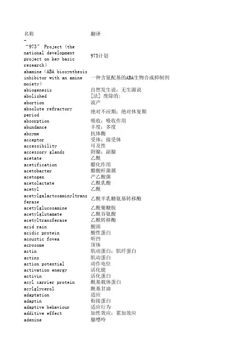
名称翻译-“973” Project (the nationa973计划abamine(ABA biosynthesis in一种含氨配基的ABA生物合成抑制剂abiogenesis自然发生说;无生源说abolished[法] 废除的;abortion流产absolute refractory period绝对不应期;绝对休复期absorption吸收;吸收作用abundance丰度;多度abzyme抗体酶acceptor受体;接受体accessibility可及性accessory glands附腺;副腺acetate乙酰acetification醋化作用acetobacter醋酸杆菌属acetogen产乙酸菌acetolactate乙酰乳酸acetyl乙酰acetylgalactosaminyltransfer乙酰半乳糖氨基转移酶acetylglucosamine乙酰葡糖胺acetylglutamate乙酰谷氨酸acetyltransferase乙酰转移酶acid rain酸雨acidic protein酸性蛋白acoustic fovea听凹acrosome顶体actin肌动蛋白;肌纤蛋白actins肌动蛋白action potential动作电位activation energy活化能activin活化蛋白acyl carrier protein酰基载体蛋白acylglycerol酰基甘油adaptation适应adaptin衔接蛋白adaptive behaviour适应行为additive effect加性效应;累加效应adenine腺嘌呤adenoviral腺病毒的 adj.adenovirus腺病毒adhesin粘附素adiantum铁线蕨adiponectin脂肪连接蛋白Adipophilin(ADFP)脂着素adipose脂肪adipsin抑渴蛋白adrenal肾上腺adrenal gland肾上腺adrenocortical肾上腺皮质adriamycin阿霉素adsorption吸附;吸附作用adventitious root不定根affinity chromatography亲和层析法agamous无性的agar琼脂;琼胶;洋菜胶agar medium琼脂培养基agar plate琼脂平面agaricales伞菌目age class龄级age distribution年龄分布age group年龄群age-dependent年龄-依赖性agenesis器官缺失畸形agglutination凝集aggregation聚集agmatine胍基丁胺agon辅基agonist拮抗因子air bubble气泡air embolism气栓air humidity大气湿度air space气室airlift bioreactor气升式生物反应器albumen(1)蛋白;蛋清,(2)清蛋白;白蛋白,(3)胚乳albumin清蛋白;白蛋白albuminous cell蛋白细胞alcohol醇alcoholic醇的alcoholic fermentation酒精发酵aldosterone醛甾酮,醛固酮alga藻;藻类algae藻类alignment联配aliquot等量样本;等分部份alkaline碱alkaline protein碱性蛋白质alkaline soil碱性土壤allantois尿囊allele frequency等位基因频率allelopathy异种克生[现象]allergen过敏素;致敏素;变应素allium葱属alloantigen同族抗原allometry异速生长allophycocyanin别藻蓝素allosteric modulator别构调节酶,别构效应物allotetraploid异源四倍体alloxan阿脲alluvial plain冲积平原alluvial soil冲积土aloe芦荟属alopecia秃发alpha helix a螺旋alpha-galactosidaseα-半乳糖苷酶altitudinal海拔alum矾,明矾aluminium oxide矾土alveolar肺泡alveolus腺泡Alzheimer disease阿尔茨海默病,老年性痴呆amaryllidaceae石蒜科amaurosis黑朦amelogenin成釉蛋白amensalism偏害共栖Amersham发玛西亚,安玛西亚Ames test埃姆斯试验amide酰胺; 胺amido black酰胺黑amino acid permease氨基酸透性酶aminoethyl氨乙基aminoglycan氨基聚糖aminolevulinate氨乙酰丙酸aminomethyl氨甲基aminopterin氨基蝶呤aminopurine氨基嘌呤aminotransferase转氨酶amitosis无丝分裂ammonium molybdate钼酸铵ammonium sulfate precipitati硫酸铵沉淀amniocentesis羊膜穿刺术amorphous无定形的amp腺苷一磷酸amphetamine安非他明;苯异丙胺amphibian两栖动物amphibians两栖amphioxus厦门文昌鱼amphipoda端足目amphiregulin双调蛋白amphisome自噬内涵体ampicillin氨芐青霉素;氨芐青霉素ampoule安瓿;针药管amur东北amylase淀粉酶[包括糖化酶和麦芽糖化酶] amylase inhibitor淀粉酶抑制剂amyotrophic肌萎缩性An Iterative Algorithm迭代算法an underestimate低估数据anaerobe厌氧生物;厌氧菌anal fin臀analogous protein类似蛋白质analytical balance分析天平analytical chemistry分析化学anatomist解剖学家anatomy(1)解剖,(2)解剖学anchor锚anchorin锚定蛋白androecium雄蕊群anemophily风媒anesthetic麻碎剂;麻碎的aneuploidy非整倍性angel天使蛋白angina咽峡炎angiogenin血管生成蛋白angiomyolipomas血管肌脂过多angiopoietin血管位蛋白angiospermae被子植物angiostatin血管稳定蛋白angle角度animal behavior动物行为animal husbandry畜牧;畜牧业animal kingdom动物界animal protein动物性蛋白质aniridia无铱症anisohydric behavior非等水行为ankyloglossia舌粘连annexin膜联蛋白annual plant一年生植物annulus环带ant plants蚁植物antagonist拮抗剂,拮抗物antenna触角anterior pituitary胸垂体前叶anterograde顺行性anther花药anther culture药培养antheridium雄器anthocyanidin花色素:;花青素anthocyanin花青苷,花色素苷anthranilate synthase邻氨基苯甲酸合成酶,邻氨基苯甲酸合酶anthrax炭疽热anthropogeny人类起源anthropoid类人猿antibody protein抗体蛋白质anticancer agent抗癌剂anticipation前发anticlinal wall垂周壁[见于植物]antigen presenting抗原呈递antigen presenting cell抗原呈递细胞,呈递抗原细胞antigenic heterogeneity抗原异质性antimicrobial agent抗微生物剂antimonate锑酸盐antioncogene,tumor suppresso抗癌基因,肿瘤抑制基因antiozonant防臭氧剂antiporter逆向运输antipyretic退热剂 ; 解热剂antiseptic防腐剂 ; 消毒剂 ; 抗菌剂antiserum抗血清antitoxin抗毒素antrum窦 ; 腔anura无尾目aortic arch大动脉弓apex顶点aphididae蚜属aphids蚜虫类apical bud顶芽apical cell顶端细胞,顶细胞apical initial顶端原始细胞Apicomplexanprotozoa顶复合器门的原生动物apicoplast质体样细胞器Apis蜜蜂属apnoea呼吸暂停aponeurosis腱膜apoplast非原质体apoptosis细胞凋亡apoptosome凋亡体; 凋亡复合体; 凋亡小体; 寡聚体apothecium子囊盘apparatus装置;器(官)apparent infection显性感染apparent viscosity表观粘度apple tree苹果树Applied Biosystems, ABI美国应用生物系统中国公司aquaculture(1) 水产养殖, (2) 溶液培养aquarius宝瓶座aquatic animals水生动物aquatic fungi水生真菌类aqueous水的,含水的araceae天南星科arachidic acid花生酸archaeology考古学Archaeopteryx始祖鸟属argininosuccinate精氨琥珀酸argon氩aridity(1) 干旱, (2) 干旱度Armadillo犰狳蛋白artemin, enovin, neublastin神经导向素arterial blood动脉血arterial pressure动脉压 ; 动脉血压arteriosclerosis动脉硬化 ; 动脉硬化症arthropoda节肢动物门artichoke朝鲜蓟articular cartilage关节软骨articular process关节突artifact人工产物artificial cell人工细胞artificial planting人工植树arylalkylamine芳烷基胺arylamine芳胺arylsulfatase芳硫酸酯酶ascocerida袋角石目ascus子囊asepsis无菌aseptically无菌地asparaginase天门冬酰胺酶asparagine天门冬氨aspartase天冬氨酸酶aspartate天冬氨酸;天冬氨酸盐、酯、根aspartic acid天冬氨酸Aspergillus曲霉属; 曲霉属assimilation同化 ; 同化作用associated virus伴随病毒asteraceae菊科astigmatism散光asymmetric PCR不对称PCR[引物用量不对称的PCR] ATCC ATCCatmospheric pollution大气污染atopy过敏症attenuation减毒作用auditory canal听道Aurelia海月水母属auricle(1) 心耳, (2) 叶耳aurora kinases极光激酶authentic真的,真实的,真正的Autoanalyzer[商]自动分析仪[Technicon公司商标] autoclave高压灭菌器; 消毒蒸锅autofluorescence自体荧光autogamy自体受精; 自核交配autograft自体组织移植片automaticity自动节律性autophagy自噬autoregulatory自调控Autosis自死亡autotic自死亡的avian鸟类的avifauna区系avirulent strain减毒株,无毒株axillary region腋区axons神经轴突Axygen爱思进azoospermia无精症azospirillum巴西固氮b cell epitope b细胞抗原决定基B cell lymphoma B细胞淋巴瘤Bacillus subtillis枯草芽胞杆菌backbone脊柱bacteria culture细菌培养bacterial motility细菌运动性bacterial virulence细菌毒力bacteriochlorophyll细菌叶绿素; 菌绿素bacteriocin细菌素bacteriophage噬菌体bacterioplankton异养浮游细菌bacteriostasis抑菌作用bacteroid类菌体balance of nature自然平衡balanced diet均衡膳食band centrifugation method层带离心法bar chart条线图 ; 棒形图 ; 条形图barbate具毛的barbiturate巴比妥酸盐barbituric acid巴比妥酸bark树皮baroreceptor reflex压力感受拼射Barr body巴氏体 ; 巴尔氏小体 ; 性染色质小体barren不孕baseplate基片basic ingredient基础成分basic number基数Basidiomycetes担子菌纲Basidiomycotina担子菌亚门basonuclin碱性核蛋白,碱核蛋白basophilic嗜碱的bast fiber韧皮纤维batch process分批工艺,分批法bathochromically红移bats蝙蝠Bdellovibrio bacteriovorus蛭弧菌beclin苄氯素bee venom蜂毒benign良性benzimidazoles苯甲亚胺醇benzyl alcohol苯甲醇bile胆bile duct胆管biliary duct胆管binding energy结合能binocular vision双眼视觉bioburden微生物量Bioceramics生物陶瓷biochip生物芯片biodegradable生物可降解的biodegradation生物降解 ; 生物降解作用Bio-energizer生物促生剂bioenergy生物能量biofeedback生物反馈biofilm生物薄膜,生物膜,生物被膜,微生物膜biogenesis生源说biogeographic region生物地理区biogeography生物地理bioinformatics生物信息学biolistic technology生物弹道技术biological glucoside生物甙Biological invasion生物入侵biological nitrogen fixation生物固氮酌biological products生物制品biological rhythm生物节律bioluminescence生物发光biomarker生物标志物Biomass Energy生物质能源biomimetic synthesis仿生合成biomimetics生体模仿学biomolecular生物大分子bionics仿生学biophysics生物物理学Bio-rad伯乐生物biosecurity生物安全biosensor生物传感器biostratigraphical生物地层的biotechnology生物技术 ; 生物科技biotelemetry生物遥测学biotic community生物群落群biotinylation生物素化biotransformation生物转化biotype(1) 生物型 ; 生物类型, (2)同型小种 ; biovar生物变型bird migration鸟迁移bird of prey猛禽类bird ringing鸟类环子birdsong鸟鸣birth出产bisexual flower两性花biting insect咬虫bitopic membrane proteins双顶膜蛋白bivalent(1) 二价的 , (2) 二价染色体blastospore芽生孢子bleaching agent漂白剂blight疫病blind spot盲点blinded study参加者不知情的研究blood circulation血循环blood flow血量blood group antigen血型抗原blood screening血液筛查blood type血型blunt end钝圆末端Bohr effect波尔效应boletus牛肝菌属bone骨,骨骼bone age骨龄bone ash骨灰bone lamella骨层板bone marrow transplantation骨髓移植bone meal骨粉bone tissue骨组织boron deficiency硼缺乏botulinum肉毒菌botulism肉毒中毒bound water结合水bract苞片brain cortex大脑皮质brainstem脑干branch point分支点[如见于RNA剪接]branchial鳃的BRAND普兰特breadths宽度breeding season繁殖期brefeldin布雷菲(尔)德菌素Brief Introduction简介broad leaved forest落叶林bromophenol blue溴酚蓝bromothymol blue溴百里酚蓝; 溴百里香酚蓝, 溴麝香草酚蓝bronchitis支气管炎bronchus支气管brown alga褐藻brownian motion布朗运动bryophyte苔藓植物 ; 苔藓类bryophytes苔藓Buchner funnel布氏漏斗buffer缓冲剂bulldog clamp动脉夹bullous大疱bupleurum柴胡属buster大体butterflies蝴蝶butyrylcholinesterase丁酰胆碱脂酶by product副产物C. elegans;Caenorhabditis e线虫,秀丽隐杆线虫cache缓冲cadherin钙粘着蛋白calciferol钙化醇calcification钙化 ; 钙化作用calcineurin钙调磷酸酶calcitonin降血钙蛋白calcium binding protein钙结合蛋白(质)calculating machine计算器calcyclin钙周期蛋白,钙(细胞)周期蛋白calibration curve校准曲线callose胼胝质,愈伤葡聚糖callosum胼胝质callus differentiation愈伤组织分化calmodulin钙调蛋白,钙调节蛋白,钙调素calnexin钙联结蛋白calpain钙蛋白酶camalexin一种植物保护素cambrian period寒武纪campaniform钟状的cancer cell癌细胞cane sugar甘蔗糖cannabinoid大麻成分capillary毛细管capping加帽,加帽反应;封闭反应;帽化,成帽反应capping protein加帽蛋白,戴帽蛋白caproyl己酸基capsid(病毒)衣壳,(病毒)壳体capsule(1) 荚膜, (2) 孢蒴, (3) 蒴果captivity笼养caragana锦鸡儿属carbamate氨基甲酸脂carbohydrate糖carbon balance碳平衡carbon dioxide fixation碳酸气固定carbonyl羰基carboxykinase羧激酶carboxylate羧酸cardiac glycoside强心苷cardiovascular心血管cardiovascular disease心血管疾病carex苔草属cargo笼carotenoid类胡萝卜素; 类叶红素carrying capacity(1) 携带力 ; 负载力 , (2) 容纳量cascade级联caspase胱冬酶caste阶级; 级castor bean蓖麻籽castration去势catabolite分解代谢物catalytic center催化中心catalytic constant催化常数,转换数[符号Kcat]catalytic mechanism催化机理catalytic subunit催化亚基catalyzer催化剂catenation链状排列catenin连环蛋白cathelicidin组织蛋白酶抑制素catheter导管cave洞穴caveolae(复)陷窝,小窝,小凹cavity腔cecropin蚕素cell adhesion molecule细胞粘着分子,细胞粘附分子cell biology细胞生物学cell fusion细胞融合 ; 细胞并合cell line细胞株cell membrance细胞膜cell membrane(细)胞膜cell migration细胞迁移cell nucleus细胞核cell polarity细胞极性cell protein细胞蛋白质cell respiration细胞呼吸cell strain细胞株cell theory,theory of cell细胞学说Cell-map proteomics细胞器蛋白质组学,细胞图谱蛋白质组学cellular respiration细胞呼吸 ; 细胞呼吸作用cellulase纤维素cellulose decomposing fungi纤维素分解真菌类cement gland黏腺center中心center of origin起源中心centrifugal separation离心分离(法)centrifuge离心机centrifuge tube离心管centriole中心粒centrum椎体cephalopod头足动物 ; 头足类cephalosporin头孢菌素cercaria摇尾幼虫; 尾蚴cereals谷粒cerebral cortex大脑皮质cerebrospinal fluid脑脊液降钙素基因相关肽CGRP,calcitonin gene related peptidechalaza(1)卵带; 卵黄系带, (2) 合点chalazal合点端chalone抑素,抑制素channel管道channel theory经络学说chaperone protein伴侣蛋白质,伴娘蛋白chara轮藻character字符characteristic性状charged particle带电粒子charophyte轮藻植物chemokine趋化因子chemotaxis趋化性chemotaxonomic分类学chenopodiaceae藜科植物Chest PA & LAT胸部正侧位chi square test检验chiasma frequency交叉频率chilling injury寒害chimeric嵌合的chimeric gene嵌合基因chimeric protein嵌合体蛋白质chiral手性的chitinase几丁酶chloral氯醛chloramphenicol氯霉素; 氯霉素chloramphenicol acetyltransf氯霉素乙酰转移酶Chlorella绿藻属; 绿球藻属chlorenchyma绿色组织chlorogenic acid绿原酸chlorophyllbody叶绿体chlorophyll-protein叶绿素蛋白chlorophyta绿藻chlorophyte绿色植物chlorpyrifos毒死蜱,氯吡硫磷cholecystokinin缩胆囊素; 胆囊收缩素cholera toxin霍乱毒素[分A、B亚基]cholesterol胆固醇cholesterol oxidase胆固醇氧化酶cholic acid胆酸choline esterase胆碱酯酶cholinesterase胆碱脂chondroitin软骨蛋白chondrosarcoma软骨瘤chorda tympani鼓索chorionic绒膜chromatin染色质chromatin condensation染色质凝聚chromatographic column层析柱chromophore生色团chromosomal aberration染色体畸变; 染色体异常chromosome mapping染色体作图,染色体定位chromosome polymorphism染色体多态性chromosome segregation染色体分离chronic慢性的chukar石鸡chymotrypsinogen胰凝乳蛋白 原cilium纤毛circadian生理节律circadian rhythm(近)昼夜节律circulation循环怜circulation of blood血循环circulatory system循环系统cisplatin[商]顺式铂氨,顺氯氨铂[一种抗癌药] citric acid柠檬酸cladistic分支系统学clasper交合突; 鳍脚class纲clavulanic acid棒酸clay粘土cleavage site切割位点climate气候climax community顶极群落clinical microbiology临床微生物学clinical trial临床实验clonal activation克隆活化clonal expansion克隆扩充clone克隆cloning无性繁殖系化closure闭合clot凝块clumped distribution丛生分布Cluster of Orthologous Group蛋白相邻类的聚簇CMP CMPcnidoblast刺细胞CNQX CNQXCNTF CNTFCoA CoAcoat protein外壳蛋白; 病毒蛋白外壳;病毒蛋白外壳;(病毒)外壳蛋白coating antigen包被抗原cocarboxylase辅羧酶coccus球菌cochlea耳蜗coding sequence编码序列codominant共显性种codon密码子codon bias密码子偏倚(性)coeliac artery腹腔动脉coenzyme辅cohesin黏连蛋白colchicine秋水仙碱colinearity共线性collagen胶原collagenase胶原酶collar襟collum颈colonization定居colony菌落color reaction呈色反应colouration着色coma昏迷commitment定型comparative morphology比较形态学Comparative proteomics analy比较蛋白质组学研究compartmentalization区域化compartmentation区域化compensation补偿compensation depth补偿深度compensation point补偿点competence感受态competent活性的,有能力的,(处于)感受态的competitive capacity竞争能力competitive inhibition竞争性抑制作用complement补体[补体系统包括C1-C9等多种成分]complementation互补(作用)complete response完全有效conazole唑类;一般是后缀中使用concatemer多联体,串联体,连环体;噬菌体]串联体concatenation连环酌concentration浓度concentration gradient浓度梯度concerted model齐变模式concretion结石condensation(1) 缩合, (2)凝聚; 凝结condiments刀conditional gene条件基因conductivity传导性conductivity detector电导率检测器cone(1) 视锥, (2) 球果 ; 球花configuration构形confluent culture铺满培养物,铺满培养congestion充血conglomerate聚块conidium分生孢子conifer针叶树; 松柏类植物conjugated bile acid结合胆汁酸conjugated protein缀合朊; 结合蛋白质;结合蛋白质;结合蛋白(质)conjugation接合connectomes连接组Connectomics连接组学consanguineous marriage血亲联婚consecutive连串的,相连续的conserved domain保存区段consortium聚生体[一种以上细菌聚集在一起,具有相对固定的依存关系] constipation便秘constriction狭窄construction构建,组建contamination污染Content of Heavy Metal重金属含量contig重叠群,毗连(序列)群[一组从基因组中克隆的毗连DNA序列] contiguity邻接continental shelf大陆架continuous passage culture连续传代培养(物)continuous variation连续变异contour line等高线contour map等高线图,等值线图control对照control gene第基因control plot对照小区conus芋螺convergence会聚[用于神经系统];趋同conzyme辅酶cooling jacket冷却套cooling tower冷却塔cooperative协同的cooperative behavior协同行为cooperative effect协同效应,合作效应coordination协调coordination bond配位键copepod桡足类copulation(1) 接合, (2) 交配cordycepin3'-脱氧腺苷;蛹虫草菌素cormorant鸬鹚Corn Straw玉米秸杆Corning康宁coronary heart disease冠心病correlation coefficient相关系数corrosion protection防腐cortex皮层corticoid类皮质激素cortin皮质激素cortisol皮质醇cortisone可的松cosmid黏粒cosmopolitan广布种costus lactone木香内酯cosurfactant助表面活性剂cotranslational integration共翻译整合cotranslational secretion共翻译分泌cotton棉花counter electrode对电极counterstaining对染(法),复染(法)[一种对比染色法] coupled偶联的coupled reaction偶联反应coupled transport偶联输送cover degree覆盖度cover glass盖玻片CpG island CpG岛cramp痉挛cre表达crecricetulus仓鼠cricket蟋蟀crickets蟋蟀CRO,contract research orgniz合同研究组织crops罪cross protection交叉保护cross section横切面crosstalk串华,通讯cruciferae十字花科植物crust地壳土壤crustacea甲壳动物cryogen冷冻剂,制冷剂cryopreservation深低温保藏[法]cryoprotectant冷冻保护剂cryptobiosis隐生cryptorchidism隐睾crystal structure晶体结构crystallization结晶(作用)cucumber黄瓜culm空心秆cultivar培养变种culture培养; 培养物culture collection培养物保藏culture tube培养(试)管cupule(1) 杯形托, (2) 胞芽杯, 93) 杯形器, (4) 壳斗current density电流密度cushion垫层cuticle角质膜cutis皮肤cuvette比色皿cyan氰cyanobacteria蓝细菌cyanobacterial蓝藻cyanobacterial toxins藻毒素cyanobacterium蓝细菌cybrid胞质杂种cycas苏铁属cycle循环cyclin细胞周期蛋白cyclophilin亲环蛋白,亲环素,环孢素A结合蛋白cymbidium兰属cystathionine胱硫醚cystoma囊瘤cytochemistry细胞化学cytochrome c oxidase细胞色素c氧化酶cytochrome oxidase细胞色素氧化酶cytokine细胞因子cytokine network细胞因子网络cytokinesis胞质分裂 ; 细胞质分裂cytokinin细胞分裂素; 细胞激动素cytological细胞学的cytomegalovirus巨细胞病毒cytometer血球计数器血球计cytometric[医] 细胞计数的;cytoneme胞管cytopathic effect(致)细胞病变(效应)cytopenia细胞减少(症)cytoplasmic membrane细胞质膜cytoplasmic tail胞质尾区[如指跨膜蛋白的胞内小区]cytosol细胞溶质cytotaxonomy细胞分类学cytotoxic T lymphocyte细胞毒性T淋巴细胞cytotrophoblast细胞滋养层dairy cow乳牛de novo n. 重新, 更始,【生物学】从头dead space无效空间decane癸烷decomposer分解者[可指具有分解动植物残体或其排泄物能力的微生物] defective缺损的,缺陷的defective virus缺损病毒,缺陷病毒defensin防卫素,防御素defined medium确定成分培养基,已知成分培养液deforestation滥伐 ; 滥伐林木deformation变形degenerate简并的degradation退化dehiscent fruit裂果dehydration脱水(作用)dehydrogenase脱氢dehydrogenation脱氢(作用)deiodinase脱碘酶deionized water去离子水delayed fluorescence迟延荧光delayed hypersensitivity迟发过敏性delayed recovery恢复延迟deletion mutagenesis缺失诱变delphinium翠雀属deme同类群 ; 混交群体demethylation脱甲基化denaturants变性剂dendron树突denitrification反硝化作用density gradient密度梯度dental齿骨dental caries龋齿dentine齿质deoxycytidine脱氧胞苷deoxysugar脱氧糖depletion排除depressed脱阻抑的deprived除去的,丧失的derivate衍生物dermatan皮层dermatitis皮炎dermatoglyphics肤纹desaturase去饱和酶desaturases去饱和酶descendants后代descent谱系description描述desertification沙漠化desmin结蛋白desmocollin桥粒(芯)胶(粘)蛋白desquamation脱鳞desulfurization脱硫(作用)detention滞留determination测定detritus碎屑; 腐质developmental genetics发生遗传学device仪器devonian period泥盆纪dextran葡聚糖,右旋糖酐dextran sulfate葡聚糖硫酸酯dextrin糊精dhamotil复方地芬诺酯片,芬诺酯和阿托品diabetes insipidus尿崩症diagnostic procedure诊断程序,诊断手续dianthus石竹属diapause(1) 滞育, (2) 休眠期diarrhea腹泻diastolic pressure舒张压; 心舒压dichotomy双歧分枝Dickkopf-1Wnt通路抑制因子dielectrophoresis介电(电)泳diester二酯differential centrifugation差速离心differential expression差异表达Differential-Display PCR, DD差别显示聚合酶链反应diffusion扩散dihybrid双基因杂种dihydrofolate二氢叶酸dihydrotestosterone二氢睾酮dihydroxyacetone二羟丙酮dihydroxyvitamin二羟维生素diluent稀释剂,稀释液dimethylarginine二甲基精氨酸dinitrophenol二硝基苯酚dinoflagellate腰鞭毛虫; 甲藻; 涡鞭藻dioecious雌雄异体的dioecism雌雄异体,雌雄异株dioxygenase双加氧酶diphtheria白候dipterocarpaceae龙脑香科discontinuous variation不连续变异dish平皿Dishevelled,Dsh或Dvl蓬乱蛋白disinfection消毒dislocation转换位置dispersal散布displacement位移disproportionation岐化(反应)disturbance of consciousness意识障碍disulfide bond二硫键disulfide linkage二硫键dna binding protein dna 结合蛋白质DNA ligase DNA 连接DNA typing DNA分型docking protein船坞蛋白,停靠蛋白,停泊蛋白dodecane十二烷dolphins海豚domain域,区域,结构域,功能域dominance显性;优势(度)dominant allele显性等位基因dominant gene显性基因dominant lethal显性致死dosimeter剂量计double chin二重颊double-reciprocal plot双倒数作图,Lineweaver_Burk作图dove鸠dragonflies蜻蜓drebrin脑发育蛋白drift漂变drosophila果蝇属drug abuse&dependence毒品滥用和依赖drug resistant strain抗药性菌株dry weight干重duality二重性duct管duplication复制dynamic balance动态平衡dynamin发动蛋白,缢断蛋白dynein动力蛋白dyslipidemia血脂异常dysregulation调节异常early gene早期基因eating habits饮食习惯ecdysis蜕皮ecdysone蜕皮素;蜕皮激素ecg心电图echinoderm棘皮类Echinodermata棘皮动物门echinoderms棘皮动物eclampsia子癫症ecological factor生态因素ecotone群落交错区;群落过渡带ecotope生态区ectoblast外胚层ectodomain胞外域ectoplasm外质effect of gene基因剂量效应elaioplast油质体elasticity弹性elastoma高弹性electric focusing电子聚焦electrofusion电融合electrolyte solution电解质溶液electromotive force电动势electron diffraction电子衍射electron microscope电(子显微)镜electron transfer电子传递,电子转移electrophoresis pattern电泳图谱electrophysiological propert电生理特性electrophysiology电生理学electroporated电穿孔electroporation电转化electrospinning电纺丝,静电纺丝electrotransformation电转化(法)element元素elevational海拔elfin矮人elicitins激发素elicitor诱导子elimination消去,消除;弃置elongated spermatids精子细胞elongation延伸eluant洗脱液eluate洗脱物,洗出液Eluted[医] 被洗脱的,被洗提的; embedding medium包埋剂embryo culture胚胎培养embryogenesis胚形成embryoid胚状体embryonal胚的emetine吐根碱[属异喹啉类生物碱] emotional reaction情写应emphysema肺气肿encephalon脑髓encephalopathy脑病变endangering濒危endemic特有种,特有的,地方性的,地方的endemic disease地方病endocrine内分泌endocuticle内表皮endocytosis胞吞endoderm内胚层endodermis内皮endogenous内源(性)的endogenous opioid peptide内源性阿片样肽endogenous protective system内生保护系统endomembrane system内膜系统endometrioid子宫内膜endopeptidase内 ; 键内切endoplasmic reticulum (ER)内质网endosymbiont内共生体endothecium药室内壁energetics能量学energy band能量带energy charge能荷energy conservation能量守恒energy crop能源作物energy transduction能量转移entomological昆虫entomophily虫媒entropy熵enucleation去核environmental conditions环境条件environmental pollution环境污染environmentalist环境问题专家enzymatically adv.enzymatically modified lecit酶改性磷脂enzyme kinetics酶动力学enzyme system酶系统eosinophilia嗜伊红粒细胞增多症eosinophils嗜伊红粒细胞ephedra麻黄属ephemeral短命ephrin蝶素epidemic流行病epidemic hemorrhagic fever流行性出血热epidemiology流行病学epidermis表皮epigenetics发育遗传学,表观遗传学epigenome表观基因组epinasty偏上性episome附加体,游离体episperm种皮epistatic gene上位基因epithelium上皮细胞epitope mapping表位作图,表位定位epitope tagging表位附加,表位追加[把附加的抗原表位融合到目的蛋白上] epoxide hydrolase环氧化物酶epoxygenase环氧化酶Eppendorf tube[商]Eppendorf离心管,微量离心管eppin细粒蛋白Equisetum木贼属equivalence等价equivalent当量ergosterol麦角固醇;麦角甾醇error correction错误校正erythroid网织红细胞erythroid differentiation fa红细胞分化因子erythropoiesis红血球生成;红血细胞生成;造红血球;造红血细胞ester酯estival夏季的estrogen雌激素estrous cycle动情周期estrus动情期ethnobotanical民族植物学ethological行为学的ethyl alcohol乙醇ethylenimine氮杂环丙烷etiolation黄化;黄化现象etoposide鬼臼亚乙苷eudicot双子叶植物;eukaryon真核eukaryotic真核生物的eutrophic lake富营养湖evolutionarily进化上exchange group可交换组[比较相似并常可互换的氨基酸残基]excimer受激子excitatory postsynaptic pote兴奋性突触后电位excreted排泄; 排出; 分泌; 排除( excrete的过去式和过去分词 ); Exendin-4唾液素4[暂名]exocyst泡外exocytosis胞吐酌exon shuffling外显子改组exonuclease核酸外切酶exosmosis外渗exosome外切体expiration呼explantation外植exponential growth指数生长expressed sequence tag序列表达标签Expression proteomics表达蛋白质组学expression vector表达载体external外的extine外壁extracellular胞外的;细胞外的extracellular fluid胞外液;细胞外液exudate渗出液eyebrow眉face shield面罩factor因子falciform镰刀状的Fallopian tube法类皮欧氏管;输卵管familial adenomatous polypos家族性多发性腺癌fang长牙fatal dose致死量fatty liver脂肪肝fatty substance脂肪物质favus白癣fecundity生育力;生殖力felling采伐female flower雌花ferment酶fermentation发酵fern蕨类ferns蕨类fertility受精能力fertilized已受精的fertilizer肥料ferulic acid阿魏酸fiber plant纤维植物fibrinogen纤维蛋白原fibrinolysis(血)纤(蛋白)溶(解)fibroblast成纤维细胞fibroblastic tumor纤维母细胞瘤fibronectin纤连蛋白fibrosis纤维化fibrous protein纤维状蛋白; 纤维状蛋白质; 纤维状朊;纤维状蛋白质;....field study实地考察;野外考察filamentous丝状的filter press压榨滤净器filtering过滤fin ray条finch雀fingerprint指纹,指纹结构[例如核酸或蛋白质的酶切消化物在双向电泳中显示的特征first leaf第一叶fish protein concentrate鱼粉; 浓缩鱼蛋的;浓缩鱼蛋的;鱼蛋白浓缩物flagellin鞭毛蛋白flanking sequence侧面序列flat bone扁平骨flats平原flavone黄酮flavonoid类黄酮flesh果肉flexion屈曲flexor muscle屈肌floor plate底板flora植物区系;植物相floral植物的floras区系florigen成花素flow瘤flow cell流动池flow cytometer流式细胞仪flower color gene花色基因flowering plant有花植物fluid mosaic model[膜的]流动镶嵌模型,流体镶嵌模型fluorescence microscopy荧光显微镜检术fluorescence recovery after 光脱色荧光恢复技术fluoroacetate氟乙酸;氟乙酸盐、酯、根fluorochrome荧光燃料flurbiprofen氟比洛芬foam cells泡沫细胞focal adhesion焦点粘连focal distance焦距focus聚焦foetus胎儿food protein食用蛋白forelimb前肢对称性formyl甲酸基formyltransferase甲酰转移酶fossa窝fractalkine曲动蛋白fraction分段fragmentation断裂frame rate帧率freeze冻结Freon[商]氟利昂frequency频度frequency index频率指数friction摩擦friction coefficient摩擦系数frizzled,Frz卷曲蛋白frontal analysis迎头(分析)法fru果糖fructofuranosidase呋喃果糖苷酶fructosamine果糖胺fructose果糖fucan岩藻多糖fucose岩藻糖fucosyltransferase岩藻糖转移酶fuel cell燃料电池fumarase延胡索酸酶fumarate延胡索酸fumaric acid反丁烯二酸Funaria葫芦藓属functional differentiation机能分化functional gene功能基团functional group官能团functional localization功能定位functional protein功能蛋白functional redundancy功能性丰余,功能丰余性functional response机能反应fundus眼底fungi真菌fungicide杀霉菌剂funiculus菌丝索furry毛皮fusion融合;并合G1 Phase G1期gadfly牛虻gain增益gait步态galactosyltransferase半乳糖基转移酶galea外叶gallbladder胆galliformes鸡形目galvanometer电疗gametogenesis配子发生ganglioside神经节苷脂garlic大蒜gastritis胃炎gastrointestinal cancer胃肠癌gastrulation原肠胚形成Gaucher disease戈谢病,高雪病gazelle羚GC content GC含量gel mobility shift assay凝胶迁移率变动分析,凝胶移位分析gelation凝胶作用gelsolin凝溶胶蛋白gena颊gene action基因作用gene analysis基因分析gene bank基因库; 基因文库;基因文库gene carrier基因携带者gene clone基因克隆gene cloning基因克隆化;基因无性繁殖gene cluster基因簇gene combination基因组合gene complex基因综合体gene content基因含量gene conversion基因转变gene copy基因复制; 基因拷贝;基因拷贝gene deletion基因缺失gene dosage基因剂量;基因量;基因数量; 基因剂量gene engineering遗传工程;基因工程gene flow基因怜;基因流; 基因流动;基因流动; &n....gene frequency基因频率;等位基因频率gene fusion基因融合gene information基因信息gene interaction基因互作; 基因相互作用;基因相互酌;基因相互作用gene location基因定位gene loci基因位点gene locus基因位点;基因座位gene map基因图;基因图谱gene mapping基因定位; 基因定位; 基因图;基因图; &....gene order基因序列gene pairs基因对gene pool基因储备;基因库; 基因文库gene recombination基因重组gene theory基因论;基因学说gene therapy基因疗法;基因治疗; 基因治疗general anesthesia全身麻醉genetic遗传的genetic distance遗传距离genetic engineering基因工程,遗传工程,遗传工程学genetic immunization基因免疫接种[采用基因疫苗进行接种]genetic screening遗传筛选genetic transformation遗传转化[例如一个品系的生物吸收另一品系生物的遗传物质,并获得后一genetic variance遗传方差genetics遗传学Genistein’,5,7三羟基异黄酮genome基因组,染色体组Genome Mismatch Scanning基因组错配扫描genomic imprinting基因组印记[配子发生过程中基因的选择性差异表达]genomics基因组学genophore基因线,基因带gentamicin艮他霉素gentianaceae龙胆科geranium老鹳草属geranylgeranyl香叶酰香叶酰germ cell胚细胞germ plasm种质, 生殖质germinal center发芽中心germination萌发;萌发作用germline生殖细胞系ghost菌蜕gibberellic acid赤霉酸;赤霉酸gibberellin赤霉素;赤霉素glacial period冰川时代glaciation冰河酌gland腺体glandular epithelium腺上皮glass beads玻璃念珠gley horizon潜育层glia胶质Global Proteomics整体蛋白质组学glomerulosclerosis肾小球硬化症glottis声门glucocorticoid糖皮质(激)素glucomannan葡甘露聚糖glucosamine葡糖胺,氨基葡糖glucose oxidase葡萄糖氧化酶glucosidase葡糖苷酶glucuronyl葡糖醛酸基glutaminase谷酰胺酶glutelin谷蛋白glycinebetaine甘氨酸甜菜碱glycoconjugates糖缀合物,糖复合物glycophorin血型糖蛋白glycoprotein糖蛋白glycosphingolipid鞘糖脂Glyoxal Oxidase乙二醛氧化酶glyoxylate cycle乙醛酸循环GM-CSF GM-CSFGMP GMPgneiss片麻岩GnRH GnRHgoblet cell杯形细胞gonads生殖腺grain protein content籽粒蛋白含量Gram stain革兰氏染液gramicidin A短杆菌肽Agramineae禾本科植物granulation tissue肉芽组织granuloma肉芽肿granulose细菌淀粉粒granzyme颗粒酶grape procyanidins葡多酚green plant绿色植物groundwater level地下水位grove林growth curve生长曲线growth promoter生长促进剂grus鹤GTP-Binding Proteins GTP结合蛋白质类guanosine鸟尿环核甙guild种群guinea pig豚鼠gull鸥gus gus酶habitat栖所haemagglutinin血球凝集素hail霰hail damage雹害hair follicle毛囊halophilic嗜盐菌halophilic bacteria嗜盐菌halosere盐生演替系列hamamelidaceae金缕梅科Hantaan virus汉坦病毒,汉滩病毒,汉他病毒harder哈德Hardy-Weinberg equilibrium哈迪-温伯格平衡hazard ratio风险比HBAg HBAgHBcAg乙型肝炎核心抗原HBeAg HBeAgHBsAg HBsAg。

![一种抗衰老的药物组合物及其制备方法和用途[发明专利]](https://uimg.taocdn.com/21f6d00d81c758f5f71f670e.webp)
专利名称:一种抗衰老的药物组合物及其制备方法和用途专利类型:发明专利
发明人:张芝庭,张涛涛
申请号:CN202110257936.2
申请日:20210310
公开号:CN112717121A
公开日:
20210430
专利内容由知识产权出版社提供
摘要:本发明公开了一种抗衰老的药物组合物,涉及医药技术领域。
包括以下重量份的原料:重组人表皮生长因子10‑16份,鸡胚胎20‑30份,小球藻提取物8‑12份和山药提取物30‑45份。
本发明提供的抗衰老的药物组合物使用安全,无任何毒副作用,能对衰老的根源予以干预,且通过将该药物组合物制备成贴膏剂,改变了传统的抗衰老产品的注射剂型,减轻了患者的疼痛,使用方便。
申请人:贵州神奇药物研究院
地址:550081 贵州省贵阳市北京路1号
国籍:CN
更多信息请下载全文后查看。
MINI REVIEW ARTICLEpublished:17September2013doi:10.3389/fonc.2013.00221 Role of epithelial-mesenchymal transition in pancreatic ductal adenocarcinoma:is tumor budding the missing link? Eva Karamitopoulou1,2*1Clinical Pathology Division,Institute of Pathology,University of Bern,Bern,Switzerland2Translational Research Unit,Institute of Pathology,University of Bern,Bern,SwitzerlandEdited by:Inti Zlobec,University of Bern, SwitzerlandReviewed by:Parham Minoo,University of Calgary, CanadaQianghua Xia,The Children’s Hospital of Philadelphia,USA*Correspondence:Eva Karamitopoulou,Clinical Pathology Division,Institute of Pathology,University of Bern, Murtenstrasse31,CH-3010Bern, Switzerlande-mail:eva.diamantis@pathology.unibe.ch Pancreatic ductal adenocarcinoma(PDAC)ranks as the fourth commonest cause of cancer death while its incidence is increasing worldwide.For all stages,survival at5years is<5%. The lethal nature of pancreatic cancer is attributed to its high metastatic potential to the lymphatic system and distant ck of effective therapeutic options contributes to the high mortality rates of PDAC.Recent evidence suggests that epithelial-mesenchymal transition(EMT)plays an important role to the disease progression and development of drug resistance in PDAC.Tumor budding is thought to reflect the process of EMT which allows neoplastic epithelial cells to acquire a mesenchymal phenotype thus increasing their capacity for migration and invasion and help them become resistant to apoptotic signals. In a recent study by our own group the presence and prognostic significance of tumor budding in PDAC were investigated and an association between high-grade budding and aggressive clinicopathological features of the tumors as well as worse outcome of the patients was found.The identification of EMT phenotypic targets may help identifying new molecules so that future therapeutic strategies directed specifically against them could potentially have an impact on drug resistance and invasiveness and hence improve the prognosis of PDAC patients.The aim of this short review is to present an insight on the morphological and molecular aspects of EMT and on the factors that are involved in the induction of EMT in PDAC.Keywords:pancreatic cancer,epithelial-mesenchymal transition,tumor budding,prognosis,biomarkerPANCREATIC CANCERPancreatic ductal adenocarcinoma(PDAC)is a common can-cer with dismal prognosis(1)that escapes early detection and resists treatment(2).Most patients have advanced stage dis-ease at presentation with a median survival of less than1year (1,3).Surgical resection is the only potentially curative treat-ment of PDAC(3).Classical histomorphological features like tumor size,blood vessel,or lymphatic invasion,and presence of lymph node metastases constitute essential prognostic deter-minants in pancreatic cancer and are invariably included in the pathology reports,with tumor stage being the most important of all(3).The lethal nature of PDAC has been attributed to the propensity of PDAC cells to rapidly disseminate to the lym-phatic system and distant organs(4).However,even patients with completely resected,node-negative PDACs eventually die of their disease.Within this context and considering the fact that the management of PDAC remains suboptimal and that adjuvant therapy has resulted to limited progress,the identification of addi-tional reliable and reproducible prognostic markers that would enable better patient stratification and eventually provide a guide toward a more successful and individualized therapy,is mandatory (1,5).EPITHELIAL-MESENCHYMAL TRANSITIONEpithelial-mesenchymal transition is a biologic process that allows epithelial cells to undergo the biochemical changes that enable them to acquire a mesenchymal phenotype,including enhanced migratory capacity,invasiveness,elevated resistance to apoptosis, and increased production of extracellular matrix(ECM)compo-nents(6,7).EMT is characterized by loss of cell adhesion,down regulation of E-cadherin expression,acquisition of mesenchy-mal markers(including N-cadherin,Vimentin,and Fibronectin), and increased cell motility(6).Both EMT and mesenchymal-epithelial transition(MET),the reversion of EMT,are essential for developmental and repair processes like implantation,embryo for-mation,and organ development as well as wound healing,tissue regeneration,and organfibrosis(8).However,EMT also occurs in neoplastic cells that have undergone genetic and epigenetic changes.These changes affect both oncogenes and tumor sup-pressor genes that enable cancer cells to invade and metastasize. Moreover,some neoplastic cells may go through EMT retaining many of their epithelial properties while other cells are becoming fully mesenchymal(9).Many molecular processes are involved in the initiation of EMT including activation of transcription factors,expression of specific cell-surface proteins,reorganization and expression of cytoskeletal proteins,production of ECM-degrading enzymes,and changes in the expression of specific microRNAs(miRNAS).The above fac-tors can also be used as biomarkers to detect cells in EMT state(10). EMT has been linked to cellular self-renewal programs of cancer stem cells and apoptosis-anoikis resistance,which are features of therapeutic resistance(11).The zincfinger transcription factors Snail,Slug,Zeb1,and Twist repress genes responsible for the epithelial phenotype and represent important regulators of EMT(6,7,12).In PDAC Snail expression has been reported to be seen in nearly80%of the cases and Slug expression in50%(13).Snail expression was inversely correlated with E-cadherin expression and decreased E-cadherin expression was associated with higher tumor grade. Similarly,poorly differentiated pancreatic cancer cell lines showed higher levels of Snail and lower levels of E-cadherin compared with moderately differentiated cell lines(13)while silencing of Zeb1leaded to up-regulation of E-cadherin and restoration of an epithelial phenotype(14).Zeb1expression in PDAC also corre-lated with advanced tumor grade and worse outcomes(14–16) and was shown to be primarily responsible for the acquisition of an EMT phenotype,along with increased migration and inva-sion in response to NF-κB signaling in pancreatic cancer cells (16).EMT AND TUMOR BUDDINGTumor budding reflects a type of diffusely infiltrative growth con-sisting of detached tumor cells or small cell clusters of up tofive cells at the invasive front of gastrointestinal carcinomas(17–22). Tumor buds represent a non-proliferating,non-apoptotic,highly aggressive subpopulation of tumor cells that display migratory and invasive capacities(23).The aim of tumor buds seems to be the invasion of the peritumoral connective tissue,the avoidance of the host’s defense andfinally the infiltration of the lymphatic and blood vessels with the consequence of local and distant metastasis. The EMT process by allowing a polarized cell to assume a more mesenchymal phenotype with increased migratory capacity,inva-siveness,and resistance to apoptosis seems to play a major role in the development of tumor buds.In fact,tumor buds are thought to result from the process of EMT.Thus,although formally tumor budding cannot be equated with EMT,several similarities between the two processes,including activation in WNT signaling,can be shown(24).The detachment of tumor buds from the main tumor body is accomplished by loss of membranous expression of the adhesion molecule E-cadherin.Activation of WNT sig-naling is further suggested by nuclear expression of b-catenin in tumor-budding cells,as well as increase of laminin5gamma2and activation of Slug and Zeb1(24,25).The presence of high-grade tumor budding has been consis-tently associated with negative clinicopathologic parameters in gastrointestinal tumors(26–30).In a previous study from our group we could show that tumor budding occurs frequently in pancreatic cancer and is a strong,independent,and reproducible, highly unfavorable prognostic factor that may be used as a para-meter of tumor aggressiveness and as an indicator of unfavorable outcome,even within this group of patients with generally poor prognosis.Moreover,tumor budding was proven to have a more powerful prognostic ability than other more classic prognostic fac-tors including TNM stage,thus adding relevant and independent prognostic information(31).EMT AND miRNAsMicroRNAS are small non-coding RNAs of18–25nucleotides, excised from60to110nucleotide RNA precursor structures (32).MiRNAs are involved in crucial biological processes, including development,differentiation,apoptosis,and pro-liferation,through imperfect pairing with target messenger RNAs of protein-coding genes and the transcriptional or post-transcriptional regulation of their expression(33,34).Recent studies illustrate the role of miRNAs on the regula-tion of gene expression and proteins in metastasis.For exam-ple,it has been shown that miR-10b,which is up-regulated by EMT transcription factor Twist,is associated with increased invasiveness and metastatic potential(35,36).Furthermore,it was shown that the miR-200family(miR-200a,miR-200b,miR-200c,miR-141,and miR-429)and miR-205play critical roles in regulating EMT by directly targeting the mRNAs encoding E-cadherin repressors Zeb1and Zeb2(37).Moreover,recent studies showed that members of the miR-200family by induc-ing EMT can regulate the sensitivity to epidermal growth fac-tor receptor(EGFR)in bladder cancer cells and to gemcitabine in pancreatic cancer cells(38).Conversely,Zeb1represses the transcription of miR-200genes by directly binding to their promoter region,thereby forming a double-negative feedback loop(39).On the other hand,miR-200family can also pro-mote the conversion of mesenchymal cells to epithelial-like cells (MET)suggesting that these miRNAs may also favor metastatic outgrowth.Recent studies aiming at the evaluation of miRNAs in pan-creatic cancer have shown that specific miRNAs are dysregulated in PDAC while the higher expression of some miRNA species was able to distinguish between benign and malignant pancre-atic tissue(40).For example,miR-21was shown to be over-expressed in79%of pancreatic cancers as opposed to27%of chronic pancreatitis(41).In resected PDAC specimens high lev-els of miR-200c expression strongly correlated with E-cadherin levels and were associated with significantly better survival rates compared with patients whose tumors had low levels of miR-200c expression(42).CHEMORESISTANCE AND EMTCells undergoing EMT become invasive and develop resistance to chemotherapeutic agents.Moreover,EMT can be induced by chemotherapeutic agents,and stress conditions such as exposure to radiation or hypoxia(43,44).Up-regulation of Twist has been shown to be associated with resistance to paclitaxel in nasopharyngeal,bladder,ovarian,and prostate cancers(45).In colorectal cancer cell lines,chronic expo-sure to oxaliplatin leaded to the development of the ability to migrate and invade with phenotypic changes resembling EMT(spindle-cell shape,loss of polarity,intercellular separa-tion,and pseudopodia formation)by the oxaliplatin-resistant cells(46).Pancreatic cancer remains today an extremely lethal disease largely because of its resistance to existing treatments(47).EMT has been shown to contribute significantly to chemoresistance in several cancers,including pancreatic cancer(30,48,49).Induction of gemcitabine resistance in previously sensitive cell lines resulted in development of an EMT phenotype and was associated with an increased migratory and invasive ability compared to gemc-itabine sensitive cells(49).Moreover,gene expression profiling ofchemoresistant cells showed a strong association between expres-sion of the EMT transcription factors Zeb1,Snail,and Twist and decreased expression of E-cadherin(39,50).Silencing of Zeb1 with siRNA resulted to MET(51)and restored chemosensitivity (14).Interestingly,maintenance of chemoresistance in cell lines that have undergone EMT is dependent on Notch and NF-κB signaling(30).Inhibition of Notch-2down regulates Zeb1,Snail, and Slug expression,attenuates NF-κB signaling,and reduces the migratory and invasive capacity of the gemcitabine resistant cells(30).Epithelial-mesenchymal transition can also confer resistance to targeted agents.For example,lung cancer cell lines that have undergone EMT,became resistant to the growth inhibitory effects of EGFR kinase inhibition(erlotinib)in vitro and in xenografts(47)as well as other EGFR inhibitors such as gefitinib and cetuximab(48)Thus,EMT can lead to resis-tance to multiple agents and result to rapid progression of the tumor.Clarifying the correlation between EMT and drug resistance may help clinicians select an optimal treat-ment.CONCLUSIONPancreatic cancer remains an extremely lethal disease partly because of the poor response to existing treatments.Accumulat-ing evidence suggests that EMT plays an important role in PDAC progression,is associated with stem cell features of the PDAC cells and seems to significantly contribute to the chemoresistance of pancreatic cancer.Moreover,is associated with more aggressive tumor characteristics and with poor patient survival.Because of its role in therapy response and tumor progression,targeting EMT could potentially reduce drug resistance and have a great impact in the survival of PDAC patients.Tumor budding thought to be the result of the EMT process is commonly observed in PDAC and high-grade tumor budding has been proven to have an independent adverse prognostic impact in the survival of PDAC patients.Figure1depicts tumor bud-ding as a possible transition between a fully epithelial and a fully mesenchymal phenotype of the tumor cells in PDAC.Moreover, cancer cells in tumor buds have been shown to have EMT and cancer stem cell characteristics.The further characterization of the budding cells at a protein and gene level in order to iden-tify a“molecular budding-promoting profile”will lead to a better understanding of the tumor-stroma interaction at the area of the invasive front and help to further elucidate the similarities between budding cells,EMT process and cancer stem cells in pancreatic cancer.Investigating these issues will allow us to gain further insight into pancreatic carcinogenesis,and provide us with a platform on which to build future studies leading to the identification of new therapeutic interventions.REFERENCES1.Hidalgo M.Pancreatic cancer.NEngl J Med(2010)362:1605–17.doi:10.1056/NEJMra09015572.Tuveson DA,Hingorani SR.Duc-tal pancreatic cancer in humans and mice.Cold Spring Harb Symp Quant Biol(2005)70:65–72.doi:10.1101/ sqb.2005.70.0403.Fernandez-del-Castillo C,JimenezRE,Steer ML.Surgery in the treatment of exocrine pancreas and prognosis.In:Tanabe KK,edi-tor(2013).Available from:www.4.Li Y,Kong D,Ahmad A,Bao B,Sarkar FH.Pancreatic cancer stem cells:emerging target for designing novel therapy.Cancer Lett(2012).Available from:/10.1016/j canlet.2012.03.018,5.Welsch T,Kleeff J,Friess H.Molecu-lar pathogenesis of pancreatic can-cer:advances and challenges.Curr Mol Med(2007)7:504–21.doi:10.2174/1566524077813870826.Kalluri R,Weinberg RA.The basicsof epithelial-mesenchymal transi-tion.J Clin Invest(2009)119:1420–8.doi:10.1172/JCI391047.Thiery JP,Acloque H,Huang RYJ,Nieto MA.Epithelial-mesenchymal transitions in development and dis-ease.Cell(2009)139:871–90.doi:10.1016/j.cell.2009.11.0078.Rasheed ZA,Yang J,Wang Q,Kowal-ski J,Freed I,Murter C,et al.Prog-nostic significance of tumorigenic cells with mesenchymal features in pancreatic adenocarcinoma.J Natl Cancer Inst(2010)102:340–51.doi:10.1093/jnci/djp5359.Iwatsuki M,Mimori K,YokoboriT,Ishi H,Beppu T,Nakamori S.Epithelial–mesenchymal transi-tion in cancer development and its clinical significance.Cancer Sci (2010)101(2):293–9.doi:10.1111/j.1349-7006.2009.01419.x10.Kalluri R.EMT:when epithelial cellsdecide to become mesenchymal-like cells.J Clin Invest(2009)119:1417–9.doi:10.1172/JCI3967511.Krantz SB,Shields MA,Dangi-Garimella S,Munshi HG,Bentrem DJ.Contribution of epithelial-to-mesenchymal transition and cancer stem cells to pancreatic cancer progression.J Surg Res(2012)173: 105–12.doi:10.1016/j.jss.2011.09.02012.Lee JM,Dedhar S,Kalluri R,Thompson EW.The epithelial-mesenchymal transition:new insights in signaling,devel-opment,and disease.J Cell Biol(2006)172:973–81.doi:10.1083/jcb.20060101813.Hotz B,Arndt M,Dullat S,BhargavaS,Buhr HJ,Hotz HG.Epithelial tomesenchymal transition:expressionof the regulators snail,slug,andtwist in pancreatic cancer.ClinCancer Res(2007)13:4769–76.doi:10.1158/R-06-292614.Arumugam T,Ramachandran V,Fournier KF,Wang H,MarquisL,Abbruzzese JL,et al.Epithe-lial to mesenchymal transition con-tributes to drug resistance in pan-creatic cancer.Cancer Res(2009)69:5820–8.doi:10.1158/0008-5472.CAN-08-281915.Buck E,Eyzaguirre A,Barr S,Thompson S,Sennello R,Y oung D,et al.Loss of homotypic cell adhe-sion by epithelial-mesenchymaltransition or mutation limits sen-sitivity to epidermal growth factorreceptor inhibition.Mol CancerTher(2007)6:532–41.doi:10.1158/1535-7163.MCT-06-046216.Maier HJ,Schmidt-Strassburger U,Huber MA,Wiedemann EM,BeugH,Wirth T.NF-kappaB promotesepithelial-mesenchymal transition,migration and invasion of pancre-atic carcinoma cells.Cancer Lett(2010)295:214–28.doi:10.1016/j.canlet.2010.03.00317.Prall F.Tumour budding in col-orectal carcinoma.Histopathology(2007)50:151–62.doi:10.1111/j.1365-2559.2006.02551.x18.Brown M,Sillah K,Griffiths EA,Swindell R,West CM,Page RD,et al.Tumour budding and a lowhost inflammatory response areassociated with a poor progno-sis in oesophageal and gastro-oesophageal junction cancers.Histopathology(2010)56:893–9.doi:10.1111/j.1365-2559.2010.03559.x19.Koike M,Kodera Y,Itoh Y,Nakayama G,Fujiwara M,Hamajima N,et al.Multivariateanalysis of the pathologic featuresof esophageal squamous cell cancer:tumor budding is a significantindependent prognostic factor.AnnSurg Oncol(2008)15:1977–82.doi:10.1245/s10434-008-9901-620.Miyata H,Y oshioka A,YamasakiM,Nushijima Y,Takiguchi S,Fujiwara Y,et al.Tumor bud-ding in tumor invasive front pre-dicts prognosis and survival ofpatients with esophageal squa-mous cell carcinomas receivingneoadjuvant chemotherapy.Cancer(2009)115:3324–34.doi:10.1002/cncr.2439021.Roh MS,Lee JI,Choi PJ.Tumorbudding as a useful prognosticmarker in esophageal squamous cellcarcinoma.Dis Esophagus(2004)17:333–7.doi:10.1111/j.1442-2050.2004.00436.x22.Ohike N,Coban I,Kim GE,Bas-turk O,Tajiri T,Krasinskas A,etal.Tumor budding as a strongprognostic indicator in invasiveampullary adenocarcinomas.Am JSurg Pathol(2010)34:1417–24.doi:10.1097/PAS.0b013e3181f0b05a23.Zlobec I,Lugli A.Epithelial mes-enchymal transition and tumorbudding in aggressive colorectalcancer:tumor budding as oncotar-get.Oncotarget(2010)1:651–61.24.Muto T,Mochizuki H,Masaki T edi-tors.Tumor Budding in ColorectalCancer:Recent Progress in ColorectalCancer Research(Horizons in Can-cer Research).(Vol.8).Tokyo:Nova(2006).25.Schmalhofer O,Brabletz S,Brabletz T.E-cadherin,beta-catenin,and ZEB1in malignantprogression of cancer.CancerMetastasis Rev(2009)28:151–66.doi:10.1007/s10555-008-9179-y26.Karamitopoulou E,Zlobec I,KölzerV,Kondi-Pafiti A,Patsouris ES,Gen-natas K,et al.Proposal for a10-high-power-fields scoring methodfor the assessment of tumor bud-ding in colorectal cancer.ModPathol(2013)26:295–301.doi:10.1038/modpathol.2012.15527.Nakamura T,Mitomi H,KikuchiS,Ohtani Y,Sato K.Evaluationof the usefulness of tumor bud-ding on the prediction of metasta-sis to the lung and liver after cura-tive excision of colorectal cancer.Hepatogastroenterology(2005)52:1432–5.28.Ueno H,Mochizuki H,HashiguchiY,Hatsuse K,Fujimoto H,HaseK.Predictors of extrahepatic recur-rence after resection of colorectalliver metastases.Br J Surg(2004)91:327–33.doi:10.1002/bjs.442929.Ueno H,Murphy J,Jass JR,Mochizuki H,Talbot IC.Tumour‘budding’as an indexto estimate the potential ofaggressiveness in rectal cancer.Histopathology(2002)40:127–32.doi:10.1046/j.1365-2559.2002.01324.x30.Wang LM,Kevans D,Mulcahy H,O’Sullivan J,Fennelly D,Hyland J,et al.Tumor budding is a strong andreproducible prognostic marker inT3N0colorectal cancer.Am J SurgPathol(2009)33:134–41.doi:10.1097/PAS.0b013e318184cd5531.Karamitopoulou E,Zlobec I,BornD,Kondi-Pafiti A,Patsouris E,Gen-natas K,et al.Tumor budding isa strong and independent prognos-tic factor in pancreatic cancer.EurJ Cancer(2013)49:1032–9.doi:10.1016/j.ejca.2012.10.02232.Calin GA,Croce CM.Micro RNAsignatures in human cancers.NatRev Cancer(2006)6:857–66.doi:10.1038/nrc199733.McShane L,Altman DG,SauerbreiW,Taube SE,Gion M,Clark GM,etal.REporting recommendations fortumor MARKer prognostic studies(REMARK).Eur J Cancer(2005)41:1690–6.doi:10.1016/j.ejca.2005.03.03234.Pasquinelli AE,Hunter S,BrachtJ.MicroRNAs:a developing story.Curr Opin Gen Dev(2005)15:200–5.doi:10.1016/j.gde.2005.01.00235.Peter ME.Let-7and miR-200microRNAs:guardians againstpluripotency and cancer progres-sion.Cell Cycle(2009)8:843–52.doi:10.4161/cc.8.6.790736.Preis M,Gardner TB,Gordon SR,Pipas JM,Mackenzie TA,Klein EE,et al.MicroRNA10b expression cor-relates with response to neoadju-vant therapy and survival in pan-creatic ductal adenocarcinoma.ClinCancer Res(2011)17:5812–21.doi:10.1158/R-11-069537.Gregory PA,Bert AG,Paterson EL,Barry SC,Tsykin A,Farshid G,et al.The miR-200family and miR-205regulate epithelial to mesenchymaltransition by targeting ZEB1andSIP1.Nat Cell Biol(2008)10:593–601.doi:10.1038/ncb172238.Burk U,Schubert J,Wellner U,Schmalhofer O,Vincan E,SpadernaS,et al.A reciprocal repressionbetween ZEB1and members of themiR-200family promotes EMT andinvasion in cancer cells.EMBO Rep(2008)9:582–9.doi:10.1038/embor.2008.7439.Wellner U,Schubert J,Burk UC,Schmalhofer O,Zhu F,Sonntag A,et al.The EMT-activator ZEB1pro-motes tumorigenicity by repress-ing stemness-inhibiting microR-NAs.Nat Cell Biol(2009)11:1487–95.doi:10.1038/ncb199840.Panarelli NC,Chen YT,ZhouXK,Kitabayashi N,Yantiss RK.MicroRNA expression aids thepreoperative diagnosis of pancre-atic ductal adenocarcinoma.Pan-creas(2012)41:685–90.doi:10.1097/MPA.0b013e318243a90541.Dillhoff M,Liu J,Frankel W,CroceC,Bloomston M.MicroRNA-21is overexpressed in pan-creatic cancer and a potentialpredictor of survival.J Gas-trointest Surg(2008)12:2171–6.doi:10.1007/s11605-008-0584-x42.Yu J,Ohuchida K,Mizumoto K,Sato N,Kayashima T,Fujita H, et al.MicroRNA,hsa-miR-200c, is an independent prognos-tic factor in pancreatic cancer and its upregulation inhibits pancreatic cancer invasion but increases cell prolifera-tion.Mol Cancer(2010)9:169.doi:10.1186/1476-4598-9-169 43.Li C,Heidt DG,Dalerba P,BurantCF,Zhang L,Adsay V,et al.Identification of pancreatic can-cer stem cells.Cancer Res(2007) 67:1030–7.doi:10.1158/0008-5472.CAN-06-203044.Lee CJ,Dosch J,Simeone DM.Pan-creatic cancer stem cells.J Clin Oncol(2008)26:2806–12.doi:10.1200/JCO45.Hong SP,Wen J,Bang S,ParkS,Song SY.CD44-positive cells are responsible for gemc-itabine resistance in pancreatic cancer cells.Int J Cancer(2009)125:2323–31.doi:10.1002/ijc.2457346.Hermann PC,Huber SL,HerrlerT,Aicher A,Ellwart JW,GubaM,et al.Distinct populations ofcancer stem cells determine tumorgrowth and metastatic activity inhuman pancreatic cancer.Cell StemCell(2007)1:313–23.doi:10.1016/j.stem.2007.06.00247.Li Y,VandenBoom TG,Kong D,Wang Z,Ali S,Philip PA,et al.Up-regulation of miR-200and let-7bynatural agents leads to the reversalof epithelial-to-mesenchymal tran-sition in gemcitabine-resistant pan-creatic cancer cells.Cancer Res(2009)69:6704–12.doi:10.1158/0008-5472.CAN-09-129848.Yang AD,Fan F,Camp ER,vanBuren G,Liu W,Somcio R,etal.Chronic oxaliplatin resistanceinduces epithelial-to-mesenchymaltransition in colorectal cancercell lines.Clin Cancer Res(2006)12:4147–53.doi:10.1158/1078-R-06-003849.Shah AN,Summy JM,Zhang J,Park S,Parikh N,Gallick GE.Development and characterizationof gemcitabine-resistant pancre-atic tumor cells.Ann Surg Oncol(2007)14:3629–37.doi:10.1245/s10434-007-9583-550.Shimono Y,Zabala M,ChoRW,Lobo N,Dalerba P,QianD,et al.Downregulation ofmiRNA-200c links breast cancerstem cells with normal stemcells.Cell(2009)138:592–603.doi:10.1016/j.cell.2009.07.01151.Conroy T,Paillot B,Francois E,Bugat R,Jacob JH,Stein U,etal.Irinotecan plus oxaliplatin andleucovorin-modulatedfluorouracilin advanced pancreatic cancer–aGroupe Tumeurs Digestives of theFederation Nationale des Centres deLutte Contre le Cancer study.J ClinOncol(2005)23:1228–36.doi:10.1200/JCO.2005.06.050Conflict of Interest Statement:Theauthor declares that the research wasconducted in the absence of anycommercial orfinancial relationshipsthat could be construed as a potentialconflict of interest.Received:24July2013;accepted:11August2013;published online:17Sep-tember2013.Citation:Karamitopoulou E(2013)Role of epithelial-mesenchymal tran-sition in pancreatic ductal adenocar-cinoma:is tumor budding the miss-ing link?Front.Oncol.3:221.doi:10.3389/fonc.2013.00221This article was submitted to Gastroin-testinal Cancers,a section of the journalFrontiers in Oncology.Copyright©2013Karamitopoulou.Thisis an open-access article distributed underthe terms of the Creative CommonsAttribution License(CC BY).The use,distribution or reproduction in otherforums is permitted,provided the origi-nal author(s)or licensor are credited andthat the original publication in this jour-nal is cited,in accordance with acceptedacademic practice.No use,distribution orreproduction is permitted which does notcomply with these terms.。
关于灵芝孢子粉的文章英文回答:Reishi Spore Powder: A Potent Medicinal Fungus with Numerous Health Benefits.Reishi spore powder is a potent natural supplement derived from the fruiting body of the reishi mushroom (Ganoderma lucidum). It has been used in traditional Chinese medicine for centuries and has gained increasing popularity in recent years due to its remarkable health benefits.Nutritional Composition.Reishi spore powder is rich in various nutrients, including polysaccharides, triterpenes, proteins, and amino acids. Polysaccharides, such as beta-glucans, are known for their immune-modulating properties. Triterpenes, such as ganoderic acids, have antioxidant and anti-inflammatoryeffects.Health Benefits of Reishi Spore Powder.1. Immune System Support.Reishi spore powder has been shown to enhance immune function by stimulating the production of natural killer cells, T cells, and macrophages. This makes it beneficial for fighting infections, boosting immunity, and reducing the risk of chronic diseases.2. Anti-Cancer Properties.Studies have suggested that reishi spore powder may have anti-cancer effects. It contains compounds that promote apoptosis (programmed cell death) in cancer cells and inhibit tumor growth.3. Anti-Inflammatory Effects.Triterpenes in reishi spore powder exert anti-inflammatory properties that can help reduce inflammation throughout the body. This makes it beneficial for conditions such as arthritis, asthma, and inflammatory bowel diseases.4. Antioxidant Activity.Reishi spore powder is a potent antioxidant that protects cells from damage caused by free radicals. Free radicals are highly reactive molecules that contribute to aging, disease, and cancer.5. Liver Support.Reishi spore powder has been shown to protect the liver from damage caused by toxins and inflammation. It can improve liver function and promote detoxification.6. Cardiovascular Health.Reishi spore powder may help improve cardiovascular health by lowering cholesterol levels, reducing bloodpressure, and improving blood flow.7. Stress and Anxiety Relief.Reishi spore powder contains compounds that promote relaxation and reduce anxiety. It can help improve sleep quality and reduce stress levels.Dosage and Usage.The recommended dosage of reishi spore powder varies depending on individual needs and health goals. It is generally recommended to start with a low dose and gradually increase it as needed. It can be taken in capsule or powder form.Safety Considerations.Reishi spore powder is generally considered safe when taken in recommended doses. However, it should not be taken by individuals with blood clotting disorders or those who are taking blood thinners. Pregnant women and nursingmothers should consult their healthcare provider before taking reishi spore powder.Conclusion.Reishi spore powder is a highly beneficial natural supplement with a wide range of health-promoting effects. It supports immune function, has anti-cancer and anti-inflammatory properties, provides antioxidant protection, supports liver and cardiovascular health, and reduces stress and anxiety. By incorporating reishi spore powder into your daily regimen, you can improve your overall well-being and enhance your health and longevity.中文回答:灵芝孢子粉,一种具有众多健康益处的强大药用真菌。
基于肿瘤微环境的中医药免疫治疗最新进展①高铭王彤杨春宇任香善(延边大学医学院病理学教研室肿瘤研究中心,延吉133002)中图分类号R730.3文献标志码A文章编号1000-484X(2021)04-0506-05[摘要]肿瘤微环境主要包括非肿瘤细胞、细胞因子、趋化因子和细胞外基质,是抗肿瘤免疫的重要部分,在肿瘤的形成和侵袭过程中发挥至关重要的作用。
中医药能够作用于肿瘤微环境中的免疫细胞、细胞因子以及免疫检查点等多个靶点,为肿瘤的治疗提供了新思路。
这篇综述以肿瘤微环境为出发点,阐释了中医药在肿瘤免疫治疗方面的最新进展。
[关键词]肿瘤微环境;中医药;免疫治疗;免疫细胞;肿瘤相关巨噬细胞Latest development of traditional Chinese medicine immunotherapy based on tumor microenvironmentGAO Ming,WANG Tong,YANG Chun⁃Yu,REN Xiang⁃Shan.Department of Pathology,Tumor Research Center,School of Medicine,Yanbian University,Yanji133002,China[Abstract]The tumor microenvironment mainly includes non-tumor cells,cytokines,chemokines and extracellular matrix.It is an important part of anti-tumor immunity and plays a crucial role in the formation and invasion of tumors.Traditional Chinese Medicine can act on multiple targets such as immune cells,cytokines and immune checkpoints in the tumor microenvironment,providing a new idea for the treatment of tumors.Starting from the tumor microenvironment,this review explains the latest progress of traditional Chinese medicine in tumor immunotherapy.[Key words]Tumor microenvironment;Traditional Chinese medicine;Immunotherapy;Immune cells;Tumor-associated macrophages肿瘤的治疗是当今世界最困扰的医疗难题,很多人“谈癌色变”,其原因在于传统的肿瘤治疗手段如手术、放疗和化疗等会产生一定的毒副作用和耐药性,很大程度上降低了患者的生存质量,因此寻求更好的治疗方法是亟待解决的问题[1]。
创必复产品介绍l创必复(生物蛋白海绵)l创必复(FGF+ Collagen)源于国家一类新药,十五重点攻关计划,863重大科技成果。
创必复是基因工程与胶原蛋白纯化两大生物工程技术相结合而制成的一种生物活性材料,属国家三类医疗器械。
它的问视为现代医学治疗创伤,特别是治疗难愈合创伤提供了最新最有意义的治疗方法。
它经过特殊工艺处理,对胶原蛋白(collagen)和成纤维细胞生长因子(FGF)进行表面修饰和改性,在发挥胶原蛋白缓释、可吸收作用的同时,FGF以胶原蛋白为载体,与其特有的受体结合,在受损组织周围维持理想浓度、活性,并与正常细胞分化相匹配,从而达到独特的治疗效果,使其生物功效得到最大发挥,克服了单独使用的不良作用及缺点。
产品特性l[创必复的特性]:l1、主动修复、恢复愈后组织功能;FGF通过促进与创伤修复有关细胞的迅速增殖而推动创面的主动修复l2、提高愈合质量,减少瘢痕形成;缩短创面愈合时间,加快伤口愈合。
l3、良好的止血性:与血液接触后能诱导血小板聚集,激发血小板释放凝血因子,激活凝血酶原,促进血凝块的形成,粘着在渗血的伤口上,在伤口表面形成凝胶层,对损伤的血管起填塞作用,能更有效地达到止血的目的。
l4、神经营养作用: FGF像神经生长因子 (NGF)一样对许多脑区的神经元有维持生存和促进生长的作用.这些脑区包括:额顶、枕叶的皮层;纹状体、隔区脑。
FGF是胶质细胞如轴突神经胶质细胞、星形胶质细胞和施旺细胞的分裂原,体内FGF具有神经营养作用.●⒌低抗原性;由于不含有色氨酸残基,酪氨酸残基的含量也很少,采用酶切技术和钴60灭菌技术制备,纯度高,分子的抗原性很弱、生物相容性优异;●⒍可生物降解性:可水解为氨基酸被人体吸收;●⒎独家使用了国家发明专利《促进脊髓组织损伤愈合的海绵材料其制备方法及应用》技术,知识产权国家保护20年;适用范围l[适用范围]l1、陈旧创面:l糖尿病性溃疡、放射性溃疡、褥疮、瘘窦、粘膜溃疡和糜烂。
附录:常用免疫学名词解释Aabsorption吸收应用特异性抗原与溶液中的抗体结合,形成不溶性复合物而除去抗体,例如用此法处理血清,即称为吸收血清。
用作吸收的抗原称为吸收剂。
accessory cell辅佐细胞特异性免疫应答需要的细胞,但不是实际介导的,通常用于描述抗原呈递细胞(APC)。
acquired immunity获得性免疫机体在生活过程中所获得的免疫力,称为获得性免疫,它与先天性免疫或天然免疫相反。
获得性免疫可分为:自动免疫,被动免疫,体液免疫与细胞免疫。
参看适应性免疫(adaptive immunity),过继性免疫(adoptive immunity),免疫耐受(immune tolerance)。
acquired immunodeficiency Sydrom(AIDS)获得性免疫缺陷综合征(艾滋病)由人类免疫缺陷病毒(HIV)所致的免疫缺陷病。
HIV感染主要引起T淋巴细胞CD4亚群的极度减少。
患者表现为迟发型超敏反应降低或消失,对机会感染菌极其易感,易发生某些少见的,如Kaposi氏肉瘤或Burkitt氏淋巴瘤。
HIV也可引起B 淋巴细胞多克隆性扩增,导致高丙种球蛋白血症。
尽管血清中免疫球蛋白量明显增加,但对抗原不能发生免疫反应。
这种综合征发生于“危险”人群,包括同性恋的男子,滥用静脉药物者,血液或血液制品的接受者,以及某些来自中非或加勒比海的人群。
在“危险”人群的异性伙伴中和AIDS母亲的婴儿中也已发现了这种综合症。
active immunization主动免疫(作用)抗原进入机体起免疫应答(自动免疫)adaptation tolerance适应性耐受生物在长期进化过程,宿主与寄生物在相互反应中,宿主的防御能力选择性地被减弱。
adaptative immunity适应免疫机体与抗原接触而发生的免疫力(包括主动体液免疫与主动细胞免疫)。
adherent cell粘附细胞在体外能粘附于表面的细胞。
银屑病中免疫抑制性细胞及中医药对其调控作用的研究进展①侯怡飞苏琳邹纯朴陈晓张慧敏②胥孜杭(上海中医药大学基础医学院,上海201203)中图分类号R751.05文献标志码A文章编号1000-484X(2021)04-0497-09[摘要]银屑病是一种常见的难治性皮肤病,以过度激活的T细胞产生的异常免疫应答为主要特征。
在机体免疫系统内,免疫抑制性细胞主要包括髓源抑制细胞(MDSCs)、调节性T细胞(Tregs)、调节性B细胞(Bregs)、巨噬细胞(Mφ)等,它们可以预防T细胞等免疫效应细胞的异常活化和增殖,维持免疫稳态。
近年来随着对银屑病研究的深入,发现银屑病中的免疫抑制性细胞存在异常表现,可能影响银屑病的发生发展,但根据文献检索发现,国内医学尤其是中医药对银屑病中免疫抑制性细胞的探讨明显匮乏,故本文以此为切入点,探讨银屑病中免疫抑制性细胞的募集和功能变化以及中医药对此类细胞的调控作用,旨在更深入和全面地了解银屑病的发病机制,为临床上延缓银屑病进程提供新的策略。
[关键词]银屑病;免疫抑制性细胞;中医药;MDSCs;TregsResearch advancements of immunosuppressive cells and its regulation by traditional Chinese medicine in psoriasisHOU Yi-Fei,SU Lin,ZOU Chun-Pu,CHEN Xiao,ZHANG Hui-Min,XU Zi-Hang.School of Basic Medical Sci⁃ence,Shanghai University of Traditional Chinese Medicine,Shanghai201203,China[Abstract]Psoriasis is a common refractory skin disease and characterized by abnormal immune responses produced by over-activated T cells.In the body's immune system,myeloid-derived suppressor cells(MDSCs),regulatory T cells(Tregs),regulatory B cells(Bregs),macrophages(Mφ),etc.are included in immunosuppressive cells,and they prevent abnormal activation and prolifera⁃tion of immune effector cells such as T cells to maintain immune homeostasis.In recent years,with in-depth research on psoriasis,it has been found that there are abnormal expressions of immunosuppressive cells in psoriasis,which may affect the occurrence and de⁃velopment of psoriasis.However,according to literature search,it is found that the research on immunosuppressive cells in psoriasis is obviously lacking in domestic medicine,especially Chinese medicine.Therefore,our article on the basis of this starting point to ex⁃plore the recruitment and functional changes of immunosuppressive cells in psoriasis and the regulatory effects of traditional Chinese medicine on them,aims at a deeper and comprehensive understanding of the pathogenesis of psoriasis and provides new potential strat⁃egies for clinical medicine to delay the progression of psoriasis.[Key words]Psoriasis;Immunosuppressive cells;Traditional Chinese medicine;MDSCs;Tregs银屑病是一种常见的、难以根治且易复发的皮肤病,影响全世界约1.25亿人,以皮肤出现明显红斑、脱屑,甚至干燥出血为主要临床表现,民间俗称“牛皮癣”,给患者造成巨大的身心负担,并严重影响其生活质量[1-2]。
免疫学导论复习知识点第一章绪论免疫(Immunity):指机体识别和排除抗原异物,维持机体生理平衡和稳定的功能。
免疫学(Immunology):研究机体免疫系统的组成(免疫器官、免疫细胞和免疫分子)、结构及其免疫生物学(生理性的和病理性的)功能的学科。
固有性免疫:又称先天免疫性,是机体先天就有的而且始终存在的防御机制。
特点:(1)出生时已具备(早)(2)可稳定性遗传给后代(3)作用广泛:无特异性(4)个体差异不大获得性免疫:是机体通过抗原诱导获得免疫应答而产生的对非自身物质的抵抗性特点:(1)出生后受抗原刺激产生(2)具有特异性(针对性)(3)一般不能遗传(4)个体差异大(5)具有记忆性中枢免疫器官 : 骨髓, 胸腺外周免疫器官:脾脏、淋巴结、皮肤相关的淋巴组织(SALT)、粘膜相关的淋巴组织(MALT)免疫系统三大功能: 1.免疫防御抗感染(immunologic defense)2.免疫稳定消除炎症或衰老细胞(immunologic homeostasis)3.免疫监视控制癌变细胞(immunologic surveillance)第二章抗原抗原:一类能刺激机体免疫系统使之产生特异性免疫应答,并能与相应的应答产物在体内外发生特异性结合的物质。
抗原的两种特性:1.免疫原性(Immunogenicity)-------刺激机体免疫系统产生特异性免疫应答的能力2.免疫反应性(Immunoreactivity)----与相应的应答产物在体内外发生特异性结合的能力免疫原(immunogen):免疫原性完全抗原(complete Ag):两种特性半抗原(hapten):免疫反应性耐受原(tolerogen):免疫耐受变应原(allergen):Ⅰ型超敏抗原决定族(Antigen Determinant,AD:抗原分子中决定抗原特异性的特殊化学集团,又称为表位(epitope)。
AD的数目、性质和空间构象决定抗原特异性;抗原以AD与相应抗原受体及抗体特异性结合功能性抗原决定簇:位于表面的、易被淋巴细胞识别的、可启动免疫应答的决定簇隐蔽性抗原决定簇:为于分子内部的,不能引起免疫应答的决定簇顺序决定簇(线性决定簇):1.一段序列相连的氨基酸片段形成2.多在抗原分子内3.主要由T识别,B也可识别构象决定簇:1.序列上不相连,由天然构象形成2.位于分子内部3.由B识别抗原的结合价:抗原分子中能和抗体分子结合的AD的数目共同抗原:在两种不同的抗原之间可以存在有相同或相似的抗原决定基,称为共同抗原。
菲律宾科学家发明治疗皮肤癌的乳霜
佚名
【期刊名称】《《中国医药指南》》
【年(卷),期】2006(000)001
【摘要】据菲律宾媒体报道,一名菲律宾科学家在皮肤癌的治疗上取得垂大突破。
经临床试验证实,他所发明的一种外用草药乳霜可使患部百分之百痊愈,而且不再复发。
【总页数】1页(P69)
【正文语种】中文
【中图分类】R739.5
【相关文献】
1.澳研究出皮肤癌新疗法有望开发治病乳霜 [J],
2.德科学家用光疗法治疗皮肤癌 [J], 方留民
3.美国科学家将外源基因移植入人体治疗恶性皮肤癌 [J], 无
4.乳霜治疗皮肤癌 [J],
5.上海科学家发现结核病治疗一线药物吡嗪酢酰胺的治病机制,有助新治疗策略的发明 [J],
因版权原因,仅展示原文概要,查看原文内容请购买。
医学英语考试题及答案一、选择题(每题2分,共20分)1. The term "cardiology" refers to the study of which organ?A. HeartB. LungsC. LiverD. Kidneys2. Which of the following is a common symptom of influenza?A. RashB. FeverC. HeadacheD. All of the above3. The abbreviation "MRI" stands for:A. Magnetic Resonance ImagingB. Medical Records IndexC. Medical Research InstituteD. Multiple Risk Indicators4. What does "diabetes" mean in medical terms?A. High blood sugarB. Low blood sugarC. High blood pressureD. Low blood pressure5. The prefix "hyper-" typically means:A. Above normalB. Below normalC. ExcessiveD. Deficient6. Which of the following is a type of cancer?A. MelanomaB. BronchitisC. PneumoniaD. Asthma7. The term "anemia" is used to describe a condition where there is:A. Too much red blood cellsB. Too few red blood cellsC. Too much white blood cellsD. Too few white blood cells8. "Hemoglobin" is a protein in red blood cells that carries:A. OxygenB. Carbon dioxideC. GlucoseD. Water9. Which of the following is a method for measuring blood pressure?A. StethoscopeB. SphygmomanometerC. OtoscopeD. Ophthalmoscope10. The suffix "-itis" usually indicates:A. InflammationB. TumorC. CongestionD. Degeneration答案:1. A2. D3. A4. A5. A6. A7. B8. A9. B 10. A二、填空题(每空1分,共10分)1. The medical term for a break in the skin is a(n) _______.2. The study of the heart and its diseases is known as_______.3. A(n) _______ is a medical professional who specializes in the diagnosis and treatment of diseases of the eye.4. The process of identifying the cause of a disease iscalled _______.5. A person who has a fear of needles is said to have a(n)_______.6. The medical term for a surgical incision is a(n) _______.7. The term "hypertension" refers to _______ blood pressure.8. The abbreviation "HIV" stands for Human _______ Virus.9. The medical term for the removal of fluid from a bodycavity is _______.10. The study of the structure of the body is called _______.答案:1. Laceration2. Cardiology3. Ophthalmologist4. Diagnosis5. Trypanophobia6. Incision7. High8. Immunodeficiency9. Aspiration10. Anatomy三、简答题(每题5分,共30分)1. Explain the difference between a virus and a bacterium.2. What is the function of the thyroid gland?3. Describe the process of blood clotting.4. What is the role of insulin in the body?5. What are the symptoms of a common cold?6. How does the respiratory system work?答案:1. A virus is a small infectious agent that can only replicate inside the living cells of an organism, while a bacterium is a single-celled microorganism that can exist independently and can be beneficial, neutral, or harmful to humans.2. The thyroid gland is a butterfly-shaped gland located in the front of the neck that produces hormones regulating the body's metabolism.3. Blood clotting is a complex process that involves platelets and clotting factors to prevent excessive bleeding when a blood vessel is injured.4. Insulin is a hormone produced by the pancreas that regulates the amount of glucose in the blood, allowing cells to take in glucose from the bloodstream for energy.5. Symptoms of a common cold include a runny nose, sorethroat, coughing, sneezing, and sometimes fever.6. The respiratory system works by taking in oxygen from the air, which is then transported to the blood via the lungs,and expelling carbon dioxide from the body through exhalation.四、翻译题(每题5分,共20分)1. 将“急性阑尾炎”翻译成英文。
1.医学免疫学名词解释部分免疫(immunity):是指机体识别“自己”与“非己”抗原,对自身抗原形成天然免疫耐受,对非己抗原发生排斥作用的一种生理功能。
正常情况下,对机体有利;免疫功能失调时,会产生对机体有害的反应。
1.固有免疫应答(innate immune response):也称非特异性或获得性免疫应答,是生物体在长期种系发育和进化过程中逐渐形成的一系列防御机制。
此免疫在个体出生时就具备,可对外来病原体迅速应答,产生非特异性抗感染免疫作用,同时在特异性免疫应答过程中也起作用。
2.适应性免疫应答(adaptive immune response):也称特异性免疫应答,是在非特异性免疫基础上建立的,该种免疫是个体在生命过程中接受抗原性异物刺激后,主动产生或接受免疫球蛋白分子后被动获得的。
3.免疫防御(immunologic defence):是机体排斥外来抗原性异物的一种免疫保护功能。
该功能正常时,机体可抵御病原微生物及其毒性产物的感染和损害,即抗感染免疫;异常情况下,反应过高会引起超敏反应,反应过低或缺失可发生免疫缺陷。
4.免疫自稳(immunologic homeostasis):是机体免疫系统维持内环境稳定的一种生理功能。
该功能正常时,机体可及时清除体内损伤、衰老、变性的细胞和免疫复合物等异物,而对自身成分保持免疫耐受;该功能失调时,可发生生理功能紊乱或自身免疫性疾病。
5.免疫监视(immunologic surveillance):是机体免疫系统及时识别、清除体内突变、畸变细胞和病毒感染细胞的一种生理功能。
该功能失调时,有可能导致肿瘤发生,或因病毒不能清除而出现持续感染。
6.MALT(mucosal-associated lymphoid tissue):即黏膜伴随的淋巴组织。
是指分布在呼吸道、肠道及泌尿生殖道的粘膜上皮细胞下的无包膜的淋巴组织。
除执行固有免疫外,还可执行局部特异性免疫。
Immunoproteasomes PreserveProtein Homeostasis uponInterferon-Induced Oxidative StressUlrike Seifert,1Lukasz P.Bialy,1,4Fre´de´ric Ebstein,1Dawadschargal Bech-Otschir,1Antje Voigt,2Friederike Schro¨ter,1 Timour Prozorovski,3Nicole Lange,1Janos Steffen,1Melanie Rieger,1Ulrike Kuckelkorn,1Orhan Aktas,3Peter-M.Kloetzel,1,*and Elke Kru¨ger1,*1Institut fu¨r Biochemie CC2,Charite´-Universita¨tsmedizin Berlin,Oudenarder Strasse16,D-13347Berlin,Germany2Medizinische Klinik fu¨r Kardiologie und Angiologie CC13,Charite´-Universita¨tsmedizin Berlin,Charite´platz1,D-10117Berlin,Germany 3Department of Neurology,Medical Faculty,Heinrich-Heine-University Dusseldorf,Moorenstrasse5,D-40225Du¨sseldorf,Germany4Present address:Department of Histology and Embryology,Center for Biostructure Research,Medical University of Warsaw, Chalubinskiego5,02004Warsaw,Poland*Correspondence:p-m.kloetzel@charite.de(P.-M.K.),elke.krueger@charite.de(E.K.)DOI10.1016/j.cell.2010.07.036SUMMARYInterferon(IFN)-induced immunoproteasomes(i-pro-teasomes)have been associated with improved pro-cessing of major histocompatibility complex(MHC) class I antigens.Here,we show that i-proteasomes function to protect cell viability under conditions of IFN-induced oxidative stress.IFNs trigger the pro-duction of reactive oxygen species,which induce protein oxidation and the formation of nascent, oxidant-damaged proteins.Wefind that the ubiquity-lation machinery is concomitantly upregulated in response to IFNs,functioning to target defective ribosomal products(DRiPs)for degradation by i-pro-teasomes.i-proteasome-deficiency in cells and in murine inflammation models results in the formation of aggresome-like induced structures and increased sensitivity to apoptosis.Efficient clearance of these aggregates by the enhanced proteolytic activity of the i-proteasome is important for the preservation of cell viability upon IFN-induced oxidative stress. Ourfindings suggest that rather than having a specific role in the production of class I antigens, i-proteasomes increase the peptide supply for antigen presentation as part of a more general role in the maintenance of protein homeostasis. INTRODUCTIONThe ubiquitin proteasome system(UPS)represents the major ATP-dependent degradation system in eukaryotes that is involved in the maintenance of cellular protein homeostasis. The central task of the26S proteasome is the degradation of poly-ubiquitylated proteins formed by a cascade of E1,E2,and E3 enzymes,which activate,conjugate,and transfer multiple ubiquitin(ub)moieties to protein substrates,targeting these for degradation.Substrates of proteasomes are short-lived regula-tory proteins involved in cell differentiation,cell-cycle regulation, transcriptional regulation,or apoptosis(Kloetzel,2001;Liu et al., 2005).The rapid elimination of proteins by the UPS is also of particular importance under stress conditions,such as heat shock or oxidative stress,that cause the production of misfolded or partially denatured proteins(Goldberg,2003).The20S proteasome,with its active site b subunits b1,b2, and b5,represents the central catalytic unit of the UPS and the catalytic core of the proteasome.The20S catalytic core associ-ates with19S regulator complexes to form the26S proteasome. In addition,the proteasome activator28(PA28)can interact with the20S complex,forming20S-PA28or hybrid proteasomes (19S-20S-PA28)(Tanahashi et al.,2000).In vertebrates,specific catalytically active b subunits,i.e.,b1i/LMP2,b2i/MECL1,and b5i/LMP7,are incorporated into nascent proteasome complexes upon interferon(IFN)-mediated induction to form an alternative proteasome composition(Kloetzel,2001).Two of these subunits (b1i/LMP2,b5i/LMP7)are encoded within the major histocom-patibility complex(MHC)class II region,which led to the term immunosubunits(i-subunits)and immunoproteasome(i-protea-some)(Aki et al.,1994).Together,thesefindings connected i-proteasome function with antigen presentation.Proteasomes are responsible for the generation of peptides derived from path-ogens or cellular proteins that are presented by MHC class I molecules on the cell surface to cytotoxic T cells(CTLs) (Yewdell,2005).i-proteasomes exhibit altered peptidase activities and cleavage site preferences that result in more efficient liberation of many MHC class I epitopes(Kloetzel and Ossendorp,2004; Kru¨ger et al.,2003).The increase in peptide supply by i-protea-somes has a positive effect on efficient MHC class I antigen presentation and seems to be important for triggering an effec-tive early CTL response(Chen et al.,2001;Deol et al.,2007; Toes et al.,2001;Van Kaer et al.,1994).Newly translated poly-peptides,a large pool of which are ubiquitylated,represent an important source for MHC class I epitopes(Reits et al.,2000;Cell142,613–624,August20,2010ª2010Elsevier Inc.613Schubert et al.,2000).It thus was proposed that this pool is mostly composed of defective proteins,and hence these proteins were called defective ribosomal products (DRiPs).The DRiP hypothesis,linking antigen processing to translation,would guarantee that the cellular immune system has access to its substrates before they reach their final destination within or outside the cell (Lelouard et al.,2004;Reits et al.,2000;Schu-bert et al.,2000;Yewdell,2005).However,the idea that a large amount of newly translated proteins should be misfolded or harbor translational errors raised some doubts with regard to the validity of the hypothesis (Vabulas and Hartl,2005;Yewdell and Nicchitta,2006).Cells rapidly shift to i-proteasome formation in response to proinflammatory cytokines produced by the innate immune system early upon infection.i-proteasome formation is a strongly accelerated and transient response (Heink et al.,2005),implying an urgent and timely need for their function.However,the absence of fully functional i-proteasomes in mice did not gener-ally reveal effects on T cell priming or on the hierarchy of known T cell epitopes (Nussbaum et al.,2005).Moreover,in pathogen-challenged tissues,noninfected cells also induce i-proteasome formation in response to cytokines without the need for improved antigen processing (Yewdell,2005).It was therefore suggested that i-proteasomes must have important non-antigen-process-ing functions (Nussbaum et al.,2005;Yewdell,2005).i-proteasome function has been mainly associated with antigen processing and MHC class I antigen presentation.Here,we show that a major function of the i-proteasome is the maintenance of protein homeostasis and the preservation of cell viability under cytokine-induced oxidative stress.RESULTSIFNs Induce a Transient Accumulation of K48-Linked Ub ConjugatesBecause IFNs enhance proteasome-dependent antigen presen-tation efficiency (Kloetzel,2001),we reasoned that IFNs may also enhance the pool of poly-ub conjugates.To test this assumption,we stimulated cells with IFN-g (Figure 1A and Figure S1C available online),-a ,or -b (Figures S1A and S1B).Monitoring of the total amount of poly-ub conjugates over time revealed a significant enrichment of poly-ub conjugates between 4and 12hr after IFN stimulation (Figure 1B and Figure S1).This enrich-ment appeared to be a transient response because the levels of detectable poly-ub conjugates initially increased and the amount of poly-ub conjugates returned to reduced levels between 24and 48hr of IFN stimulation.The accumulation of poly-ub conjugates was independent of the cell type and the type of IFN used and was detectable in both mouse and human cells (Figures 1A and 1B and Figure S1).Concomitant with the induc-tion of i-proteasomes,as determined by increased b 5i/LMP7levels,the amount of poly-ub conjugates gradually declined (Figures 1A and 1B and Figure S1).The strong decrease in poly-ub conjugates after 48hr was due to enhanced protea-some-dependent protein turnover because the poly-ub conju-gates were stabilized by the proteasome inhibitor epoxomicin (Figure 1C and Figure S1E).Previously,it was shown that the rate-limiting pool of free ub can be compensated by depletion of ubiquitylated histone H2A (ubH2A)(Dantuma et al.,2006).Indeed,at time points displaying strongly enriched levels of ub conjugates in response to IFN-g ,we observed decreased levelsC 8 24 48 8 24 48 IFN-γ(h)---+ + +epoxomicin K48K630 2 12 0 2 12 0 2 12 0 2 12 IFN-γ(h)free ubGAPDH K63 to R poly-ub0 1 2 3 4 6 8 12 24 48 IFN-γ(h)Amurine B8fibroblastsD 0 2 4 6 CHX (h)-+-+-+-+ 12h IFN-γIP poly-ubGAPDH β5i/LMP7GAPDHpoly-ub long12γ(h)E f o l d i n c r e a s e u b -s t a i npoly-ub shortK48 to R free ububH2Apoly-ub long poly-ub shortFigure 1.Accumulation of K48Poly-Ub Conjugates Is Caused by IFN-g(A)Murine fibroblasts were stimulated with IFN-g for up to 48hr.Accumulation of poly-ub conju-gates was visualized by immunoblotting (long and short exposure).Transiently decreased levelsof ubH2A and free ub were obtained with the same experimental setup.Increase in LMP7levels served as controls for IFN-induced i-proteasomeformation and GAPDH as a loading control.(B)A significant accumulation of poly-ub conju-gates was visible at 8hr compared to 48hr of IFN stimulation (densitometric evaluation of fourindependent series,±standard deviation [SD];p values from t test).(C)Murine fibroblasts were exposed to IFN-g for 8,24,and 48hr and were either treated with protea-some inhibitor epoxomicin for the last 4hr of theincubation period or left untreated.Proteasomeinhibition resulted in a significant accumulation ofpoly-ub conjugates.(D)In vitro ub conjugation was demonstrated after IFN-g treatment of murine fibroblasts with modifiedforms of His-tagged ub as indicated.Poly-ub conju-gates stimulated by IFN-g are K48linked as shown inthe left panel (using only K48-linked ub chain)and in the right panel (using K63ub modified to arginine at position 63but containing K48-linked ub chain).(E)Ubiquitylated proteins were immunoprecipitated with FK2ub antibody from pulse-labeled and IFN-g -stimulated murine fibroblasts (lane 0:cycloheximide [CHX]).CHX chases demonstrated the dependency of poly-ub protein levels on translation.See also Figure S1.614Cell 142,613–624,August 20,2010ª2010Elsevier Inc.of ubH2A,and slightly decreased free ub levels (Figure 1A and Figure S1C).The functional role of ubiquitylation and thus the fate of the modified proteins largely depend on the type of linkage between the ub moieties within a poly-ub chain (Mukho-padhyay and Riezman,2007).To study which type of poly-ub conjugates was induced in IFN-g -treated cells,we performed in vitro ub conjugation assays using modified forms of epitope-tagged ub.Substitution of the K48residue by arginine (R)almost completely abrogated the formation of poly-ub conjugates,demonstrating that IFN-g preferentially stimulated the synthesis of poly-ub chains with K48linkages (Figure 1D),targeting these ub conjugates for degradation.The inhibitory effect of the K48R ub mutant on IFN-g stimulated ub conjugation was observed despite the presence of other functional K residues.Thus,other K residues within the ub molecule do not significantly contribute to IFN-g -induced polyubiquitylation.A slight stimulation of K63poly-ub chains by IFN-g is negligible,because the IFN-induced ub conjugation can be completely restored using the K63R ub mutant containing a functional K48residue (Figure 1D).As expected,IFN-g treatment induced translation via the mTOR pathway (Kaur et al.,2008).After 2hr of IFN stimulation,we detected increased levels of the phosphorylated ribosomal S6subunit,a marker for increased translational activity (Fig-ure S1F).To determine whether the accumulated poly-ub conjugates derive from newly synthesized proteins,we pulse-labeled IFN-g -treated cells and immunoprecipitated ub-modi-fied proteins.IFN-g induced a strong increase in the amount of poly-ub conjugates (Figure 1E,‘‘0CHX’’)suggesting that primarily newly translated proteins shape the pool of poly-ub substrates in response to IFN-g .In support,cycloheximide0 1 3 48 IFN-γ(h)poly-ub0 2 3 4 6 8 12 24 48 IFN-γ(h)UBE2L6AB20000400006000080000100000120000140000048141825324248IFN-γ(h)U B E 2L 6 (c o p i e s /µl )free Ub 020406080γ%u b -s t a i nGAPDHpoly-ubGAPDHUBE2L6RT-PCR C--+ + 8h IFN-γco si co siUBE2L6170100230Figure 2.Accumulation of Poly-Ub Conju-gates Is a Result of Increased Ub Conjuga-tion Activity in IFN-Stimulated Cells(A)In vitro polyubiquitylation activity was deter-mined in cellular lysates after treatment with IFN-g for up to 48hr with His-tagged ub.Increased polyubiquitylation was detected at early time points after IFN-g induction (1hr)and stable for up to 48hr.(B)Real-time RT-PCR analysis and immunoblot illustrating UBE2L6expression in HeLa cells induced by IFN-g (two independent series ±SD).(C)siRNA knockdown of E2enzyme UBE2L6was performed in HeLa cells treated for 8hr with IFN-g or left untreated.Silencing of UBE2L6(UBE2L6si versus control si)impairs IFN-g -induced upregula-tion of poly-ub conjugates as shown (right panel).UBE2L6knockdown was confirmed by RT-PCR and immunoblotting (GAPDH,loading control;lower panel,densitomeric evaluation of three independent series ±SD).See also Figure S2and Table S1.chase experiments demonstrated that the increase in poly-ub protein levels was strongly dependent on translation (Figure 1E).Thus,the enhanced polyubi-quitylation of newly synthesized proteins in response to IFN-g is accompanied byaugmented de novo protein synthesis.Furthermore,the same experiments also revealed that poly-ub proteins are degraded significantly faster in IFN-g -treated than in untreated cells (see also Figure 4).The Ub Conjugation Machinery Is Remodeled in Response to IFNsTo determine whether the accumulation of poly-ub conjugates is the result of IFN-induced enhanced polyubiquitylation activity,we performed in vitro ubiquitylation experiments with cellular lysates of IFN-g -stimulated cells.Enhanced polyubiquitylation activity was detected already at early time points and did not significantly decrease over the time period analyzed (Figure 2A).This prompted us to study whether IFNs also exert a more general effect on the ubiquitin-proteasome system (UPS).There-fore,we investigated the expression of UPS-related messenger RNAs (mRNAs)in response to IFN-g in a focused microarray approach.IFNs specifically induced 20E3ub ligases.Of the E2ub-conjugating enzymes,only UBE2L6was strongly induced (Table S1),and its mRNA was upregulated more than 50-fold in response to 4hr of IFN treatment (Figure 2B and Figure S2).To study the role of UBE2L6in IFN-induced ub conjugation,we silenced expression of UBE2L6by small interfering RNA (siRNA)to interfere with the thioester cascade.Depletion of UBE2L6significantly abolished the increase in total poly-ub conjugates 8hr after IFN-g stimulation,whereas no differences could be detected in untreated cells (Figure 2C).Thus,UBE2L6-dependent ub conjugation activity plays an important role in accumulation of ub conjugates during the early IFN response.Together,our experiments demonstrate that increased levelsCell 142,613–624,August 20,2010ª2010Elsevier Inc.615of polyubiquitylated proteins in response to IFNs are at least in part the result of augmented ub conjugation activity.Proteasome Activity Is Transiently Decreased in Response to IFNsIn addition to enhanced ubiquitylation activity,a cytokine-dependent partial impairment of proteasomal degradation capacity may also explain the transient increase in cellular levels of poly-ub conjugates.Therefore,we assayed the proteasomal chymotrypsin-like (CT-L)peptide-hydrolyzing activity in total cell lysates of IFN-g -treated HeLa cells (Figure 3A).Indeed,IFN stimulation resulted in a transient decrease of proteasomal CT-L activity 4–12hr after induction,which was restored and even augmented after 24–48hr.This result was confirmed by measurement of in-gel CT-L activity in native polyacrylamide gel electrophoresis (PAGE)and could be assigned to 26S and to PA28-associated proteasome pools (Figures 3B–3D and Figures S3D and S3E).Concomitant with the decreased proteolytic activity between 8and 12hr,we detected transiently lower 26S proteasome levels in response to IFN-g (Figure 3C).To define this phenom-enon in more detail,we immunoprecipitated proteasomes from glycerol gradient fractions enriched for 26S complexes,and immunodetection of the 19S regulator subunit Rpn2fol-lowed.In agreement with the measured decrease in proteolytic activity,we observed a transiently reduced association of 19S regulators with 20S complexes (Figure 3E).Total amounts of 20S complexes,as monitored by the a 4subunit levels,remained unchanged,suggesting that IFN-g induces a transient dissocia-tion of the 20S complexes from the 19S regulator.Next,we studied the kinetics of 26S i-proteasome formation by incorporation of the b 1i/LMP2subunit into the complexes by using native PAGE (Figure 3F).In agreement with the increase in proteasomal activity after 24hr of IFN-g exposure,i26S proteasome levels were strongly enhanced at this time point.Reorganization of proteasomes upon IFN exposure was also shown by immunoprecipitation experiments or two-dimensional separation of purified 20S complexes (Figures S3A–S3C).Impor-tantly,the time frames exhibiting the highest accumulation of poly-ub conjugates corresponded exactly with the decline of proteasomal peptidase activity and coincided with the time period required for the formation of i26S proteasomes.turnover of Suc-LLVY-AMC R F UA0 8 12 24 48 IFN-γ(h)BIP:β7 Ab from 26S gradient fractions0 4 8 12 24 IFN-γ(h)α4Rpn2Cα6A bβ1i /L M P 2A bDE0 8 12 24 48 IFN-γ(h)γ(h)substrate overlay0 812 24 48 IFN-γ(h)immunoblot26S 20S +PA2820S20S + PA2826S26S20S20S + PA28F26S R p n 2A bP A 28βA bhybrid 20S + PA280 8 12 24 48 IFN-γ(h)Figure 3.Altered Proteasome Activity and Composition in Response to IFNs(A)Total 26S chymotryptic peptide-hydrolyzing activity was analyzed in HeLa cells exposed to IFN-g .After a transient decrease in activity between 4and 8hr of IFN-g treatment,chymotryptic activity increases again and peaks at 48hr of IFN-induction (three independent series ±SD).(B–D)Cellular proteasome pools were separated on native PAGE,proteasome population was analyzed by in-gel chymotryptic activity (B)and by immunoblot with a 6,Rpn2(C),and PA28b antibodies (D).(E)26S proteasomes were immunoprecipitated from glycerol gradient fractions with a b 7antibody.Association of 19S subunit Rpn2to 20S proteasome is transiently decreased between 4and 8hr of IFN-g induction.Levels of the constitutive subunit a 4were determined as control.(F)Analysis of 26S i-proteasome amount after IFN-g stimulation on native PAGE with b 1i/LMP2antibody.See also Figure S3.616Cell 142,613–624,August 20,2010ª2010Elsevier Inc.i-Proteasomes Are Required for Efficient Degradation of Polyubiquitylated ProteinsThe above experiments suggest that i-proteasomes may be more efficient in degrading poly-ub substrates than standard proteasomes (s-proteasomes).We therefore examined the removal of poly-ub conjugates in mouse embryonic fibroblasts (MEFs)and human cell lines (T1,T2)with either a fully functional i-proteasome (WT,T1)or cells deficient for one or two of the i-proteasomes subunits (LMP7À/À,T2)after IFN challenge (Figures 4A and 4B and Figure S4A).Furthermore,to study the requirement of i-proteasomes for the degradation of IFN-g induced poly-ub conjugates in vivo,we induced inflammation in WT mice and in i-proteasome-deficient mice by using lipopo-lysaccaride (LPS)and analyzed i-proteasome function in experimental autoimmune encephalomyelitis (EAE)(Figures 4C and 4D).In WT MEFs as well as in LPS-induced acute inflammation of liver tissue in mice (Figures 4A–4C)or in human T1cells (Fig-ure S4A),we observed a transient accumulation of poly-ub conjugates that returned to basal levels at 48hr after the challenge (see also Figure 1).In contrast,in LMP7À/ÀMEFs,that are impaired in i-proteasome formation (Griffin et al.,1998)(Figures 4A and 4B)or in human T2cells that are devoid of i-proteasomes (Figure S4A),the accumu-lation of poly-ub conjugates persisted over the entire period without returning to basal levels.Most strikingly,inflammation-challenged liver (LPS)or brain (EAE)displayed a significantly increased accumulation of poly-ub conjugates in LMP7-deficientAB0 4 8 12 24 480 4 8 12 24 48 IFN-γ(h)wt MEFsLMP7-/-MEFsβ5i/LMP70 30 60 120 mins26Si26SDUb 5LMP7-/-wt brain:EAE diseased poly-ubGAPDHβ5i/LMP7LMP7-/-wt liver:LPS 24h C57BL/6liver:0 24 48 LPS (h)poly-ub CGAPDHβ5i/LMP701248h IFN-γp i x e l d e n s i t y u b -s t a i n-/-p i x e l d e n s i t y u b -s t a i np i x e l d e n s i t y u b -s t a i npoly-ubGAPDH β5i/LMP7* p< 0.005* p< 0.005GAPDHELPS EAE E n z y m e a c t i v i t y0h2h4h6h CHX chase%r a d i o a c t i v i t y o f u b -c o n j u g a t e s100 61 35 16 %100 35 19 8 %poly-ub short exp.FFigure 4.Poly-Ub Conjugates Are Degraded by i-Proteasomes(A and C)Comparison of C57BL/6WT MEFs and LMP7À/ÀMEFs treated with IFN-g up to 48hr (long and short exposure)(A)or C57BL/6mice treated with LPS orleft naive (C)were sacrificed after 24or 48hr and processed for ub conjugates in livers by immunoblotting.Representative results out of four independent series are shown (GAPDH,loading control;b 5iLMP7,IFN and knockout control).(B)In samples expressing i-proteasomes (WT)accumulation of ub conjugates returns to basal levels after 48hr of IFN-g treatment.Statistic evaluation of ub staining out of four independent series (±SD;p values from t test).(D)Inflammation-challenged liver (left)or brain tissue (right)of C57BL/6WT or LMP7À/Àmice stained for poly-ub conjugates by anti-ub antibody in immunoblots.Ub conjugate formation is higher in cells lacking LMP7(LMP7À/À).Representative results are shown (GAPDH,loading control;b 5iLMP7,IFN and knockout control;statistic evaluation of ub staining of four animals ±SD;p values from t test).(E)In vitro degradation by s26S and i26S proteasomes was analyzed with a Ub 5-Muc 8model substrate and immunodetection with the mucin antibody.Quantitative evaluation of Ub 5-Muc 8digestion by i26S proteasomes showed a 2-fold increase in substrate turnover compared to s26S proteasomes (enzyme activity in ng/min $m g proteasome ±SD,n =5).(F)Quantitative evaluation of the pulse chase experiment in Figure 1E (±SD,n =2)showing that i-proteasomes degrade poly-ub conjugates faster than do s-proteasomes.See also Figure S4.Cell 142,613–624,August 20,2010ª2010Elsevier Inc.617mice compared to the WT(Figure4D).In agreement with these data,the prevention of i-proteasome formation by knockdown of the proteasome maturation protein POMP(Heink et al.,2005) resulted in a considerably slower turnover of poly-ub conjugates (Figure S4B).Together,these experiments show that i-protea-some-deficient cells are unable to efficiently eliminate the accumulating poly-ub conjugates in response to IFN-g,both in cell culture studies and in in vivo inflammation models.The observation of an accelerated degradation of poly-ub conjugates dependent on i-proteasomes suggests that i-protea-somes may possess enhanced poly-ub substrate turnover activity.To investigate this in more detail,we performed in vitro degradation assays with affinity-purified s-and i-proteasomes by using the established poly-ub model substrate Ub5-Muc8 (Bech-Otschir et al.,2009).Indeed,in vitro degradation of Ub5-Muc8revealed an approximately2-fold higher substrate turnover with i-proteasomes than in experiments using s-protea-somes(Figure4E and Figures S4C–S4E),thus explaining at least in part the observed accelerated degradation of accumulated poly-ub conjugates upon IFN-g stimulation.Moreover,the turn-over of i k B a as a known proteasomal substrate in response to TNF-a signaling was faster in cells stably expressing i-protea-somes than in untransfected cells(Figures S4F and S4G).In support of these experiments,the turnover of radiolabeled poly-ub conjugates occurs approximately3-fold faster in response to IFN-g(Figures1E and4F).Impaired i-Proteasome Function Resultsin the Formation of Protein AggregatesTo avoid potentially toxic effects of accumulated poly-ub conju-gates,cells attempt to form aggresomes or aggresome-like induced structures(ALISs).Such ub-rich cytoplasmic inclusions are formed in response to cellular stresses such as heat shock, proteasome inhibition,oxidative stress,transfection,or starva-tion(Kopito,2000;Lelouard et al.,2004;Szeto et al.,2006).To determine whether ALISs are formed in response to IFN,we visualized poly-ub conjugates in different cell types by immuno-cytochemistry.Interestingly,coinciding with the strongest accumulation of poly-ub conjugates and independent of the cell type analyzed,ALISs were transiently formed in response to IFN-g(Figure5A and Figure S5A).In striking contrast to the WT,but in agreement with the inability of LMP7À/ÀMEFs to remove poly-ub conjugates in response to IFNs(Figure5B),we detected ALISs throughout the IFN-g induction time course (Figures5B and5C and Figure S5B).Thus,we observed the highest accumulation of ALISs in i-proteasome-deficient cells at time points at which WT cells were already cleared from ALISs (Figure5and Figure S5B).In support,LMP7-deficient cells dis-played a continuous depletion of ubH2A from nuclei,because ub pools were occupied by ALIS formation,whereas in WT MEFs ubH2A was only transiently depleted from nuclei as long as ALISs were present(Figure S6).Supporting our data obtained in cell culture studies,inflamma-tion-challenged liver(LPS)or brain(EAE)tissue revealed pro-nounced higher staining of poly-ub in LMP7À/Àmice compared to the WT(Figure5D),demonstrating that the observation made in cultured cells can be also applied for the in vivo situa-tion.Thus,at least in part because of their higher substrate-turn-over capacity,i-proteasomes efficiently remove poly-ub conju-gates and either remove ALISs or prevent their formation. Under conditions of IFN challenge,such as during an inflamma-tory response,cells that are impaired in i-proteasome formation fail to cope with the increased formation of poly-ub conjugates and consequently produce ALISs.i-Proteasomes Protect Cells against IFN-Induced Oxidative StressIFNs are important triggers for the formation of reactive oxygen species(ROSs)that cause oxidative stress(Pearl-Yafe et al., 2003;Sasaki et al.,2008;Watanabe et al.,2003).Indeed,ROS levels increased up to a2-fold higher level after48hr in response to IFN-g in HeLa and T1cells(Figure S7A),suggesting that i-pro-teasomes may possess an important role in IFN-induced oxida-tive stress response.Endogenous ROS formation may cause oxidant-damage to proteins.Therefore,by detection of carbonyl groups,we tested whether IFN stimulation results in increased levels of oxidized proteins in HeLa(Figure6A)and T1and T2 (Figure S7C)cells.The amount of oxidized proteins coincided with increased amounts of poly-ub conjugates and was signifi-cantly higher in samples of cells lacking i-proteasomes,such as T2cells,whereas the levels were decreased in samples that exhibited high i-proteasome activity(Figure6A and Figures S7B and S7C).To study whether the oxidant-damaged proteins are indeed polyubiquitylated,we immunoprecipitated ub-modi-fied proteins and stained the precipitates for oxidized proteins. Oxidized proteins could be precipitated from lysates of IFN-stim-ulated cells with an anti-ubiquitin antibody(Figure6B).Further-more,a known specific substrate of proteasomes,i k B a,was also oxidant damaged(Figure S7D).Thus,our results suggest that IFNs trigger oxidation of proteins,which would then be polyubiquitylated and degraded by i-proteasomes.The correla-tion between oxidant damage and ub modification was supported through the use of the antioxidant sulforaphane, which impaired the IFN-induced accumulation of both poly-ub conjugates and oxidant-damaged proteins(Figure6C).To investigate the impact of i-proteasomes on degradation of oxidant-damaged protein,WT and LMP7-deficient cells were compared with respect to levels of oxidized proteins upon IFN-g exposure.Unchallenged WT skinfibroblasts displayed low levels of oxidized proteins,which were transiently increased between8and24hr of IFN-g exposure(Figure6D).Notably, such a transient induction of oxidant-damaged proteins was also detectable in liver tissue in response to LPS-induced inflam-mation(Figure6E).In contrast,LMP7-deficientfibroblasts ex-hibited significant amounts of oxidized proteins throughout the time course,with the highest accumulation at48hr upon IFN-g exposure(Figures6D).This was also observed in LMP7-deficient MEF cells(Figures S7E and S7F)and T2cells(Figure S7C).Simi-larly,inflammation resulted in a strong induction of oxidized proteins in vivo as revealed by the analysis of liver and brain tissue from inflammation-challenged mice(Figure6F).Thus, proteins that are oxidant damaged by IFN-induced ROSs are polyubiquitylated and become a substrate of i-proteasomes. Because IFN-induced oxidative stress may result in enhanced cell death,we tested whether LMP7-deficient cells are more susceptible to apoptosis in response to IFNs than WT cells.618Cell142,613–624,August20,2010ª2010Elsevier Inc.。