GASMET傅立叶红外仪器说明书
- 格式:pdf
- 大小:1.08 MB
- 文档页数:43

傅里叶红外光谱操作说明傅里叶红外光谱(Fourier Transform Infrared Spectroscopy,简称 FTIR)是一种广泛应用于化学、材料科学、生物学等领域的非破坏性分析技术。
它基于样品对红外光的吸收特性,通过测量不同波数下样品所吸收的红外辐射能量,来确定样品的化学成分和结构。
下面是傅里叶红外光谱的操作说明,包括样品准备、仪器调节和实验数据处理等方面的内容。
一、样品准备1.确定所需测试的样品类型,如固体、液体、气体等,并准备相应的样品。
2.对于固体样品,通常需要将其制备成均匀的薄膜或粉末样品,并将其放置在透明的红外透射窗口上。
确保样品的均匀性和透明性。
3.对于液体样品,取适量样品倒入红外吸收池。
注意避免空气中的水分对样品的影响。
4.对于气体样品,将气体引入光谱仪,需要使用特定的采样装置和气体密封系统。
二、仪器调节1.打开傅里叶红外光谱仪,并进行预热,通常需要预热20-30分钟。
2.调节光谱仪的偏振器以确保样品能够吸收穿过样品的平行或垂直入射的光。
3.校正仪器的基线,确保仪器的零点和灵敏度能够准确显示。
4.调节光谱仪的干涉仪以获得所需的光谱范围和分辨率。
5.根据样品的特性和预期的光谱范围,选择适当的光源和检测器。
三、实验操作1.将样品放入光谱仪的样品池中,并将其固定在适当的位置。
2.设置所需的光谱参数,例如扫描范围、信号平均次数和扫描速度等。
3.点击仪器软件上的"开始"按钮,开始数据采集。
4.采集完整的红外光谱数据。
通常每个波数点需要进行多次光谱扫描并取平均值,以提高数据的准确性。
5.完成数据采集后,保存数据并进行后续分析。
四、数据处理1.使用专业的光谱分析软件打开采集到的数据文件。
2.对数据进行基线校正,去除仪器背景所导致的扰动。
3.进行光谱峰的识别和解析。
与标准光谱数据库进行比对,确定样品的成分和结构。
4.如果需要,可以对数据进行定量分析,例如计算样品中其中一种成分的相对含量。
Nicolet 670 FTIR傅里叶变换红外光谱操作使用说明书注意事项:1.保持测试环境的干燥和清洁。
2.不可在计算机上进行与实验无关的操作。
3.拷贝数据请使用新软盘。
4.认真填写实验记录、红外光谱基本原理红外光谱(Infrared Spectrometry IR)又称为振动转动光谱,是一种分子吸收光谱。
当分子受到红外光的辐射,产生振动能级(同时伴随转动能级)的跃迁,在振动(转动) 时伴有偶极矩改变者就吸收红外光子,形成红外吸收光谱。
用红外光谱法可进行物质的定性和定量分析(以定性分析为主),从分子的特征吸收可以鉴定化合物的分子结构。
傅里叶变换红外光谱仪(简称FTIR)和其它类型红外光谱仪一样,都是用来获得物质的红外吸收光谱,但测定原理有所不同。
在色散型红外光谱仪中,光源发出的光先照射试样,而后再经分光器(光栅或棱镜)分成单色光,由检测器检测后获得吸收光谱。
但在傅里叶变换红外光谱仪中,首先是把光源发出的光经迈克尔逊干涉仪变成干涉光,再让干涉光照射样品,经检测器获得干涉图,由计算机把干涉图进行傅里叶变换而得到吸收光谱。
红外光谱根据不同的波数范围分为近红外区( 13330-4000 cm-)、中红外区(4000-650 cm-)和远红外区(650-10 cm-)。
Nicolet 670 FTIR光谱仪提供中红外区的分测试。
、试样的制备1.对试样的要求(1)试样应是单一组分的纯物质;(2)试样中不应含有游离水;(3)试样的浓度或测试厚度应合适。
2 •制样方法(1)气态试样使用气体池,先将池内空气抽走,然后吸入待测气体试样。
(2)液体试样常用的方法有液膜法和液体池法。
液膜法:沸点较高的试样,可直接滴在两片KBr盐片之间形成液膜进行测试。
取两片KBr盐片,用丙酮棉花清洗其表面并晾干。
在一盐片上滴1滴试样,另一盐片压于其上,装入到可拆式液体样品测试架中进行测定。
扫描完毕,取出盐片,用丙酮棉花清洁干净后,放回保干器内保存。
傅里叶红外光谱仪操作方法傅里叶红外光谱仪操作方法傅里叶红外光谱仪是一种用于分析样品中分子结构的科学仪器。
它可以通过分解样品中的红外光谱曲线,得到分子的振动频率和结构特征,从而了解样品的化学组成和结构信息。
在实际应用中,傅里叶红外光谱仪的操作方法非常关键,下面将为大家介绍操作步骤及相关注意事项。
操作步骤:1. 准备样品:将所需要检测的样品放置于透明晶体压片机中,制成薄片或固体粉末。
压片时应保证样品是均匀的、无气泡的,并避免使用表面粗糙的晶体。
2. 打开仪器:将均衡恒温器加热并保持温度恒定,打开傅里叶红外光谱仪电源并等待仪器进入工作状态。
3. 校正仪器:在开始测量之前,需要进行仪器校正。
首先进行线性光学校正,即对仪器进行背景扫描和黑体校准,以保证仪器灵敏度和精度。
4. 开始测量:打开样品室,将制备好的样品放置于样品架上,并对样品进行定位。
选择测量模式和光谱范围,并打开激光,开始扫描样品。
5. 小结和保存数据:等待测量结果稳定后,可以将数据保存并进行分析。
在保存数据时,应注意标注样品信息和仪器参数等重要信息。
操作注意事项:1. 操作前应熟悉仪器的结构、性能和使用方法,遵循相关操作规范。
2. 样品制备应遵循标准方法,样品厚度应保证在0.1-10微米之间。
3. 装入样品时应避免过度压实和过度拉伸,以免影响测量结果。
4. 使用前应检查仪器的灵敏度和稳定性,以保证测量结果的准确性。
5. 测量之前应先扫描空气或空白样品,将样品与空气或空白样品的红外光谱曲线做对比,以剔除环境或其他影响因素带来的干扰。
总之,掌握傅里叶红外光谱仪的操作方法和注意事项,能够确保仪器的稳定性和精度,并为科学研究和实验分析提供可靠的数据支持。
傅里叶红外光谱仪操作规程傅立叶红外光谱仪是一种常用的分析仪器,用于研究和分析物质的结构和成分。
为了确保仪器的正常运行和实验的准确性,下面是傅里叶红外光谱仪的操作规程。
一、仪器准备1.打开仪器电源,并确保仪器处于稳定的工作温度和湿度环境中。
2.检查仪器的外部连接,确保所有的电缆和管路都连接稳固,并且没有松动的现象。
3.检查光源和探测器的工作状态,确保它们正常工作。
二、样品准备1.根据需要,选择合适的样品,确保样品的纯度和质量。
2.将样品放置在透明的夹具中,并确保夹具紧密固定,不会产生晃动。
三、仪器操作1.打开软件界面,并选择相应的仪器控制模式。
2.将样品夹具放置在仪器的样品台上,并轻轻扣紧。
3.调整仪器的工作模式和参数,如扫描范围、分辨率等。
4.点击“开始”按钮,启动扫描程序,并观察波谱的生成过程。
5.在扫描过程中,需密切观察仪器的工作状态,确保它运行正常。
6.扫描结束后,保存并导出所得到的光谱数据。
四、注意事项1.在操作过程中,注意保持实验室的安静和整洁,确保光谱信号的准确性。
2.注意避免样品夹具的阻碍,以免影响光束的传递和扫描的准确性。
3.在更换样品时,应彻底清洁夹具,避免不同样品的污染和混杂。
4.在操作过程中,应遵循安全操作规程,注意保护眼睛和皮肤,避免接触有害物质。
5.定期对仪器进行维护保养,并定期进行校准,以确保仪器的准确性和稳定性。
五、仪器的关闭1.结束实验后,停止扫描程序,并关闭仪器的软件。
2.关闭红外光源和探测器的电源。
3.清理和整理实验现场,确保仪器和实验室的整洁。
六、故障排除1.在发现仪器故障时,不要私自修理,应及时向仪器维修人员报告,以确保仪器的安全和正常使用。
2.在操作过程中遇到问题或疑问时,及时请教实验指导人员或专业人士的意见和建议。
以上就是傅里叶红外光谱仪的操作规程。
通过按照规程进行操作,可以保证仪器的正常运行和实验的准确性,为科学研究和分析提供准确的数据基础。
同时,还应注意安全操作,确保实验人员的人身和设备的安全。
傅里叶红外光谱仪使用说明书明书
傅里叶红外光谱仪是一种用于分析有机化合物和无机物质的仪器。
使用此仪器需要遵守以下说明:
一、仪器准备:
1. 将傅里叶红外光谱仪放在水平台面上;
2. 将电源线插入电源插座,按下电源开关;
3. 等待仪器启动完成后,将样品盖打开,将样品放入样品室中;
4. 关闭样品盖,关闭样品室。
二、操作步骤:
1. 打开傅里叶红外光谱仪软件;
2. 选择样品类型、检测方式、峰位和扫描速度等参数;
3. 点击扫描按钮,等待扫描完成;
4. 分析扫描结果,确定样品的成分和结构。
三、注意事项:
1. 使用前仔细阅读使用说明书明书;
2. 样品区域不要受到外部光线的干扰;
3. 样品应清洁,避免灰尘和杂质影响结果;
4. 操作过程中注意安全,避免触电或火灾等事故。
总之,使用傅里叶红外光谱仪需要仔细阅读使用说明书明书、注意仪器准备和操作步骤,以及遵循安全操作规程,才能正确快速地获得样品的有关成分和结构信息。
傅里叶红外光谱仪操作指南说明书第一章:引言引言部分主要介绍傅里叶红外光谱仪的背景和作用,阐明该仪器在分析化学和材料科学等领域的重要性。
同时,简要介绍了本操作指南的目的和结构。
第二章:仪器概述本章对傅里叶红外光谱仪的基本原理、结构和主要组成部分进行了详细阐述。
包括光源系统、样品室、光学系统等各个方面的功能和特点。
读者通过本章可以对仪器有一个整体的了解。
第三章:仪器准备本章主要介绍在使用傅里叶红外光谱仪之前需要进行的准备工作。
包括保持仪器的清洁、检查仪器的运行状态、校正和调试等内容。
同时,也提供了常见故障排除的方法,以便用户在需要的时候能够及时解决问题。
第四章:操作步骤本章对使用傅里叶红外光谱仪的详细操作步骤进行了全面的介绍。
包括打开仪器电源、选择合适的工作模式、调节光源和检测器等各个环节。
此外,还针对不同类型的样品提供了不同的操作指导,以确保用户能够正确地进行实验。
第五章:数据分析与结果解读本章重点介绍如何对傅里叶红外光谱仪获得的数据进行分析和结果解读。
包括数据处理软件的使用、常见光谱图的解读方法、定性和定量分析等技巧。
此外,还提供了一些典型实例和应用案例,以方便用户更好地理解和运用所学知识。
第六章:维护与保养本章对傅里叶红外光谱仪的日常维护与保养进行了详细的说明。
包括仪器的清洁、存放和运输注意事项、常见故障处理等。
通过正确的维护与保养,可以延长仪器的使用寿命,保证其正常运行。
第七章:常见问题与解答本章罗列了一些用户常遇到的问题,并给出了相应的解答。
这些问题涉及到仪器操作、数据处理和故障排除等方面,能够帮助用户更好地理解和使用傅里叶红外光谱仪。
结尾部分最后,感谢读者认真阅读并使用本傅里叶红外光谱仪操作指南说明书。
希望本操作指南能够为用户提供正确、有效的操作指导,使其能够顺利地进行实验和数据分析。
如需进一步了解或有其他疑问,请参阅仪器附带的详细说明书或联系相关技术人员。
附录部分本操作指南还附有一些实验数据图表和相关文献资料,供读者参考和进一步学习使用傅里叶红外光谱仪的相关知识。
傅里叶红外光谱仪器使用方法
1.开机前先检查各个部件是否连接好,处于零点状态。
2.打开稳压电源开关,稍等片刻,当电压稳定在220V后,打开主机电源,预热一至二小时方可进行正常实验操作。
3.实验时固体样品可用压片法先制样,KBr与样品按100:1的质量比混合后用玛瑙研钵于红外灯下研细,然后移入压片机中压片,将片子固定在样品架上方可测试。
4.液体样品可用液膜法测定,将1-2滴试样直接滴放在可拆池的一块盐片上,然后盖上另一块盐片,借助池架上的固紧螺丝拧紧两盐片后方可测试。
5.打开响应的软件,先采集背景值,然后将样品架插入样品池中采集样品值,红外扫描32秒后,将谱图切入当前窗口对其进行处理。
6.傅里叶红外光谱仪实验完毕后,关闭电源,使仪器恢复原状,并进行必要的整理和清洁工作。
傅里叶红外光谱仪详细使用方法
傅里叶红外光谱仪是一种分析样品中红外光谱的仪器,它可以检测样品中特定的化学键和它们的位置。
以下是傅里叶红外光谱仪的详细使用方法:
1. 开启傅里叶红外光谱仪并让它加热,通常需要预热20-30分钟。
2. 准备样品,并使用一种样品支持材料,如KBr盘,以便制备一个非常薄的样品。
3. 使用反射模式或透射模式,将样品放在傅里叶红外光谱仪的样品台上。
4. 在样品上扫描一定的范围,以便收集样品中不同波长的红外光谱。
5. 完成扫描后,可以使用傅里叶变换将数据转换为谱图,并利用谱图分析样品中含有的不同化学键。
6. 对于更高级的应用,可以使用一些傅里叶变换算法,如傅里叶变换红外差分光谱和傅里叶变换拉曼光谱,来获得更详细的信息。
7. 最后,可以通过与傅里叶变换光谱库比较,确定样品中存在的特定化学物质,并量化样品中每种成分的含量。
总之,傅里叶红外光谱仪是一种非常有用的工具,它可以帮助人们分析化学样品中的成分和结构信息。
但需要注意的是,在使用时应该遵循正确的实验室安全规范,以确保实验带来的安全。
傅里叶红外光谱操作指导书
傅里叶红外光谱是一种非常常用的物质分析技术,以下是傅里叶红外光谱操作指导书:
1. 准备样品
准备一个足够的样品,将其放置在红外光谱仪制备室中静置一段时间,使其温度达到室温。
2. 放置样品
将样品置于红外光谱仪的样品仓中,确保其摆放平稳并与仪器的分析光线完全对齐。
3. 打开仪器
打开仪器电源,等待红外光谱仪预热至设定温度。
4. 设置仪器参数
根据样品类型和测试要求,设置仪器参数。
这些参数包括扫描速度、积分时间、处理方式以及波数范围等。
5. 扫描样品
在设定好仪器参数后,开始扫描样品。
在扫描过程中,仪器会输出数据,并且在显示屏上显示样品的红外光谱图像。
6. 分析数据
对扫描到的红外光谱数据进行分析。
这可能涉及对图像的放大、平移或旋转以便更好地观察和分析数据。
7. 结论和报告
最后,通过对分析数据的分析,得出结论并报告样品的状况。
这个过程可能需要批准或辅导,特别是对于初学者来说。
总之,这是傅里叶红外光谱分析的一般步骤。
希望本指导书能够对学习红外光谱分析的人们提供帮助。
傅里叶红外光谱仪说明书
傅里叶红外光谱仪是一种能够分析和检测样品中分子的红外光谱的仪器,其主要原理是利用样品吸收红外辐射后产生的振动和转动能量差异,通过傅里叶变换将其转换为可视化的光谱图。
傅里叶红外光谱仪的主要组成部分包括样品室、光源、单色器、干涉仪、检测器和计算机控制系统。
在使用之前需要先进行光谱仪的校准和优化,调整光路以确保仪器的精度和准确性。
为了获得高质量的红外光谱,使用者应该注意以下事项:
1. 样品的准备和处理需要严格按照要求,样品的纯度和数量都会影响结果的准确性。
2. 样品的加载应该均匀和透明,避免空气和水汽的干扰。
3. 发射光源的强度和频率需要根据样品的性质进行相应的调整,避免过弱或过强的光源对样品造成损伤。
4. 需要进行仪器的常规清洁和保养,以确保仪器的稳定性和长寿命。
5. 在分析和解释光谱时,需要结合样品的性质和实际情况进行综合考虑,避免
错误的结论。
以上就是傅里叶红外光谱仪的说明书,希望能够对使用者有所帮助。
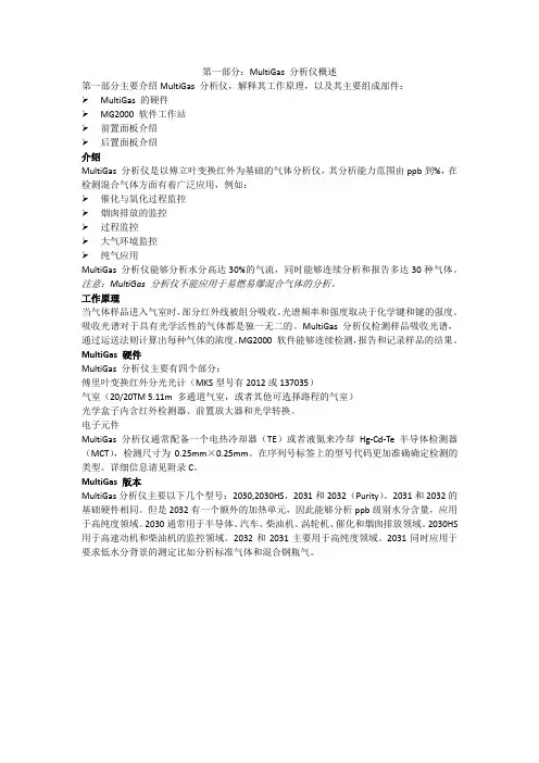
第一部分:MultiGas 分析仪概述第一部分主要介绍MultiGas 分析仪,解释其工作原理,以及其主要组成部件:MultiGas 的硬件MG2000 软件工作站前置面板介绍后置面板介绍介绍MultiGas 分析仪是以傅立叶变换红外为基础的气体分析仪,其分析能力范围由ppb到%,在检测混合气体方面有着广泛应用,例如:催化与氧化过程监控烟囱排放的监控过程监控大气环境监控纯气应用MultiGas 分析仪能够分析水分高达30%的气流,同时能够连续分析和报告多达30种气体。
注意:MultiGas 分析仪不能应用于易燃易爆混合气体的分析。
工作原理当气体样品进入气室时,部分红外线被组分吸收。
光谱频率和强度取决于化学键和键的强度。
吸收光谱对于具有光学活性的气体都是独一无二的。
MultiGas 分析仪检测样品吸收光谱,通过运送法则计算出每种气体的浓度。
MG2000 软件能够连续检测,报告和记录样品的结果。
MultiGas 硬件MultiGas 分析仪主要有四个部分:傅里叶变换红外分光光计(MKS型号有2012或137035)气室(20/20TM 5.11m 多通道气室,或者其他可选择路程的气室)光学盒子内含红外检测器、前置放大器和光学转换。
电子元件MultiGas分析仪通常配备一个电热冷却器(TE)或者液氮来冷却Hg-Cd-Te半导体检测器(MCT),检测尺寸为0.25mm×0.25mm。
在序列号标签上的型号代码更加准确确定检测的类型。
详细信息请见附录C。
MultiGas 版本MultiGas分析仪主要以下几个型号:2030,2030HS,2031和2032(Purity)。
2031和2032的基础硬件相同。
但是2032有一个额外的加热单元,因此能够分析ppb级别水分含量,应用于高纯度领域。
2030通常用于半导体、汽车、柴油机、涡轮机、催化和烟囱排放领域。
2030HS 用于高速动机和柴油机的监控领域。
傅里叶变换红外光谱仪操作说明书一、简介傅里叶变换红外光谱仪是一种基于傅里叶变换原理的分析仪器,广泛应用于材料分析、生物化学、环境监测等领域。
本操作说明书旨在详细介绍傅里叶变换红外光谱仪的组成、操作流程及常见故障处理方法,以帮助用户熟练操作并解决操作过程中可能遇到的问题。
二、仪器组成傅里叶变换红外光谱仪由以下几个主要部分组成:1. 光源:提供红外光源,常用的有红外灯。
2. 采样系统:负责将待测样品与光源进行交互作用,并将反射或透射的光信号收集到检测器中。
3. 干涉仪:由干涉仪和光谱仪构成,用于将入射光分解为不同波长的光束,并通过傅里叶变换将光信号转换为频谱信号。
4. 检测器:接收并转换频谱信号为电信号。
5. 数据采集与处理系统:负责采集、处理和输出检测到的光谱数据。
三、操作流程请按照以下步骤操作傅里叶变换红外光谱仪:1. 打开仪器电源,确保仪器处于正常工作状态。
2. 准备待测样品,将样品放置在采样系统上。
3. 调节样品位置,使样品与光源充分接触,确保信号采集的准确性。
4. 启动数据采集与处理系统,进入光谱采集界面。
5. 设置光谱采集参数,包括采样时间、波数范围等。
6. 点击开始采集按钮,系统开始采集并处理光谱数据。
7. 采集完成后,保存数据并进行必要的数据处理,如光谱峰识别、峰面积计算等。
8. 根据实际需求,可以进行多组数据的比较和分析。
9. 关闭仪器电源,清理和保养仪器,确保仪器处于良好状态。
四、常见故障处理方法在使用傅里叶变换红外光谱仪时,可能会遇到一些常见故障,下面是一些常见故障处理方法:1. 仪器无法开机:检查电源是否接通,确保电源供电正常。
2. 光谱信号杂乱:检查光源是否完好,采样系统是否正确安装。
3. 数据采集异常:检查数据采集与处理系统的连接是否稳定,重新启动系统。
4. 光谱峰形模糊:检查采样系统是否干净,样品是否合适。
5. 仪器响应速度慢:检查仪器是否需要清洁和维护,及时进行保养。
傅里叶红外光谱仪使用说明书一、引言傅里叶红外光谱仪是一种常用的分析仪器,广泛应用于化学、生物、医药等领域。
本使用说明书将详细介绍傅里叶红外光谱仪的操作方法和注意事项,旨在帮助用户正确使用和维护该仪器,以确保获得准确可靠的实验结果。
二、仪器概述傅里叶红外光谱仪是一种利用傅里叶变换原理分析物质的仪器。
它主要由光源、光谱仪、样品室、探测器和计算机等组成。
用户在使用时应了解各部分的功能和特点,并正确连接和调整仪器。
三、操作步骤1. 准备工作在操作傅里叶红外光谱仪之前,确保仪器周围环境整洁干净,并检查设备是否处于良好的工作状态。
同时,保证所需样品和试剂准备完备,并遵守相关安全操作规范。
2. 打开仪器首先,接通仪器的电源并按照操作面板上的指示启动傅里叶红外光谱仪。
在启动过程中,用户可以根据需要设置相关参数,比如光源强度和波长范围等。
3. 校准仪器为了获得准确的测试结果,用户需要定期校准傅里叶红外光谱仪。
具体的校准方法可以参考仪器的说明书或相关技术文献,根据实际情况进行操作。
校准完成后,保持仪器的稳定状态。
4. 放置样品将待测试的样品放置于样品室中,并尽量保证样品的均匀性和稳定性。
可以根据实验需要选择适当的样品盛装容器,并注意避免样品与仪器内部零件的接触。
5. 开始测试在仪器操作界面上选择相应的测量模式和参数设置,并点击开始测量。
傅里叶红外光谱仪会自动进行采样和测量,用户只需等待测试完成。
6. 数据处理测试完成后,仪器将生成一份原始的红外光谱图像。
用户可以利用计算机软件对图像进行处理和分析,提取所需的数据信息,并进行进一步研究和应用。
四、注意事项1. 使用时注意安全操作傅里叶红外光谱仪时,用户应注意安全,避免接触高温部件和有害化学物质。
使用过程中如发现异常情况或故障,请立即停止操作,并及时向仪器维护人员报告。
2. 仪器保养定期对傅里叶红外光谱仪进行检查和保养,确保仪器各部件的正常工作和长期使用。
注意保持仪器和周围环境的清洁,避免灰尘、水分等污染物进入仪器。
傅里叶变换红外光谱仪的操作说明书一、概述傅里叶变换红外光谱仪是一种高精度的光学仪器,广泛应用于材料科学、化学分析等领域。
本操作说明书旨在为使用者提供清晰、详细的操作指导,以确保仪器的正常运行和准确测试结果。
二、仪器组成傅里叶变换红外光谱仪主要由以下部分组成:1. 光源:提供红外光源,确保测试的稳定性和准确性;2. 样品室:放置待测试样品的区域,要求密封性良好;3. 透射系统:用于将红外光从光源传递至样品室;4. 干涉系统:利用干涉原理对样品室内的红外光进行分析;5. 探测器:接收经干涉系统分析后的光信号,并将其转换为电信号;6. 数据处理系统:对接收到的电信号进行处理和分析。
三、操作流程及注意事项1. 打开电源:接通电源并确保仪器正常启动,注意检查电源线是否连接稳固。
2. 预热:根据仪器规格要求,预热一定时间,以保证仪器的稳定性。
3. 样品准备:将待测试样品放入样品室中,确保样品密封良好,避免外界污染。
4. 选择测试模式:根据实验需求选择透射模式或者反射模式,并调整相关设置。
5. 扫描参数设置:输入所需的扫描参数,如波数范围、采样间隔等。
6. 开始扫描:点击开始扫描按钮,观察仪器是否正常工作,注意观察扫描过程中的任何异常现象。
7. 数据处理:扫描完成后,将仪器采集到的数据进行导出、分析和编辑。
8. 关闭仪器:关闭仪器电源,并按照清洁指南对仪器进行清理和维护。
注意事项:1. 操作人员应接受相关培训,了解仪器的基本原理和操作要点,以确保操作的准确性和安全性。
2. 在操作仪器前,应仔细阅读仪器的技术手册和操作指南,了解操作流程和安全注意事项。
3. 严格按照操作指引进行操作,避免在仪器运行过程中进行任何不必要的操作。
4. 保持仪器干净整洁,定期清理样品室和光学部件,避免影响测试结果和仪器寿命。
5. 定期进行仪器的校准和维护,以确保仪器的性能和测试结果的准确性。
四、故障排除在使用过程中,可能会遇到一些常见的故障,以下是一些建议的故障排除方法:1. 若仪器未正常启动,请检查电源是否接通、电源线是否连接良好。
傅里叶红外变换光谱仪使用说明
傅里叶红外变换光谱仪(FTIR)是一种用于分析物质结构和化学成分的分析仪器。
下面是使用FTIR的基本步骤:
1.打开FTIR: 前置需要确保FTIR设备已经连接到电源,并在FTIR获取软件软件上选择合适的仪器类型。
2.准备样品: 准备好代表物质的化合物样品,并确保样品足够小。
3.将样品放入质谱室: 样品要放置于红外光线可以透过的样品支架上,确保样品与电探针完全接触。
4.选择适当的红外光源: 根据样品的性质选择适当的红外光源,并按照设备指南设置。
5.收集样品光谱: 运行样品的光谱,并确保光谱中包含最小噪音。
(这可以通过取平均光谱或选择合适的时间来实现)
6.分析光谱: 通过软件分析光谱,将光谱与已有的对照光谱进行比较,以确定化合物的结构和组成。
7.保存数据: 将分析数据保存到计算机中,以备后续使用和分析。
需要注意的是,在使用傅里叶红外变换光谱仪时,应该严格遵守设备操作规程,并且在光谱分析时应该采取足够的注意力,以确保获得精确的结果。
傅里叶变换红外光谱仪的操作指南红外光谱技术是一种重要的化学分析方法,在有机物质的表征、质谱分析、生物医学研究等领域有着广泛的应用。
而傅里叶变换红外光谱仪作为红外光谱技术的重要仪器,其正确的操作和使用方法对于保证实验结果的准确性和可靠性至关重要。
本文将介绍傅里叶变换红外光谱仪的基本操作步骤和注意事项,以便读者能够了解如何正确操作和使用这一仪器。
一、准备工作在进行实验之前,首先需要对傅里叶变换红外光谱仪的相关设备进行准备和检查。
确保仪器整体干净,各部件完好无损。
检查仪器中的红外光源、检测器和光栅是否正常工作,并及时更换或修复需要维护的部件。
二、样品制备在进行红外光谱实验前,需要准备待测样品。
样品制备的方法和要求会根据不同的实验目的和样品类型有所差异。
一般来说,样品应尽量避免与环境中的杂质接触,可以采用透明盖片、石英池等样品容器来装载样品。
对于不同形态的样品,如固体、液体或气态样品,可采取不同的样品制备方法。
对于液体样品,应进行适当的稀释和混匀处理,确保实验时的准确性和可重复性。
三、仪器调试在进行实验之前,应对傅里叶变换红外光谱仪进行仔细的调试。
首先需要校准仪器的波数刻度,以保证实验结果的准确性。
接下来,需要调整仪器的参考(参比)峰,即选择一个已知波数和强度的特征峰作为参考,以进行波数的校准。
此外,还需调整仪器的背景光谱,以保证实验信号的清晰度和准确性。
四、实验操作在调试完傅里叶变换红外光谱仪之后,可以开始进行实验操作。
首先,将已经制备好的样品放入样品室,确保样品与仪器之间充分接触。
然后,选择合适的光谱扫描范围和扫描数目,开始采集红外光谱信号。
为了保证实验数据的准确性,建议进行多次扫描,并取其平均值作为最终结果。
五、数据处理和分析在完成红外光谱信号的采集之后,还需要进行数据处理和分析工作。
将所得的光谱信号进行傅里叶变换,得到傅里叶红外光谱图。
通过观察和分析傅里叶光谱图中的各峰和吸收带,可以得到样品中的化学成分、官能团和分子结构等信息。
傅立叶变换红外光谱仪使用方法一.打开电源,黄灯亮表示电源接通.二.将仪器心脏部分(偏振器)插入(注意: 突出部分向里, 必须全部放入槽口), 放入后按原样把盖盖上并拧紧螺丝。
三.开机及启动软件1. 仪器前部面板上的电源开关;2. 打开计算机,再双击桌面上IRsolution 快捷键,出现界面。
四.选择仪器及初始化1. 选择菜单上的Enviroment(环境) > Instrument Preferences(仪器参数选择) > Irprestige-21,然后确定。
2. 单击measure,然后选择菜单条上的Measurement(测量) > initialize(初始化),初始化仪器至四只绿灯两起,即可进行测量。
五.其它参数都已设置好,一般可直接进行测量。
如果需要改变参数,可在界面上进行设置(如波长范围,扫描次数等)。
六.测量方法1. 选择面板上的<BKG>键,进行背景扫描,背景为空气。
2. 插入标准样品”聚苯乙烯”进行仪器检测,再与样品板上的谱图对比,基本相同是可以进行所需的样品测量。
七.数据保存选择测量出来的数据, 单击(File) 中的(Save as), 将数据保存到你所需要的位置。
八.关机1. 选择File>Exit, 退出程序。
2. 关闭仪器电源。
分析测试中心仪器分类及使用方法(暂行)为了更好的发挥现有大型精密贵重仪器的作用,引入开放、竞争、服务新机制,进一步提高仪器的使用率,更好地为全校教学和科研服务,本测试中心根据仪器的特性和我校仪器台件数的现状,将现有仪器进行初步的分类。
不同类型的仪器将提供不同的使用方法。
一. 仪器分类第一类仪器:紫外、电子万能试验机、强力实验机、工业显微镜、复合纺丝机、纤维拉伸机气相色谱、薄层扫描仪、TOC、转矩流变仪、扩展流变仪、热变型维卡软化点测定、热机分析仪、原子发射光谱、原子吸收光谱、激光散射粒度分布仪、扫描电镜、离子色谱第二类仪器:傅立叶变换红外光谱仪、核磁、气—质联用仪、质谱仪、液相色谱、扫描量热仪、差示扫描量热仪、微机差热天平、热重差热分析仪、动态热机械分析仪、X光衍射仪、能谱仪二、使用方法1.第一类仪器属于半开放操作仪器设备。
GASMET TM DX-4010 FT-IR Gas AnalyzerON-SITE SeriesInstruction & OperatingManual2002-03-15WARRANTY STATEMENTThis warranty applies to the GASMET TM brand name products sold with this warranty statement. This warranty is applicable in all countries and may be enforced in any country where Temet Instruments Oy or its authorised service providers offer warranty service subject to the terms and conditions set forth in this warranty statement.Temet Instruments Oy shall not be liable for technical or editorial errors or omissions contained herein. The information in this document is provided “as is” without warranty of any kind and is subject to change without notice. Should you find any error, we would appreciate if you notified us.Temet Instruments Oy quarantees that all products manufactured and sold by it are free of defects in materials and workmanship under normal use during the warranty period.Temet Instruments’ products are manufactured using new materials or new and used materials equivalent to new in performance and reliability. Spare parts may be new or equivalent to new.Temet Instruments Oy agrees to either replace or repair free of charge (Ex Works Helsinki, Incoterms 2000), any such defective product or part, that is returned to its repair facility within one (1) year of the delivery date. All parts or products removed under this warranty become the property of Temet Instruments Oy. The replacement product or part takes on the warranty status of the removed product or part.The warranty does not extend to any product from which the serial number has been removed or that has been damaged or rendered defective (a) as a result of accident, misuse, abuse, normal wear of components or other external causes; (b) by operation outside the usage parameters stated in the user documentation that is provided with the product; (c) by the use of parts not manufactured by Temet Instruments Oy; or (d) by modification or service by anyone other than Temet Instruments Oy.Temet Instruments Oy is not liable for any damages caused by the product or the failure of the product to perform, including any loss of profits or savings, incidental damages, or consequential damages.GASMET TM and CALCMET TM are trademarks of Temet Instruments OyCONTENTS1INTRODUCTION (8)2PRINCIPLE OF MEASUREMENT (8)2.1P RINCIPLES OF I NFRARED S PECTROSCOPY (9)2.2C OMPONENTS OF A F OURIER T RANSFORM I NFRARED S PECTROMETER (11)2.3Q UANTITATIVE A NALYSIS OF FT-IR S PECTRA (13)2.4M ULTICOMPONENT A NALYSIS (14)2.5R ESOLUTION OF FT-IR A NALYSIS (15)2.6D ESCRIPTION OF THE GASMET TM DX-4010 (16)2.6.1 Applications of the GASMET TM DX-4010 Analyzer (16)2.6.2 The GASMET TM DX-4010 Analyzer Performance (17)2.6.3 Structure of the GASMET TM DX-4010 Analyzer (17)2.7GASMET TM DX-4010 T ECHNICAL D ATA (18)2.7.1 General Parameters (18)2.7.2 Spectrometer (18)2.7.3 Sample Cell (18)2.7.4 Measuring parameters (19)2.7.5 Electrical connectors (19)2.7.6 Gas Inlet and Outlet Conditions (19)2.7.7 Computer and Electronics (19)2.7.8 Enclosure (19)3INSTALLATION (21)3.1S UPPLY S CHEDULE (21)3.1.1 Package (21)3.1.2 Contents of the GASMET TM Package (21)3.1.3 Settings (21)3.2A MBIENT C ONDITIONS (22)3.2.1 Storing and Transporting the GASMET TM (22)3.2.2 Installation location (22)3.2.3 Explosion Protection (22)3.3S AMPLE G AS S UPPLY (23)3.4G AS C ONNECTORS (24)3.5O N-B OARD S AMPLE P UMP (25)3.6P OWER C ONNECTION (25)3.6.1 Power Supply (25)3.6.2 Fuses (25)3.7S IGNAL C ONNECTIONS (26)3.7.1 Optional Analog Output Signals (26)3.7.2 RS232C-Interface (26)3.7.3 Sample sequencing control (26)3.7.3.1Valve Dry Contact (27)3.7.3.2Pump Dry Contact (27)4MAINTENANCE (28)4.1S AFETY P RECAUTIONS (28)4.2M AINTENANCE P LAN (28)4.3V ISUAL I NSPECTION (29)4.4S AMPLE C ELL I NSPECTION (29)4.5R EPLACEMENT OF O PTOELECTRONIC C OMPONENTS (29)5START-UP (30)5.1C ONNECTIONS (30)5.2I NSTALLING THE SOFTWARE (30)5.2.1 Installing Calibration Files (30)5.3S WITCHING THE ANALYZER’S POWER ON (31)5.4O PERATION (31)5.4.1 Using the CALCMET Software (31)5.4.2 Setting the Measuring Times (34)5.4.3 Zero Calibration (34)5.4.4 Measuring the Sample Spectrum (35)5.4.4.1Single Measurement (35)5.4.4.2Continuous Measurement (36)5.4.5 Checking the Hardware (37)5.4.6 Analyzing the Sample Spectrum (37)5.5S AMPLE CALCMET SESSION (37)5.5.1 Step 1: Select the Components that will be analyzed (37)5.5.2 Step 2: Measure Background (37)5.5.3 Step 3: Measure and Analyze the Sample (38)5.5.4 Step 4: Verify the Results (38)6ANALYZER INSPECTION (39)6.1GASMET FT-IR G AS A NALYZER I NSPECTION S HEET (40)FIGURESFigure 1 Normal modes of vibration of carbon dioxide CO2 (9)Figure 2 An absorbance spectrum of Sulfur dioxide SO2 (11)Figure 3 Basic components of a FT-IR spectrometer (11)Figure 4 Michelson interferometer (12)Figure 5 A typical intereferogram (12)Figure 6 An example of spectra for multicomponent analysis (15)Figure 7 A basic structure of GASMET TM DX-4010 analyzer (17)Figure 8 Dimensionnal Drawing of the Analyzer Enclosure (20)Figure 9 Gas Connectors of the GASMET DX-4010 (24)Figure 10 Flow schematics of the on-board sample pump package (25)Figure 11 GASMET TM DX-4010 Connector unit (26)Figure 11 Welcome to Calcmet dialog (32)Figure 12 A typical background spectrum (35)TABLESTable 1 GASMET fuses. Physical size of each fuse is 5*20 mm (26)Table 2 Maintenance plan (29)Table 3 Welcome to Calcmet system parameters (33)Table 4 Commands for setting measuring times (34)Table 5 Zero Calibration measurement commands (34)Table 6 Single measurement commands (36)Table 7 Continuous measurement commands (36)Table 8 Commands for checking hardware status (37)PREFACEThank you for choosing GASMET TM, the state-of-the-art FT-IR gas analyzer manufactured by Temet Instruments. The GASMET TM is a high-tech product made of high quality components.Temet Instruments’ substantial investment in R&D is targeted at innovative, customer-driven solutions. Working closely with customers and global distribution network, the company offers extensive technical applications support services. Along with high reliability, Temet products offer easy operation and consistent and accurate results, together with competitive pricing.To develop the powerful technology of FT-IR has required uncompromising commitment and expertise in several fields of high technology. As a result, Temet products are not only superior in performance, but simple to operate and maintain. Temet Instruments’ continuing philosophy is to provide reliable measurements in a variety of industrial applications now and in the future. Whatever your applications are, we hope you will find the GASMET TM gas analyzer fast, accurate, reliable, and easy to use.1 INTRODUCTIONThis instructions manual provides information of the GASMET TM DX-SERIES Fourier Transform Infrared (FT-IR) Gas Analyzer. Please read this manual carefully prior to using the analyzer. Improper use of the analyzer may damage the equipment.Chapter 2, "Error! Not a valid bookmark self-reference.", discusses theoretical aspects of infrared spectroscopy. These fundamentals help understand the physical principles the GASMET TM system is based on. “Description of the Gas Analyzer” provides information about the GASMET TM DX-SERIES hardware and technical specifications.Chapter 3, “Installation”, provides information of installing the GASMET TM DX-SERIES Gas Analyzer. Use this paragraph to check the contents of the GASMET TM package and to get the GASMET TM from storage into operational condition.Chapter 4, “Maintenance”, describes the maintenance operations that may be necessary in the long run.Chapter 5, "Start-Up", provides some basic information of the operation of the GASMET TM DX-SERIES. This chapter is recommended to read, before any operation.Chapter 6, “Error! Reference source not found.”, describes what shloud be done when the analyzer is used first time. This chapter includes inspection sheet, what should be filled within 30 days from the date of delivery, in order that the warranty is in full valid.To learn how to operate the GASMET TM analyzer with the CALCMET TM controlling and analyzing software, refer to the CALCMET User’s Guide.This instructions manual is copyrighted 2001 by Temet Instruments. All rights reserved. No part of this manual may be reproduced in whole or in part in any form without the prior permission of Temet Instruments Oy.2 PRINCIPLE OF MEASUREMENT2.1 Principles of Infrared SpectroscopyWhen infrared radiation is passed through a sample of gaseous molecules, it can be observed that certain wavelengths of the infrared radiation are not transmitted through the gas very well. That is, the gas absorbs some specific wavelengths of the infrared radiation. What happens is that the infrared radiation interacts with the gas molecules. The gas molecules get energy from the infrared radiation and start to vibrate or rotate with increasing amplitude. This energy transfer from the infrared radiation to the gas molecules can be seen as decreased intensity of some wavelengths of the transmitted infrared radiation. If the infrared source sends a broad band of wavelengths of infrared radiation through the sample, some of the wavelengths will be partly absorbed by the gas sample.Figure 1shows how a gas molecule may behave when it interacts with infrared radiation. All different vibrations, rotations, and their combinations result in absorption of specific wavelengthsof the infrared radiation.CONTRACTSTRETCHSTRETCHSTRETCH C OO C OO COOOO υ1υ2υ34Figure 1Normal modes of vibration of carbon dioxide CO 2.An absorption spectrum shows graphically to what extent the different wavelengths of the infrared radiation are absorbed by the sample gas. The spectrum shows the transmission of the infrared radiation through the gas as a function of wavelength. For each wavelength, thetransmittance T is the intensity of the infrared radiation that has passed through the sample gas, divided by the intensity of the infrared radiation that has entered the sample gas. When there is no absorption, the value of transmittance T is 1 (or 100 %), which indicates that 100 % of the infrared radiation at that wavelength goes through the sample gas. If the intensity of the radiation entering the sample is I 0 and the intensity of the radiation that has passed through the sample is I, the transmittance T can be expressed as:T I I =/0T transmittanceI =intensity entering the sample I =intensity that has passed through the sample0=Besides using transmittance T, the absorption of the infrared radiation can be presented using the absorbance scale. Absorbance A is given by the logarithm of the transmittance reciprocal: A T =log 10(/)1A absorbance T transmittance ==The advantage of using the absorbance scale is that the value of absorbance is directlyproportional to the thickness of the sample gas (absorption path length), and the concentration of the sample gas.The infrared absorption spectrum is unique to all different gas molecules. It is possible to identify any gas component from the spectrum of the sample. An example of infrared absorbance spectrum is shown in Figure 2.Figure 2 An absorbance spectrum of Sulfur dioxide SO2.2.2 Components of a Fourier Transform Infrared SpectrometerThere are some basic parts that are typical for all FT-IR spectrometers. First of all, there is an infrared source that emits a broad band of different wavelengths of infrared radiation. The infrared radiation goes through an interferometer that modulates the infrared radiation. The interferometer performs an optical inverse Fourier transform on the infrared radiation entering the interferometer. The modulated infrared beam passes through the gas sample. Finally, the intensity of the infrared beam is detected by a detector. The detected signal is digitized and Fourier transformed by the computer to get the IR-spectrum of the sample gas.Figure 3 Basic components of a FT-IR spectrometer.The unique part of a FT-IR spectrometer is the interferometer. A Michelson type plane mirror interferometer is displayed in Figure 4. Infrared radiation from the source is collected and collimated before it strikes the beamsplitter. The beamsplitter ideally transmits one half of the radiation, and reflects the other half. Both transmitted and reflected beams strike mirrors, which reflect the two beams back to the beamsplitter. Thus, one half of the infrared radiation that finallythen back to the beamsplitter. The other half of the infrared radiation going to the sample has first gone through the beamsplitter and then reflected from the fixed mirror back to the beamsplitter. When these two optical paths are reunited, interference occurs at the beamsplitter because of the optical path difference caused by the scanning of the moving mirror.Radiation to the sample gas and detector Moving mirrorFixed mirrorFigure 4 Michelson interferometer.The optical path length difference between the two optical paths of a Michelson interferometer is two times the displacement of the moving mirror. The interference signal measured by the detector as a function of the optical path length difference is called the interferogram. A typical interferogram produced by the interferometer is shown in Figure 5. The graph shows the intensity of the infrared radiation as a function of the displacement of the moving mirror. At the peak position, the optical path length is exactly the same for the radiation that comes from the moving mirror as it is for the radiation that comes from the fixed mirror.Figure 5 A typical intereferogram.The spectrum can be computed from the interferogram by performing a Fourier transform. The Fourier transform is performed by the same computer that ultimately performs the quantitative analysis of the spectrum.2.3 Quantitative Analysis of FT-IR SpectraThe basic law for spectroscopic quantitative analysis is Beer's law. Beer's law shows how the concentration of the sample gas is related to the measured absorbance of the sample spectrum. Beer's law is commonly expressed as:log(/)log(/)I I T A abc 01===I =intensity entering the sampleI =intensity that has passedthrough the sampleA absorbance T transmittancea a()absorptivityb optical path lengthc =sample concentration0=====vThe absorptivity a is a constant that characterizes the capacity of the molecule to absorb infrared radiation. The value of a varies from one molecule to another and one wavelength to another, but is constant for a given molecule at a given wavelength. The quantity b is the optical path length, that is, the distance the infrared radiation beam traverses in the gas sample. The quantity cindicates the concentration of the sample gas molecules in the sample. If the optical path length is held constant, Beer's law states that the absorbance is directly proportional to the concentration of the sample gas at a given wavelength. Since Beer's law is additive, absorbance A is equal to the sum of the values of the a , b , and c for each gas component.Two conditions are implied in the derivation of Beer's law. The radiation being measured is monochromatic, and the sample absorptivity does not vary with concentration. If the sample concentration range is wide, the change in the sample environment can cause absorptivitychanges and deviations from Beer's law. For narrow concentration ranges and for low absorbance values a plot of concentration versus absorbance is nearly linear.The changes in the gas pressure may cause broadening in the absorption line shape of the sample gas. The rotational fine structure of gas-phase bands are broadened by collisions between the molecules of the component being measured. All IR-bands are broadened.The changes in the gas temperature may cause 'hot bands' in the spectrum of the sample gas. As the temperature increases, the distribution of the molecules in different energy levels changes. The changes in the temperature cause changes in the absorption line shape of the sample gas. To analyze the concentration of a gas compound, the GASMET TM calculates the number of gas molecules in the sample cell. The number of the gas molecules in the sample cell depends linearly on both the gas pressure and the volume of the sample cell, and reciprocally on the gas temperature:pV nRT =p=pressureV=volumen=number of the gas moleculesT=temperatureR=Ideal gas constantAny changes in sample gas temperature or pressure in the sample cell directly affect on the measured gas compound concentration. In addition, gas pressure or temperature changes may effect the line shape of the measured absorbance spectrum, and thus the accuracy of the analysis results.2.4 Multicomponent AnalysisThe degree of absorption of infrared radiation at each wavelength is quantitatively related to the number of absorbing molecules in the sample gas. Since there is a linear relationship between the absorbance and the number of absorbing molecules, multicomponent quantitative analysis of gas mixtures is feasible.To perform multicomponent analysis we start with the sample spectrum. In addition, we need reference spectra of all the gas components that may exist in the sample, if these components are to be analyzed. A reference spectrum is a spectrum of one single gas component of specific concentration. In multicomponent analysis we try to combine these reference spectra with appropriate multipliers in order to get a spectrum that is as close as possible to the sample spectrum. If we succeed in forming a spectrum similar to the sample spectrum, we get the concentration of each gas component in the sample gas using the multipliers of the reference spectra, provided that we know the concentrations of the reference gases.For example, suppose we have a sample spectrum and reference spectra like those shown in Figure 6. In this case, we know that the sample gas consists of gases Reference 1 and Reference 2. We have the reference spectra available and we know that these reference spectra represent concentrations of 10 ppm and 8 ppm respectively. To find out the concentration of each component in the sample gas, we try to form the measured sample spectrum using a linear combination of the reference spectra. We find out that if we multiply the spectrum Reference 1 by 5 and the spectrum Reference 2 by 2, and combine these two spectra, we get a spectrum that is similar to the sample spectrum. Accordingly, the sample gas contains reference gas 1 at five times the amount in the reference spectrum 1, and reference gas 2 at two times the amount in the reference spectrum 2. The analysis indicates that the sample indeed consists of these two reference gases. The concentration of the reference gas 1 in the sample is found to be 50 ppm, and the concentration of the reference gas 2 in the sample is 16 ppm.increases and makes the analysis less precise.Furthermore, the information content of the measurement is not maximized by using as high a resolution as possible. Scientific research1 has proved that when the resolution is reduced by the some factor 1/k, signal-to-noise-ratio can be increased by a factor of k3/2 in the same registration time. This makes the use of low resolution quite worthwhile. To achieve precise quantitative analysis results, a low resolution measurement should be used provided that the spectral overlap problem can be taken care of.There are two reasons for the increase of the signal-to-noise ratio:• In an FT-IR spectrometer, the length of the measured interferogram determines the resolution. The longer the interferogram, the more data points, and the higher the resolution.In a high resolution study the time required by a single measurement is also high, so that we are able to co-add less measurements in a fixed time, leading to higher noise level. As a result, the longer the measured interferogram is, the lower signal-to-noise-ratio results.• High resolution requires narrow slit or a small aperture to prevent a phenomenon called aperture broadening. Narrow slit, in turn, reduces the signal and thus signal-to-noise-ratio ratio.1 See for example, P Saarinen & J Kauppinen. 1991. Multicomponent Analysis of FT-IR Spectra. Applied Spectroscopy. Volume 45, Number 6. Pages 953 - 963.2.6 Description of the GASMET TM DX-4010GASMET TM ON-SITE SERIES includes portable and transportable multicomponent gas analyzers for demanding applications. The GASMET TM DX-4010 incorporates a Fourier Transform Infrared, FT-IR spectrometer. The analyzer offers versatility and high performance for all users.The GASMET TM DX-4010 in designed for on site measurements. It is an ideal tool to environmental applications. Sample cell absorption path length is 9.6 m and the temperature 50 °C.The GASMET TM DX-4010 incorporates advanced hardware and software to provide a portable high-speed gas analysis system. The GASMET TM DX-4010 brings analytical laboratory performance in non-laboratory conditions. The GASMET TM DX-4010 eliminates the equipment, calibration, and service costs associated with multiple, single component gas analyzers. GASMET TM DX-4010 allows simple calibration using only single component calibration gases, the user can easily configure the analyzer for a new set of compounds.The software of GASMET TM is based on the powerful multicomponent analysis method CALCMET TM. The CALCMET TM analysis method is used to compute the concentrations and error limits of the different components present in the sample gas. The reference spectra needed in the analysis are stored on the fixed disk of computer and are simply loaded from the library when used in the analysis of an unknown sample. CALCMET for Windows software is described in the “CALCMET for Windows Software Manual”2.6.1 Applications of the GASMET TM DX-4010 AnalyzerThe GASMET TM DX-4010 enables identification and quantification of multiple gaseous compounds simultaneously and accurately, with results available in seconds.The GASMET TM DX-4010 is a versatile gas analyzer that can be used in several applications. The GASMET TM DX-4010 is specially designed for:• Environmental emissions monitoring• Quality control• Workplace air monitoring2.6.2 The GASMET TM DX-4010 Analyzer Performance• Lowest detectable concentrations: 0.2-2 ppm (depends on the application).• Accuracy: 2 % of the measurement range (depends on the application)• Precision: 0.01 % of the measurement range (depends on the application)• Diatomic homonuclear gases (i.e. N2, O2, H2, Cl2) and noble gases (i.e. He, Ne, Ar, Kr) do not absorb infrared radiation. They are the only gases that cannot be measured by the GASMET TM2.6.3 Structure of the GASMET TM DX-4010 AnalyzerFigure 7 outlines a complete gas analysis system. The IR source produces broad band IR radiation which is modulated in the interferometer. The interferometer actually performs an optical inverse Fourier transform. The modulated IR radiation passes through the sample cell where sample gas absorbs certain wavelengths of the IR radiation. The detector detects the transmitted IR radiation. The signal is digitized by A/D converter. The computer performs a mathematical Fourier transform on the digitized modulated signal, and a spectrum is obtained. The GASMET TM uses a Fast Fourier Transform to compute the spectrum from the interferogram. The transmittance spectrum is computed as a ratio of the sample spectrum to the background spectrum. The absorbance spectrum is computed from the transmittance spectrum. The GASMET TM uses the CALCMET TM software to compute the concentrations of the components present in the sample gas from the absorbance spectrum.Figure 7 A basic structure of GASMET TM DX-4010 analyzer.The data and signal processing electronics control the operation of the GASMET. An external PC compatible computer controls the GASMETTM DX-4010.A unique part of the GASMET is the Temet Carousel interferometer. The Carousel interferometer is rugged and withstands the demanding environmental conditions of non-laboratory environment. The Carousel interferometer modulates the infrared radiation coming from the infrared source, as discussed previously.The modulated light passes through the temperature controlled sample cell. The gas sample is introduced into the gas cell through standard gas line connectors. The transmitted infrared radiation is finally detected by a thermoelectrically cooled detector.2.7 GASMET TM DX-4010 Technical Data2.7.1 General ParametersMeasurement principle:FT-IR (Fourier Transform Infrared)Performance: Simultaneous analysis of up to 50 gas compuondsResponse time: <120 s, depending on the gas flow and measurements timeOperating temperature: 20 ± 20°C, optimum 15 - 25°C non condensingStorage temperature: -20 - 60°C, non condensingPower supply: 12 VDC or 100-240 VAC / 50 - 60 Hz2.7.2 SpectrometerInterferometer: Temet© Carousel InterferometerResolution: recommended 8 cm-1 or 4 cm-1Scan frequency: 10 spectra/sAperture: 1’’Detector: Thermo-electrically cooled MCT or DTGSIR-source Ceramic, SiC, 1550 K temperatureBeamsplitter: ZnSeWindow material: ZnSeWavenumber range: 700 – 4200 cm-12.7.3 Sample CellStructure: Multi pass, fixed path length 1.2m, 2.5m, 5.0m, or 9.8mMaterial: 100 % Gold coater aluminiumMirrors: fixed, protected gold coatingVolume: 1.07 lConnectors: Swagelok 6 mm or 1/4"Gaskets: Teflon® coated Viton® or Kaltrez® O-ringsTemperature: 50 °C, maximumMaximum sample gas pressure: 2 barsFlow rate 1-5 l/minResponse time: <60 sSample condition: non condensing2.7.4 Measuring parametersZero point calibration: 24 hours calibration with nitrogen (4.0 or higher N2 rekomended)Zero point drift: 2 % of smallest measuring range per zero point calibration interval Sensitivity drift: noneLinear derivation: 2 % of smallest measuring rangeTemperature drift: 2 % of smallest measuring range per 10 °C temperature changePressure influence: 1 % change of measuring value for 1 % sample pressure change2.7.5 Electrical connectorsDigital interface: RS-232 C, 9-pole D-Connectors. CR4000/2000/1000 is connected to anexternal computer via RS-232 C cable. The external computer controls theGASMET.Power connection: Standard plug CEE-22Relay connection: External PCAnalog input/output External PC2.7.6 Gas Inlet and Outlet ConditionsGas temperature: non-condensing, the sample gas temperature should be the same as the samplecell temperatureSample pump: InternalFlow rate: 120-160 l per hourGas filtration: filtration of particulates (2µ) requiredSample gas pressure: ambient2.7.7 Computer and ElectronicsA/D converter: dynamic range 95 dBSignal Processor: 32-bit floating point DSP 120 MFLOPS speedComputer: External, not includedOperating system: Windows 95 or 98Analysis software: CALCMET for windows 95/982.7.8 EnclosureMaterial: AluminumDimensions (mm): 433 ∗ 185 ∗ 425Weight: 16 kgCE label: according to EMI guideline 89/336/ECCE mark indicates compliance with directive 89/336/EEC for electromagnetic Compability. The CE marked GASMET models are certified by the manufacturer as follows:。