cDNA-AFLP Protocol
- 格式:pdf
- 大小:301.50 KB
- 文档页数:25
AFLP分子标记的发展及应用分子标记是指能反映生物个体或种群间基因组中某种差异特征的DNA片段,直接体现基因组DNA 间的差异。
在过去30多年,DNA 分子标记得到了迅速发展。
扩增片段长度多态性(Amplified Fragment Length Polymorphism, AFLP) 技术为第二代分子标记技术,是在随机扩增多态性(Random Amplified Polymorphic DNA, RAPD) 和限制性片段长度多态性(Restriction Fragment Length Polymorphism, RFLP) 技术上发展起来的DNA 多态性检测技术,同时拥有RFLP的高重复性和RAPD简便快捷的特点。
同其他以PCR 为基础的标记技术相比,AFLP 技术能同时检测到大量的位点和多态性标记,具有覆盖面广、高保真性、高效性、高分辨率、DNA用量少、事先勿需知道序列任何信息、可在全基因组产生标记、标记的分离遵循孟德尔遗传规律等优点,自1995年由Zabeau和Vos创建以来,已在动物、植物和微生物的分子系统学研究中得到应用,具有广阔的发展前景。
1 AFLP 标记的基本原理、操作流程及技术关键1.1 AFLP 标记的原理AFLP 标记的原理是对基因组总DNA 酶切后经PCR 进行选择性扩增。
先将基因组DNA 用两种限制性内切酶酶切成大小不等的片段,并与含有粘性末端的人工接头相连,形成酶切位点不同的限制性酶切片段,然后用与接头和位点相匹配的引物进行预扩增,预扩增产物作为进一步PCR 扩增的模板,再选用特异引物进行选择性扩增,扩增后的产物经聚丙烯酰胺凝胶电泳将特异的限制性片段分离。
由于不同来源DNA 的酶切片段存在差异,因而产生了扩增产物的多态性。
1.2 操作流程AFLP 标记的具体操作流程为: ①DNA 提取和质量检测;②双酶切和酶切片段连接;③酶切连接片段的预扩增;④选择性扩增;⑤扩增产物在聚丙烯酰胺变性凝胶上电泳; ⑥将电泳后的凝胶进行显影检测及数据分析。
中国农业科学2003,36(5):557-560利用cDNAAFLP技术分析白菜核雄性不育两用系的表达差异王永勤, 曹家树,符庆功RNA指纹技术可以揭示基因调控在RNA水平的表现,并进一步阐明其调控方式。
Liang等根据高等生物成熟mRNA带有polyA尾巴的特性,用特异的锚定引物反转录后,进行PCR扩增,建立了mRNA差异显示技术(mRNAdifferentialdisplay,mRNADD)[1]。
但该技术存在假阳性高、重复性差、3′端引物倒数第二个碱基的简并性使得某些差异条带不能被扩增等缺点。
1996年Bachem等发展了一种新的方法,将AFLP用于mRNA表达差异分析,即cDNA-AFLP。
cDNA-AFLP指纹技术重复性高、假阳性低,可准确地反映基因间表达量的差异,可广泛用于植物分子生理过程的基因表达特性的研究[2]。
白菜(BrassicacampestrisL.ssp.chinensisMakino)为典型的异花授粉作物,具有明显的杂种优势。
虽然可用自交不亲和系生产F1代种子,但由于亲本繁殖困难、自交系多代生活力下降及易受环境影响等原因,很难保证100%的杂交率,自交不亲和系的应用受到一定的制约。
雄性不育系是白菜等十字花科植物生产一代杂种的理想系统。
因此,雄性不育的选育及其应用研究倍受重视。
白菜核不育两用系自20世纪70年代培育成功以来,已在生产一代杂种中发挥了重要作用[3]。
但是,对其遗传规律的认识还停留在形态学和细胞学水平,产生雄性不育的分子机理还不清楚,获得不育系还需要通过转育、选择配合力测定等一系列工作,费时、费工,限制了雄性不育系在生产上的进一步应用[4]。
因此,从分子水平研究白菜雄性不育机理具有重要的实践和理论意义。
本研究采用cDNAAFLP对白菜核雄性不育两用系可育株群与不育株群的不同时期、不同部位的mRNA的表达差异进行比较分析,为揭示芸薹属等植物雄性不育的分子机理提供证据。
运用cDNA-AFLP技术初步鉴定两种方法合成的cDNA的指纹图谱史胜青;张守攻;李春秀;汪阳东;齐力旺【期刊名称】《分子植物育种》【年(卷),期】2006(4)1【摘要】在植物基因克隆以及构建cDNA文库时,都需要合成高质量的双链cDNA,并要达到一定的数量。
然而研究者经常受到试验样品量的限制。
特别是比较稀少的植物材料,例如植物的根尖、茎尖或花的雌、雄蕊等,难以获得足够的RNA,以致影响cDNA的合成量,无法开展下游的实验。
以PCR为基础合成第二链cD-NA的Smart技术(LD-PCR),能够以50ng的总RNA为反转录模板合成高质量的双链cDNA。
但研究者对第二链采用PCR方法是否有些基因信息丢失或丰度上发生很大的变化存有疑虑。
针对以上存在的问题,通过置换合成和长距离PCR(LD-PCR)两种方法合成3个月的梭梭幼苗茎尖双链cDNA,EcoRI和MseI限制性内切酶双酶切后,用16对选择性扩增引物对两种cDNA进行cDNA-AFLP的指纹图谱分析。
结果表明,置换合成和LD-PCR两种方法合成的cDNA指纹图谱中,分别有条带约495条和470条。
其中,相同的条带共计433条,不同的条带分别有62条和37条,分别为各自合成方法总指纹数的12.5%和7.87%,相同的指纹信息达87%以上。
这说明两种方法合成的cDNA存在着较大的差异,合成方法对cDNA合成质量的影响较大,为研究人员选用何种cDNA合成方法提供了借鉴。
【总页数】4页(P79-82)【关键词】梭梭(Haloxylon;Ammodendron);置换合成法;LD-PCR法;cDNA-AFLP【作者】史胜青;张守攻;李春秀;汪阳东;齐力旺【作者单位】中国林业科学研究院林研所细胞生物学实验室【正文语种】中文【中图分类】S513;S565.4【相关文献】1.利用cDNA-AFLP技术鉴定菊花品种‘紫荷’的抗白锈病相关基因 [J], 黄河;王顺利;戴思兰2.用cDNA-AFLP技术构建白菜转录图谱 [J], 范淑英;乐建刚;成广杰;吴才君3.利用cDNA-AFLP技术鉴定甘蓝显性核不育基因相关表达序列 [J], 娄平;王晓武;Guusje.Bonnema;方智远4.用cDNA-AFLP技术构建基因组转录图谱 [J], 付凤玲;李晚忱5.cDNA-AFLP技术在植物耐盐基因鉴定上的应用 [J], 胡淑英;张春红;王小敏;黄涛;吴文龙;李维林因版权原因,仅展示原文概要,查看原文内容请购买。
cDNA-AFLP技术分离蓖麻矮化相关基因的开题报告
一、研究背景
蓖麻是重要的工业油料作物,具有高产、高油、强抗旱、适应性强
等优点,因此在全球范围内得到广泛的种植。
但是,蓖麻的高度却限制
了其种植面积和产量的进一步提高。
因此,探究蓖麻矮化的相关基因,
对于提高蓖麻的产量和种植面积具有重要的意义。
二、研究目的
本研究旨在通过cDNA-AFLP技术筛选出与蓖麻矮化相关的候选基因,并进一步验证其功能,为蓖麻的育种和种植提供理论依据。
三、研究方法
1. 样品处理
从蓖麻幼苗中分离总RNA,并进行纯化和定量。
使用反转录酶转录
一链cDNA,并用扩增引物扩增cDNA。
2. cDNA-AFLP技术
使用EcoRI和MseI限制酶对cDNA进行限制性酶切,再使用EMR
和MSE扩增引物扩增AFLP片段,得到AFLP产品。
将AFLP片段分离并
进行纯化,然后进行克隆测序和比对分析,找出与蓖麻矮化相关的候选
基因。
3. 基因功能验证
将候选基因克隆到表达载体中,转化到大肠杆菌中表达检测基因功能,并利用基因敲除和基因表达调控等方法,进一步验证其功能。
四、研究意义
本研究通过cDNA-AFLP技术及其辅助方法,可以筛选出与蓖麻矮化相关的基因,为蓖麻种植和育种提供理论基础。
同时,研究方法也可以为其他作物的功能基因研究提供借鉴和参考。
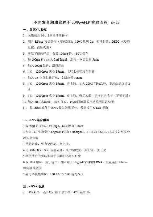
不同发育期油菜种子cDNA-AFLP实验流程Ver 2.0一、总RNA提取1. 采集花后不同日期的油菜种子2. 用具RNase灭活处理(玻璃器皿:160℃烘烤2h;塑料制品:DEPC水浸泡过夜,高压灭菌)3. 液氮下研磨样品,分装100mg/管,-80℃保存4. 每100mg样品加入1ml Trizol,混匀,室温温育5min5. 加入200μl氯仿,剧烈震荡6. 4℃,12000rpm离心15min,上层水相转移至新管7. 加入0.5倍体积异丙醇,室温静置10min8. 4℃,12000rpm离心15min,弃上清,加入200μl 75%乙醇,重悬洗涤沉淀2次9. 4℃,12000rpm离心15min,弃上清,吸尽乙醇,超净台内吹干(不要干透)10. 加入50μl水溶解,-80℃保存。
1%琼脂糖凝胶电泳检测提取结果注:若Trizol对种子RNA提取效果不佳,考虑改用CTAB提取二、RNA结合磁珠1.取20ul总RNA(约2ug),65℃温育10min2.加入1ul 生物素化oligo(dT)引物(700ug/ul),1.1ul 20×SSC,轻轻混匀至完全冷却至室温3.重悬磁珠,磁力架收集,弃上清。
4.用300ul 0.5×SSC重悬磁珠,磁力架收集,弃上清。
洗三次5.将清洗后的磁珠重悬于100ul 0.5×SSC中6.取10ul磁珠,置于管中,加入结合oligo(dT)引物的RNA,室温温育10min,保持磁珠悬浮7.磁力架收集磁珠,100ul 0.1×SSC清洗四次三、cDNA合成1. cDNA第一链合成:按下表加样,42℃温育2h磁珠--- biotinylated oligo(dT) primer (700μg/μl)1μl H2O 24μl 5× First Strand Buffer 8μl0.1M DTT 4μleach 10mM dNTP mix 2μl 2.5mM Superscript II (200U/μl) 1μl M-MLV RTase, RNase H-Final V olume 40μl 或20ulT10E0.1重悬磁珠,H2O 4ul5× First Strand Buffer:(-20℃)250mM Tris-HCl (pH 8.3)15mM MgCl2375mM KCl2. 磁力架收集磁珠,仔细吸弃上清,用5×第二链buffer平衡磁珠,磁力架收集,弃上清3. cDNA第二链合成:按下表加样,12℃温育1h,22℃温育1h磁珠--- 5× Second Strand Buffer 20μl 10×buffer:10μl10mM dNTP mix 2μl0.1M DTT 4μlE.Coli DNA ligase (10U/μl) 1μl 10UDNA Polymerase I (10U/μl) 3μl 30U RNase H (5U/μl) 1μl 5U H2O 69μl Final V olume 100μl或20ulT10E0.1重悬磁珠,H2O 49ul5× Second Strand Buffer: (-20℃)7.0)(pH100mMTris-HCl20mM MgCl2450mMKCl750μM NAD+50mM (NH4)2SO44. 磁力架收集磁珠,仔细吸弃上清,用1×RL-Buffer清洗磁珠,磁力架收集(或20ulT10E0.1清洗)四、Bst YI 酶切1. 向磁珠加入10U BstYI (1μl,10U/μl),4μl 10×RL-Buffer,35μl H2O,共40μl,混匀。
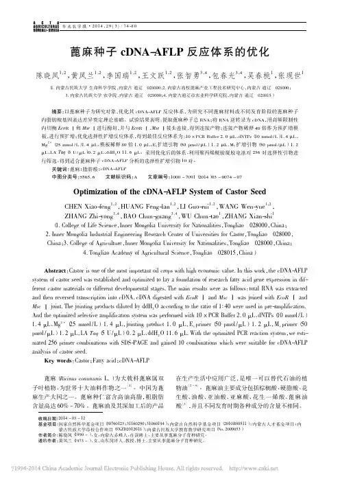
华北农学报·2014,29(3):74-80收稿日期:2014-03-12基金项目:国家自然科学基金项目(30760123;31160290;31060194);内蒙古自然科学基金项目(2010BS0511);内蒙古人才基金项目;内蒙古民族大学市校合作项目(SXZD2012018);内蒙古民族大学教育教学研究项目(No.2009053)作者简介:陈晓凤(1990-),女,内蒙古赤峰人,在读硕士,主要从事蓖麻分子育种研究。
通讯作者:黄凤兰(1973-),女,山东菏泽人,教授,博士,主要从事蓖麻分子育种研究。
蓖麻种子cDNA-AFLP 反应体系的优化陈晓凤1,2,黄凤兰1,2,李国瑞1,2,王文跃1,2,张智勇3,4,包春光3,4,吴春桃1,张现世1(1.内蒙古民族大学生命科学学院,内蒙古通辽028000;2.内蒙古高校蓖麻产业工程技术研究中心,内蒙古通辽028000;3.内蒙古民族大学农学院,内蒙古通辽028000;4.内蒙古通辽市农业科学研究院,内蒙古通辽028015)摘要:以蓖麻种子为研究对象,优化其cDNA-AFLP 反应体系,为研究不同蓖麻材料或不同发育阶段的蓖麻种子内脂肪酸基因表达差异奠定理论基础。
试验结果表明:提取蓖麻种子总RNA ;将RNA 逆转录为cDNA ,用高频限制性内切酶Eco RⅠ和Mse Ⅰ进行酶切,并与Eco RⅠ、Mse Ⅰ接头连接,得到连接产物;连接产物稀释40倍作为预扩增模板,进行预扩增;优化选择性扩增反应体系,得到最佳反应体系为:10ˑPCRBuffer 2.0μL 、dNTPs (10mmol /L )1.4μL 、Mg 2+(25mmol /L )1.4μL 、模板稀释80倍1.0μL 、E x 扩增引物(50pmol /μL )1.2μL 、M y 扩增引物(50pmol /μL )1.2μL 、LA Taq (5U /μL )0.2μL 、ddH 2O 11.6μL 。
cdna-aflp原理
cDNA-AFLP(cDNA-Amplified Fragment Length Polymorphism)是一种用于研究基因表达的分子生物学技术。
它结合了cDNA(互补DNA)合成和AFLP(Amplified Fragment Length Polymorphism)
技术,用于研究不同组织或条件下基因表达的差异。
cDNA-AFLP的原理涉及以下几个步骤:
1. cDNA合成,首先,从不同组织或条件下的RNA中合成cDNA。
这通常涉及使用逆转录酶(reverse transcriptase)将RNA转录成cDNA。
这样可以获取基因表达的信息,因为cDNA是由RNA转录而来的,反映了细胞中的基因表达情况。
2. cDNA片段的选择性扩增,接下来,利用AFLP技术中的限制
性内切酶对cDNA进行限制性酶切。
然后使用适配器引物(adapter primers)对酶切后的cDNA片段进行扩增。
这一步能够产生大量的cDNA片段,这些片段的长度和序列会反映出基因表达的差异。
3. 片段分析,扩增后的cDNA片段通过聚丙烯酰胺凝胶电泳进
行分离和检测。
这样可以得到cDNA片段的长度和数量信息。
通过对不同样本中cDNA片段的长度和数量进行比较分析,可以发现在不同条件下基因的表达差异。
这种技术可以用于寻找与特定生物学过程或环境条件相关的基因表达差异,从而揭示基因调控的机制和生物学功能。
总的来说,cDNA-AFLP技术结合了cDNA合成和AFLP技术,能够全面地分析基因表达的差异,对于研究基因调控和生物学功能具有重要意义。
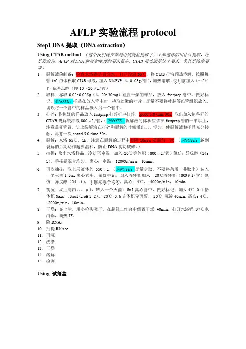
AFLP实验流程protocolStep1 DNA提取(DNA extraction)Using CTAB method (这个我们现在都是用试剂盒提取了,不知道你们用什么提取,还是发给你,AFLP对DNA纯度和浓度的要求很高,CTAB很难满足这个要求,尤其是纯度要求)1.裂解液的制备:检查水浴锅是否有水,打开设温65℃,将CTAB母液预热溶解,按照每管1ml的体积取CTAB母液,加入3%PVP(即0.03g/管),加热溶解,使用前...加入1~2%β-巯基乙醇(即10~20μl/管)2.取样:称取0.02~0.025g(即20~30mg)硅胶干燥的样品,放入fastprep管中,做好标记。
※NOTE:样品在放入管中时,挑取幼嫩的叶片,尽量不要将叶脉等维管组织放入,切误将一个管中的样品溅入另一个管中。
3.打碎:将称好的样品放入fastprep打碎机中打碎,speed 5.0 time 30s, 取出加入制备好的CTAB裂解缓冲液800μl/管,(※NOTE:裂解液的体积应该在fastprep管的一半以上,注意盖好管冒,防止裂解液在打碎和裂解的时候溢出。
),混匀,使裂解液和样品充分接触,再打一次speed 5.0 time 30s。
4.裂解:水浴65℃,1h,注意在裂解的过程中每隔10min要混匀一次,(※NOTE:越到裂解的后期动作越要温和,防止DNA剪切破碎。
)5.抽提:取出水浴样品,冷却至室温.....,加入-20℃等体积(800μl/管)氯仿:异戊醇(24:1),手摇晃混合均匀.......,离心:室温,12000r/min,10min。
6.再次抽提:取上层液体约550μl,(※NOTE:尽量少取,不要将杂质一并取出)转入一个灭菌1.5ml离心管中,做好标记,加入等体积加入-20℃等体积(800μl/管)氯仿:异戊醇(24:1),手摇晃混合均......匀.,离心:4℃,14000r/min,10min。
2005年第40卷第7期生物学通报59对不同细胞或者同类细胞在不同的生理状态、不同的发育阶段的基因表达状况进行分析,可以为研究生命活动的过程提供十分重要的信息,人们已经把基因表达差异的研究拓展到了生命科学的各个领域之中。
现阶段基因组学研究已经进入后期的功能基因组学(functionalgenomics)的研究阶段,对基因在特定时期转录水平上的表达丰度及差异表达等情况进行研究,由此分析基因的表达方式及作用已经形成了功能基因组学的重要方面———转录组学(transcriptomics)。
目前已有多种可在转录水平对差异表达基因筛选分析的方法。
最早发展起来的差别筛选技术和扣除杂交方法由于灵敏度底、过程繁琐、工作量大已经被逐渐淘汰。
Liang等在1992年建立的mRNA差异显示反转录PCR技术(differentialdisplayreversetranscriptase-PCR,DDRT-PCR)得到广泛应用,但也遇到许多问题。
主要是其采用的是随机引物和相对较低的退火温度,引物可在多个位点结合,扩增产物很大程度上依赖引物和模板的结合情况而不是仅与cDNA的初始浓度有关。
这些使得DDRT-PCR面临着可重复性较差,以及50%~70%的高假阳性的问题,并且由于PCR进行时使用3′端polyT锚定引物和5′端寡核苷酸随机引物,扩增产生的cDNA片段产物倾向于mRNA的3′非翻译区,包含的差异表达基因的信息量相对减少,很难通过与现有数据库比对进一步获得相应片段的有价值信息。
虽然DDRT-PCR自建立以来又经历了不少的改进和优化,但是这些明显的不足是由其原理本身所决定的,仅仅根据技术上的改进无法克服。
cDNA-AFLP是Bachem等在一种应用于基因组DNA的选择性扩增限制性片段即AFLP(Amplifiedfragmentlengthpolymorphism)的基础上发明出来的用于RNA指纹分析的方法。
它将RT-PCR和AFLP结合起来,由于在PCR中使用较高的退火温度和应用特定引物对包含信息较多的内部特异片段进行选择性扩增,增加了反应条件的严谨度,使其比DDRT-PCR的可靠性和可重复性大大提高。
for a PDF version of this protocol click herecDNA/AFLPA tool for transcriptome analysis12th June 2002CWB BACHEM1.RNA Isolation1.1.Plant tissue material is harvested and immediately frozen in liquid N2. The material isthen ground to a fine powder in a pre-cooled pestle and mortar under liquid N2. (This material can be stored at -80°C indefinitely). To proceed with extraction, transfer around 1g of powdered material into pre-cooled 50 ml plastic tubes (usually around 5 ml volume).1.2.Transfer 10 ml of a hot (80°C) 1:1 mixture of RNA extraction buffer and phenol (basevolume: 5 ml) into the tube containing plant material. Be sure that no liquid N2remains in the sample when adding the extraction buffer. Vortex or manually shake the samples vigorously for at least 30 seconds.1.3.To the mixture add a base volume of chloroform (5 ml) and shake vigorously. Toseparate the phases, centrifuge the samples for 20 minutes (3500 rpm in a large swing-out rotor). Repeat chloroform extraction with one base volume (5ml) until no inter-phase can be detected (at least twice).1.4.Transfer resultant cleared aqueous phase to a new 50 ml tube (ca. 5.5 ml; this materialcan be stored frozen for 24 h). Add 1/3 volume of ice cold 8M LiCl and precipitate for at least 3 h at 0°C (or over night). Centrifuge RNA at 0°C for 20 minutes (3500 rpm) and resuspend in 500 µl H2O after extensive washing of the pellet with 75% Ethanol (ice cold). Take a small aliquot of this sample or determine concentration, integrity and purity of the RNA (1µl for agarose gel and 1µl for spectrophotometer).1.5.Precipitate RNA in 2 volumes 96% Ethanol in the presence of 1/10 volume 3 M NaAcetate pH 5.3 for 1h and centrifuge. Resuspend the RNA pellet thoroughly in H2O (heat for 5 minutes at 65°C to help resuspension if necessary) to obtain a 1 µg/µl RNA solution.2.Bead preparation (poly d[T]25V beads)(Dynabeads M-280 streptavidin, Dynal Biotech)e 0.5 mg (50 µl) of bead suspension per 50 µg total-RNA. Wash the beads twice in anequal volume of STEX buffer. Resuspend the beads in 2 vol. of 2x-STEX buffer (100 µl).2.2.Add 2 vol (100 µl) of biotinylated d[T]25V oligonucleotide (Oligo00) [10ng/µl] (20 ngoligo00/µl beads). 1 mg of streptavidine beads binds 200 pM of biotinylated d[T]25V oligonucleotide (Oligo00) thus, 2000 ng of Oligo00 gives 1.5 excess. Incubate at room temperature for 30’.2.3.Wash the beads three times with 2 vol of STEX buffer (100 µl) to eliminate unboundOligo0. After the last wash, resuspend beads in 1 vol (50 µl) of 2x-STEX buffer.3.1Poly-A+RNA enrichment and cDNA synthesis3.3.Take 50 µg of total RNA [1 µg/µl] and add to the 50 µl beads prepared previously.Incubate at room temperature for 10’and subsequently on ice for 5’.3.4.Wash bead / RNA mixture 3 times in STEX buffer (100 µl). Eliminate all traces ofSTEX buffer by centrifugation after the last wash. Finally, resuspend the beads in 20 µl of H2O.3.5.Incubate beads at 65°C for 5 min and immediately place in MPC and transfer the 20µlpoly-A+ solution into a new tube. (Usually < 20 µl). (Alternatively, two elutions with 10 µl H2O at 65ºC can be done instead).1Poly-A+ RNA isolation is not required for the procedure, especially when isolating RNA from small amounts of tissue. However, when total RNA is used for template preparation, DNaseI treatment is required.3.6.Prepare the following first strand cDNA synthesis reaction mix:20.00 µl Poly A+ RNA1.00 µl primer d[T]25V (from 100 ng/µl stock)6.00 µl 5 x cDNA I buffer (Superscript II buffer)1.00 µl dNTPs (25 mM)1.00 µl Reverse transcriptase (200 U, Invitrogen SuperScript II)2.00 µl DTT (100 mM)----------31.00 µl Incubate for 1 h at 42°C3.7.To the first strand mix add the following:15.00 µl 10 x cDNA II buffer3.50 µl DNA Polymerase I (10U/µl) (Invitrogen)1.50 µl RNase H (2U/µl) (Invitrogen)1.00 µl dNTPs (25 mM)98.00 µl H2O----------150.00 µl Incubate for 2 h at 16°C.e a 5µl aliquot of this sample and check on an agarose gel: a clear DNA-smearbetween 500-4000 bp should be visible (see figure). Phenol-Chloroform extract the rest of the cDNA sample (145µl) and precipitate with 0.6 vol. isopropanol.3.9.Resuspend the cDNA in 40µl H2O. Half this amount can then be used for templatepreparation and half stored for future use.Single and double stranded cDNA;4.Template preparation4.3.Mix the following restriction digests:20.00 µl cDNA1.00 µl Enzyme (1) – Taq I (10U)8.00 µl R/L buffer (5 x)11.00 µl H2O----------40.00 µlIncubate at the temperature appropriate for the Enzyme (1) for 1 h. (65°C).4.4.To the first digest mix add the following:1.00 µl R/L buffer (5 x)2.00 µl Enzyme (2) - Ase I (10U)7.00 µl H2O (add 10 µl / tube)----------50.00 µl Incubate at 37°C for 1 h.4.5.Anchor (adapter) preparation: Take 25 µg of the top strand oligo (61) and 22 µg of thebottom strand oligo (62) (1 µg/µl stocks) and increase volume to 100 µl H2O. This givesa 50 pM stock of Taq I anchor.Oligo61(T-top)/Oligo62 (T-bottom) 5’-GACGATGAGTCCTGACTACTCAGGACTGGC- 5’ Take 2.6 µg of the top strand oligo (41) and 2.0 µg of the bottom strand oligo (42) (1µg/µl stocks) and increase volume to 100 µl H2O. This gives a 5 pM stock of Ase I anchor.Oligo41(A-top)/Oligo42 (A-bottom) 5’-CTCGTAGACTGCGTACCCTGACGCATGGAT- 5’4.6.For the ligation of the anchors add the following:1.00 µl anchor (1) (50 pM: Taq I)1.00 µl anchor (2) (5 pM: Ase I)1.00 µl 10 mM ATP0.50 µl 1,4 All buffer (10 x)1.00 µl T4 DNA ligase (1U/µl)0.50 µl H2O (add 5 µl / tube)--------55.00 µl Incubate for 2 h at 37°C.The ligation product is termed primary template and is used directly for pre-amplification.5.Pre-amplification of the template10.00 µl 10 x dilution Primary template1.00 µl Primer 1 (oligo43/A00, 100 ng/µl stock)1.00 µl Primer 2 (oligo63/T00, 100 ng/µl stock)5.00 µl PCR buffer (10x Superbuffer, SphaeroQ)0.50 µl dNTPs (25 mM)0.25 µl Taq-Pol (SuperTaq 5U/µl)32.25 µl----------50.00 µlCycle: [94°C, 30 sec; 55°C, 30 sec; 72°C, 60 sec] x 25 cyclesCheck the products on an agarose gel and estimate the concentration. The material should appear as a smear between 50-700 bp (see Panel II below). There should be no visible smearing above 2 kb and no residue around the well (as in Panel I below). At this stage, products of abundant transcripts may be visible as prominent bands in the smear.Dilute the material to a concentration of about 1ng/µl (usually 20 - 50 x dilution). This material (secondary template) is used as a template in the active PCR using the AFLP protocol (see Gel Sheet below).Notes:- Quality control for first-time users:1) Check the total RNA for integrity and concentration (all samples). rRNAs should be clear and nodegradation products visible.2) Check presence of poly A+ RNA on gel controlling size distribution and enrichment level (somerRNA always remains).3) Double stranded cDNA should show a size distribution between about 0.5 - 5KB with the bulk of thesmear around 1.5 KB. Some tissues produce prominent transcripts that appear as bands in the cDNA.4) Complete restriction enzyme digestion can be monitored using control DNA.5) Ligation of anchors (adaptors) can be best monitored by PCR of the primary template (ligation mix)and should yield a strong smear between 600 bp - 50 bp.- Apart from the total-RNA that should also be checked photospectrometrically, the assessment of the material can be done on agarose gel electrophoresis. Although these checks are a good idea in the beginning later, only an analysis of the "pre-amp" is necessary (the amplification of the primary template with primers lacking selective extensions).- Another restriction enzyme pair (REP) commonly used with good results is Eco RI/Mse I. Data from our lab and others indicates that a wide range of REPs yield similar fingerprints in terms of quality and fragment numbers.- To obtain just one TDF per transcript a restriction enzyme with a 5-bp recognition sequence can be used.The protocol involves cutting with the 5-bp cutter and then capturing the 3’-ends of the cDNAs on magnetic beads. After elimination of the free 5’-fragments TDFs can be released from the beads with the frequent cutter. This method can also achieve a higher transcript visualisation level.- Protocols for verification of band identity are available (see Ron van der Hulst or Bart Brugmans) and should be used where TDFs are to be isolated from gels.- Very small quantities of RNA can be successfully used as a starting point for cDNA-AFLP. The amounts mentioned in the protocol should however be proportionally scaled down. Scaling down the procedure also allows the use of micro-titre pate (96 well) format making high throughput applications possible.- All glassware, disposables and solutions that come into contact with RNA should be RNase free.Autoclaving usually gives enough protection and DEPC treatment or use of RNase inhibitors is unnecessary.6. Buffers:– 2 x STEX (autoclave) 2.0 M NaCl20.0 mM Tris-HCl2.0 mM EDTA0.2 % Triton X-100 pH 8.0–RNA extraction buffer (autoclave) 100.0 mM Tris-HCl100.0 mM LiCl10.0 mM EDTA1.0 % SDS pH 8.0–10 x cDNA II buffer (use sterile components; filter sterilise) 200.0 mM Tris-HCl750.0 mM KCl100.0 mM (NH4)2SO450.0 mM MgCl210.0 mM DTT pH 7.5–R/L buffer (x5) (10x)-“One-For-All” buffer 500 µlBSA purified (10mg/ml) 25 µlDTT (1M) 25 µlµlMilli-Q4507. Selective amplification with IRD 700 / 800 labelled primersIRD 700IRD 80020x / 50x dilution secondary template 5.00 µl 5.00 µl Labelled-Primer 1 (Ase I (+2), 1 pmol /µl stock)0.5 µl 0.6 µl Primer 2 (Taq I (+2), 50 ng/µl stock) 0.3 µl 0.3 µl 10x PCR buffer (Superbuffer, SphaeroQ) 1.0 µl 1.00 µl dNTPs (5 mM) 0.40 µl 0.40 µlTaq-Pol (SuperTaq 5U/µl; SphaeroQ) 0.04 µl 0.04 µl H 2O 2.80 µl 2.70 µl ---------- ----------10.00 µl10.00 µlTouch-down profile: (94ºC, 30 sec; 65 – 56ºC (decrease 0.7ºC each cycle), 30 sec; 72ºC, 60sec) x 12Continue with :(94ºC, 30 sec; 56ºC, 30 sec; 72ºC, 60 sec) x 248. Protocol for preparing LI-COR gels (follow manufacturer’s instructions)8.1. 6% Long Ranger gel (7M Urea/1.2x TBE):252 g Urea72 ml Long Ranger 50% gel solution 72 ml 10x TBEAdd distilled water to a target weigh of 675 gUse 20 ml/gel (248 x 254 mm plates, 0.25 mm spacers). Polymerise with 150 µl 10% APS and 15 µl TEMED.8.2. To the selective amplification product add an equal vol (10µl) of formamide loadingbuffer (98% formamide, 10 mM EDTA pH8.0 and 0.1% Bromophenol blue). Vortex andcentrifuge for 3 minutes at 1000 rpm. Denature the samples by heating 5 minutes at 94°C. The samples are placed on ice until loaded.8.3.Pre-warm the gel to 45°C. Load 0.5-0.8 µl sample.9.Preparation of the IRD 700 / 800 labelled SEQUAMARK™ 10 bp ladder29 µl Milli Q10 µl Circumvent polymerase buffer (10x, NEB)48 µl dNTP/ddTTP mix (720µM ddTTP; 30µM dATP; 100µM dCTP; 100µM dGTP; 33µM dTTP)5 µl Vent (exo-)DNA polymerase (NEB)5 µl IRD700 or IRD800 primer (2 pmol/µl)Subdivide in 4 PCR tubesPCR: 94ºC, 5 min.; [94ºC, 30 sec; 56ºC, 30 sec; 72ºC, 30 sec] x 35 cycles; 72ºC, 7 min10.Selective amplification with radioactively labelled primers20x / 50x dilution secondary template 5.00 µlµl* 0.25+2)Labelled-Primer 1 (Ase IPrimer 2 (Taq I (+2), 50 ng/µl stock) 0.3 µl10x PCR buffer (Superbuffer, SphaeroQ) 1.0 µl0.40µlmM)(5dNTPs0.04µl5U/µl))(SuperTaqTaq-PolH2O 3.01 µl----------10.00 µlTouch-down profile:(94ºC, 30 sec; 65 – 56ºC (decrease 0.7ºC each cycle), 30 sec; 72ºC, 60sec) x 12 Continue with :(94ºC, 30 sec; 56ºC, 30 sec; 72ºC, 60 sec) x 24* Labelling of the primer: 0.05 µl γ33P label (10µCi/µl γ33P-ATP)ng/µl)primer(500.05µlT4 Kinase (10U/µl)0.01µlT4 Kinase buffer0.025µl 10xµl H2O0.115----------0.25µlIncubate for 60 min. at 37ºC. Terminate the reaction by heating at 70ºC for 10 min.11.Protocol for preparing cDNA/AFLP polyacrylamide gels (BRL S2 system)11.1.New glass plates are treated with 10 M NaOH to remove all dirt. The treatment iscompleted when the liquid stays as a film on the glass, and does no longer break into droplets. The short glass plate is siliconized or treated with Rain-X.11.2.Clean both glass plates tap water, brush and soap. Rinse thoroughly and dry with tissue.Make sure that the water will stay as a film on long plate; the water should pearl on the short plate. When repeated cleaning with water and soap cannot achieve film or pearls respectively, then the NaOH or Rain-X treatment should be repeated. Clean with ethanol.11.3.Put the spacers (thickness 0.4 mm) between the clean glass plates with the cushionsagainst the edge of the shorter glass plate. Attach the clamps. Place the comb with the teeth upward and test it is neither tight nor loose.11.4.Seal both sides with yellow tape (Scotch electrical tape). Press the tape firmly on theglass. Seal the bottom of the plates and make straight folds around the corners.11.5.BRL S2 sequencing gels need approximately 80 ml of 5% polyacrilamide gel solution,to which 400 µl of 10% APS (w/v) and 80µl of TEMED is added. Store monthly fresh made APS and TEMED in refrigerator. Gel solution from the refrigerator will not polymerise as fast as gel solution at room temperature. Crystallisation of Urea will re-dissolve already after slightly increasing the temperature.11.6.Pour the 5% acrylamide gel and place the comb (shark tooth) with the teeth upwards, tocreate a straight `running` front. The depth of the placement of the comb will determine the sample volume that can be loaded. It is recommended to align the edge of the holesin the comb with the edge of the glass (holes outside). Check the front, because irregularities will disturb the banding pattern. Put extra clamps to squeeze the comb between the glass, because the slightest amounts of acrylamide between glass and comb will remain as rubble in the wells.Allow the gel to polymerise for at least two hours.Wrap the gel with Saran against drying in case of o/n polymerisation.11.7.Remove the comb and rinse the glass with tap water to remove with a soft brush allspilled acrylamide from of the glass and well. Mount the gel on the apparatus. Pre-run for approximately 1/2 hour with 1xTBE as running buffer. The 1x TBE buffer in the lower compartment is supplemented with 0.5 M NaAc, creating a salt gradient. This will slow down the small fragments in the lower part of the gel while improving the separation of the larger fragments in the upper part of the gel. This mimics a gradient gel electrophoresis.11.8.After the pre-run rinse the well from acrylamide rubble and diffused Urea with a beamof liquid (buffer) using a syringe. Place the comb with the teeth downwards (just on top of the gel surface; teeth just for 0.5 mm in the gel).11.9. To the 10 µl active PCR product add 20 µl formamide dye (98% formamide, 10 mMEDTA, 0.025% bromophenol blue and 0.025% xylene cyanol FF). Denature the samples with the formamide dye by heating them for 5 min. at 92ºC. Place samples on ice and load 2 to 5 µl. Leakage from the end wells can be prevented by loading a counterweight of loading buffer in the adjacent wells.11.10.Electrophoresis is performed at constant power of 80 Watt per gel to give a constantheat development during running of the samples. The temperature should be 55ºC. After2.5 hours of running (The Bromophenol Blue is compressing at the bottom or justrunning off. The Xylene Cyanol is at approx. 10 to 15 cm of the bottom) the run will be stopped and dismount the gel.11.11.Remove the tape from both sides, and use a wedge to open the glass. The gel iscovered by a Whatmann 3MM paper and attached to the paper it is removed from the glass. The other side of the gel is covered with Saran Wrap. Work fast because the Whatmann will swell from water, which will cause rims in the paper during drying on a standard slab dryer at 80 C for two hours. Use several sheets of filter paper to keep the gel dryer support plate free from activity.11.12.Place the dry gels are placed on X-ray films (Kodak X-omat LS); make marks on thegel and film so that you will be able to align them afterwards, and developed after an appropriate exposure (1-3 days).12.2Protocol for the isolation of amplified fragments from poly-acrylamide gels12.1.Mark the film and the gel to orientate it while in the cassette. Develop the film normally.12.2.Place the dried film onto the gel, lining up the orientation marks and identify the bands tobe isolated (usually done best by placing film and gel on a light box and pencil marking band positions on the Whatman paper. Cut out the band and monitor the activity.12.3.Incubate in 200 µl TE buffer for 2h at room temperature. Use 5 µl for direct PCR usingsame primers and conditions as in the pre-amplification.Alternatively:12.3.Cut out pieces of Whatman DE81 paper to the size of the gel fragments and insert bothinto incisions in a 2% agarose gel. Electro-elute at 100 V for 15 min.12.4.Take out DE81 paper and remove excess TEA buffer on Whatman 3MM (do not dry out).Monitor activity on both gel fragment and DE81 paper - 90% of the activity should now be on the DEAE paper.12.5.If necessary trim off excess paper and place in a 500 µl tube with a small hole punched inthe base. Insert tube into a 1.5 ml eppendorf tube. Spin off remaining TEA buffer and wash twice with 100 µl LSB.12.6.Elute DNA with 50 µl HSB at R/T for 15 min (for fragments larger that 500 bp, heat to56°C for 10 min). Spin down supernatant into a new eppendorf tube. Check activity on the remaining paper. If activity is left on the paper, repeat elution.12.7.Precipitate the DNA with 2.5 volumes of ethanol.2Since fragment isolation from PAGE can give rise to various artefacts, it is important to verify band identity using the appropriate protocols.ing the same PCR conditions and primers re-amplify fragment for ca. 25 cycles - morecycles may be necessary for weak or small fragments.12.9.The amplified fragment may be purified after separation on a 1.2% agarose gel or cloneddirectly.LSB Buffer 10.0 mM TRIS pH 7.51.0 mM EDTA100.0 mM LiClHSB Buffer 10.0 mM TRIS pH 7.51.0 mM EDTA1.0 M LiCl20.0 % Ethanol13.Ase I/Taq I PCR primers used for cDNA-AFLPPOLY D[T]25V, BIOTINYLATION AT 5' END5’- TTTTTTTTTTTTTTTTTTTTTTTTVTotal number of bases is: 26.DNA sequence composition: 0 A; 0 C; 0 G; 25 T; 1 OTHER; Sequence name: OLIGO00-----------------------------------------TOP STRAND ADAPTOR - ASEI5’- CTCGTAGACTGCGTACCTotal number of bases is: 17.DNA sequence composition: 3 A; 6 C; 4 G; 4 T; 0 OTHER; Sequence name: OLIGO41/ A-Top-----------------------------------------ADAPTOR BOTTOM STRAND - ASEI5’- TAGGTACGCAGTCTotal number of bases is: 13.DNA sequence composition: 3 A; 3 C; 4 G; 3 T; 0 OTHER; Sequence name: OLIGO42 / A-bottom-----------------------------------------ASEI - + 05’- CTCGTAGACTGCGTACCTAATTotal number of bases is: 21.DNA sequence composition: 5 A; 6 C; 4 G; 6 T; 0 OTHER; Sequence name: OLIGO43 / A00-----------------------------------------ASEI, + 2 – AA5’- GACTGCGTACCTAATAATotal number of bases is: 17.DNA sequence composition: 6 A; 4 C; 3 G; 4 T; 0 OTHER; Sequence name: OLIGO44 / A11-----------------------------------------5’- GACTGCGTACCTAATACTotal number of bases is: 17.DNA sequence composition: 5 A; 5 C; 3 G; 4 T; 0 OTHER; Sequence name: OLIGO45 / A12-----------------------------------------ASEI, + 2 - AG5’- GACTGCGTACCTAATAGTotal number of bases is: 17.DNA sequence composition: 5 A; 4 C; 4 G; 4 T; 0 OTHER; Sequence name: OLIGO46 / A13-----------------------------------------ASEI, + 2 - AT5’- GACTGCGTACCTAATATTotal number of bases is: 17.DNA sequence composition: 5 A; 4 C; 3 G; 5 T; 0 OTHER; Sequence name: OLIGO47 / A14-----------------------------------------ASEI, + 2 - CA5’- GACTGCGTACCTAATCATotal number of bases is: 17.DNA sequence composition: 5 A; 5 C; 3 G; 4 T; 0 OTHER; Sequence name: OLIGO48 / A15-----------------------------------------ASEI, + 2 - CC5’- GACTGCGTACCTAATCCTotal number of bases is: 17.DNA sequence composition: 4 A; 6 C; 3 G; 4 T; 0 OTHER; Sequence name: OLIGO49 /A16-----------------------------------------5’- GACTGCGTACCTAATCGTotal number of bases is: 17.DNA sequence composition: 4 A; 5 C; 4 G; 4 T; 0 OTHER; Sequence name: OLIGO50 / A17-----------------------------------------ASEI, + 2 - CT5’- GACTGCGTACCTAATCTTotal number of bases is: 17.DNA sequence composition: 4 A; 5 C; 3 G; 5 T; 0 OTHER; Sequence name: OLIGO51 / A18-----------------------------------------ASEI, + 2 - GA5’- GACTGCGTACCTAATGATotal number of bases is: 17.DNA sequence composition: 5 A; 4 C; 4 G; 4 T; 0 OTHER; Sequence name: OLIGO52 / A19-----------------------------------------ASEI, + 2 - GC5’- GACTGCGTACCTAATGCTotal number of bases is: 17.DNA sequence composition: 4 A; 5 C; 4 G; 4 T; 0 OTHER; Sequence name: OLIGO53 / A20-----------------------------------------ASEI, + 2 – GG5’- GACTGCGTACCTAATGGTotal number of bases is: 17.DNA sequence composition: 4 A; 4 C; 5 G; 4 T; 0 OTHER; Sequence name: OLIGO54 / A21-----------------------------------------5’- GACTGCGTACCTAATGTTotal number of bases is: 17.DNA sequence composition: 4 A; 4 C; 4 G; 5 T; 0 OTHER; Sequence name: OLIGO55 / A22-----------------------------------------ASEI, +2 - TA5’- GACTGCGTACCTAATTATotal number of bases is: 17.DNA sequence composition: 5 A; 4 C; 3 G; 5 T; 0 OTHER; Sequence name: OLIGO56 / A23-----------------------------------------ASEI, +2 - TC5’- GACTGCGTACCTAATTCTotal number of bases is: 17.DNA sequence composition: 4 A; 5 C; 3 G; 5 T; 0 OTHER; Sequence name: OLIGO57 / A24-----------------------------------------ASEI, +2 - TG5’- GACTGCGTACCTAATTGTotal number of bases is: 17.DNA sequence composition: 4 A; 4 C; 4 G; 5 T; 0 OTHER; Sequence name: OLIGO58 / A25-----------------------------------------ASEI, + 2 - TT5’- GACTGCGTACCTAATTTTotal number of bases is: 17.DNA sequence composition: 4 A; 4 C; 3 G; 6 T; 0 OTHER; Sequence name: OLIGO59 / A26-----------------------------------------ADAPTOR, TOP STRAND-TAQI5’- GACGATGAGTCCTGACTotal number of bases is: 16.DNA sequence composition: 4 A; 4 C; 5 G; 3 T; 0 OTHER; Sequence name: OLIGO61 / T-Top-----------------------------------------ADAPTOR, BOTTOM STRANS - TAQI5’- CGGTCAGGACTCATTotal number of bases is: 14.DNA sequence composition: 3 A; 4 C; 4 G; 3 T; 0 OTHER; Sequence name: OLIGO62 / T-bottom-----------------------------------------TAQI, + 05’- GACGATGAGTCCTGACCGATotal number of bases is: 19.DNA sequence composition: 5 A; 5 C; 6 G; 3 T; 0 OTHER; Sequence name: OLIGO63 / T00-----------------------------------------TAQI, + 2 - AA5’- GATGAGTCCTGACCGAAATotal number of bases is: 18.DNA sequence composition: 6 A; 4 C; 5 G; 3 T; 0 OTHER; Sequence name: OLIGO64 / T11-----------------------------------------TAQI, + 2 - AC5’- GATGAGTCCTGACCGAACTotal number of bases is: 18.DNA sequence composition: 5 A; 5 C; 5 G; 3 T; 0 OTHER; Sequence name: OLIGO65 / T12-----------------------------------------5’- GATGAGTCCTGACCGAAGTotal number of bases is: 18.DNA sequence composition: 5 A; 4 C; 6 G; 3 T; 0 OTHER; Sequence name: OLIGO66 / T13-----------------------------------------TAQI, +2 - AT5’- GATGAGTCCTGACCGAATTotal number of bases is: 18.DNA sequence composition: 5 A; 4 C; 5 G; 4 T; 0 OTHER; Sequence name: OLIGO67 / T14-----------------------------------------TAQI, +2 - CA5’- GATGAGTCCTGACCGACATotal number of bases is: 18.DNA sequence composition: 5 A; 5 C; 5 G; 3 T; 0 OTHER; Sequence name: OLIGO68 / T15-----------------------------------------TAQI, +2 - CC5’- GATGAGTCCTGACCGACCTotal number of bases is: 18.DNA sequence composition: 4 A; 6 C; 5 G; 3 T; 0 OTHER; Sequence name: OLIGO69 / T16-----------------------------------------TAQI, +2 - CG5’- GATGAGTCCTGACCGACGTotal number of bases is: 18.DNA sequence composition: 4 A; 5 C; 6 G; 3 T; 0 OTHER; Sequence name: OLIGO70 / T17-----------------------------------------5’- GATGAGTCCTGACCGACTTotal number of bases is: 18.DNA sequence composition: 4 A; 5 C; 5 G; 4 T; 0 OTHER; Sequence name: OLIGO71 / T18-----------------------------------------TAQI, +2 - GA5’- GATGAGTCCTGACCGAGATotal number of bases is: 18.DNA sequence composition: 5 A; 4 C; 6 G; 3 T; 0 OTHER; Sequence name: OLIGO72 / T19-----------------------------------------TAQI, +2 - GC5’- GATGAGTCCTGACCGAGCTotal number of bases is: 18.DNA sequence composition: 4 A; 5 C; 6 G; 3 T; 0 OTHER; Sequence name: OLIGO73 / T20-----------------------------------------TAQI, +2 - GG5’- GATGAGTCCTGACCGAGGTotal number of bases is: 18.DNA sequence composition: 4 A; 4 C; 7 G; 3 T; 0 OTHER; Sequence name: OLIGO74 / T21-----------------------------------------TAQI, +2 - GT5’- GATGAGTCCTGACCGAGTTotal number of bases is: 18.DNA sequence composition: 4 A; 4 C; 6 G; 4 T; 0 OTHER; Sequence name: OLIGO75/ T22-----------------------------------------5’- GATGAGTCCTGACCGATATotal number of bases is: 18.DNA sequence composition: 5 A; 4 C; 5 G; 4 T; 0 OTHER; Sequence name: OLIGO76 / T23-----------------------------------------TAQI, +2 - TC5’- GATGAGTCCTGACCGATCTotal number of bases is: 18.DNA sequence composition: 4 A; 5 C; 5 G; 4 T; 0 OTHER; Sequence name: OLIGO77 / T24-----------------------------------------TAQI, +2 - TG5’- GATGAGTCCTGACCGATGTotal number of bases is: 18.DNA sequence composition: 4 A; 4 C; 6 G; 4 T; 0 OTHER; Sequence name: OLIGO78 / T25-----------------------------------------TAQI, +2 - TT5’- GATGAGTCCTGACCGATTTotal number of bases is: 18.DNA sequence composition: 4 A; 4 C; 5 G; 5 T; 0 OTHER; Sequence name: OLIGO79 / T26。