
收稿日期:2004-05-10
3
This work was supported by the National Natural Science Foundation of China (30270464,30325026),and by the Chinese Academy of Sciences
(KSCX2-SW -221) Corresponding author :Luo Yuejia ,E 2mail :luoyj @https://www.doczj.com/doc/558892532.html,
K nowing that You K now and K nowing that You don ’t K now :A f MRI Study on
Feeling 2of 2K nowing (FO K )
3
Luo Jing 1,Kazuhisa Niki 2,Ying Xiaoping 3,Luo Yue 2jia
1
(1Key L aboratory of Mental Health ,Instit ute of Psychology ,Chi nese Academy of Sciences ,Beiji ng 100101,Chi na )
(2Neuroscience Research Instit ute ,A IS T ,Tsukuba 305-8568,Japan )
(3U niversity of Tsukuba ,Tsukuba 305-8568,Japan )
Abstract Neural correlates of feeling 2of 2knowing (FO K )were investigated by event 2related fMRI and unrelated word 2pairs in a standard Recall 2J udgment 2Recognition (RJ R )procedure.According to performance in post 2scan criterion test ,FO K trials were categorized as ”PP ”(positive 2FO K ,positive/”hits ”2recognition ),”NN ”(negative 2FO K ,negative/”misses ”2recognition ),”NP ”(negative 2FO K ,negative 2recognition ),and ”PN ”(positive 2FO K ,negative 2recognition ).Contrasts between accurate FO K predictions (PP ,NN )and inaccurate ones (NP ,PN )revealed no difference.Further analysis indicated PP and NN were different ;combining them together might conceal differences.S pecifically ,PP was as 2sociated with left prefrontal activities in BA 8or BA 47relative to NN or NP respectively.This observation queried the conventional view that regarded PP and NN as the same kind of ”accurate FO K predictions ”,and called for dissociations between feeling 2of 2knowing (PP )and feeling 2of 2not 2knowing (NN ).K ey w ords feeling 2of 2knowing ,event 2related fMRI ,metamemory.
1 Introduction
Hart [1]studied feeling 2of 2knowing (FO K )in 1965.In a typical Recall 2J udgment 2Recognition (RJ R )paradigm of FO K ,subjects were asked to an 2swer a specific question (e.g.,the capital of a coun 2try )or to recall the ”target word ”that had been asso 2ciated with the ”cue word ”in learning phase.If sub 2jects failed to recall the correct answer ,they were asked to estimate the possibility to recognize the cor 2rect answer among several lures.Finally ,subjects performed a criterion recognition test in which they recognized the targets from the lures.There were sig 2nificant (but not very high )correlations between sub 2jects ’FO K judgments and their recognition perfor 2mance.This implied subjects could still have a feeling of knowing on the stored memories even when they could not directly access them.Since Hart ,there have been substantial researches on this topic [2].Two major theories were proposed to account for FO K ,”trace 2access mechanism ”and ”inferential mechanism ”
[2]
.The typical version of ”trace 2access mechanism ”
proposed that FO K was based on partial retrieval of target information (the ”partial retrieval hypothesis ”[3]
),whereas the typical version of ”inferential mecha 2nism ”proposed that FO K was based on the familiari 2
ty with the cue (”cue 2familiarity hypothesis ”[4]
.However ,the neural correlates of FO K is still un 2known.Shimamura and Squire found subjects of K or 2sakoff ’s syndrome were impaired on FO K predic 2
tions [5]
.Since K orsakoff patients are known to suffer from general cerebral atrophy ,and frontal atrophy in particular ,it is reasonable to propose FO K is based on frontal functions.Souchay et al [6]also found the function of frontal cortex was closely related to FO K accuracy.But there were also evidences showing brain areas other than the frontal cortex (i.e.,tem 2poral lobe )subserving FO K [7,8]
. In the present research ,participants were scanned by fMRI when they did cued 2recall and FO K judgments to the cue words ,after they learned the list of cue 2target word pairs.The trials of positive 2
心 理 学 报 2004,36(4):426~433 Acta Psychologica Sinica
426
FO K,positive/”hit”2recognition[PP]and trials of negative2FO K,negative/”miss”2recognition[NN] (these two kinds of trials were regarded as”accurate FO K predictions”)were contrasted with the trials of positive2FO K,negative2recognition[PN]and trials of negative2FO K,positive2recognition[NP](these two kinds of trials were regarded as”inaccurate FO K predictions”).Furthermore,detail contrasts were calculated among trials of PP,NN,and NP.
2 Materials and Methods
2.1 P articipants
Six healthy,right2handed undergraduates(20~22years,three females)recruited from University of Tsukuba participated in this experiment.They were interviewed several days before they attended the fM2 RI experiment and provided informed consent in ac2 cordance with the MRI ethics committee of Elec2 trotechnical Laboratory(ETL,now reorganized as AIST).
2.2 Cognitive tasks
The experiment procedure followed the RJ R paradigm.There were three phases in the entire ses2 sion:learning phase,cued2recall and FO K phase,and recognition phase.Imaging was carried out in the cued2recall and FO K phase.To familiarize subjects with the procedure and speed of this task,they were trained with another set of similar materials in the same procedure before the formal experiment.
L earni ng Phase.Subjects learned80unrelated Japanese Kanji word pairs which consisted of two characters,low frequency words,at a pace of2.5sec per pair(2sec for item presentation,0.5s for un2 filled delay)in the learning phase.Subjects learned the list twice in a randomized order and then instruct2 ed to memorize each word pair in order to recall the target word(the right word of each pair)when given the cue word(the left word).
Cued2recall and FO K j udgments Phase.Seven minutes after the end of the learning phase,the cued2 recall and FO K phase started with fMRI scanning. The cue words were presented at a pace of6.6sec per item(2sec for cue presentation,4.6sec for cross viewing).During the presentation of the cue word, subjects were asked to recall the target word that had been paired with it.If they successfully recalled the corresponding target word,the subjects were asked to press the left key of the response box with the right index finger,which was attached to right leg.When they could not recall the target,they were to press the middle key with right middle finger if they felt they would be able to recognize the correct answer a2 mong several candidates later(the positive/yes FO K),or,to press the right key by right ring finger if they felt they wouldn’t(the negative/no FO K). In the key2pressing condition in which an asterisk was presented at the same speed as in cued2recall and FO K judgment;subjects were asked to press the left,mid2 dle,and right keys alternatively,that is,to press the left key to the first asterisk they saw,the middle key to the second,the right key to the third,and the left key again to the fourth etc.Four asterisk items pre2 sented successively and this formed a key2pressing block.Ten cued2recall items presented successively and formed a cued2recall and FO K block.There were 16blocks in all.Eight of them belonged to the cued2 recall and FO K condition,eight of them to the key2 pressing condition.
Cues were presented in a randomized order rela2 tive to the order in the learning phase.The types of responses,but not the response time(R T),were recorded during scanning.
Recognition Phase.Seven minutes after the end of the cued2recall and FO K phase,subjects went out from the MRI machine and did a recognition test,in which298word pairs were presented in a randomized order and subjects made old/new judgments to these items.80items of them were targets,218were lures.Among the lures,80were”new cue,new tar2 get”pairs,40were”new cue,old target”pairs,40 were”old cue,new target”pairs,and58were”in2 correctly combined old cue,old target”pairs.These interference lures were used to increase the sensitivity of the criterion test in detecting the FO K predictabili2 ty.
Sorti ng of the items.Based on types of respons2 es in the cued2recall and FO K phase(a.successful cued2recall, b.unsuccessful cued2recall and positive FO K,and c.unsuccessful cued2recall and negative FO K)and in the recognition phase(”hits”or”miss2 es”),items in the cued2recall and FO K phase were categorized into5types:PP items(positive2FO K, positive/”hit”2recognition);NN items(negative2 FO K,negative/”miss”2recognition);PN items(pos2 itive2FO K,negative2recognition);NP items(nega2
4期罗 劲等:“知道自己知道”与“知道自己不知道”427
tive 2FO K ,positive 2recognition );and SC items (suc 2cessful cued 2recall ,positive 2recognition ).In addition to the KP items (key 2pressing baseline ),there were 6types of events in all.2.3 f MRI scans
All scanning was performed on a 3.0T MRI Scanner (GE Signa )equipped with EPI capability.Eighteen axial slices (5.5mm thick ,interleaved )were prescribed to cover the whole brain.A T2weighted gradient echo EPI was employed.The imaging parameters were TR =3sec ,TE =32ms ,FA =70degrees ,FOV =2020cm (6464mesh ).To avoid head movement ,subjects wore a neck brace and were asked not to talk or move during scanning.Motion correction was also performed in a standard realign process in SPM99.2.4 Image analysis
Images were pre 2processed (timeslice adjusted ,realigned ,normalized and smoothed )by SPM99.Then ,imaging data of six subjects were estimated by fixed effect model ,using the Event Related Analysis of SPM99.Because there was no enough number of trials in PN and SC (less than ten )in some subjects ,the main effects of accurate FO K predictions were ex 2amined by collapsing across PP and NN (the accu 2rately predicted trials ),and PN and NP (the inaccu 2rately predicted trials )respectively.The events of PP ,NN ,and NP ,which have provided enough number of trials in each subject (≥11),were con 2trasted with each other and with KP baseline respec 2tively.The threshold was set at p <0.05(corrected for multiple comparisons )with ten or more continues
voxels (T ≥4.19,K ≥10voxels ).The SPM coor 2dinates for the standard brain from Montreal Neuro 2logical Institute (MN I )were converted to Talairach coordinates by a non 2linear transform method (Image Homepage ,//www.mrc https://www.doczj.com/doc/558892532.html,/Imaging/mnispace.html )and projected into Talairach pictures (Talairach Space Utility Homepage ,http ://www.ihb.spb.ru/~pet-lab/TSU/TSUMain.html )
3 Results
3.1 B ehavioral results
FO K judgments had significant predictability on the later recognition performance.The G amma corre 2lation between the FO K judgments and the recogni 2tion performance on later criterion test is 0.55.It was significantly above chance level (95%CI Upper :0.76;95%CI Lower :0.35).3.2 Image results
The general neural net w orks underlyi ng PP ,N N ,and N P.Relative to the KP baseline ,PP was associated with activations in left superior frontal gyrus (Brodmann area [BA ]8and 10),left middle frontal gyrus (BA 9,6,and 11),bilateral inferior frontal gyrus (BA 45and 47),bilateral lingual gyrus (BA 18and 19),and left cuneus (BA 18);NN was associated with activations in left superior frontal gyrus (BA 6and 10),left middle frontal gyrus (BA 46),and left inferior frontal gyrus (BA 47);NP was associated with activations in left superior frontal gyrus (BA 6),left inferior frontal gyrus (BA 45),and left middle frontal gyrus (BA 6).See Table 1and Figure
1.
Figure 1 Rending of group 2averaged activities revealed in the contrasts of ”PP minus KP ”(above ),”NN minus KP ”(middle ),and ”NP minus KP ”(p <0.05,corrected ,K E ≥10).The bar denotes T value.
428 心 理 学 报36卷
T able1 List of activations
Contrast K E T value x y z Area
PP-KP2908.612305621Left Superior Frontal Gyrus,BA10
4.342186710Left Superior Frontal Gyrus,BA10
7168.601653Left Superior Frontal Gyrus,BA8
5007.252502132Left Middle Frontal Gyrus,BA9
5.712532619Left Inferior Frontal Gyrus,BA45
208 6.952381253Left Middle Frontal Gyrus,BA6
362 6.6324432212Left Middle Frontal Gyrus,BA11
5.9623425210Left Inferior Frontal Gyrus,BA47
122 5.7401929Right Inferior Frontal Gyrus,BA47
20 4.862026424Right Lingual Gyrus,BA19
24 4.822027223Left Lingual Gyrus,BA18
38 4.77242778Left Cuneus,BA18
NN-KP348 6.92222056Left Superior Frontal Gyrus,BA6
77 5.79230581Left Superior Frontal Gyrus,BA10
40 4.912462323Left Middle Frontal Gyrus,BA46
31 4.7724227211Left Inferior Frontal Gyrus,BA47
NP-KP144 5.62221449Left Superior Frontal Gyrus,BA6
178 5.152482421Left Inferior Frontal Gyrus,BA45
15 4.512361155Left Middle Frontal Gyrus,BA6
PP-NN18 4.602401253Left Middle Frontal Gyrus,BA8
PP-NP40 5.252463629Left Middle Frontal Gyrus,BA47
NN-PP No super-threshold activation
NN-NP No super-threshold activation
NP-PP No super-threshold activation
NP-NN No super-threshold activation
Note:(cluster2level[K E],local maxima T scores,stereotactic coordinates[x,y,z],and approximate anatomical localizations)revealed in each con2 trast(p<0.05corrected,K E≥10).
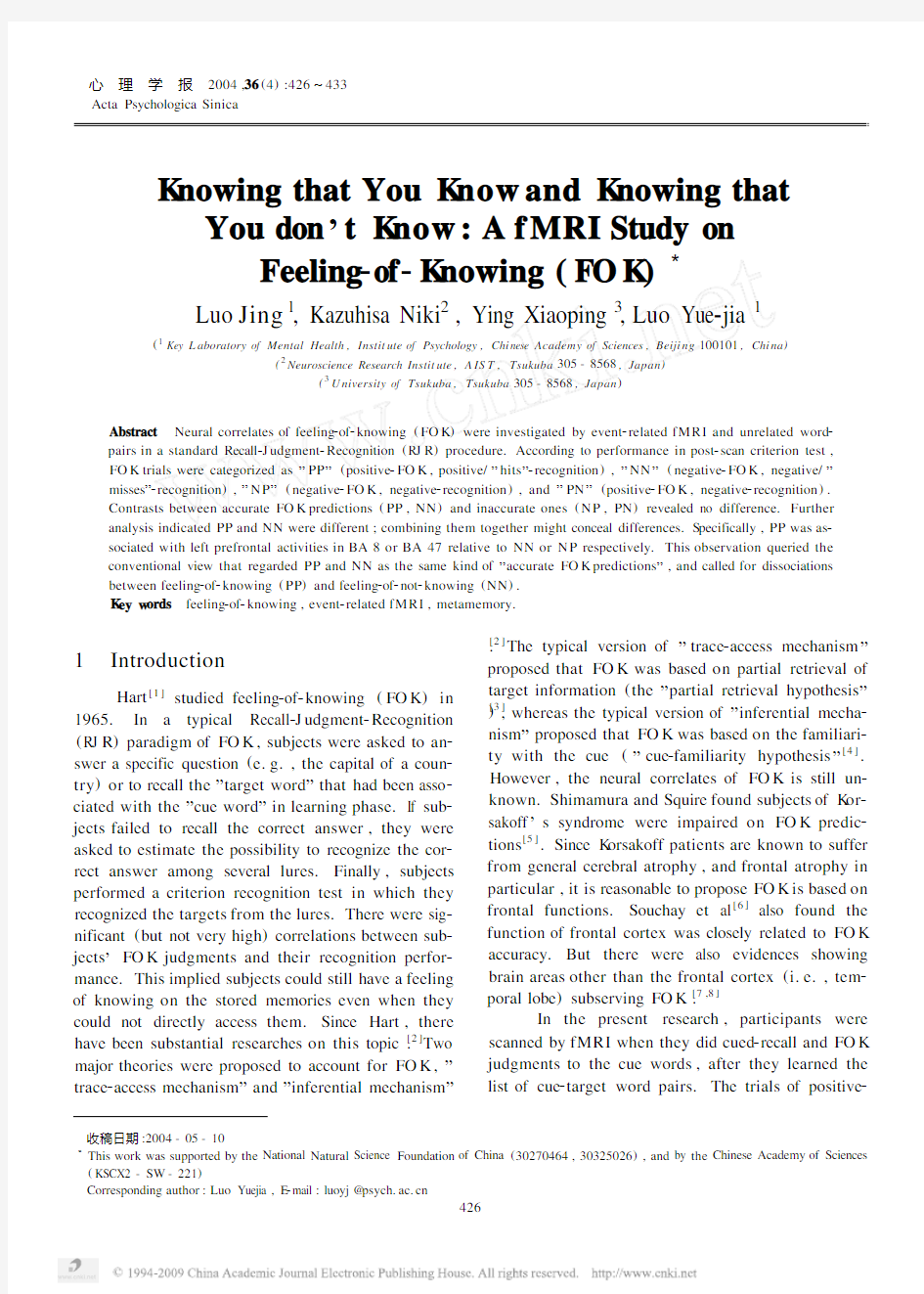
Neural correlates of accurate FO K prediction. The contrast between the accurately predicted events (PP,NN)and the inaccurately predicted events (PN,NP)revealed no significant difference.Then, the contrast of”PP versus NN”,”PP versus NP”, and”NP versus NN”were calculated respectively (Table1).Two left prefrontal activities were exhib2 ited.Relative to NN,PP was associated with an ac2 tivity in left middle frontal gyrus peaked in BA8and extended into BA6and9(Figure2).Relative to NP,PP was associated with an activity in left middle frontal gyrus peaked in BA47(Figure3).
4 Discussion
4.1 The general neural net w orks underlying cued2 recall and FOK judgments
Different from Maril et al.′s observation that the tip2of2tongue(TO T)items were associated with right2lateralized prefrontal activities relative to the no2 TO T items[9],our results revealed robust left2lateral2 ized prefrontal activities not only when the cued2recall and FO K trials were contrasted with the key pressing baseline trials(”PP2KP”,”NN2KP”,and”NP2 KP”),but also when the cued2recall and FO K trials were contrasted with each other(”PP2NN”and”PP 2NP”).Compared with the semantic knowledge re2 trieval in Maril et al.’s,the episodic retrieval in this experiment might call for more reflective and system2 atic processing,and lead to more left prefrontal activ2 ities[10].Specifically,the left ventral prefrontal cor2 tex(BA47/11)exhibited in”PP2KP”and”NN2 KP”has been proved to subserve semantic selec2 tions[11].The left dorsolateral prefrontal cortex(BA 46/9)observed in”PP2KP”,”NN2KP”,and”NP
4期罗 劲等:“知道自己知道”与“知道自己不知道”429
2KP ”has been observed to be associated with recall of non 2imaginable word relative to recall of imaginable
word [12],recognition of
deeply
Figure 2 Group 2averaged left prefrontal activation revealed in the contrast of ”PP minus NN ”.Thresholded at p <0.001(uncorrected )for demonstrations.Left :the SPM coronal section showing the territory of activation ;the blue cross marks the voxel that has the peak value in the con 2trast.Talairach coronal section showing the territory of activation is also given in the right bottom of the picture.The bar denotes t value.Right :the event 2related plots showing the averaged signal change (%)of the best 2fitting canonical hemodynamic response function (HRF )from the peak voxel (X =-40,Y =12,Z =53)marked by the blue cross in the left
picture.
Figure 3 Group 2averaged left prefrontal activation revealed in the contrast of ”PP minus NP ”.Thresholded at p <0.001(uncorrected )for demonstrations.Left :the SPM coronal section showing the territory of activation ,the blue cross marks the voxel that has the peak value in the con 2trast.Talairach coronal section showing the territory of activation is also given in the right bottom of the picture.The bar denotes t value.Right :the event 2related plots showing the averaged signal change (%)of the best 2fitting canonical hemodynamic response function (HRF )from the peak voxel (X =246,Y =36,Z =29)marked by the blue cross in the left picture.
studied items relative to recognition of shallowly stud 2ied word [13],and rejection of the conjunction lures that made from a conjunction of two previously learned items (e.g.,the probe ”nosedive ”after
studying ”nosebleed ”and ”skydive ”)relative to rejec 2tion of novel negative probes [14]
.The left anterior prefrontal cortex (BA 10)observed in ”PP 2KP ”and ”NN 2KP ”(a more posterior and inferior activation peaked in BA 47but near BA 10was also observed in ”PP 2NP ”,see Figure 3)was associated with ”reflec 2tive ”processes engaged in episodic retrieval ,left an 2terior prefrontal cortex was proved to be responsive to
430 心 理 学 报36卷
the demands of retrieving perceptually detailed infor2 mation about studied objects,and generally,to be re2 lated to monitoring and evaluation of specific memory characteristics at retrieval process that is critical for accurate episodic remembering[15].These areas high2 lighted the neural network that was sensitive to the process of episodic memory retrieval and metamemory monitoring.
4.2 Feeling2of2knowing and feeling2of2not2knowing Our image results did not reveal any detectable difference between accurately predicted trials(PP, NN)and the inaccurately predicted trials(PN,NP), although the behavioral results confirmed that the FO K judgments did have above2chance predictability on later recognition performance.
One possibility was that although both PP and NN were accurately predicted trials,they might be mediated by distinct brain mechanisms.To regard them as the same type of trials(i.e.,collapsing across PP and NN in analysis)might conceal some differences.There were several lines of evidences in agreement with this possibility.First,relative to NP,PP highlighted the inferior part of left prefrontal (BA47),whereas NN did not show any super2 threshold activity.Second,relative to KP,PP was associated with activities in right inferior frontal gyrus (BA47),bilateral lingual gyrus(BA18and19), and left cuneus(BA18),whereas NN(relative to KP)was not.Third,relative to KP,PP exhibited larger volumes of activities than NN(relative to KP) in left anterior prefrontal cortex(BA10)(290voxels in”PP KP”versus77voxels in”NN KP”),left ven2 tral prefrontal cortex(BA47/11)(362versus31 voxels),left dorsolateral prefrontal cortex(BA46/9) (500versus40voxels),and left superior frontal gyrus(BA8)(716versus348voxels).Finally,the direct contrast of PP and NN revealed significant ac2 tivity in left middle frontal gyrus(BA8).
To consider PP and NN as being subserved by different brain processes has important implications on FO K studies.Contrary to the conventional point of view that regards PP and NN as the same type of”accurate FO K predictions”,the differential neural correlates between PP and NN implied feeling2of2 knowing(PP)and feeling2of2not2knowing(NN) might be subserved by distinct metamemory process2 es.
The more involvement of left prefrontal cortex in PP relative to NN implied that feeling2of2knowing was realized by a more systematic retrieval process than feeling2of2not2knowing.In particular,the left middle frontal gyrus(peaked in BA8and extended into BA6and9)highlighted in the contrast of”PP minus NN”was observed in free recall[16],cued2re2 call[17],orally repeating of recognized old words[18], and retrieval success[19].But activity in this area was less consistently activated in episodic memory retrieval than in verbal working memory retrieval[20].Recent neuroimaging studies have bridged the gap between the episodic and working memory,based on the ob2 servation that specific prefrontal regions contributed to both episodic and working memory.It was pro2 posed that prefrontal activation during episodic mem2 ory might reflect the recruitment of specific working memory processes in the service of episodic learning and remembering[21].G iven the function of left supe2 rior/middle frontal gyrus(BA8)in working memory is”active maintenance”of abstract representation that goes beyond the stimulus itself[22],we proposed the activity of left middle frontal gyrus(BA8)in PP subserved the”active maintenance”of information e2 voked by the retrieval cues,and embodied the manip2 ulation of metamemory monitoring.The activation of BA8did not imply there were more eye movement in PP than in NN,given that a)the location of left middle frontal gyrus(BA8/9)observed in the pre2 sent research was much anterior than the observed frontal eye field in humans[23];b)the sensorimotor requirements in PP and NN were kept equivalent; and c)no difference between PP and NN was detect2 ed in the parieto2occipital regions that was usually more sensitive to the difference in eye movement.
Relative to KP,NN activated fewer and smaller cortex areas than PP did.It was possible that feeling2 of2not2knowing was realized by a”null activity hy2 pothesis”,that is,the brain made a judgment of feel2 ing2of2not2knowing based on the fact that there was no much activity associated with the specific cue. This was not only consistent with K olers and Palef’s observation that FO K judgments could be made very rapid for negative(not knowing)judgments[24],and with K lin et al.’s observation that subjects responded to implicit”don’t know”questions(i.e.,no infor2 mation regarding the answers was provided)faster
4期罗 劲等:“知道自己知道”与“知道自己不知道”431
than to explicit”don’t know”questions(i.e.,narra2 tives explicitly stated that something was un2 know n)[25],but also generally consistent with the hypothesis that cue familiarity heuristic plays an im2 portant role in FO K judgments[3]in that feeling2of2 not2knowing can be achieved by the simple process of cue familiarity/novelty heuristic.
5 Conclusion
People could still have feeling2of2knowing of stored memories even when they could not directly access these memories.Through neuroimage method, this research revealed how the brain could realize this function.Frontal activities,in particular left superi2 or,inferior,and middle frontal gyrus,were observed to be associated in this metamemory process.The re2 sults also implied that feeling2of2knowing(PP)and feeling2of2not2knowing(NN)could be mediated by the distinct neural and cognitive processes.This ob2 servation queried the conventional view that regarded PP and NN as the same type of”accurate FO K pre2 dictions”and called for dissociations between feeling2 of2knowing and feeling2of2not2knowing in future re2 search.
R eference
1 Hart J T.Memory and the feeling2of2knowing experience.Journal of Educational Psychology,1965,56:208~216
2 Nelson TO,G erler D,Narens L.Accuracy of feeling2of2knowing judgments for predicting perceptual identification and relearning.
Journal of Experimental psychology:G eneral,1984,113:282~300
3 Schacter DL.Feeling of knowing in episodic memory.Journal of Experimental Psychology:Learning,Memory and Cognition, 1983,12:452~460
4 Metcalfe J.Novelty monitoring,metacogntion,and control in a composite holographic associative recall model:Implications for K o2 rsakoff amnesia.Psychological Review,1993,100:3~22
5 Shimamura A,Squire L.Memory and metamemory:A study of the feeling2of2knowing phenomenon in amnesic patients.Journal of Experimental Psychology:Learning,Memory and Cognition, 1986,12:452~460
6 Souchay C,Isingrini M,Espagnet L.Aging,episodic memory feeling2of2knowing,and frontal functioning.Neuropsychology, 2000,14:299~309
7 Prevey ML,Delaney RC,Mattson RH et al.Feeling2of2knowing in temporal lobe epilepsy:monitoring knowledge inaccessible to conscious recall.Cortex,1991,27:81~92
8 Prevey ML,Delaney RC,Mattson RH.Metamemory in temporal lobe epilepsy:self2monitoring of memory functions.Brain and Cog2
nition,1988,7:298~311
9 Maril A,Wagner AD,Schacter DL.On the tip of the tongue:an event2related fMRI study of semantic retrieval failure and cognitive conflict.Neuron,2001,31:653~660
10 Nolde SF,Johnson M K,Raye CL.The role of prefrontal during test of episodic memory.Trends in Cognitive Sciences,1998,2: 399~406
11 Thompson2Schill SL,D’Esposito M,Aguirre GK et al.Role of left inferior prefrontal cortex in retrieval of semantic knowledge:a reevaluation.Proceedings of the National Academy of Sciences of the United States of America,1997,94:14792~14797
12 Fletcher PC,Shallice T,Frith CD et al.Brain activity during memory retrieval.The influence of imagery and semantic cueing.
Brain,1996,119:1587~1596
13 Rugg MD,Walla P,Schloerscheidt AM et al.Neural correlates of depth of processing effects on recollection:evidence from brain po2 tentials and positron emission tomography.Experimental Brain Re2 search,1998,123:18~23
14 McDermott K B,Jones TC,Petersen SE et al.Retrieval Success is Accompanied by Enhanced Activation in Anterior Prefrontal Cortex During Recognition Memory:An Event2Related fMRI Study.
Journal of Cognitive Neuroscience,2000,12:965~976
15 Ranganath C,Johnson M K,D’Esposito M.Left anterior pre2 frontal activation increases with demands to recall specific perceptu2 al information.The Journal of Neuroscience,2000,20:1~5
16 Andreasen NC,O’Leary DS,Cizadlo T et al.II.PET studies of memory:novel versus practiced free recall of word lists.Neuro Im2 age,1995,2:296~305
17 Buckner RL,Raichle ME,Petersen SE.Dissociation of human prefrontal cortical areas across different speech production tasks and gender groups.Journal of Neurophysiololgy,1995,74:2163~2173
18 Fujii T,Okuda J,Kawashima R et al.Different roles of the left and right parahippocampal regions in verbal recognition:a PET study.NeuroReport,1997,8:1113~1117
19 Tulving E,Markowitsch H J,Craik FE et al.Novelty and familiar2 ity activations in PET studies of memory encoding and retrieval.
Cerebral Cortex,1996,6:71~79
20 Cabeza R,Nyberg L.Imaging cognition II:An empirical review of 275PET and fMRI studies.Journal Cognitive Neuroscience, 2000,12:1~47
21 Wagner AD.Working memory contributions to human learning and remembering.Neuron,1999,22:19~22
22 Zurowski B,G ostomzyk J,Gron G et al.Dissociating a common working memory network from different neural substrates of phonological and spatial stimulus processing.Neuro Image,2002, 15:45~57
23 Petit L,Courtney SM,Ungerleider L G et al.Sustained activity in the medial wall during working memory delays.The Journal Neu2 roscience,1998,18:9429~9437
24 K olers PA,Palef SR.Knowing not.Memory&Cognition,1976, 4:553~558
25 K lin CM,Guzman AE,Levine WH.Knowing that you don’t know:metamemory and discourse processing.Journal of Experi2 mental Psychology:Learning,Memory and Cognition,1997,23: 1378~1393
432
心 理 学 报36卷
“知道自己知道”与“知道自己不知道”
———一项有关知道感(FO K )的脑成像研究罗 劲1 仁木和久2 应小萍3 罗跃嘉1
(1中国科学院心理研究所心理健康重点实验室,北京100101)
(2产业技术综合研究所,筑波305-8568,日本)
(3筑波大学,筑波305-8568,日本)
摘 要 采用事件相关功能性磁共振成像技术(event 2related fMRI )以及在FO K (feeling 2of 2knowing )研究中常用的
“线索回忆2FO K 判断2再认测验”
(Recall 2J udgment 2Recognition ,RJ R )程序,以汉字词对为识记材料,研究了FO K 判断中的大脑活动区域。根据FO K 判断的正负以及其后的再认测验的对错,实验将FO K 判断的项目分为四类:PP 项目(正性FO K 判断,正确再认),NN (负性FO K 判断,错误再认),NP (负性FO K 判断,正确再认)以及PN (正性FO K 判断,错误再认)。脑成像的分析结果显示:准确的FO K 预测(即PP 与NN 项目)与不准确的FO K 预测(即NP 与NP 项目)在脑活动上没有显著的差异。而进一步分析表明,这种“无差异”的现象可能是由于PP 项目与NN 项目激活了不同的脑活动模式所造成的。具体地讲,相对于NN 项目而言,PP 项目伴随有明显的左侧前额叶(BA 8区)的活动。这一观察提示我们:知道感(PP )与不知道感(NN )可能是由不同的脑神经网络所支持、并通过不同的认知过程来实现的。关键词 知道感(FO K ),事件相关功能性磁共振成像,元记忆。分类号 B842
4期罗 劲等:“知道自己知道”与“知道自己不知道”433
第四节脑功能成像技术1 语言神经认知机制研究是语言科学研究的重要内容,它主要研究语言与大脑的关系,简单的说就是研究语言在人脑中的理解与产生的过程。但是人脑被一层厚厚的颅骨所包围,因此仅凭肉眼无法判断大脑处理语言时的情况。认知语言学通过语言理论的假设来构建语言认知模型,心理语言学则通过行为学方法,通过测试量表来研究具体语言结构的反应时间和正确率。但是,这两种研究方向都不能直接观察大脑实时处理语言的情况。随着科学技术的发展,新的语言科学研究技术已经被广泛用于语言研究中,其中PET和fMRI尤其是fMRI技术又是神经认知科学研究被最广泛应用的一种新的技术手段。 一脑功能成像技术简介 PET(Positron Emission Tomography,PET)即正电子发射断层扫描技术,其基本原理是:刺激作用于大脑会产生血流变化,利用血液中注射的放射性示踪物质来和脑活动的某些脑区进行对比,从而确定刺激任务与特定脑区之间的关系。fMRI是functional Magnetic Resonance Imaging的简称,中文名称为功能性磁共振成像。其实质就是在磁共振成像的基础上获取大脑活动的功能图像,以获取被试对所给语言、图形、声音等刺激材料进行加工时产生的fMRI信号并加以分析,以确定这些刺激材料与对应脑区的关系,从而分析其脑机制。赵喜平(2000)认为所谓的fMRI就是利用MRI对组织磁化高度敏感的特点来研究人脑功能,特别是大脑各功能区划分或定位的无创伤性检测技术。由于PET技术在技术要求以及资金需求方面的原因,用于认知任务的研究越来越少,现在主要的脑成像技术就是fMRI,因此这里主要介绍fMRI技术以及实验数据的处理和对实验数据的解读。 1.1 fMRI的发展及其原理 MRI(Magnetic Resonance Imaging,磁共振成像)产生于上个世纪70年代。1970年,美国纽约州立大学的Raymond Damadian发现正常组织的NMR(Nuclear Magnetic Resonance)信号与病变组织的信号明显不同。这以后Paul Lauterbur、Peter Mansfield 和Graunell发展了各种成像方法。1976年 Mansfield得到了第一幅人体断层像,1977年世界上第一台名为indomitable 的全身磁共振成像装置诞生,1978年的图像质量已经接近CT,1980年磁共振成像设备用于商业用途,这之后,磁共振成像技术开始进入一个飞速发展的时期。美国Technicare公司、GE公司、 1 为了使读者能够直观的了解脑功能成像的实验过程,本章节的图片除了引用编著者已经发表的陈国之外,还参考了https://www.doczj.com/doc/558892532.html,/afni、https://www.doczj.com/doc/558892532.html,/products/e-prime/网站的部分图片。
2、认知神经科学脑成像技术简介、当前应用及展望 答: CT、MRI——癫痫的病因 SPECT——脑血流灌注 PET——脑代谢 MRS——脑内生化物质的改变 fMRI——血氧水平依赖(BOLD)描述大脑内神经元激活的区域 脑电图—主要体现在对于癫痫源或者功能区的定位帮助,并不能用于诊断病人是否患有癫痫 【以上是一个课件中得到的,我不是很明白说的意思。】 下面是关于fMRI的应用发展 1. 功能核磁共振的概念、特点及工作原理 (1)概念 功能磁共振成像(functionalmagneticresonanceimaging,fMRI)的突出特点是:可以利用超快速的成像技术,反映出大脑在受到刺激或发生病变时脑功能的变化。它突破了过去仅从生理学或病理生理学角度对人脑实施研究和评价的状态,打开了从语言、记忆和认知等领域对大脑进行探索的大门。 传统的磁共振成像(MRI)与功能磁共振成像(fMRI)之间的主要区别是它们所测量的磁共振信号有所不同。MRI是利用组织水分子中的氢原子核处于磁场中发生的核磁共振现象,对组织结构进行成像,而fMRI所测量的是在受到刺激或发生病变时大脑功能的变化。根据所测量的脑功能信号的不同,磁共振功能成像主要有以下四种工作方式:①血氧水平依赖功能磁共振成像(blood-oxygen-level-dependentfMRI,BOLD-fMRI),它主要是通过测量区域中氧合血流的变化(或血流动力学的变化),实现对不同脑功能区域的定位;②灌注功能磁共振成像(perfusionfMRI),又称为灌注加权成像(perfusionweightedimaging,PWI)。这种成像方法主要用于测量局部脑血流和血容积;③弥散加权功能磁共振成像(diffusion-weightedfMRI),这种方法主要用于测量水分子的随机运动;④磁共振波谱成像(MRIspectroscopy),该方法用于测量脑的新陈代谢状态以及参加到新陈代谢中的某些物质(如磷和氧)的含量。目前,临床上和脑科学研究中一般都是用第一种方式,文献中出现的fMRI,如果不做特别说明,一般都是指BOLD-fMRI,简称为fMRI。 (2)工作原理 BOLD 技术是fMRI 的理论基础。当大脑在执行一些特殊任务或受到某种刺激时,某个脑区的神经元的活动就会增强。增强的脑活动导致局部脑血流量的增加,从而使得更多的氧通过血流传送到增强活动的神经区域,使该区域里的氧供应远远超出了神经元新陈代谢所需的氧量,导致了血流中氧供应和氧消耗之间的失衡,结果造成了功能活动区血管结构中氧合血红蛋白的增加,而脱氧血红蛋白的相对减少。脱氧血红蛋白是一种顺磁性物质,其铁离子有四个不成对电子,磁距较大,有明显的T2缩短效应,因此在某一脑区脱氧血红蛋白的浓度相对减少将会造成该区域T2。信号的相对延长,使得该区域中的MR信号强度增强,在脑功能成像时功能活动区的皮层表现为高信号,利用EPI快速成像序列就可以把它检测出来。
右脑王英语学习机详细介绍 来源:问学堂官网https://www.doczj.com/doc/558892532.html, 右脑王英语学习机,是透过特殊的设备,营造全封闭英语环境,传递「声」「光」多重频率 组合讯号,快速地与脑波产生共振,直接引导脑细胞活动效能(NEQ)至最佳的大脑状态, 唤醒右脑巨大学习潜能,促进大脑运作,并同时形成唯一英语通道,以 2-4 倍速度大量输 入英语课程,将英语信息直接传送至右脑潜意识,达到学习高效而轻松学习英语的目的。 右脑王英语学习机是一款能让使用者在最短时间内迅速提升英语水平的电 子产品,通过视觉光导以及声波共振的作用,对使用者的脑电波进行控制、 迅速激活右脑潜能,让使用者打开右脑语言学习区,调整大脑至最佳的英 语学习状态。结合倍速播放的英语学习课程,充分利用右脑的超强记忆能 力和超强的计算能力,通过右脑英语学习方法进行英语学习,从而令使用 者在极短的时间内,迅速地提升英语水平。 1:右脑王快速地与脑波产生共振,直接引导脑细胞活动效能(NEQ) 2:最佳的右脑意识状态,以2—4倍的速度海量输入及处理英语信息 3:美籍专家团队专为右脑学习需求而研发的高品质课程,更实用,地 道 4:躺在舒适的软椅上,不用任何基础,只要会说中国话,你就会说英 语 5:不背单词,不抠语法,立竿见影,永久记忆,人人轻松掌握 6:4天1000单词,30天轻易突破中小学英语,四六级,商务英语,GRE 7:4G内存,游戏,摄影,音乐,视频,电子书精彩不断,寓教于乐 1:激发脑力:营造全封闭英语环境,传递“声”“光”多重频率组合 讯号,与脑波共振,让大脑快速达到最佳状态,唤醒右脑潜能,轻松征服 英语。
2:全英语教材:海量音频英语课程,精选视频教材,TXT电子文档阅 读,营造全真英语学习情境,从单词到短文阶梯型教学 3:在线更新:在线扩展课程,随产品发放注册码,海量下载最新课程4:多循环模式:配备多种循环模式供使用者选择,完整巩固学习效果。 5:全新设计:2.8/26万色真彩TFT超大显示屏,滑盖设计,新颖独特的钢琴黑。 6:升级功能:免驱U盘驱动,直接连接电脑进行文件操作。支持固件升级,支持TF卡。 7:全动画菜单:直观动画菜单,配合相应文字显示,使用起来得心应手。 8:多媒体播放格式:支持JPEG、GIF图片浏览,支持MP3、WMA、FLAC、APE、RMVB、AVI、 RM、MP4等格式的音频、视频英语课程文件播放。 9:录音功能:内置麦克风,超长时间录音。 10:任务功能:支持多任务功能,能在进行右脑王英语课程学习的同时阅读对应的英语教材。 11:照片随心拍:可调节分辨率,设置多种拍照风格,支持手动拍照、 定时拍照。 12:超长录像功能:超长时间录像,使用过程非常方便。 1:符合右脑学习意识状态下的需要: 通过右脑王英语学习机的声光调节,使用者进入右脑学习意识状态,其百万倍的录音记忆被打开,此时需要通过右脑王英语学习机反复的加速输入英语信息。我们的课程根据深度听觉的特性,甄选了最适合进阶学习的优质课程,更把每个课程分成常速、2倍速、4倍速不同的版本,供使用者逐步提升。当您掌握了4倍速课程,你就可以轻易应对所有英语问题。 2:超人性化,地道,实用: 我们课程的每个句子和单词都非常实用,语音地道标准,你只需要在右脑记忆激活的情况下认真听,无 意识跟读,轻松、简单、高效!我们每个要素都由精心挑选的超实用内容组成。模拟您在国外可能碰到
心理科学进展2009, V ol. 17, No. 2, 349–355 Advances in Psychological Science 一般流体智力的脑成像研究述评 张小将1刘昌1刘迎杰2 (1南京师范大学教育科学学院暨认知神经科学实验室,南京 210097)(2江西师范大学教育学院,南昌,330022) 摘要对一般流体智力的脑成像研究有助于探明智力的脑机制。从结构成像与功能成像两个方面阐述了相关的研究。已有研究表明,一般流体智力与总的脑体积、额叶或其他多个脑区的灰质体积中等正相关。一般智力(g)因素负荷高的任务比负荷低的任务引发特定脑区更大的激活。较早的研究发现一般智力水平个体差异与脑区的激活呈负相关,但此后的一些研究却得到相反的结论。未来的研究应加强对个体差异的研究,将脑成像技术与EEG或ERP技术结合起来,整合多水平(心理测量、遗传学等)的研究结果,进一步阐明一般流体智力的脑机制。 关键词一般流体智力;脑成像;磁共振成像;功能磁共振成像;正电子发射断层扫描 分类号 B845 一般智力(或者g)的概念是Spearman于1904年首次提出的,通常是指对一组智力测验进行因素分析时第一个未旋转的主成分。不同的智力测验可以根据其对一般智力的预测能力而区分为不同的g 因素负荷。Spearman的一般智力与Cattell的流体智力(fluid intelligence)在心理测量的内容上较为相似。因素分析的研究表明,流体智力的g因素负荷甚至高达1.0(Keith & Wolfle, 1995)。由于二者之间的相似程度较高,研究者通常将二者统合起来,称之为一般流体智力(general fluid intelligence gF)。 虽然一般智力因素可以从多个测验分数中提取,但是却没有纯粹测量一般智力的测验,每个测验都或多或少地包含除一般智力之外的特定能力。因此,对于一般流体智力的评估主要是以较高g因素负荷的测验或任务为指标。总体上来看,已有的研究中对一般流体智力的评估主要有三种方式:(1)以某些公认的一般流体智力测验的成绩作为指标,如瑞文推理测验(Raven’s Progressive Matrices)、卡特文化公平智力测验(Cattell’s Culture Fair Intelligence test)及修改版的韦克斯勒成人智力测验中的全量表分测验(WAIS-R中的FSIQ分测验,g 因素负荷约为0.9)。(2)以某些较高g因素负荷的任务(如抽象的图形推理任务)的成绩作为指标。(3)也有部分的研究将包含多个子测验的IQ分数作为一般智力指标。从实践来看,一般智力和IQ的 收稿日期:2008-05-26 通讯作者:刘昌,E-mail: cglew@https://www.doczj.com/doc/558892532.html, 差异不大,甚至可以在概念上互换;但是从统计上来看,它们还是存在一定的差别。一般智力(g)是通过因素分析从多个IQ子测验中提取的共同因素,而IQ则是所有子测验分数简单相加的总和。因此,本文所讨论的研究主要是以前两种方式为一般智力指标的研究。 早期关于智力的研究主要集中于智力理论、智力结构及其认知基础等方面。近年来,随着认知神经科学的兴起,更多的研究者试图借助新兴的技术手段来探索智力的神经机制。其中,脑成像技术凭借其较高的空间分辨率及无损伤性的特点而受到人们的青睐。总体上看,对一般流体智力的脑成像研究主要从两个方面来探讨智力的脑机制:一种是结构性成像,主要是以磁共振成像(MRI)技术探讨一般流体智力与脑结构(主要是脑体积)的关系;另一种是功能性脑成像,以功能磁共振成像(fMRI)和正电子发射断层扫描(PET)技术来研究一般流体智力与脑活动的关系。本文将围绕这两方面对一般流体智力的脑成像研究进行梳理,并提出一些自己的观点。 1 一般流体智力与脑结构 早在100多年前,研究者们就试图将智力与脑的大小联系起来以探讨其生理基础,虽然这一领域的研究曾饱受批评,但大量的行为研究证实了智力与人脑的大小存在较小的正相关(约为0.1~0.3)(Wickett, Vernon, & Lee, 1994)。由于这些研究主要以外部测量的头围作为脑大小的指标,容易受无关
如何使用我们的大脑-大学英语作文_1500字 如何使用我们的大脑 How to Use Our Brain 1.人脑的重要性及作用。 2.如何科学用脑。 [写作导航]先写大脑的特性和作用(复杂、智能的器官,使人区别于动物等);再写大脑越用越灵,但过度使用也会出问题,为第三段作铺垫;第三段从两方面说明如何合理用脑:一是劳逸结合,手脑交替;二是从科学的角度,利用数据,说明过度用脑可能给大脑带来的伤害。
[范文] Human brain is the most complex and intelligent mechanism in the world. It is the major factor that distinguishes man from animals. With our brain we get to know the world and make a good use of the world to our benefit. Our brain is a product of constant use through millions of years. Other things can be used up, but used properly, our brain can never be exhausted. In fact, the more we use it, the more capable and efficient it will become. Excessive use of the brain, however, will causea lot of problems. So it is useful to know how to use our brain wisely. First, handwork or physical labor is good exercise as well
Regions abbr. Precentral gyrus PreCG.L 中央前回 Precentral gyrus PreCG.R 中央前回 Superior frontal gyrus, dorsolateral SFGdor.L 背外侧额.比回 Superior frontal gyrus, dorsolateral SFGdor.R 背外侧额上回 Superior frontal gyrus, orbital part ORBsup.L 眶部额上回 Superior frontal gyrus, orbital part ORBsup.R 眶部额比回 Middle frontal gyrus MFG.L 额中回 Middle frontal gyrus MFG.R 额中回 Middle frontal gyrus, orbital part ORBmid.L 眶部额中回 Middle frontal gyrus, orbital part ORBmid.R 眶部额中回 Inferior frontal gyrus, opercular part IFGoperc.L 岛盖部额下回 Inferior frontal gyrus, opercular part IFGoperc.R 岛盖部额下回 Inferior frontal gyrus, triangular part IFGtriang.L 三角部额下回 Inferior frontal gyrus, triangular part IFGtriang.R 三角部额下回
Inferior frontal gyrus, orbital part ORBinf.L 眶部额下回 Inferior frontal gyrus, orbital part ORBinf.R 眶部额下回 Rolandic operculum ROL.L 中央沟盖 Rolandic operculum ROL.R 中央沟盖 Supplementary motor area SMA.L 补充运动区 Supplementary motor area SMA.R 补充运动区 Olfactory cortex OLF.L 嗅皮质 Olfactory cortex OLF.R 嗅皮质 Superior frontal gyrus, medial SFGmed.L 内侧额上回 Superior frontal gyrus, medial SFGmed.R 内侧额_上回 Superior frontal gyrus, medial orbital ORBsupmed.L 眶内额上回 Middle frontal gyrus, orbital part ORBmid.R 眶部额中回 Inferior frontal gyrus, opercular part IFGoperc.L 岛盖部额下回 Inferior frontal gyrus, opercular part IFGoperc.R 岛盖部额下回 Inferior frontal gyrus, triangular part IFGtriang.L
脑功能成像技术 近20年来,随着现代物理、电子与计算机技术的迅速发展,脑功能成像技术(functional brain imaging)取得了长足的进步,一批功能强大的无创性脑功能成像手段相继诞生。这促使研究者们对脑功能成像技术及其在认知过程、情绪过程中的应用产生了浓厚的兴趣,将它们迅速应用到认知神经科学以及心理学的各个领域中,并取得了许多突破性成果,促进了这些领域研究的深入化进程。 (一)使用脑功能成像技术的理由 研究者进行脑功能成像技术进行实验,最明显的目的是为了将脑的结构与其功能联系起来。我们已经知道,脑的许多功能都是定位于大脑的神经组织结构之中的;基于此,研究者们开始试图成像出那些参与到不同脑结构激活中的基本过程。现代神经成像假定,我们可以根据组成复杂心理过程的一些基本操作的结合来对其进行最好的描述,这些基本过程并不是定位于大脑中的某个单一部位的,而通常是神经元网络共同作用的结果。神经成像的这一假定自然而然地导致了人们对与基本心理过程相伴随着的脑激活的探讨。而将这些基本过程成像到大脑中的区域和功能性网络就是现代脑成像研究的主要目标。 对不同脑结构的功能的详细成像可以为我们提供关于基本心理过程的可靠证据。一旦我们能够确定,特定的脑区与某一心理过程有关系,就可以超越这种结构与功能的简单对应关系,而使用统计技术(如区域间相关、因素分析、结构方程建模等)来进一步考察与复杂心理任务有关的激活环路,分析出心理任务中包含了哪些基本过程的结合。这样,通过考察激活模式,我们就能从简单到复杂,并能了解在某一模式中所激活的结构所具有的功能。此外,在脑损伤研究中,还能帮助我们推测受其影响何种脑功能会丧失。 使用脑成像技术的另一个原因是:它可以分离心理过程。如果我们能够获得不同心理任务所导致的激活模式的数据,就可以用它来检验这两个任务是否存在双重分离(Smith和Jonides, 1995)。这种分离的原理是:假设某特定脑区A处理某认知过程a ;类似地,某特定脑区B处理某认知过程b 。假设有1、2两种心理任务。任务1需要心理活动a参与而不需要b ;任务2需要心理活动b参与而不需要a 。如果我们在被试完成这两种任务时对其
现代脑成像技术的发展历史 现代脑成像技术在19世纪末初具雏形,在德国神经学家Emil Dubois-Reymond发现神经活动可引起电信号改变的基础上,1875年Richard Caton首次记录了兔脑神经的电活动;1929年Hans Berger首次借助置于头皮的电极,成功测量到脑部的电活动,成为脑电(EEG)发展的里程碑;Adrian等于1934年,Jasper等于1935年也观察并证实了Berger的观察。从此,EEG的客观存在才得到了科学界的一致认同。20世纪60年代以后科学家们开始记录同执行任务相关的EEG,将与刺激事件相关的,并在时间上与刺激锁定的EEG信号平均起来,观察到一系列的事件相关电位(ERPs),这些电位提供了关于认知活动的脑内信息,而且具有毫秒级的时间分辨率,被称为事件相关电位技术。 在脑电发展的同时期,1890年开始,人们就知道血流与血氧的改变(两者合称为血液动力学)与神经元的活化有着紧密的关系;20世纪70年代磁共振成像(MRI)技术的发明,成为医学影像学发展史上的一次革命;1990年,基于传统的MRI技术,美国Bell实验室的Seiji Ogawa等人根据脑功能活动区氧合血红蛋白(HbO2)含量的增加导致磁共振信号增强的原理得到了关于人脑的功能性磁共振图像,发明了功能性磁共振成像技术(fMRI),该技术的发展极大地推动了医学、神经生理学和认知神经科学的迅速发展。 另外,随着“洞见五脏症结”的要求出现,在1973年,Godfrey Hounsfield发明了X线计算机辅助断层成像(CT)技术,这是现代临床医学发展史上的重要里程碑事件。此后,人们对CT技术进行了扩展,70年代产生了单光子发射计算机断层成像技术(SPECT)以及正电子发射断层成像(PET),PET扫描技术在脑功能定位上的效果比SPECT好,它可以三维高空间分辨地对脑活动区进行定位,出现后迅速成为神经科学界的宠儿。
大脑结构名词中英文对照 安蒙氏角Ammoms horn 白质white matter 背内侧丘脑dorsalmedial thalamus 背外侧通路dorsallateral pathway 背外侧膝状核dorsal lateral geniculate nucleus 被盖tegmentum 被盖背束dorsal tegmental bundle 被盖腹区ventral tegmental area 边缘皮质limbic cortex 边缘系统limbic system 布洛卡区Broca%26;s area 苍白球globus pallidus 侧脑室lateral ventricle 齿状回dentate gyrus 穿质通路perforant path 传出纤维efferent fibers 传入纤维afferent fibers 大脑半球cerebral hemisphere 大脑导水管cerebral aqueduct 导水管四周灰质periaqueductal grey matter 第三脑室third ventricle 第四脑室fourth ventricles 顶盖tectum 顶盖脊髓束tectospinal tract 顶盖枕核系统tectopulvinar system 顶叶parietal lobes 豆状核lentiform nucleus 端脑telecephalon
额眶皮质orbital frontal cortex 额叶frontal lobe 额叶岛盖frontal operculum 耳蜗核cochlear nucleus 二级投射区sencondary projection area 非特异性投射系统nonspecific projecting system 缝际核raphe nucleus 伏核nucleus accumbens 腹内侧通路ventraomedial pathway 腹内侧下丘脑ventromedial hypothalamus 隔区septal area 弓状核arcuate nucleus 弓状束arcuate fasciculus 沟sulcus 钩束uncinate faciculus 孤束核solitary nucleus 海马hippocampus 海马结构hippocampus formation 海马伞fimbria 黑质substantia nigra 黑质纹状体束nigrotriatal bundles 红核red nucleus 红核束rubrospinal tract 后脑metencephalon 灰质gray substance 基底神经节basal ganglia 基底外侧核群basolateral nuclear group 间脑diencephalon 交叉decussation 角回angular gyrus
1.无创脑成像技术有哪些? 答:无创脑成像技术有10种: (1)X射线断层成像(CA T) (2)近红外光学成像(DOI) (3)事件相关光学信号成像(EROS) (4)光声效应成像 (5)磁共振成像(MRI) (6)功能磁共振成像(fMRI) fMRI成像的物理学基础是核磁共振现象:自旋磁矩不为零的原子核(如氢原子核)在外界静磁场中发生磁化,环绕静磁场的纵轴拉莫进动,产生静磁矩,在一定频率(拉莫共振频率)的射频脉冲作用下,吸收能量发生能级的跃迁,而射频脉冲停止后,跃迁的原子核通过弛豫回复到原来的能级状态,同时释放出能够被记录到的能量信号。选择不同的成像周期的重复时间参数和成像的回波时间参数,可以得到不同参数依赖的加权图象,如T1加权像,T2*加权像和质子密度像。
fMRI成像的时间可以短至几十毫秒,空间分辨率可以达到1毫米,能同时提供大脑结构像和功能像获得准确的空间定位,可以无创性地多次重复实验。但fMRI测量的信号不是直接的神经活动信号,其测量的血氧变化信号一般滞后于神经活动(4~8秒)响应延迟,目前能够达到的时间分辨率最多只能在数百毫秒数量级。 (7)脑电图(EEG) 脑电图是通过脑电图描记仪将脑自身微弱的生物电放大记录成为一种曲线图,以帮助诊断疾病的一种现代辅助检查方法.它对被检查者没有任何创伤。 (8)脑磁图(MEG) 脑磁图是一种完全无侵袭,无损伤的脑功能检测技术,可广泛地用于大脑功能的开发研究和临床脑疾病诊断。MEG的检测过程,是对脑内神经电流发出的极其微弱的生物磁场信号的直接测量,同时,测量系统本身不会释放任何对人体有害的射线,能量或机器噪声。在检测过程中,MEG探测仪不需要固定在患者头部,测量前对患者无须作特殊准备,所以准备时间短,检测过程安全、简便,对人体无任何副作用。 (9)正电子发射断层扫描(PET) 正电子发射断层成像(Positron Emission Tomography) 系统是利用正电子同位素衰变产生出的正电子与人体内
TR(time of repetition,TR)又称重复时间。MRI的信号很弱,为提高MR的信噪比,要求重复使用同一种脉冲序列,这个重复激发的间隔时间即称TR。 弛豫(relaxation,经常被误写为“驰豫”)是指在核磁共振和磁共振成像中磁化矢量由非平衡态到平衡态的过程。在统计力学和热力学中,弛豫时间表示系统由不稳定定态趋于某稳定定态所需要的时间。在协同学中,弛豫时间可以表征快变量的影响程度,弛豫时间短表明快变量容易消去。这个系统可以是具体或抽象的,比如弹性形变消失的时间可称为弛豫时间,又比如光电效应从光照射到射出电子的时间段也称为弛豫时间,政策实施到产生效果也可称为弛豫时间。 弛豫时间有两种即T1和T2。 T1 T1为自旋一晶格或纵向驰豫时间,纵向磁化强度恢复的时间常数T1称为纵向弛豫时间(又称自旋-晶格弛豫时间)。 T2 T2为自旋一自旋或横向弛豫时间,横向磁化强度消失的时间常数T2称为横向弛豫时间(又称自旋-自旋弛豫时间)。 T2* 在理想的状态下,在同一磁场下,给定的化学环境中,所有的核以同一频率进动。但是在实际系统中,各个核的化学环境有细微的不同。 1/T2* = 1/T2 + 1/T (inhomo) = 1/T2 + γΔB0 不像T2,T2*受磁不均匀性的影响,T2*总是比T2短。 T1总是比T2长吗? 一般来说,2T1 ≥ T2 ≥ T2*。在大部分情况下,T1比T2长。 常见弛豫时间值 以下为常见健康人体组织的两个弛缓时间常数大概数值,仅供参考。 1.5特斯拉主磁场之下 组织类型T 1 大约值(毫秒) T 2 大约值(毫秒) 脂肪组织 240-250 60-80 全血(缺氧血) 1350 50 全血(带氧血) 1350 200 脑脊髓液(类似纯水) 2200-2400 500-1400 大脑灰质 920 100
大脑结构名词中英文对照 汉英版: 安蒙氏角Ammom's horn 白质white matter 背内侧丘脑dorsalmedial thalamus 背外侧通路dorsallateral pathway 背外侧膝状核dorsal lateral geniculate nucleus 被盖tegmentum 被盖背束dorsal tegmental bundle 被盖腹区ventral tegmental area 边缘皮质limbic cortex 边缘系统limbic system 布洛卡区Broca's area 苍白球globus pallidus 侧脑室lateral ventricle 齿状回dentate gyrus 穿质通路perforant path 传出纤维efferent fibers 传入纤维afferent fibers 大脑半球cerebral hemisphere 大脑导水管cerebral aqueduct 导水管周围灰质periaqueductal grey matter 第三脑室third ventricle 第四脑室fourth ventricles 顶盖tectum 顶盖脊髓束tectospinal tract 顶盖枕核系统tectopulvinar system 顶叶parietal lobes 豆状核lentiform nucleus 端脑telecephalon 额眶皮质orbital frontal cortex 额叶frontal lobe 额叶岛盖frontal operculum 耳蜗核cochlear nucleus 二级投射区sencondary projection area 非特异性投射系统nonspecific projecting system 缝际核raphe nucleus 伏核nucleus accumbens 腹内侧通路ventraomedial pathway 腹内侧下丘脑ventromedial hypothalamus 隔区septal area 弓状核arcuate nucleus
FMRI 人们越来越执著于对客观、确凿的大脑真相的追寻,现在有了一种非常优秀的大脑成像技术,那就是功能磁共振成像(FMRI)。空间编码是磁共振成像的关键技术。 自上世纪90年代初问世至2007年底,这种技术已出现在12000多篇科学论文中,而且这个数字至今还在以每周30至40篇的速度增长。人们之所以对它如此重视,那是因为比起现有其他大脑功能成像技术,fMRI在“观察活动中的大脑”时,不仅时间分辨率更高,就连空间分辨率也可达到毫米水平。借助fMRI,对大脑的研究便可扩展至记忆、注意力、决定……在某些情况下,fMRI技术甚至能够识别研究对象所见到的图像或者阅读的词语。对个人内心世界的这些揭示不禁让人期待在大脑中鉴别谎言这种复杂状态的可能性。 人脑是人体最重要的器官之一,对于人脑功能的探求无疑是非常有意义的事情。长久以来,科学家们就注意到这样的事实:即人脑的功能反映在大脑皮层是按空间分区的,在脑内次级结构也是按空间分隔的。研究脑功能映射(Function Brain Mapping)有许多成功的模式(Modality),例如正电子发射断层扫描(Positron Emission Tomography,PET),在向脑内注射15O水后,通过测量局部脑血流(rCBF)的方法来检测大脑的活动。脑电图(EEG)和脑磁图(MEG)也可检测大脑对诱发刺激响应的电或磁信号,但很难对活动区作准确的空间定位。在众多的模式中,用于脑功能定位的磁共振成像(Magnetic Resonance Imaging,MRI)技术,或功能磁共振成像(Functional MRI)是一种非常有效的研究脑功能的非介入技术,已经成为最广泛使用的脑功能研究手段。最早起源于1991年春天,美国麻省总医院(Massachusetts General Hospital,MGH)的磁共振研究中心利用磁共振成像生成反映脑血流变化的图像。它虽然是一种非介入的技术,但却能对特定的大脑活动的皮层区域进行准确、可靠的定位,空间分辨率达到1mm,并且能以各种方式对物体反复进行扫描。 fMRI的另一个特点是:能实时跟踪信号的改变。例如在仅几秒钟内发生的思维活动,或认知实验中信号的变化,时间分辨率达到1s。大批的脑科学研究人员已经开始从事磁共振功能神经成像的研究,并将它应用于认知神经科学。医学领域的迫切需求也进一步促使fMRI技术的发展,一些在病理方面的应用已初见端倪,例如利用扩散(Diffusion)成像和灌注(Perfussion)成像技术对大脑局部缺血进行诊断等。 物理基础
脑功能成像原理和技术 翁旭初贾富仓 (中国科学院心理研究所脑高级功能研究实验室,北京100101) 目录 引言 第一节 常用脑功能成像技术简介 1.1测量脑内化合物技术 1.2测量脑局部代谢和血流变化的技术 1.3测量脑内神经元活动的技术 第二节 功能磁共振成像原理与技术 2.1物理原理和成像技术 2.2实验设计 2.3数据处理 第三节研究实例 问题与展望 参考文献
引言 20世纪70年代以来,相继诞生了各种无创伤或创伤性较小的测量活体人脑结构和功能的技术,其中大多数能把测量的结果用通过图像形式显示出来,这些技术统称为脑成像技术。脑成像技术总体上可分为两大类。一类主要用于脑结构静态特征的测量,如已在临床普遍应用的计算机辅助X线断层显像(CT, computerized tomography)和磁共振成像(MRI, magnetic resonance imaging)技术,两者均可显示正常头颅和脑组织的结构以及病变的直接或间接特征。脑结构成像技术不但在临床实践中得到了广泛应用,而且可以借助该技术研究脑结构损伤和认知功能缺陷之间的的关系,为理解认知功能的脑结构基础提供了重要的研究手段。但不管这些技术如何发展,本质上只能提供脑结构的静态信息,应用于认知神经科学研究有一定局限性。 另一类脑成像技术就是最近受到认知神经科学家普遍重视的脑功能成像技术。与脑结构成像不同的是,这些技术可以动态地检测知体脑的生理活动,对当代认知神经科学的发展产生了深刻而巨大的影响。脑功能成像技术发展非常迅速,迄今进入实用阶段的已有十几种。根据所测量的内容,可以把脑功能成像技术分为三大类。第一类是各种活体脑内化合物测量技术,这些技术也可看作特殊的神经化学研究技术,它们可定位、定量(或半定量)地测量活体人脑内各种生物分子的分布和代谢;第二类是非侵入性电生理技术,可实时测量活体脑内神经元的活动,但现有的技术只能测量大群神经元的总体活动,空间分辨率有限;第三类脑功能成像技术则通过测量神经元活动引起的次级反应(如局部葡萄糖代谢和血流、血氧变化等)研究与行为相关性的脑局部神经元的活动情况,这类技术的时间和空间分辨率已能在一定程序上满足认知神经科学研究的需要,受到了普遍的关注,这些技术也正是本章将要重点介绍的内容。 本章首先概要性地介绍各种较常脑功能成像技术的原理、特点和应用范围,然后以功能磁共振成像(fMRI,functional magnetic resonance imaging)为例,较为详细地讨论利用脑功能成像研究认知神经科学问题的一些较为实用的技术细节,最后介绍一个研究实例并简要讨论现有脑功能成像技术面临的一些问题和发展展望,希望这种安排有助于读者对脑功能成像在认知神经科学中的应用有一个较为具体的认知。
脑功能成像 文章目录*一、脑功能成像的基本信息1. 定义2. 专科分类3. 检查分类4. 适用性别5. 是否空腹*二、脑功能成像的正常值和临床意义1. 正常值2. 临床意义*三、脑功能成像的检查过程及注意事项1. 检查过程2. 注意事项*四、脑功能成像的相关疾病和症状1. 相关疾病2. 相关症状*五、脑功能成像的不适宜人群和不良反应1. 不适宜人群2. 不良反应 脑功能成像的基本信息 1、定义脑功能成像技术是一类无创的神经功能活动测量一成像技术。脑功能研究主要探索认知和情绪的神经基础,而脑功能成像是十分重要。神经功能区内部或周围出现有肿瘤,神经元活动弱,可能涉及某些神经疾病。 磁共振脑功能成像(fMRI)是通过刺激特定感官,引起大脑皮层相应部位的神经活动(功能区激活),并通过磁共振图像来显示的一种研究方法。 fMRI 最初是采用静脉注射增强剂等方法等来实现的。 1990 年美国贝尔实验室学者Ogawa 等首次报告了血氧的 T2*效应。在给定的任务刺激后,血流量增加,即氧合血红蛋白增加,而脑的局部耗氧量增加不明显,即脱氧血红蛋白含量相对降低。脱氧血红蛋白具有比氧合血红蛋白T2*短的特性,另一方面, 脱氧血红蛋白较强的顺磁性破坏了局部主磁场的均匀性,使得局
部脑组织的T2*缩短,这两种效应的共同的结果就是,降低局部磁共振信号强度。由于激活区脱氧血红蛋白相对含量的降低,作用份额减小,使得脑局部的信号强度增加,即获得激活区的功能图像。由于这种成像方法取决于局部血氧含量,故称为血氧水平依赖功能成像。 2、专科分类神经 3、检查分类核磁共振 4、适用性别男女均适用 5、是否空腹非空腹 脑功能成像的正常值和临床意义 1、正常值各神经功能活动正常。 2、临床意义异常结果:神经功能区内部或周围出现有肿瘤,神经元活动弱,可能涉及某些神经疾病。 需要检查人群:神经功能损害者,老年痴呆症。
脑功能成像分析软件(AFNI)的使用介绍 北京生物医学工程 1999年第1期第0卷应用软件介绍 作者:孙沛刘景文 单位:孙沛刘景文中国科学院高能物理研究所二室(北京100039) 引言 随着科学技术的发展,人们现在可以对大脑进行无损的结构和功能成像,其中主要包括计算机断层扫描(CT)、磁共振成像(MRI)等结构成像方法;脑电图(E EG)、单光子发射断层扫描 (SPECT)、正电子断层扫描(PET)、功能磁共振成像(FMRI)、脑磁图(MEG)等功能成像方法。目前各种结构和功能成像方法已普遍应用于临床诊断和研究之中。而近十几年发展起来的正电子断层扫描、功能磁共振成像和脑磁图等功能成像方法由于其具备较好的空间和时间分辨率,也可以对人类的认知活动进行研究。 在对大脑认知功能进行脑功能成像研究之中,由于其方法本身的特殊性,图像后处理是其中重要的组成部分。本文主要介绍有关功能磁共振成像的图像后处理方法。 随着磁共振成像技术的发展,人们现在可以通过磁共振成像,无损地观测与神经活动增加的相应位置关联的脑部皮质血容积、血流和血氧合的变化,对人类的认知活动进行脑功能成像的研究[1]。 在脑功能成像实验中,一般有10~20个实验序列,一个序列可以取到50~100次扫描,每次扫描中对大脑的多个层面同时进行观测,一般在4~10层。整个实验所收集的数据量很大。由于实验结果数据量较大以及脑功能成像实验本身的特殊性,在数据分析中就有必要发展相应的处理软件。目前国外大多数脑成像研究中心都致力于发展自己的分析软件。 脑功能成像分析软件(analysis of functional neuroimages, AFNI)是由 美国Wisconsin医学院生物物理研究所开发研制的,其主要开发者为Cox博士。 AFNI是一个交互式的脑功能成像数据分析软件,它可以将低分辨率的脑功 能成像的实验结果叠加在具有较高分辨率的结构脑图像上进行三维显示;通过选择一些特定的特征点,它可以将实验数据转换到立体定位(talairach-tournoux)坐标;它可以同时在屏幕上显示三个正交的平面图像,显示的图像可以在各种功能和解剖数据之间互相转换;其附加的程序包可以对三维图像数据集进行操作和融合[2]。
The Brain Hello, everyone. Before I start my speech, I want to play a game with you. Or rather, it is a test of intelligence. OK, let’s look at the screen: Can you speak out the color of each word? Please pay attention . We should speak out the color of each word instead of the word. Who can try? OK, have a try~ Please sit down. As we can feel just now, it seems a little difficult to read the color of each word on screen. Why? Because our right brains try to read the color, but our left brains insist to read the word. After doing the test, can you guess what my topic is? Yes, my topic is “The Brain”. The Brain is one of the most famous and amazing programs on TV, which is broadcasted on Jiangsu TV. And it’s also my favorite television show. The slogan of The Brain is “Let science popular!”.It aims at strengthening our scientific awareness.In addition,the wonderful performance of contestants and fierce competition between Chinese team and foreign countries’ teams also deeply a ttract me. The Brain makes up by six members:The presenter,Jiang Changjian,is a gentle and polite man with a suit wearing.Next,the scientific judge,Doctor.Wei, is recognized as the most handsome scientist.His most popular pet phrase is : Science is the sole criterion for judging everything! And the other four judges' duty is to grade for these contestants.You