Combined Cerenkov luminescence and nuclear ima
- 格式:pdf
- 大小:1.13 MB
- 文档页数:9
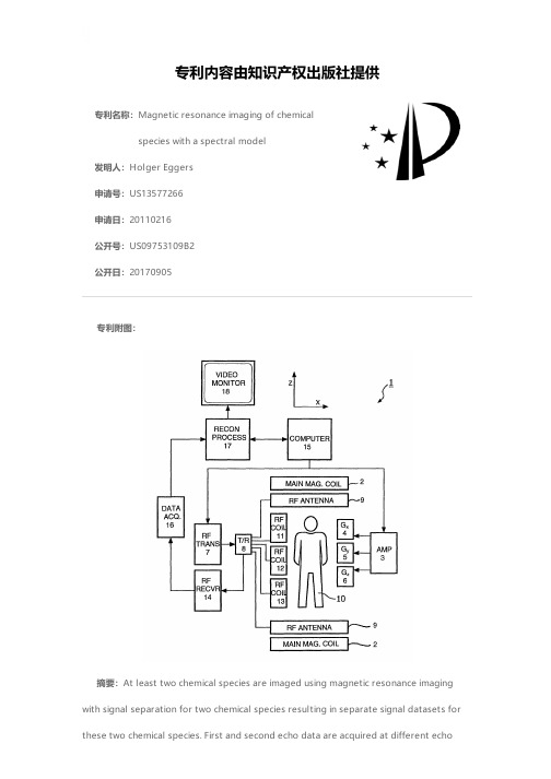
专利名称:Magnetic resonance imaging of chemicalspecies with a spectral model发明人:Holger Eggers申请号:US13577266申请日:20110216公开号:US09753109B2公开日:20170905专利内容由知识产权出版社提供专利附图:摘要:At least two chemical species are imaged using magnetic resonance imaging with signal separation for two chemical species resulting in separate signal datasets for these two chemical species. First and second echo data are acquired at different echotimes resulting in a first and second acquired complex dataset. The first and second acquired datasets are modelled by employing a spectral signal model of at least one of the chemical species. The modelling results in a first and second modelled complex dataset. The first and second modelled datasets include a first and second phase error and the separate signal datasets for the two chemical species. From the first and second acquired dataset and the first and second modelled dataset the separate signal datasets for the two chemical species are determined.申请人:Holger Eggers地址:Ellerhoop DE国籍:DE更多信息请下载全文后查看。
专利名称:改良的嵌合毒素受体蛋白和用于治疗和预防炭疽的嵌合毒素受体蛋白
专利类型:发明专利
发明人:劳埃德·M·于,基思·L·威科夫,詹姆斯·W·拉里克
申请号:CN200680035956.9
申请日:20060802
公开号:CN101277970A
公开日:
20081001
专利内容由知识产权出版社提供
摘要:描述了具有与免疫球蛋白复合物联合的毒素受体的嵌合毒素受体蛋白,所述免疫球蛋白复合物具有至少部分免疫球蛋白重链和至少部分免疫球蛋白轻链。
所述嵌合毒素受体蛋白与缺乏轻链的嵌合毒素受体蛋白相比,具有改善的稳定性。
还描述了具有增高的稳定性的炭疽和肉毒杆菌嵌合毒素受体蛋白。
申请人:行星生物技术有限公司
地址:美国加利福尼亚州
国籍:US
代理机构:中科专利商标代理有限责任公司
代理人:王旭
更多信息请下载全文后查看。
![抗微生物剂、制备抗微生物剂的方法以及包含抗微生物剂的物品[发明专利]](https://img.taocdn.com/s1/m/f54bebcfa5e9856a57126058.png)
专利名称:抗微生物剂、制备抗微生物剂的方法以及包含抗微生物剂的物品
专利类型:发明专利
发明人:罗纳德·H·巴内,塞缪尔·M·法拉,宋礼
申请号:CN200980116623.2
申请日:20090511
公开号:CN102083310A
公开日:
20110601
专利内容由知识产权出版社提供
摘要:本公开内容涉及制备抗微生物剂的方法,其包括加热二醛多糖。
该方法包括对二醛多糖(例如二醛淀粉或二醛纤维素)进行加热和/或超声处理一段时间。
本文还提供了包含所制备的二醛多糖的抗微生物组合物。
所述抗微生物组合物可在短时间内有效杀伤微生物物质(例如病毒和细菌)。
申请人:佛罗里达大学研究基金会有限公司
地址:美国佛罗里达
国籍:US
代理机构:北京集佳知识产权代理有限公司
更多信息请下载全文后查看。

两种心衰标志物荧光免疫层析联合检测技术研究①魏治静 裴晓萌 宋温婷② 侯亚璐③ 王静 柳峰松③ 赵梅红④ 吴萌(河北省科学院生物研究所,石家庄 050081)中图分类号 R446.6 文献标志码 A 文章编号 1000-484X (2023)07-1494-06[摘要] 目的:研制一种NT -proBNP 与可溶性ST2(sST2)的荧光免疫层析联合检测试剂盒。
方法:采用双抗体夹心法结合荧光免疫层析定量检测技术制备免疫层析联合检测试剂盒,并对试剂盒的空白限(LoB )、检测限(LoD )、回收率、精密度、交叉反应、稳定性及临床样本相关性进行评价。
结果:研制试剂盒NT -proBNP 的LoB 及LoD 分别为6.8 pg/ml 、19.0 pg/ml ,平均回收率为108%,批内CV<15%、批间CV<20%,200 ng/ml sST2对检测无干扰,试纸条室温或37 ℃存放14 d 稳定性良好,与罗氏NT -proBNP 电化学发光法试剂盒平行检测50例临床血清样本的相关性良好;sST2的LoB 及LoD 分别为0.47 ng/ml 、1.67 ng/ml ,平均回收率为103%,批内CV<15%、批间CV<20%,7 500 pg/ml NT -proBNP 对检测无干扰,试纸条室温或37 ℃存放14 d 稳定性良好,与Critical Diagnostics ST2 ELISA 试剂盒平行检测30例临床血清样本的相关性良好。
结论:研制了一种NT -proBNP 与sST2的荧光免疫层析联合定量检测试剂盒,为心力衰竭的临床快速检测提供了一种简便、快捷的方法。
[关键词] 心力衰竭;NT -proBNP ;可溶性ST2;荧光免疫层析;定量检测Study on fluorescence immunochromatography for combined detection of two heart failure biomarkersWEI Zhijing , PEI Xiaomeng , SONG Wenting , HOU Yalu , WANG Jing , LIU Fengsong , ZHAO Meihong , WU Meng. Biology Institute , Hebei Academy of Sciences , Shijiazhuang 050081, China[Abstract ] Objective :To develop a fluorescence immunochromatographic kit for combined detection of NT -proBNP and solu‐ble ST2 (sST2). Methods :The double antibody sandwich method combined with fluorescence immunochromatography quantitative detection technology was used to prepare the immunochromatography combined detection kit , and the limit of blank (LoB ), limit of detection (LoD ), recovery rate , precision , interference experiment , stability and coincidence rate with the comparison kit were evalu‐ated. Results :The LoB and LoD of the developed NT -proBNP kit were 6.8 pg/ml and 19.0 pg/ml. The average recovery rate was 108%, intra -batch CV was less than 15% and inter -batch CV was less than 20%. There was no cross reaction with 200 ng/ml sST2. The strip was stable when stored at room temperature or 37 ℃ for 14 days , and had a good correlation with Roche NT -proBNP electrochemilumi‐nescence kit when parallel detected 50 clinical serum samples. The LoB and LoD of sST2 were 0.47 ng/ml and 1.67 ng/ml , the average recovery rate was 103%, the intra -batch CV was less than 15%, and the inter -batch CV was less than 20%. There was no cross reac‐tion with 7 500 pg/ml NT -proBNP. The test strip had good stability when stored at room temperature or 37 ℃ for 14 days , and the coin‐cidence rate with the Critical Diagnostics ST2 ELISA kit was good , when parallel tested 30 clinical serum samples. Conclusion :A combined quantitative detection kit of NT -proBNP and sST2 by fluorescence immunochromatography was developed , which provides asimple and fast method for clinical rapid detection of heart failure.[Key words ] Heart failure ;NT -proBNP ;Soluble ST2;Fluorescence immunochromatography ;Quantitative detection心力衰竭又称心衰,是心功能受损后,各种心血管疾病发展的终末期[1]。

姜黄素联合富硒酵母对小鼠急性酒精性肝损伤的保护作用作者:梁婵华莫丽田重阳唐玉涵来源:《食品安全导刊·下》2024年第03期摘要:目的:探讨姜黄素联合富硒酵母对急性酒精性肝损伤的保护作用。
方法:将60只C57B6L/J小鼠随机分为空白组、模型组、姜黄素组(33.33 mg·kg-1 bw)、富硒酵母組(7.50 mg·kg-1 bw)和联合组(姜黄素33.33 mg·kg-1 bw+富硒酵母7.50 mg·kg-1 bw),每组12只;干预30 d后,一次性灌胃50%乙醇溶液(12 mL·kg-1 bw)建立急性酒精性肝损伤模型;观察小鼠体重、摄食、行为变化;检测小鼠肝脏指数与丙二醛(Malondialdehyde,MDA)、还原型谷胱甘肽(Glutathione,GSH)和甘油三酯(Triglyceride,TG)水平;油红染色观察小鼠肝脏组织病理学变化。
结果:与空白组比较,模型组小鼠表现出肝脏肿大、肝细胞中脂滴大量堆积病理损伤,肝脏组织MDA和TG水平显著升高(P<0.05);姜黄素联合富硒酵母能显著降低小鼠肝脏中MDA和TG表达,提高GSH水平(P<0.05),减轻小鼠肝脏脂肪沉积。
结论:姜黄素联合富硒酵母能够改善急性酒精灌胃诱导的肝脏脂质异常堆积,减轻氧化应激水平,对急性酒精性肝损伤具有良好的保护作用。
关键词:姜黄素;富硒酵母;急性酒精性肝损伤Protective Effect of Curcumin and Selenium-Rich Yeast on Acute Alcoholic Hepatic Injury in MiceAbstract: Objective: Exploring the protective effect of curcumin combined with selenium-rich yeast on acute alcoholic liver injury. Method: 60 C57B6L/J mice were randomly divided into blank group, model group, curcumin group (33.33 mg·kg-1 bw), selenium-rich yeast group (7.50 mg·kg-1 bw) and combination group (curcumin33.33 mg·kg-1 bw+selenium-rich yeast 7.50 mg·kg-1 bw), 12 mice in each group; 30 days after the intervention, a model of acute alcoholic liver injury was established by one-time gavage 50% ethanol solution (12 mL·kg-1 bw); the weight, feeding and behavior changes of mice were observed; liver index and the levels of malondialdehyde (MDA), glutathione (GSH) and triglyceride (TG) were detected, and the histopathological changes of the liver were observed by oil red staining. Result: Compared to control group, the model mice showed liver enlargement, massive accumulation of lipid droplets in liver cells, and significantly increased MDA and TG levels (P<0.05) in liver tissues, while curcumin combined with selenium-rich yeast could significantly reduce the expression of MDA and TG in the liver, increase the level of GSH (P<0.05), and reduce the fat deposition in the liver. Conclusion: Curcumin combined with selenium-rich yeast can improve abnormal accumulation of liver lipids induced by acute alcohol gavage, reduce the level of oxidative stress, and have a good protective effect against acute alcoholic liver injury.Keywords: curcumin; selenium-rich yeast; acute alcoholic liver injury酒精性肝病(Alcoholic Liver Disease,ALD)是由于长期大量摄入酒精引起的一系列肝脏疾病,包括以甘油三酯积聚为特征的酒精性脂肪肝,以肝细胞损伤、气球样变、炎症为主要病理改变的酒精性慢性肝炎,最终发展为肝硬化和肝细胞癌[1]。

秦楠,王辉敏,杨金梅,等. 复合沙棘原液对高脂血症大鼠的降脂作用[J]. 食品工业科技,2023,44(7):352−358. doi:10.13386/j.issn1002-0306.2022050091QIN Nan, WANG Huimin, YANG Jinmei, et al. Lipid-lowering Effect of Compound Seabuckthorn Concentrate on Hyperlipidemic Rats[J]. Science and Technology of Food Industry, 2023, 44(7): 352−358. (in Chinese with English abstract). doi: 10.13386/j.issn1002-0306.2022050091· 营养与保健 ·复合沙棘原液对高脂血症大鼠的降脂作用秦 楠1,王辉敏1,杨金梅1,张娜郡1,陈 超1,李冠文1,曹 满2,范光柱2(1.山西中医药大学中药与食品工程学院,山西晋中 030619;2.山西献果源生物科技有限公司,山西右玉 037200)摘 要:目的:探究复合沙棘原液对高脂血症模型大鼠的降血脂作用的影响。
方法:将60只Wistar 大鼠随机分为空白对照组、高脂模型组、阳性对照组(洛伐他汀胶囊1.80 mg/kg·bw )、复合沙棘原液低(3.57 mL/kg·bw )、中(7.14 mL/kg·bw )、高(14.28 mL/kg·bw )剂量组,并通过高脂饲料喂养法构建高脂血症大鼠模型。
空白对照组和高脂模型组灌胃等量生理盐水,实验组大鼠分别灌胃相应剂量的复合沙棘原液及洛伐他汀混悬液。
给药28 d 后,观察与分析各组大鼠的体重、Lee’s 指数、血清血脂水平、抗氧化水平、肝脏指数及肝脏病理形态。
结果:与空白对照组相比,高脂模型组大鼠体重、Lee’s 指数均极显著升高(P <0.01),表明模型构建成功;阳性对照组、复合沙棘原液高剂量组与高脂模型组相比血清中总胆固醇(total cholesterol ,TC )、甘油三酯(triglyceride ,TG )、低密度脂蛋白胆固醇(low-density lipoprotein cholesterol ,LDL-C )、丙二醛(malondialdehyde, MDA )含量均降低,高密度脂蛋白胆固醇(high-density lipoprotein cholesterol ,HDL-C )显著含量上升,超氧化物歧化酶(super-oxide dismutase, SOD )活性显著增加(P <0.01,P <0.05);各实验组的肝脏肿胀程度相对减小,胞质中脂肪空泡也明显减少。
课程名称课程英文名称发展社会学专题Development Sociology中国概况 A Brief Introduction of “The General Situation of China”英美经典短篇小说赏析 A Guide to Classic Short Stories in British and AmericanLiterature对策论 A Primer in Game Throry对策论 A Primer in Game Throry植物蛋白研究进展Aadvance of Vegetable Protein Research植物蛋白研究进展Aadvance of Vegetable Protein Research作物遗传育种专业英语Academic English作物遗传育种专业英语(必修)Academic English会计学Accounting高等农业机械化管理与模拟Adanvced Agricultural Mechanization Management and System Simulation高等农业机械化管理与系统模拟Adanvced Agricultural Mechanization Management and System Simulation调整型抽样Adjusting Sampling行政法Administrative Law高等动力学Advaced Dynamics动物传染病学专题Advance in Animal Infectious Diseases动物传染病学Advance in Animal Infectious Diseases动物传染病专题Advance in Animal Infectious Diseases动物病理学进展Advance in Animal Pathology动物病理学进展Advance in Animal Pathology植物病害生物防治进展Advance in Biological Control of Plant Diseases植物病害生物防治Advance in Biological Control of Plant Diseases植物逆境信号传递研究Advance in Plant Stress Signaling植物逆境信号传递研究Advance in Plant Stress Signaling先进制造技术Advance Manufacture Technology蛋白质互作的研究方法进展Advance of Methods for Analysis of Protein-protein Interaction 国际农药残留分析进展Advance of Pesticide Residue Analysis in Foreign Countries国际农药残留分析进展Advance of Pesticide Residue Analysis in Foreign Countries果蔬采后生理研究进展Advance of Postharvest Physiology of Fruit and Vegetable果蔬采后生理研究进展Advance of Postharvest Physiology of Fruit and Vegetable资源环境科学进展Advance of Recources and Enviromental Science高级建筑设计Advanced Garden Building Design高级园林建筑设计Advanced Garden Building Design高级生物气象学Advanced Biometeorology高级生物气象学Advanced Biometeorology高级会计理论与实务Advanced Accounting Theory and Practice高等农业机械学Advanced Agricultural Machinery高等农业机械学Advanced Agricultural Machinery高等农业机械学Advanced Agricultural Machinery高等农业机械学Advanced Agricultural Machinery高等农业机械化管理Advanced Agricultural Mechanization Management高等农业机械化管理Advanced Agricultural Mechanization Management农业机械化工程新技术讲座Advanced Agricultural Mechanization New TechnologyLectures农业机械化工程新技术讲座Advanced Agricultural Mechanization New TechnologyLectures人工智能Advanced Artificial Intelligence高级人工智能Advanced Artificial Intelligence高级审计理论与实务Advanced Auditing Theory and Practice高级生物化学Advanced Biochemistry高级生物化学Advanced Biochemistry高级生物信息SEMI.Advanced Bioinformatics Seminar高级生物信息学Seminar Advanced Bioinformatics Seminar高级害虫生物防治Advanced Biological Control of Insect Pests高级害虫生物防治Advanced Biological Control of Insect Pests高级蔬菜育种学Advanced Breeding of Vegetable Crops高级蔬菜育种学Advanced Breeding of Vegetable Crops高级财务管理Advanced Corporate Finance高级财务管理Advanced Corporate Finance高级园林植物遗传育种学Advanced Course of Ornamental Plant Breeding高级园林植物遗传育种学Advanced Course of Ornamental Plant Breeding高级作物育种学I Advanced Crop Breeding I高级作物育种学ⅠAdvanced Crop Breeding I高级作物育种学II Advanced Crop Breeding II高级作物育种学ⅡAdvanced Crop Breeding II作物生态学Advanced Crop Ecology高级作物生态学Advanced Crop Ecology高级作物生理学Advanced Crop Physiology高级细胞遗传学Advanced Cytogenetics高级发展学Advanced Development Studies高级发展学Advanced Development Studies高等结构动力学Advanced Dynamics of Structures高级计量经济学Advanced Econometrics高级计量经济学Advanced Econometrics高级园林植物生理生态学Advanced Eco-physiology of Ornamental Plants高级园林植物生理生态Advanced Eco-physiology of Ornamental Plants高等工程热力学Advanced Engineering Thermodynamics高等工程热力学Advanced Engineering Thermodynamics高级试验设计与数据分析Advanced Experimental Design and Data Analysis 高级试验设计与数据分析Advanced Experimental Design and Data Analysis 兽医免疫高级实验Advanced Experiments of Veterinary Immunology 兽医免疫高级实验Advanced Experiments of Veterinary Immunology 高级饲料分析技术Advanced Feed Analysis Technology高级饲料分析技术Advanced Feed Analysis Technology高级财务管理理论与实务Advanced Financial Management食品微生物学专题Advanced Food Microbiology动物遗传工程Advanced Gene Engineering高级基因工程Advanced Gene Engineering高级葡萄生理与分子生物专题Advanced Grape Physiology and Molecular Biology 高级葡萄生理与分子生物学专题Advanced Grape Physiology and Molecular Biology 高级昆虫生理生化Advanced Insect Physiology and Biochemistry高级昆虫生理生化Advanced Insect Physiology and Biochemistry高级昆虫毒理学Advanced Insect Toxicology高级昆虫毒理学Advanced Insect Toxicology高等内燃机学Advanced Internal-combustion Engine高等内燃机学Advanced Internal-combustion Engine高级实验动物学Advanced Laboratory Animal Science高级实验动物学Advanced Laboratory Animal Science高级园林设计Advanced Landscape Design高级园林设计Advanced Landscape Design高级园林工程Advanced Landscape Engineering高级环境绿地规划Advanced Landscape Planning高级宏观经济学Advanced Macroeconomics高级宏观经济学Advanced Macroeconomics高级管理会计理论与实务Advanced Management Accounting管理科学与工程专业Seminar Advanced Management Science and Engineering (Ph.D)管理科学与工程专业Seminar Advanced Management Science and Engineering (Ph.D)高级市场营销学Advanced Marketing高级市场营销Advanced Marketing高等金属学Advanced Metal高等金属学Advanced Metal高级微生物遗传学Advanced Microbial Genetics高级微生物遗传学Advanced Microbial Genetics高级微生物学进展Advanced Microbiological Seminar高级微生物学进展(全年)Advanced Microbiological Seminar高级微观经济学Advanced Microeconomics高级微观经济学Advanced Microeconomics高级运筹学Advanced Operations Research高级运筹学Advanced Operations Research高级果树生理学Advanced Physiology of Fruit Trees高级果树生理学Advanced Physiology of Fruit Trees高级植物与细胞生物学Seminar Advanced Plant and Cell Biology Seminars高级植物与细胞生物学Seminar Advanced Plant and Cell Biology Seminars高级植物营养学Advanced Plant Nutrition高级植物营养学Advanced Plant Nutrition高级植物生理生态Advanced Plant Physiological Ecology高级植物生理生态Advanced Plant Physiological Ecology高级植物生理学专题Advanced Plant Physiology高级植物生理学Advanced Plant Physiology高级植物生理学Advanced Plant Physiology高级植物生理学专题Advanced Plant Physiology高级观赏植物采后生理Advanced Postharvest Physiology of Ornamental Plants 高级观赏植物采后生理Advanced Postharvest Physiology of Ornamental Plants 高级植物营养进展Advanced Progress in Plant Nutrition高级设施园艺学Advanced Protected Horticulture高级设施园艺学Advanced Protected Horticulture高级可再生资源工程专题Advanced Renewable Resource Engineering现代可再生资源工程学Advanced Renewable Resource Engineering国际食品研究进展Advanced Research of Food Science植物细胞信号转导研究中的反向遗传学与细胞生物学研究技术与方法Advanced Reverse Genetic and Cell Biological Approaches to Study Signal Transduction in Plant高级生物化学与分子生物学Seminar Advanced Seminar for Biochemistry and Molecular Biology高级生物化学与分子生物学SeminarAdvanced Seminar for Biochemistry and Molecular Biology高级遗传学Seminar Advanced Seminar for Genetics高级遗传学Seminar Advanced Seminar for Genetics高级生物质工程Seminar Advanced Seminar on Biomass Engineering高级社会统计Advanced Social Statistics高级社会统计Advanced Social Statistics高级生化专题Ⅲ(生物膜)Advanced Topics in Biochemistry:Biomembrane 高级生化专题Ⅲ(生物膜)Advanced Topics in Biochemistry:Biomembrane高级生化专题IV(酶学及代谢调控)Advanced Topics in Biochemistry:Enzymology and Metabolism Control高级生化专题Ⅳ(酶学与代谢调控)Advanced Topics in Biochemistry:Enzymology and Metabolism Control高级生化专题II(核酸化学)Advanced Topics in Biochemistry:Nucleic Acid高级生化专题Ⅱ(核酸化学)Advanced Topics in Biochemistry:Nucleic Acid高级生化专题Ⅰ(蛋白质化学)Advanced Topics in Biochemistry:Protein高级生化专题Ⅰ(蛋白质化学)Advanced Topics in Biochemistry:Protein农产品物料干燥技术特论Advanced Topics in Drying Technology:Drying of PorousMedia高级分子生物学专题Advanced Topics in Molecular Biology高级分子生物学专题Advanced Topics in Molecular Biology高级城市规划Advanced Urban Planning高级蔬菜生理学Advanced Vegetable Physiology高级蔬菜生理学Advanced Vegetable Physiology高级杂草学Advanced Weeds高级杂草学Advanced Weeds高级兽医寄生虫学Advanceds Veterinary Parasitology高级兽医寄生虫学Advanceds Veterinary Parasitology高级兽医微生物学Advances in Veterinary Microbiology高级兽医微生物学Advances in Veterinary Microbiology作物栽培新技术专题Advances in 4H Crop Cultivation作物分子生理与生物技术Advances in Agricultural Biotechnology农业水土工程研究进展Advances in Agricultural Water-soil Research农业水土工程研究进展Advances in Agricultural Water-soil Research动物育种专题Advances in Animal Breeding动物育种专题Advances in Animal Breeding动物病理生理学专题Advances in Animal Pathophysiology动物病理生理学专题Advances in Animal Pathophysiology动物科学研究进展Advances in Animal Science动物科学研究进展Advances in Animal Science害虫生物防治理论与实践新进展Advances in Biological Control of Insect Pests害虫生物防治理论与实践新进展Advances in Biological Control of Insect Pests细胞生物学进展Advances in Cell Biology细胞生物学进展Advances in Cell Biology农副产品化学进展Advances in Chemistry of Agricultural Byproducts农副产品化学进展Advances in Chemistry of Agricultural Byproducts作物营养与水分生理专题Advances in Crop Nutrition and Water Physiology作物光合、产量与品质生理专题Advances in Crop Photosynthesis,Yield and Quality能源作物与生物质工程专题Advances in Crop Physiology and Ecology作物科学研究进展Advances in Crop Science作物科学研究进展Advances in Crop Science作物逆境生理专题Advances in Crop Stress Physiology发育生物学进展Advances in Developmental Biology数字农业研究进展Advances in Digital Agriculture Research 农作制度理论与技术专题Advances in Farming System Science果树学进展讨论Advances in Fruit Sciences果树学进展讨论Advances in Fruit Sciences现代果树遗传学研究进展Advances in Genetics of Fruit Crops分子遗传学进展Advances in Molecular Genetics病毒学进展Advances in Molecular Virology营养科学研究进展Advances in Nutritional Sciences营养科学技术研究进展Advances in Nutritional Sciences杀菌剂药理学及抗药性研究进展Advances in Pharmacology and Fungicide Resistance in Phytopathogen药理学与毒理学专题Advances in Pharmacology and Toxicology药理学与毒理学专题Advances in Pharmacology and Toxicology植物同化物运输高级讲座Advances in Photoassimilate Transport Mechanisms 植物同化物运输高级讲座Advances in Photoassimilate Transport Mechanisms 植物生物学进展Advances in Plant Biology植物激素与化学控制专题Advances in Plant Hormones and Chemical Regulation 植物病毒学进展Advances in Plant Virus Research植物病毒学进展Advances in Plant Virus Research家禽营养与饲养技术(案例)Advances in Poultry Nutrition and feeding Technology 种子病理学进展Advances in Seed Pathology种子病理学进展Advances in Seed Pathology兽医免疫学进展Advances in Veterinary Immunology兽医免疫学进展Advances in Veterinary Immunology兽医科学进展Advances in Veterinary Medicine兽医科学进展Advances in Veterinary Medicine水资源研究进展专题Advances in Water Resource Science水资源研究进展专题Advances in Water Resource Science分子植物病理学研究进展Advances of Molecular Plant Pathology分子植物病理学研究进展Advances of Molecular Plant Pathology生物环境与能源工程综合专题Seminar Advances on Agricultural and Bioenvironmental Engineering农业生物环境与能源工程研究进展Advances on Agricultural and Bioenvironmental Engineering 食品保藏技术研究进展Advances on Food Preservation Technology食品保藏技术研究进展Advances on Food Preservation Technology水土保持与荒漠化防治新技术研究进展Advances on Soil and Water Conservation and Deforestation Control水土保持与荒漠化防治研究进展Advances on Soil and Water Conservation and DeforestationControl结构工程研究新进展Advances on Structure Engineering城镇与区域规划Advances on Urban and Regional Planning城镇与区域规划研究进展Advances on Urban and Regional Planning近代水文学及水资源研究进展Advances on Water Concervancy Project水利工程研究进展Advances on Water Concervancy Project农业商务管理Agri-business Management农业产业组织Agribusiness Organization核技术农业应用基础Agricultural Application Foundation of Nuclear Technology 核技术农业应用基础Agricultural Application Foundation of Nuclear Technology 核技术农业应用基础Agricultural Application Foundation of Nuclear Technology农业可控管理技术Agricultural Controllable Management Technology农业可控管理技术Agricultural Controllable Management Technology农业发展经济学Agricultural Development Economics农业经济理论与政策Agricultural Economics: Theory and Policy农业经济理论与政策Agricultural Economics: Theory and Policy农业装备开发与设计Agricultural Equipment Development and Design农产品期货市场Agricultural Futures Markets农产品期货市场Agricultural Futures Markets农业历史文献选读Agricultural History Literature农业历史文献选读(必修)Agricultural History Literature农业信息系统工程Agricultural Information and System Engineering农业信息系统工程Agricultural Information and System Engineering农业保险Agricultural Insurance农产品市场分析Agricultural Market Analysis农产品市场分析Agricultural Market and Analysis农产品市场分析Agricultural Market and Analysis有害生物治理的原理与方法Agricultural Pests Prevention and Control农业有害生物的预防与控制Agricultural Pests Prevention and Control农业资源与利用Agricultural Resources and Utilization核技术农业应用专论Agricultural Specialized Application of Nuclear Technology 核技术农业应用专论Agricultural Specialized Application of Nuclear Technology 农业系统工程Agricultural Systems Engineering农村技术创新与知识系统Agricultural Technology Innovation and Knowledge System 农村技术创新与知识系统Agricultural Technology Innovation and Knowledge System 农业与食品企业管理Agriculture and Food Corporate Managemnt农业信息学Agriculture Informatics农业科技与“三农政策”Agriculture Technology and Rural Development农业装备机电一体化技术Agricutural Equipment Mechantronics农业项目的计划与管理Agricutural Project Plan and Management农业工程项目规划Agricutural Project Plan and Management农产品国际贸易实务Agri-goods International Trade Practice农业生态系统分析Analysis and Simulation of Ecosystem生态系统分析与模拟Analysis and Simulation of Ecosystem农业关联产业分析Analysis of Agribusiness国情分析和发展战略Analysis of Country Situation and Development Stratagem 兽医临床病例分析Analysis of Veterinary Clinical Cases兽医临床病例分析Analysis of Veterinary Clinical Cases现代食品分析技术Analytic Technology of Modern Food Science现代食品分析技术Analytic Technology of Modern Food Science古汉语Ancient Chinese古汉语Ancient Chinese动物病理剖检诊断技术Animal Autopsy Technique for Pathological Diagnosis克隆动物与转基因动物Animal Cloning and Transgensis克隆动物与转基因动物Animal Cloning and Transgensis动物源食品卫生检验技术Animal Derived Food Inspection Technique动物实验方法Animal Experiment Technology动物消化道微生物Animal Gastrointestinal Tract Microbiology动物消化道微生物Animal Gastrointestinal Tract Microbiology动物遗传资源Animal Genetic Resource 动物卫生行政法学Animal Health Management 动物卫生行政法学Animal Health Management 畜牧工程Animal Husbandry Engineering 动物营养代谢病Animal Metabolic Diseases 动物营养代谢病专题Animal Metabolic Diseases 人类疾病模型的构建与应用Animal Models for Human Diseases 动物分子病毒学Animal Molecular Virology 动物分子病毒学Animal Molecular Virology 动物神经生物学Animal Neurobiology 动物神经生物学Animal Neurobiology 动物营养与免疫专题Animal Nutrion and Immunology 动物营养与免疫专题Animal Nutrion and Immunology 动物保护与福利Animal Protection and Welfare 动物生殖内分泌学Animal Reproduction and Endocrinology 动物生殖内分泌学Animal Reproduction and Endocrinology 动物繁殖学SeminarAnimal Reproduction Seminar 动物繁殖学SeminarAnimal Reproduction Seminar动物繁殖理论与现代生物技术(案例)Animal Reproduction Theory and Modern Biotechnology 动物生殖生理学实验Animal Reproductive Physiology 动物生殖生理学Animal Reproductive Physiology动物生殖生理学实验Animal Reproductive Physiology Experiment 动物生殖生理学实验Animal Reproductive Physiology Experiment 动物功能基因组学Animl Functional Genomics 动物功能基因组学Animl Functional Genomics 人类学与中国社会研究Anthropology and Chinese Society 人类学与中国社会研究Anthropology and Chinese Society 植物抗菌化合物专题Antimicrobial Compounds from Plants 植物抗菌化合物专题Antimicrobial Compounds from Plants 3S 技术农业应用Application of 3S in Agriculture 3S 技术在水利工程中的应用Application of 3S Techniques on Soil and Water Conservation生物多样性与应用Application of Biodiversity 生物多样性与利用Application of Biodiversity 生物多样性与利用Application of Biodiversity 3S 技术应用Application of GIS, GPS and RS 3S 技术应用Application of GIS, GPS and RS应用数理统计Application of Mathematical Statistics 应用数理统计Application of Mathematical Statistics 分子生物学在昆虫学中的应用Application of Molecular Biology to Entomology 分子生物学在昆虫学中的应用Application of Molecular Biology to Entomology 植物生理生态仪器Application of Plant Physiology and Ecology 植物生理生态仪器Application of Plant Physiology and Ecology 3S 在水文模拟中的应用Applications of "3S" to Hydrology Simulation电力系统最优化技术Applications of Optimization Method in Electrical Power System 电力系统最优化技术Applications of Optimization Method in Electrical Power System 稳定同位素在生态环境研究中的应用Applications of Stable Isotopes in Studies of Environment and Ecology 稳定同位素在生态环境研究中的应用Applications of Stable Isotopes in Studies of Environment and Ecology 应用数理统计Applied Mathematical Statistics应用经济学Seminar Applied Economics Seminar应用地质地貌与土地资源Applied Geology Geomorphology and Land Resource应用地质地貌与土地资源Applied Geology Geomorphology and Land Resource应用植物生物技术Applied Plant Biotechnology农业应用随机过程Applied Stochastic in Agriculture应用随机过程Apply Stochastic Processes园林设计研究进展Approach of Landscape Architecture风景园林研究进展Approach of Landscape Architecture观赏植物生理生态研究进展Approach of Ornamental Horticulture观赏园艺研究进展Approach of Ornamental Horticulture英语教学法Approaches and Methods in Language Teaching英语教学法Approaches and Methods in Language Teaching水生昆虫学Aquatic Entomology水生昆虫学Aquatic Entomology艺术设计Art Design人工智能原理Artificial Intelligence人工智能技术Artificial Intelligence结构抗震减震分析原理Aseismic Analysis Principle of Structure兽药安全评价技术Assessment Technique of Veterinary Drug Safety不对称合成Asymmetric Synthesis不对称合成Asymmetric SynthesisAutoCAD 二次开发技术Auto CAD Customization自动控制技术Automatic Control Technology自动控制理论Automatical Control Theory自动控制理论Automatical Control Theory自动机械设计Automatical Machine Design禽类生理学Avian Physiology禽类生理学Avian Physiology遗传分析原理Basic Concepts of Genetic Analysisi基础分子生物学实验Basic Experiment of Molecar Biology基础分子生物学实验Basic Experiment of Molecar Biology新能源发电技术基础Basic of Renewable Energy Generation Technology交通规划理论与方法Basic Theory and Method of Traffic-layou放射卫生防护知识Basical Knowledge of Radiation Protection放射卫生防护知识Basical Knowledge of Radiation Protection放射卫生防护知识Basical Knowledge of Radiation Protection土壤物理与作物学基础Basics of Soil Physics and Crop土壤物理与作物学基础Basics of Soil Physics and Crop篮球Basketball生化分析Biochemical Analysis生化分析Biochemical Analysis生物气候模型与信息系统Bioclimatological Model and Information System生物气候模型与信息系统Bioclimatological Model and Information System畜牧生物工程专业硕士生Seminar Bio-engineering Seminar in Animal配子与胚胎生物工程Bio-engineering Technology in Animal Gamete and Embryo 配子与胚胎生物工程Bio-engineering Technology in Animal Gamete and Embryo 植物小RNA的生物合成和功能Biogenesis and Function of Small RNAs in Plant生物地球化学Biogeochemistry生物地球化学Bio-geochemistry生物信息学Bioinformatics生物信息学Bioinformatics生物信息学Bioinformatics生物信息学算法Bioinformatics Algorithm生物信息学算法Bioinformatics Algorithm生物信息检测与处理专题Bioinformatics Detection and Processing Topic生物信息学SEMI.Bioinformatics Seminar生物信息学Seminar Bioinformatics Seminar植物病害生物防治Biological Control of Plant Diseases植物病害生物防治Biological Control of Plant Diseases生物饲料加工与利用Biological Feed Processing and Application植物病原细菌生物学Biology of Plant Pathogenic Bacteria植物病原细菌生物学Biology of Plant Pathogenic Bacteria生物质工程Bio-mass Engineering Theory生物质工程原理Bio-mass Engineering Theory生物膜与信号转导Biomembrane and Signal Transduction生物膜与信号传导Biomembrane and Signal Transduction生物物理学BiophysicsBioprocessing and Food Quality Bioprocessing and Food QualityBioprocessing Engineering Bioprocessing Engineering生物生产自动化与机器人Bio-production and Robot生物生产自动化与机器人Bio-production and Robot生化反应动力学与反应器Bioreaction Engineering生化反应动力学与反应器Bioreaction Engineering生物修复Bioremediation生物修复Bioremediation农业生物安全Biosafty for Agriculture生物系统动力学(Biosystem)Biosystem Dynamics生物系统动力学Biosystem Dynamics观赏植物生物技术Biotechnology of Ornamental Plants蔬菜生物技术Biotechnology to Vegetable Science植物源杀虫剂及作用机理Botanic Pesticide植物源杀虫剂及其作用机理Botanic Pesticide边界元法Boundary Element Method材料成型中的计算机应用Brief Introduction of Computer Application in Plastic Working 经济学流派Brief Introduction of Economics Schools经济学流派Brief Introduction of Economics Schools商务英语Business EnglishC语言程序设计 C LanguageC语言程序设计 C Language基于PRO-E的CAD/CAM集成制造技术CAD/CAM Based on Creo资本运营Capital Operation汽车网络通讯技术Car Network Communication Technology汽车网络通讯技术Car Network Communication Technology碳水化合物化学Carbohydrate Chemistry碳水化合物化学Carbohydrate Chemistry农业水土工程设计案例Case Analysis for Agricultural Soil and Water Engineering国内外食品安全案例辨析Case Studies on Food Safety Incidents企业管理案例分析Case Study for Corporation Management企业管理案例分析Case Study for Corporation Management电子商务案例Case Study for E-Commerce细胞生物学Cell Biology细胞生物学Cell Biology细胞生物学Cell Biology细胞生物学Cell Biology细胞培养基础和实验Cell Culture Basics and Experiments细胞培养基础和实验Cell Culture Basics and Experiments动物细胞工程Cellular Engineering Technology in Animal动物细胞工程原理与方法(原细胞Cellular Engineering Technology in Animal工程 )英语六级CET-6化学生态学Chemical Ecology临床化验及病理诊断技术Chemical Examination and Pathological Diagnostic Technique 化学计量学Chemistry Metrology化学计量学Chemistry Metrology中国概况China Panorama粮食经济Grain Economy粮食经济Grain Economy中国化的马克思主义研究Chinalized Marxism Studies中国化的马克思主义研究Chinalized Marxism Studies土木工程抗灾原理Civil Engineering Disaster Prevention Theory土木工程材料专论Civil Engineering Material Monogragh土木工程材料专论Civil Engineering Material Monogragh传统文化与现代企业管理系列专题Classical Cutlure and Modern Enterprise Management期货品种专题Classification of Futures Commodities气候与农业减灾研究专题Climate Resource and Agriculture Disaster Reduction Seminar 气候与农业减灾研究专题Climate Resource and Agriculture Disaster Reduction Seminar 临床病理诊断技术Clinical Pathological Diagnostic Technique临床兽医学专业Seminar Clinical Veterinary Medicine Seminar公共经济学理论专题Colloquium of Public Economics公共经济学理论专题Colloquium of Public Economics组合优化Combination Optimization组合优化Combination Optimization组合数学Combinatorial Mathematics组合数学Combinatorial Mathematics农业传播技术与应用Communication for Rural Development社区基础的自然资源管理Community-based Natural Resource Management社区基础的自然资源管理Community-based Natural Resource Management社区基础的自然资源管理Community-based Natural Resource Management社区基础的自然资源管理Community-based Natural Resource Management动物生殖生理学Comparative Animal Breeding动物比较育种学Comparative Animal Breeding比较基因组学与分子进化Comparative Genomics and Molecular Evolution比较政府与政治Comparative Government and Politics比较病理学Comparative Pathology比较病理学Comparative Pathology中外教育比较评价Comparative Studies of Sino-foreign Eduction 中外教育比较研究Comparative Studies of Sino-foreign Eduction 竞争情报研究Competitive Intelligence Research 竟争情报研究Competitive Intelligence Research 竞争情报研究Competitive Intelligence Research 综合测评Comprehensive Assessment 农产品加工与贮藏综合实验Comprehensive Experiments of Agricultural Products Processing and Storage 营养与食品卫生综合大实验Comprehensive Experiments of Nutrition and Food Hygiene 营养与食品安全综合大实验Comprehensive Experiments of Nutrition and Food Hygiene 营养与食品安全综合实验Comprehensive Experiments of Nutrition and Food Hygiene 生物质综合利用与转化Comprehensive Utilization and Conversion of Biomass Resources 计算流体力学Computation Fluid Dynamics 计算流体力学Computation Fluid Dynamics 两相流与多相流计算Computation of Two-phase and Multi-phase Flow 统计计算方法Computational Method for Statistic 统计计算方法Computational Method for Statistic 农业生物信息计算机采集与处理Computer Acquisition Agricultural Biological Information and Processing 计算机算法设计Computer Algorithm Design 英语多媒体网络教育Computer Assisted Language Learning 英语多媒体网络教学Computer Assisted Language Learning 计算机图形学Computer Graphics 计算机图形学Computer Graphics 计算机图形学Computer Graphics 计算机网络体系结构Computer Network Architecture 计算机网络体系结构Computer Network Architecture 现代干燥技术Computer Simulation for Farm Product Drying 现代干燥技术Computer Simulation for Farm Product Drying 土壤水土过程模拟Computer Simulation of Soil Physical ProcessesComputer simulation of soil-water processesComputer Simulation of Soil Physical Processes 计算机支持的协同工作与网络计算Computer Supported Cooperative Work and Grid Computing 计算机支持的协同工作与网格计算Computer Supported Cooperative Work and Grid Computing 机械系统计算机虚拟样机技术Computer Virtual Prototyping Technology for Mechanical System 基于PRO_E 的CAD/CAM 集成制造技术Computer Virtual Prototyping Technology for MechanicalSystem 机械系统计算机虚拟样机技术Computer Virtual Prototyping Technology for Mechanical System 计算机视觉Computer Vision 计算机视觉技术Computer Vision 家畜育种中的计算方法Computing Algorithms in Animal Breeding 家畜育种中的计算方法Computing Algorithms in Animal Breeding 混凝土结构原理Concrete Structure Theory 混凝土结构理论Concrete Structure Theory 共轭曲面理论Conjugate Surface Theory 生物质工程导论Conspectus of Biomass Engineering 工程材料本构关系Constitutive Relation of Engineering Materials 工程材料本构关系Constitutive Relation of Engineering Materials 信息资源建设与评价Construction and Evaluation of Information Resources 土木工程质量事故处理与分析Construction Quality Accident Analysis and Treatment中外城市建设史Constructual History on China and Foreign cities农产品需求分析Consumer Demand Analysis for Food and AgriculturalProducts现代区域发展规划与管理Contemporary Regional Development Planning andManagement现代区域发展规划与管理Contemporary Regional Development Planning andManagement可持续机械化生产系统Continuable Mechanization Production System可持续机械化生产系统Continuable Mechanization Production System连续介质力学Continuity Mechanics连续介质力学Continuity Mechanics连续型抽样Continuous Sampling畜禽寄生虫病防治Control of Parasite infection in Poultry and Animals 公司金融理论Corporate Finance Theory公司法与现代企业制度Corporation Law and Modern Enterprise System公司法与现代企业制度Corporation Law and Modern Enterprise System机电产品创新设计Creative Design of Mechantronic Product机电产品创新设计Creative Design of Mechantronic Product英美文学专题Critical Approaches to Literature发展研究前沿Critical Development Studies区域经济学前沿Critical Regional Economics作物生理与分析研究技术Crop Analysis Technology作物育种理论与案例分析Crop Breeding and Cases Analysis作物抗逆机理与育种Crop Breeding for Tolerance作物抗逆机理与育种Crop Breeding for Tolerance作物生理学SEMI.Crop Physiology Seminar作物生理学Seminar Crop Physiology Seminar作物生产系统分析与模拟Crop System Analysis and Simulation作物生产系统分析与模拟Crop System Analysis and Simulation当代国际贸易体制Current International Trading System当代国际贸易体制Current International Trading System现代生物学技术Current Techniques in Biological Sciences数据拟合Data Fitting数据拟合Data Fitting数据挖掘Data Mining数据挖掘Data Mining数据挖掘Data Mining数据挖掘Data Mining数据结构Data Structure数据结构Data Structure数据结构Data Structure数据、模型与决策Data, Model and Decision-making数据库与空间数据管理Database and Spatial Data Management数据库与空间数据管理Database and Spatial Data Management数据库原理与技术Database Principle and Technology数据库原理与技术Database Principle and Technology决策分析Decision Making and Analysis决策分析Decision Making and Analysis生态工程设计与应用Design and Application of Ecological Engineering绿地工程设计与建设Design and Construction of Landscape Project绿地工程设计与建设Design and Construction of Landscape Project试验设计与多元分析Design of Experiment and Mult Variate Statistical Anlylisis 试验设计与多元分析Design of Experiment and Mult Variate Statistical Anlylisis 有机合成设计Design of Organic Synthesis设计模式Design Patterns农业建筑结构设计原理Design Principle on Agricultural Structure车辆工程专业进展Development of Vehicle Engineering车辆工程专业进展Development of Vehicle Engineering信息资源开发利用技术Development and Utilization of Information ResourcesTechnology信息资源开发利用技术Development and Utilization of Information ResourcesTechnology发展经济学专题Development Economics发展经济学Development Economics发展研究方法Development Methods发展研究方法Development Methods发展研究方法Development Methods发展研究方法Development Methods中国发展模式Development Models of China食品科学技术研究进展Development of Food Science Technology Research食品科学技术研究进展Development of Food Science Technology Research葡萄酒化学进展Development of Wine Chemistry葡萄酒化学进展Development of Wine Chemistry农业工程专业Seminar Development of Agricultural Engineering农业工程专业Seminar Development of Agricultural Engineering农业装备工程专业进展Development of Agricultural Equipment Engineering农业装备工程专业进展Development of Agricultural Equipment Engineering农业机械化工程专业进展Development of Agricultural Mechanization Engineering农业机械化工程专业进展Development of Agricultural Mechanization Engineering农产品加工工程进展Development of Farm Product Processing Engineering农产品加工工程专业进展Development of Farm Product Processing Engineering食品质量管理进展Development of Food Quality Management食品质量管理进展Development of Food Quality Management食品安全研究进展Development of Food Security Research食品安全研究进展Development of Food Security Research物流工程技术进展Development of Logistics Engineering机械设计及理论专业进展Development of Mechanical Design and Theory机械设计及理论专业进展Development of Mechanical Design and Theory机械工程技术进展Development of Mechanical Engineering机械制造及其自动化专业进展Development of Mechanical Manufacturing and Automation 现代制造前沿技术与进展Development of Modern Manufacturing Technology现代制造前沿技术与进展Development of Modern Manufacturing Technology发展规划Development Planning发展项目管理Development Project Management发展社会学Development Sociology发展社会学Development Sociology发展社会学Development Sociology发展理论与实践Development Theories and Practices自然辩证法Dialectics of Nature微分几何Differential Coefficient Geometry微分几何Differential Coefficient Geometry。
Environmental Toxicology and Pharmacology22(2006)97–103Cultivated microalgae and the carotenoid fucoxanthin fromOdontella aurita as potent anti-proliferative agentsin bronchopulmonary and epithelial cell linesDimitri Moreau a,∗,Christophe Tomasoni a,Catherine Jacquot a,Raymond Kaas b,Roland Le Guedes b,Jean-Paul Cadoret b,Arnaud Muller-Feuga b,Ioanna Kontiza c,Constantinos Vagias c,Vassilios Roussis c,Christos Roussakis aa Laboratoire de Pharmacologie Marine,ISOMer,Facult´e de Pharmacie de Nantes,1rue Gaston Veil,BP92208,44322Nantes Cedex03,Franceb Laboratoire de Physiologie et Biotechnologie des Algues,IFREMER,rue de l’ˆıle d’Yeu,BP1105,44311Nantes Cedex03,Francec University of Athens,Department of Pharmacy,Division of Pharmacognosy and Chemistry of Natural Products,Panepistimiopolis Zografou,Athens15771,GreeceReceived10November2005;accepted9January2006Available online6March2006AbstractThe antiproliferative activities of several extracts from cultivated microalgae in France have been studied against bronchopulmonary and epithelial cell lines,respectively(A549,NSCLC-N6and SRA01/04).The algal extracts,of Diatomae(Odontella aurita,Chaetoseros sp.),as well as of Haptophyceae:Isochrisys aff.galbana,appeared as the most active among all the assayed species,expressing a broad spectrum of in vitro antiproliferative activity of well-differentiated pathologic cells such as NSCLC-N6by terminal differentiation.Bio-guided fractionation of the above referred extracts,led us to the isolation,of the carotenoid fucoxanthin.Fucoxanthin has been structurally determined,through modern spectral means and has been studied separately for its activities.©2006Elsevier B.V.All rights reserved.Keywords:Odontella aurita;Antiproliferative activities;Bronchopulmonary carcinoma;Secondary cataract;Apoptosis;Fucoxanthin1.IntroductionMarine microalgae comprise the largest group of living organisms in the oceans,constituting an estimated10,000 species.Algae are at the base of entire aquatic food chain.There-fore,it is not surprising that the microalgae,which compose the phytoplankton,play a vital role in the rearing of aquatic animals like molluscs;shrimps andfish.Moreover,there are numerous applications for molecules from these phototropic microorganisms in human and animal food,health and cosme-tology(Muller-Feuga,2000).In recent years,there has been a growing interest in functional foods,that is,foods able to provide additional physiological benefits for human health,other than the basic nutritional and∗Corresponding author.Tel.:+33251125672;fax:+33251125690.E-mail address:dimitri.moreau@univ-nantes.fr(D.Moreau).energetic requirements(Bidlack,1994).Often,functional foods are traditional foods enriched with an ingredient able to pro-vide or promote a specific beneficial action for human health. These are called functional ingredients.These ingredients are preferred to have a natural origin,such as plants or perhaps algae and/or microalgae.These types of marine sources are receiving increasing attention mainly for their content in,for example,polyunsaturated fatty acids and,-carotene and other pigments(antioxidants),sulphated polysaccharides and sterols (antimicrobials).One of the main interests in our laboratories is to assess the suitability obtained from extracts and pure compounds from cul-tivated microalgae,like the ones which they have been studied, as food antioxidants and preventative agents against secondary cataracte and cancer.In this work,a preliminary screening of ten marine and fresh water species from different orders(Diatomophyceae, Rhodophyceae,Haptophyceae,Cryptophyceae,Prasinophyceae1382-6689/$–see front matter©2006Elsevier B.V.All rights reserved. doi:10.1016/j.etap.2006.01.00498 D.Moreau et al./Environmental Toxicology and Pharmacology22(2006)97–103Table1Detail of the different strains studied,with the source and the optimal condition of growingOrder Species Source Medium pH T(◦C) Diatomophyceae Odontella aurita IFREMER Conway7.520 Chaetoceros ap1010/11Conway7.520Porphyridium purpureum SAG111/79Hemerick720 Rhodophyceae Rhodella violacea SAG115/79Conway724 Galdieria sulphuraria a074W Galdi245 Chlorophyceae Chlamydomonas reinhardtii a PG27MMG/TAP724 Haptophyceae Isochrysis affinis galbana IFREMER Conway720 Cryptophyceae Rhodomonas salina ccap978/24Conway722 Prasinophyceae Tetraselmis suecica ccmp904Conway720 Dinophyceae Heterocapsa triquetra IFREMER ESP7.822 G.sulphuraria source:Institut f¨u r biologie,Freie Universit¨a t,Berlin.a Fresh water microalgae.and Dinophyceae)were investigated as natural source of antipro-liferative agents in vitro against asynchronous cells of human non-small-cell bronchopulmonary carcinoma line(NSCLC-N6) (Roussakis et al.,1991),human lung epithelial cell line(A549)and against a proliferative human lens epithelial cell line(SRA 01/04).Bio-guided fractionation of the extracts,which appeared as the most active,led us to the isolation of the carotenoid fucox-anthin,which has been also thoroughly assayed.In all cases,theTable2Composition of the different media used for algal cultureProducts MediumHemerick Conway Galdi MMG/TAP ESP NaNO3(g L−1) 1.70.10.07 (NH4)2SO4(g L−1) 1.5NH4Cl(g L−1)0.4K2HPO4,3H2O(g L−1)0.1750.106KH2PO4(g L−1)0.1750.30.053NaH2PO4,H2O(g L−1)0.02Na2C3H7O6P,5H2O(g L−1)0.01 Na2SiO3,5H2O(g L−1)0.1FeEDTA0.050.014Na2EDTA,2H2O(g L−1)0.0490.0450.008 CaCl2,2H2O(g L−1) 1.470.020.05KCl(g L−1)0.75MgSO4,7H2O(g L−1)12.30.30.1MgSO4,H2O(g L−1)0.00082 NaCl(g L−1)29.0Tris(g L−1)0.1Co(NO3)2,6H2O(g L−1)0.08CoCl2,6H2O(g L−1)20.080 2.927CoSO4,7H2O(g L−1)0.09124 CuSO4,5H2O(g L−1)0.0820.0160 2.855Fe(NH4)2(SO4)2,6H2O(g L−1)3510 FeCl3,6H2O(g L−1) 1.3245 FeSO4,7H2O(g L−1)9.073H3BO3 2.033.6572020.735700 MnCL2,4H2O(g L−1) 1.80.3636409.2Mo7O24(NH4)6,4H2O(g L−1)209.0260 2.0NaVO3,4H2O(g L−1)80O5SV,5H2O(g L−1)0.043ZnCl2(g L−1)21ZnSO4,7H2O(g L−1)0.2130.444011000 Vitamin B12(g L−1)102 Thiamin(g L−1)200100 Biotin(g L−1)1QSP L(g L−1)FW MW FW FW MW Mineral nutrient and their concentration(1×).D.Moreau et al./Environmental Toxicology and Pharmacology22(2006)97–10399 Table3IC50for all assayed microalgal extracts,with different solvents,against three cell linesSpecies Cell linesSRA A549NSCLC-N6EtOH CH2Cl2H2O EtOH CH2Cl2H2O EtOH CH2Cl2H2OO.aurita18.4±1.418.2±0.49.1±1.3––20.6±0.555.8±3.736.9±1.542.6±5.8 Chaetoceros sp.15.5±0.513.5±0.97.8±0.3–41.5±3.861.1±8.6–13.5±2.4–P.purpureum––0.5±0.1––13.9±0.4–– 6.3±0.5 R.violacea–26.7±2.550.8±2.6––––––G.sulphuraria41.3±3.810±0.6<0.366±0.05–37.3±2.59.1±1.3–18.7±2.5 4.8±0.5 C.reinhardtii––11.1±0.3––16±2.2–36.6±0.624.6±2 I.affinis galbana20.7±3.425.4±1.726.2±1.424.6±1.340.7±2.6–21.6±4.314.5±1.817.3±2.8 R.salina28.1±1.58.2±1.5–––––––T.suecica–12.3±2.5–––––45.3±10.117.6±5 H.triquetra19.7±1.436.8±1.58.2±1.6–––21.4±2.4–13.8±0.3 The results are expressed ing/ml.studied organisms were isolated from natural population and cultured in controlled manner in laboratory(Muller-Feuga et al.,2003).In the literature,there are several reports on the fatty acid, lipid,amino-acids and sugar composition of almost all microal-gae used in mariculture(V olkman et al.,1989;Servel et al.,1989; Brown,1991),because mostly of the importance of these data, for determining the nutritional value of the microalgae as food for animals in mariculture.Especially for the diatom Odontella aurita,the isolation and structure elucidation of a new sterol sul-fate has been published(Toume and Ishibashi,2002).No studies have been reported,to our knowledge,on any other chemical constituents of the assayed microalgae.2.Materials and methods2.1.Microalgal materials,culture conditions and chemicalsStudied species were isolated from their natural environment and were cul-tured in our laboratory.All parameters of culture(different sources of strains, culture media,pH,growing temperature conditions)are presented in details in Table1.Details on the composition of several media used are presented in Table2.Unialgal cultures were carried out in batch conditions in10l glass bot-tles,under constant light and aeration(air/CO2mixture,99:1).Every4days the medium was supplemented with1×nutrient(Table2),until the cell concentra-tion achieved the stationary phase.Water in all cases was sterile and distilled and solvents were analytical grade Cyclohexane(Lab-scan),EtOAc(Lab-scan),MeOH...Acetone,DMSO,etc.2.2.Preparation of algal extractsAll algae were recovered from culture,in the stationary phase,by centrifuga-tion at low speed and low temperature(4◦C).The algal residue was freeze dried before extraction and then it was then re-suspended in ethanol100%(400ml/g of dried weight),dichloromethane(400ml/g of dried weight)and water,respec-tively,so that three extracts of different polarities to be prepared.All extracts werefiltered,and evaporated under vacuum at low temperature(<45◦C).All organic extracts were dissolved with dimethyl sulfoxide(DMSO)and diluted in water at1mg/ml,before testing on cancer cell line.Thefinal concentration of DMSO used to dissolve extracts did not exceed0.2%and had no effect on the proliferation of the cells(results not shown).Aqueous extracts were directly diluted in water at1mg/ml.2.3.Strains and media,cell lines ad cultureThe NSCLC-N6-L16cell line(Roussakis et al.,1991),derived from a human non-small-cell bronchopulmonary carcinoma(moderately differentiated,rarely keratinizing,classified as T2N0M0),and A549obtained from ATCC collection reference CCL6185(Giard et al.,1973),were used for all experiments.Both cell lines were cultured in RPMI1640medium with5%fetal calf serum,to which were added100IU penicillin ml−1,100g streptomycin ml−1and2mM glutamine,at37◦C in an air/carbon dioxide(95:5v/v)atmosphere.In these conditions,cell doubling time was48h.Cells used in all experiments never exceeded35passages.Human Lens Epithelial cell line,SRA01/04,which was established by transfection with large T-antigen of SV40(Ibaraki et al.,1998)was cultured in antibiotic-free Dulbecco’s modified Eagle’s medium(DMEM)(Biochrom KG) supplemented with4%foetal calf serum and incubated in the same conditions described for L16and A549.2.4.Cytotoxicity determinations:continuous drug exposureExperiments were performed in96wells microtiter plates(105cells ml−1 for NSCLC-N6,2×104cells ml−1for A549and3×104cells ml−1for SRA). Cell growth was estimated by a colorimetric assay based on the conservation of tetrazolium dye(MTT)to a blue formazan product by live mitochondria (Mosmann,1983).Eight repeats were performed for each concentration.Control growth was estimated from eight determinations.Optical density at570nm Fig.1.Chemical structure of fucoxanthin.100 D.Moreau et al./Environmental Toxicology and Pharmacology22(2006)97–103corresponding to solubilized formazan was read for each well on a Titertek Multiskan MKII.2.5.Cytotoxicity determinations:discontinuous drug exposureCells were incubated for72h in96wells microtiter at the concen-tration of5×104cells ml−1for NSCLC-N6,104cells ml−1for A549and 1.5×10⁴cells ml−1for SRA in the culture conditions described above,and in the presence or absence of the drug.After72h medium was removed,cells were washed with phosphate-buffered saline to eliminate drug traces,and then100l fresh medium containing no drug were placed in each wells.Cell growth was evaluated by the colorimetric assay of Mosmann using MTT(Mosmann,1983).2.6.Extractions and solvent fractionationDue to the results of cytotoxic assays,the dichloromethane extract of O. aurita was primary selected for the bio-guided fractionation and the isolation and determination of the active compounds.Then total dichloromethane extract wasfirst subjected to silica gel column chromatography(CC),using mixtures of CH2Cl2/acetone/EtOH(from CH2Cl2100%to EtOH100%),to affordfive frs.:fr.1has been eluted with CH2Cl2,fr.2with100%CH2Cl2,fr.3with CH2Cl2/acetone(70:30),fr.4with CH2Cl2/acetone(50:50),andfinally fr.5with 100%EtOH.After testing on NSCLC-N6cell lines,the only active fraction(fr.3) was further purified after has been subjected to vacuum column chromatography on silica gel(VLC,60H,Merck).Fifteen fractions were obtained(named frs.3.1–3.15),using100ml of mixtures of cyclohexane/EtOAc/MeOH of increasing polarity.After a new test of the purified extract,the identification of active compound was done through modern spectral means.The[M]+ion of compound3.10atm.m/z658,in combination with the 13C NMR data required a C42H58O6molecular formula.Characteristic were the overlapping peaks atδ6.09–6.69ppm consisted with conjugated double bonds intergrading for nine protons.Obvious were four singlets at1.92,1.97, 1.79,1.97resulting from four vinylic methyls,as well as a multiplet peak at5.36due to an oxidized methine.Moreover characteristic was at the13C NMR spectrum the presence of an allene moiety[δc117.5(C-6 ),202.4(C-7 ), 103.5(C-8 ).These evidences lead to the assumption that compound3.10was (3S,5R,6S,3 S,5 R,6 R)-fucoxanthine,also according to international literature (Haugan et al.,1992).2.7.Detection of apoptotic cells in fucoxanthinThe detections of apoptotic cells were performed for all three cell lines under the same conditions.Cells were incubated in the presence of15g/ml fucoxan-thin for72h;for the studied of DNA fragmentation,the DNA was extract with a classical phenol/chloroform protocol.Then an electrophoresis was performed on agarose gel for all the DNA extracts.For the observation of apoptotic cells, they were stained with10g/ml of acridin orange for15min in the dark.The results were observed using afluorescence microscope,Olympus AX70®,with exciterfilter BP450-480.3.Results and discussion3.1.Inhibitory effect of algal extracts on cell linesFor each one of the tested extracts,the concentration required to reduce cell growth by50%(IC50)was determined and results are shown in Table3for all cell lines.Almost a73%of the assayed extracts showed moderate to strong activity against SRA cell line,while only53%and30%exhibited activity against NSCLC-N6and A549cell lines,respectively.3.2.Chemical composition of active compound fromO.auritaThe concentration of frs.3and3.10required to reduce L16 cell growth by50%was determined as previously described,and their IC50were found8.5and7g ml−1,respectively,after72h of treatment.Through the NMR spectra as well through inter-national literature it has been identified that fr.3.10was pure fucoxanthin Fig.1.The thin layer chromatography of Chaeto-ceros sp.and I.aff.galbana dichloromethane extracts show a wide quantity of fucoxanthin as for O.aurita,certainly respon-sible of the activities observed.3.3.Growth inhibition by continuous and discontinuousdrug exposureFig.2illustrated the growth kinetics of NSCLC-N6cells in the presence(from5to20g ml−1)and absence(control)of the fucoxanthin.The inhibitory effect of fucoxanthin wasdose-Fig.2.Effect of the carotenoid fucoxanthin on the cell growth of NSCLC-N6 (a);A549(b)and SRA(c)cell-lines:growth kinetics versus time after continuous exposure to drug at different concentrations.The proliferation is reduced for the three cell line in a dose dependant manner and we can observe a plateau after 24h of treatment.D.Moreau et al./Environmental Toxicology and Pharmacology22(2006)97–103101Fig.3.Effect of fucoxanthin on the cell growth of the NSCLC-N6(a),A549 (b),SRA(c)cell lines:growth kinetics versus time after discontinuous exposure to drug at different concentrations.The cell growth is apparently blocked after 72h of treatment for the three cell lines at different concentration. dependent and only observed with a continuous drug exposure. The profile obtained was typical of cytostatic activity.Fig.3 depicted the growth pattern of NSCLC-N6cell line in a drug free medium with populations pre-treated for72h in the pres-ence(10–25g ml−1)and absence(control)of fucoxanthin. This results show that the effect of fucoxanthin is irreversible and confirm its cytostatic activity against NSCLC-N6cell-line since10g/ml.3.4.Induction of apoptosisFig.4has been showed that the treatment by fucoxanthin induces a DNA fragmentation typical of apoptotic cells.It can be seen a typical fragment around185bp(Au et al.,1997). The electrophoresis showed also,the same DNA ladder for the bronchopulmonary cell lines treated by fucoxanthin.The Fig.4.Agarose gel electrophoresis of the DNA extract from witness and treated cells with15g/ml ne(1):DNA marker(SmartLadder®,euro-gentec),lanes(2,3)NSCLC-N6,lanes(4,5)A549.exposure of the cells to fucoxanthin for72h,clearly induced morphological change such as rounding up,reduction of cell volume,chromatin condensation,nuclei fragmentation and for-mation of apoptotic bodies for the two bronchopulmonary cells lines(Figs.4and5).For SRA no apoptosis induction has been observed.Indeed,the greatest interests of microalgae,is that they can be produced in controlled condition with a low cost,and that they are adapted to a wide variety of environments favour to an exceptional biochemical production.In this study,a screening of new antiproliferative compounds from10different species of microalgae,has been taken place.So,a preliminary study of their crude extracts wasfirst necessary to evaluate the presence of new potent cytotoxic and/or cytostatic compounds.Then,the results have been demonstrated,the real extraordinary potential of the microalgae for the discovery of active extracts.Through this screening,it has been already permitted to identify more than10 extracts active on three cell lines and two different pathologies.After the identification,through the bioguided fractionation, of the carotenoid fucoxanthin as the responsible agent of the activity observed by the dichloromethane extracts of the diatoms microalgae,it has been also investigated the cytostatic activ-ity of this molecule.The expressed antiproliferative effect of this compound,has been studied by characterizing the kinetics of cell growth induced by continuous and acontinuous treat-ment and observing the induced apoptosis.Through all these parameters observed,can be suggested that fucoxanthin could trigger the terminal differentiation of cancerous cells in vitro. It has been already reported that fucoxanthin induces apoptosis in human leukemia,prostate and colon cancer cells(Kotake-Nara et al.,2001;Hosokawa et al.,1999,2004).Furthermore, it has been previously also demonstrated,an inhibitory effect of fucoxanthin on N-myc expression and on cell cycle progres-sion for human neuroblastoma cell line(Okuzumi et al.,1990). But it is thefirst time that it has been demonstrated an arrest in102 D.Moreau et al./Environmental Toxicology and Pharmacology 22(2006)97–103Fig.5.Observation of cells stained with acridin orange.Morphological comparison by fluorescence microscopy of treated cells by fucoxanthin and control cells (a,b)NSCLC-N6;(c,d)A549.G0/G1phase of the GOTO cells by fucoxanthin as well as that induces apoptosis in lung cancer and in human lens epithelial cells.The structures of carotenoids are of great interest,in the reduction of growth as well as in apoptosis induction,against cancer cells.Many studies have been also reported the antiox-idant activity of fucoxanthin (Nomura et al.,1997;Murakami et al.,2000).In contrast,the pro-oxidant action of carotenoids is shown to induce apoptosis through the production of reac-tive oxygen species (Palozza et al.,2003).This suggests that carotenoids act either as an antioxidant or as a pro-oxidant,in dependence on their environment.In conclusion,the studied microalgae appeared to be effi-cient and safe antiproliferative agents.Interestingly,the species O.aurita ,Chaetoceros sp.and I.aff.galbana are proved as rich sources of the carotenoid fucoxanthin.This molecule exhibited cytostatic activity and this effect could have important implica-tions for the application of mixtures of this kind of microalgae in food manufacturing and the formulation of ocular implant products used in cataract treatment.ReferencesAu,J.L.-S.,Panchal,N.,Li,D.,Gan,Y .,1997.Apoptosis:a new pharmaco-dynamic endpoint.Pharmaceut.Res.14,1659–1671.Bidlack,W.R.,1994.Functional foods:designer foods,pharmafoods,nutraceuticals.In:Goldberg,I.(Ed.),Trends in Food Science and Tech-nology,vol.6.Chapman and Hall,pp.66–67.Brown,M.R.,1991.The amino-acid and sugar composition of 16species ofmicroalgae used in mariculture.J.Exp.Mar.Biol.Ecol.145,79–99.Giard,D.J.,Aaronson,S.A.,Todaro,G.J.,Arnstein,P.,Kersey,J.H.,et al.,1973.In vitro cultivation of human tumors:establishment of cell lines derived from a series of solid tumors.J.Natl.Cancer Inst.51,1417–1423.Haugan,A.,Englert,G.,Glinz,E.,Liaaen-Jensen,S.,1992.Acta Chem.Scand.46,389–395.Hosokawa,M.,Wanezaki,S.,Miyauchi,K.,Kurihara,H.,Kohno,H.,et al.,1999.Apoptosis-inducing effect of fucoxanthin on human leukemia cell HL-60.Food Sci.Technol.Res.5,243–246.Hosokawa,M.,Kudo,M.,Maeda,H.,Kohno,H.,Tanaka,T.,Miyashita,K.,2004.Fucoxanthin induces apoptosis and enhances the antiproliferative effect of the PPAR[gamma]ligand,troglitazone,on colon cancer cells.Biochim.et Biophys.Acta (BBA)-Gen.Sub.1675,113–119.Ibaraki,N.,Chen,S.C.,Lin,L.,Okamoto,H.,Pipas,J.M.,Reddy,V .,1998.Human lens epithelial cell line.Exp.Eye Res.67,577–585.Kotake-Nara,E.,Kushiro,M.,Zhang,H.,Sugawara,T.,Miyashita,K.,Nagao,A.,2001.Carotenoids affect proliferation of human prostate cancer cells.J.Nutr.131,3303–3306.Mosmann,T.,1983.Rapid colorimetric assay for cellular growth and survival:application to proliferation and cytotoxicity assay.J.Immunol.Methods 65,55–63.Muller-Feuga,A.,2000.The role of microalgae in aquaculture:situation and trends.J.Appl.Phycol.12,527–534.Muller-Feuga,A.,Le Guedes,R.,Pruvost,J.,2003.Benefits and limitations of modeling for optimization of Porphyridium cruentum cultures in an annular photobioreactor.J.Biotechnol.103,153–163.Murakami,A.,Nakashima,M.,Koshiba,T.,Maoka,T.,Nishino,H.,et al.,2000.Modifying effects of carotenoids on superoxide and nitric oxide generation from stimulated leukocytes.Cancer Lett.149,115–123.Nomura,T.,Kikuchi,M.,Kubodera,A.,Kawakami,Y .,1997.Proton-donative antioxidant activity of fucoxanthin with 1,1-diphenyl-2-picrylhydrazyl (DPPH).Biochem.Mol.Biol.Int.42,361–370.Okuzumi,J.,Nishino,H.,Murakoshi,M.,Iwashima,A.,Tanaka,Y .,et al.,1990.Inhibitory effects of fucoxanthin,a natural carotenoid,on N-myc expression and cell cycle progression in human malignant tumor cells.Cancer Lett.55,75–81.D.Moreau et al./Environmental Toxicology and Pharmacology22(2006)97–103103Palozza,P.,Serini,S.,Torsello,A.,Di Nicuolo,F.,Piccioni,E.,et al.,2003.Carotene regulates NF-B DNA-binding activity by a redox mechanism in human leukemia and colon adenocarcinoma cells.J.Nutr.133,381–388.Roussakis,C.,Gratas,C.,Audouin,A.F.,Le Boterff,J.,Dabouis,C.,et al.,1991.Study of in vitro drug sensitivity on a newly established cell line from a primary bronchial epidermoid carcinoma of human origin (NSCLCN6).Anticancer Res.11,2239–2244.Servel,M.-O.,Claire,C.,Derrien,A.,Coiffard,L.,De Roeck-Holtzhauer,Y., 1989.Fatty acid composition of some marine microalgae.Phytochemistry 36,691–693.Toume,K.,Ishibashi,M.,2002.5[alpha],8[alpha]-Epidioxysterol sulfate froma diatom Odontella aurita.Phytochemistry61,359–360.V olkman,J.K.,Jeffrey,S.W.,Nichols,P.D.,Rogers,G.I.,Garland,C.D.,1989.Fatty acid and lipid composition of10species of microalgae used in mariculture.J.Exp.Mar.Biol.Ecol.128,219–240.。
专利名称:岩藻黄素在制备与神经退行性疾病相关的神经保护作用以及改善记忆的产品中的应用
专利类型:发明专利
发明人:李艳梅,李良
申请号:CN201080069412.0
申请日:20100930
公开号:CN103228276A
公开日:
20130731
专利内容由知识产权出版社提供
摘要:本发明公开了岩藻黄素在制备与神经退行性疾病相关的神经保护作用以及改善记忆的产品中的应用。
本发明还公开了一种用于与神经退行性疾病相关的神经保护作用的产品。
本发明的岩藻黄素可抑制细胞的氧化应激,具有预防或治疗阿尔茨海默病以及改善记忆的作用。
申请人:北京绿色金可生物技术股份有限公司
地址:100081 中国北京市海淀区中关村南大街12号中国农业科学院科海福林大厦三层
国籍:CN
代理机构:北京康信知识产权代理有限责任公司
更多信息请下载全文后查看。
各高校考研细胞生物学试题总揽考研细胞生物学试题总揽北京大学1991硕士入学细胞生物学试题一. 名词解释细胞学说核小体端粒桥粒核纤层信号肽,导肽当家基因,奢侈基因二. 填空1.一般真核细胞的细胞膜含水分约占------,细胞膜干物质中脂类约占------左右。
2.跨膜蛋白主要依靠------以及------相互作用力,而嵌入脂双层膜上。
3小分子物质通过脂双层膜的速度主要取决于------和------。
4协助扩散和主动运输的主要相同之处在于------,主要差别在于------。
5溶酶体中的酶是在细胞的------合成的.溶酶体内部的PH为------左右。
6人的rRNA前体,45S rRNA是在------合成的,在------装配成核糖体的亚单位,完整的核糖体是在----形成的。
7脂肪酸的氧化发生在线粒体的------部位,ATP的合成是在线粒体的------进行的。
线粒体中核糖体的蛋白质主要是在------合成的,线粒体的rRNA是在------合成的。
8中等纤维的直径为------,按成分可分为-------、------、------和------等几种类型。
9在细胞周期中染色体的凝集与去凝集作用的主要调节因子是------和------ ,它们分别在细胞周期的------期和------期活性最强。
10植物细胞在形态结构上与动物细胞的主要差别是------、------、------。
11构成染色体的关键序列DNA是------、------、------。
12细胞表面糖蛋白是在细胞的------合成的,在------糖基化,经------加工包装后转运到细胞表面。
13构成细胞外基质的主要成分是------、------、------、------。
14微管的主要成分是------,目前发现的与微管结合的主要动力蛋白是------、------。
15光面内质网是主要合成------的场所,除此之外它还具有------功能。
Endocrine Journal 2011, 58 (7), 575-583Molecular imaging methods have been widely used to investigate biological events in vivo , because they enable researchers to study diseases non-inva-sively in living subjects at the molecular level [1-5]. A variety of molecular imaging modalities have been developed to provide functional and anatomical infor-mation of diseases in living small animals and patients. Such modalities could be included in nuclear imaging such as PET and SPECT, optical imaging (biolumines-cence and fluorescence), magnetic resonance imaging, ultrasound, and computed tomography [1, 2, 6].Optical imaging is a molecular imaging procedure in which light-producing molecules designed to attachCombined Cerenkov luminescence and nuclear imaging of radioiodine in the thyroid gland and thyroid cancer cells expressing sodium iodide symporter: Initial feasibility studyShin Young Jeong 1), Mi-Hye Hwang 1), Jung Eun Kim 1), Sungmin Kang 1), Jeong Chan Park 2), Jeongsoo Yoo 2), Jeoung-Hee Ha 3), Sang-Woo Lee 1), Byeong-Cheol Ahn 1) and Jaetae Lee 1)1) Department of Nuclear Medicine, Kyungpook National University School of Medicine, Daegu 700-422, Korea 2)Department of Molecular Medicine, Kyungpook National University School of Medicine, Daegu 700-422, Korea 3)Department of Pharmacology, Kyungpook National University School of Medicine, Daegu 700-422, Koreaabstract. Radioiodine (RI) such as 131I or 124I, can generate luminescent emission and be detected with an optical imaging (OI) device. To evaluate the possibility of a novel Cerenkov luminescence imaging (CLI) for application in thyroid research, we performed feasibility studies of CLI by RI in the thyroid gland and human anaplastic thyroid carcinoma cells expressing sodium iodide symporter gene (ARO-NIS). For in vitro study, FRTL-5 and ARO-NIS were incubated with RI, and the luminometric and CLI intensity was measured with luminometer and OI device. Luminescence intensity was compared with the radioactivity measured with γ-counter. In vivo CLI of the thyroid gland was performed in mice after intravenous injection of RI with and without thyroid blocking. Mice were implanted with ARO-NIS subcutaneously, and CLI was performed with injection of 124I. Small animal PET or γ-camera imaging was also performed. CLI intensities of thyroid gland and ARO-NIS were quantified, and compared with the radioactivities measured from nuclear images (NI). Luminometric assay and OI confirmed RI uptake in the cells in a dose-dependent manner, and luminescence intensity was well correlated with radioactivity of the cells. CLI clearly demonstrated RI uptake in thyroid gland and xenografted ARO-NIS cells in mice, which was further confirmed by NI. A strong positive correlation was observed between CLI intensity and radioactivity assessed by NI. We successfully demonstrated dual molecular imaging of CLI and NI using RI both in vitro and in vivo . CLI can provide a new OI strategy in preclinical thyroid studies.Key words : Cerenkov radiation, Radioiodine, Optical imaging, Thyroid gland, NISto specific cells or molecules are injected into the body and then detected by an optical imaging device. The advantages of this approach are its high sensitiv-ity, low-cost, ease of use, relatively high-throughput, and short acquisition time. Recent advances in optical imaging instruments and molecular probes have made it an excellent tool for small animal research [1, 7]. Despite of these advantages, optical imaging has limi-tations in terms of clinical applications because of poor tissue-penetrating abilities and the limited number of optical imaging probes approved by the Food and Drug Administration. On the other hand, nuclear imaging modalities are advantageous in terms of their high sen-sitivity, good tissue penetration, excellent quantifica -tion and easy translation into clinical application [1, 8]. Nuclear imaging modalities and radioiodine (RI) has been widely used in clinical thyroidology and thyroid research for the past several decades. However, nuclearReceived Feb. 2, 2011; Accepted Apr. 11, 2011 as K11E-051Released online in J-STAGE as advance publication May 7, 2011Correspondence to: Jaetae Lee, M.D, Ph.D., Department of Nuclear Medicine, Kyungpook National University Hospital, Samduk 2-ga 50, Jung-gu, Daegu, 700-721, Korea. E-mail: jaetae@knu.ac.kro riginal©The Japan Endocrine Society576Jeong et al.NIS and GFP proteins were sorted with flow cytometry (FACSorter BD Biosciences, San Jose, CA, USA). Rat thyroid cells (FRTL-5), and ARO cells stably expressing the NIS gene (ARO-NIS) were prepared for cell studies. The isolation, TSH-dependent growth, and basic characteristics of FRTL-5 cells have been previ-ously described elsewhere [13]. Briefly, FRTL-5 cells were cultured in Coon’s modified Ham’s F-12 medium supplemented with 6 hormones (TSH, 2U/L; insulin, 246mU/L; somastatin, 10mg/L; hydrocortisone, 10nM; transferrin, 5mg/L; glycyl-histidyl-lysine, 2.5 mg/L; Sigma, St. Louis, MO, USA), 5% fetal bovine serum (Hyclone, Logan, UT, USA), and 1% penicillin-strep-tomycin (GIBCO, Carlsbad, CA, USA). Cells were maintained at 37˚C in a 5% CO 2 atmosphere with a change of medium every 2 to 3 days, and were pas-saged by every 7 days. ARO-NIS cells were grown in RPMI media (Hyclone, Logan, UT, USA) containing 10% fetal bovine serum and 1% penicillin-streptomy-cin at 37˚C in a 5% CO 2 atmosphere.Assessment of NIS gene expressionFor confocal microscopic analysis, cells were seeded at 2 x 10⁴ cells in a Laboratory-Tek German borosil-icate cover glass with 8 chambers (Nunc; Nagel Inc. Roskilde, Denmark) and incubated for 24 hr in growth medium. Cells were subsequently washed with phos-phate buffered saline (PBS) and fixed with 1X B/D cytofix (BD Pharmingen, NJ, USA) at 4˚C for 30 min. Cells were washed with 200 µL of 1X BD wash buf -fer and then blocked for 1 hr with PBS containing 5% bovine serum albumin. The cells were subsequently incubated with the primary antibody (mouse anti-hNIS, 1:200 dilution, Molecular Probes, Inc., Eugene, OR, USA) at room temperature (RT) for 1 hr, rinsed 3 times with PBS, incubated with fluorescence-labeled secondary antibody (Alexa 588 goat- anti-mouse, 1:150, Millipore, Bedford, MA, USA) at room temper-ature for 40 min, and then rinsed 3 times with PBS. The slides were mounted with Vectashield Mounting Medium (Vector Laboratories, Burlingame, CA, USA), covered with glass cover slips, and examined by use of a laser confocal scanning system with Leica TCS SP2 (Leica, Wetzlar, Germany).To evaluate the functional expression of NIS gene in FRTL-5 and ARO-NIS cells, cellular uptake of I-125 was measured in triplicate using a modified ver -sion of the procedure described by Weiss et al. [14]. Briefly, FRTL-5 and ARO-NIS cells were plated at 2 ximaging modalities also have several drawbacks such as low spatial and temporal resolution, and high cost to purchase and maintain instruments. Therefore, its accessibility to basic researchers is limited.While nuclear and optical imaging are complemen-tary in many aspects, there would be additive advan-tages if we are able to perform optical imaging using a single imaging probe that also can be used for nuclear imaging. Also, there has been no available optical imaging tool yet for use in basic and preclinical thyroid research, even though these strategies could overcome the weakness of each modality [9]. Optical imaging combined with RI in thyroid research can have the advantages of both modalities and would be a new ave-nue for small animal thyroid imaging research, as well as for imaging patients in clinic.Cerenkov radiation, a well-known phenomenon, is generated when charged particles travel with a veloc-ity at a speed greater than the phase velocity of light in the given medium [10]. The charged particle induces a local polarization along its path through the medium, and radiation is emitted during the return to equilibrium. Recently, I-131 and I-124, which are commonly used for thyroid imaging, were reported to have sufficient energy to result in Cerenkov radiation that can be visualized with sensitive optical imaging equipment [11, 12].In order to apply bioluminescence imaging in the thyroidology field, we first tried to demonstrate that RI taken into cultured thyroid cells can produce detectable light photons, and that the visible light from RI in the thyroid gland and NIS-expressing tumor can be imaged in living animals with a standard optical imaging tech-nique. The relationship between the light photon emis-sion detected by optical imaging and the radioactivity measured by nuclear imaging technique was investi-gated both in vitro and in vivo .Materials and MethodsEstablishment of stable cells expressing NIS gene Anaplastic thyroid cancer (ARO) cells were pre-pared for transfection with the recombinant lentivirus co-expressing the sodium-iodide symporter (NIS) and green fluorescent protein (GFP) genes driven by phos -phoglycerate kinase (PGK) promoter (pLenti/PGK-NIS-IRES-GFP). After transfection, the cells were cul-tured for several days and then the GFP fluorescence was analyzed with fluorescent microscopy (Nikon Eclipse Ti-S, Nikon Inc., Japan). Cells expressing both577Cerenkov luminescence imaging of thyroid In vitro luminometric assay and luminescence imag-ing of radioiodine uptake in FRTL-5 and ARO-NIS cells after incubation with I-124The day before the iodine uptake, FRTL-5 and ARO-NIS cells were plated in 100-mm or 150-mm plates at 1 x 10⁶ to 8 x 10⁶ cells/dish. Radioactivity of the cells was then determined by incubating the cells with 5 mL of HBSS containing 0.5 % bHBSS and 740 kBq of car-rier-free I-124 and 10 μmol/L sodium iodide at 37 °C for 30 min. After incubation with I-124, the cells were washed twice with ice-cold bHBSS and detached with 2 mL trypsin-EDTA. Detached cells were harvested for the in vitro study in a 24-well plate. A 24-well plate containing FRTL-5 and ARO-NIS cells was scanned for 30 sec using a bench top luminometer. The light intensities were calculated by measuring the RLU sig-nal. Using the luminescence setting with no light inter-ference from the excitation lamp, luminescence imag-ing of Cerenkov radiation was acquired from 24-well plates containing 2 mL of a solution of each cell num-ber at various radioactivity levels. Luminescence sig-nals were expressed as photon/sec within an ROI as described previously. After the luminometric assay and CLI, the radioactivity of the cell lysate was mea-sured using an automatic γ-counter.Animal modelsAnimal experiments were performed with the approval of the University A nimal Research Committee. Six-week-old female Balb/c nude mice (JA animal Lab, Korea) were maintained under specific pathogen-free conditions. The nude mice were further divided into two groups for the thyroid imaging study: perchlorate blocking and non-blocking groups.For tumor xenograft mouse model, ARO-NIS cells suspended in PBS were implanted subcutaneously into the right fore flank (0.3x 10⁶ cells), left hind flank (0.9 x 10⁶ cells), and right hind flank (2.7x 10⁶ cells) of each nude mouse. One day after tumor implantation, optical imaging and small-animal PET imaging were performed.In vivo nuclear and optical imaging of the thyroid and NIS-expressing tumorFor PET imaging of I-124, the mice were placed in a spread-prone position at 6 hr after injection of I-124 (7.4 MBq/0.2 mL of 0.9% NaCl) into the tail vein, and scanned with a small-animal PET scanner (micro-PET ® R4, CTIMI, Knoxville, TN, USA) for 20 min.105 cells per well in 24-well plates before uptake test. I-125 uptake was determined by incubating the cells with 500 μL of Hank’s balanced salt solution contain -ing 0.5% bovine serum albumin (bHBSS) and 3.7 kBq carrier-free I-125 and 10 μmol/L sodium iodide (spe -cific activity of 740 MBq/mmol) at 37 °C for 30 min. After incubation, the cells were washed twice with ice-cold bHBSS and lysed with 2% sodium dodecyl sul-fate. The radioactivity of the cell lysates was mea-sured using an automatic γ-counter (Cobra II: Canberra Packard, Packard Bioscience, Dreieich, Germany). To identify the relationship between iodine uptake and cell number, the cells were plated in 24-well plates at 0.125 x 10⁶ to 1 x 10⁶ cells/well, and the iodine uptake was measured as described above. For the blocking study, cells were incubated in I-125 medium with or without 100 mM potassium perchlorate. The radioactivity of the cells was then normalized to the total protein con-tent, which was measured using a bicinchoninic acid (BCA) Protein Assay Kit (Pierce, Rockford, IL). The RI uptake was expressed as pmole/mg of protein. In vitro luminometric assay and luminescence imag-ing of I-131 and I-124Light signal from Cerenkov radiation of I-124 and I-131 was measured first by a luminometric assay and further imaged with optical imaging device (Xenogen IVIS 100, Caliper, Hopkinton, MA, USA). I-131 and I-124 solutions of various concentrations were pre-pared by serial dilution of a stock solution. A 96-well plate containing 100 µL of each RI solution at differ-ent concentrations (46.25, 92.5, 185, 370, 740 kBq) was scanned for 30 sec using a bench top luminometer (SpectraMax L, MDS Analytical Technologies, USA). The light intensity of RI was calculated by measuring the relative luminescence units (RLU). Luminescence imaging was further performed using the optical imag-ing device for a 96-well plate containing 100 µL of I-124 or I-131 at different radioactivities (46.25, 92.5, 185, 370, 740 kBq) at the luminescence setting with no light interference from the excitation lamp. A region-of-interest (ROI) was selected manually over the sig-nal of the well. The area of the ROI was kept constant, and luminescence signals were expressed as photon per second (photon/sec) within an ROI. The radioactivity of the RI was measured using an automatic γ-counter.578Jeong et al.cells. (R 2 = 0.997 for FRTL-5 and 0.998 for ARO-NIS, Figs. 1B and D)In vitro luminometric assay and luminescence imag-ing of I-131 & I-124Cerenkov radiation from I-124 or I-131 was eas-ily detected with the luminometer, even with a small activity (37 kBq). I-124, the positron emitter, revealed strong light signals and I-131, the ß- emitter, also pro-duced sufficient luminescence signals, although not as strong as I-124 (Fig. 2A). Luminometric assay revealed dose-dependent increases in luminometric intensity of I-131 and I-124. The light intensity measured by lumi-nometer (RLU) was well correlated with the radioac-tivity measured with the γ-counter (counts per minute; CPM). The emitted light intensity was directly propor-tional to the radioactivity by linear regression analysis. (R 2= 0.990 for I-124 and 0.997 for I-131)Light emission by Cerenkov radiation of I-124 or I-131 was well visualized by optical imaging device (Fig. 2B). The minimal activity of I-124 required to be detected clearly by luminescence imaging was 18.5 kBq/mL. The luminescence signal from I-124 was higher than that from the same dose of I-131. Luminescence signal intensities of ROI were well cor-related with the radioactivity for both I-131 and I-124, respectively. Linear regression analysis revealed a strong positive correlation between the luminescence intensity and the radioactivity (R 2 = 0.989 for I-124 and 0.990 for I-131).In vitro luminometric assay and luminescence imag-ing of FRTL-5 and ARO-NIS cells after incubation with I-124Cerenkov radiation from I-124 taken in FRTL-5 cells was detected with the luminometer. Light inten-sity measured by luminometer increased with increas-ing number of FRTL-5 cells. Light intensity (RLU) and the radioactivity (CPM) of FRTL-5 cells were pos-itively correlated in FRTL-5 cells by linear regression analysis (R 2 = 0.993).Light signals from I-124 taken in ARO-NIS cells were also clearly visualized by optical imaging device, and the luminescence signal intensity was well cor-related with the radioactivity of ARO-NIS cells (Fig. 3). The luminescence signals measured over ROI in the optical images were increased by increases in cell number for ARO-NIS cells. There was a strong posi-tive correlation between the intensity of the light signalThe acquired 3-dimensional emission data were recon-structed to temporally framed sinograms by use of Fourier rebinning and an ordered-subsets expectation maximization reconstruction algorithm without attenu-ation correction. Image reconstruction and quantifica -tion were performed with ASIPro software (Concorde Microsystems Inc., Knoxville, TN, USA).For γ-camera imaging of I-131, the mice were placed in a spread-supine position at 24 hr after the injection of I-131 (7.4 MBq/0.2 mL of 0.9% NaCl) via the tail vein, and planar static images were obtained for 5 min with a dual-head γ-camera equipped with a pinhole collimator (Infinia; GE Medical Systems, Milwaukee, WI, USA). A ROI was drawn over the anterior neck, including the thyroid gland, using Xeleris software (General Electric Medical Systems, Milwaukee, WI, USA). Thyroid radioactivity was corrected by the background activity. Immediately after the PET or γ-camera imaging, biolu -minescence imaging was performed with the IVIS100, equipped with a cooled charged couple detector cam-era for 3 min. The nude mice were anesthetized and placed in a spread-supine position in the dark chamber of the optical molecular imager. Pseudocolor images indicating photon counts were overlaid on photographs of the mice using the Living Image software v. 2.25 (Caliper, Hopkinton, MA, USA). A ROI was selected manually over the signal intensity. Bioluminescence signals are expressed as photons per second per cubic centimeter per steradian (p/sec/cm 2/sr) within an ROI. The area of the ROI was kept constant for all nuclear and optical images. During nuclear or optical imaging, mice were anesthetized by inhalation of a mixture of isoflurane and oxygen.resultsNIS expression in FRTL-5 and ARO-NIS cellsNIS-specific reactivity was detected in cell membrane of FRTL-5 and ARO-NIS cells by immunofluorescence staining, but not in parental ARO cells. The combined image of membrane staining for NIS and DAPI staining for nucleus is presented in Figs. 1A and C.The I-125 uptake test, which was performed to evalu-ate functional expression of NIS gene in cells, revealed that I-125 uptake in ARO-NIS cells was about 31-fold higher than in ARO cells and was almost completely inhibited by potassium perchlorate (data not shown). The degree of increase in I-125 uptake was positively correlated with cell number in FRTL-5 and ARO-NIS579Cerenkov luminescence imaging of thyroid further confirmed by nuclear imaging (γ-camera and small-animal PET) (Figs. 4A & 5A). In mice treated with perchlorate, luminescence imaging revealed a faint light signal in the mouse thyroid and nuclear image also showed similar finding (Figs. 4B & 5B). Luminescence intensity and radioactivity measured from the same ROI drawn over thyroid gland were well correlated by linear regression analysis (R 2 = 0.926 for I-131 and 0.991 for I-124).and the radioactivity of ARO-NIS cells (R 2 = 0.998).Optical and nuclear imaging of the mouse thyroid using I-131 and I-124Luminescence imaging successfully detected the light emission from neck of each mouse after intrave-nous injection of I-131 and I-124. The region where light emission was detected was supposed to be thy-roid gland on the optical image, and that was finding Fig. 1 Establishment of stable cells expressing NIS and validation of functional activity of NIS (A) Immunofluorescent staining of NISin FRTL-5 cells assessed by confocal microscopy. The cell nuclei were visualized with DAPI staining (left upper, blue). NIS expression was mainly detected on the cell membrane by immunostaining with anti-NIS/PE-conjugated secondary antibody (left lower, red). The combined image (right lower) clearly shows the localization of NIS. (B) In vitro radioiodine uptake of FRTL-5 cells. I-125 uptake was measured after incubation in bHBSS containing 3.7kBq carrier-free I-125 and 10 µM NaI. I-125 uptake of FRTL-5 cells increased with increased cell number. (C) Immunofluorescent staining of NIS and EGFP in ARO-NIS cells assessed by confocal microscopy. EGFP expression (upper right, green) was observed throughout cells while NIS expression was mainly detected on the cell membrane by immunostaining with anti- hNIS followed Alexa 568-conjugated goat anti-mouse IgG Ab (lower left, red). The cell nuclei were visualized with DAPI staining (upper left, blue). The combined image (lower right) shows the localization of NIS which was expected to be on the cell membrane. (D) In vitro radioiodine uptake of ARO-NIS cells. I-125 uptake was measured after incubation in bHBSS containing 3.7 kBq carrier-free I-125 and 10 µM NaI. I-125 uptake of ARO-NIS cells increased with increased cell number.580Jeong et al.In vivo I-124 imaging of implanted tumor cells expressing NIS in miceLuminescence signals from implanted ARO-NIS cells were clearly visualized in the right fore and both hind flanks by optical imaging (Fig. 6A). Luminescence intensity from tumor cells increased with increasing cell number. Light intensity of the tumor in the left hind flank (0.9x10⁶ cells) was 3 times higher than that of the right fore-flank (0.3x10⁶ cells), and that of the right hind flank (2.7x10⁶ cells) was 3 times higher than that of the left hind flank (0.9x10⁶ cells) by quantita-tive analysis. Small animal PET imaging with I-124 also demonstrated photon uptake in implanted ARO-NIS tumor cells in the right fore and both hind flanks, as well as physiological RI accumulation in the thyroidgland and stomach (Fig. 6B).Fig. 2 In vitro luminometric assay of I-131 and I-124 (A) Bar graph of relative luminescence units according to radioactivity ofradioiodine. Light intensity increased with increasing radioactivity of radioiodine (I-131 and I-124). (B) Cerenkov luminescence image of 96-well plates containing I-131 and I-124.Fig. 3 In vivo Cerenkov lumine-scence imaging of radio-iodineuptake in cells after incubation with I-124. Cerenkov luminescence image of 24-well plates containing ARO-NIS cells after I-124 inoculation.581Cerenkov luminescence imaging of thyroid Fig. 4 (A) Luminescence and γ-camera images of normal mouse at 24 hr after injection of I-131 (7.4 MBq). Thyroid uptake of I-131 wassuccessfully demonstrated by both imaging modalities. (B) Luminescence and γ-camera image of thyroid blocked mouse using perchlorate at 24 hr after injection of I-131 (7.4 MBq). Faint thyroid uptake of I-131 was visualized in both imaging modalities.Fig. 6 Luminescence (A) and small animal PET (B) images of nude mouse implanted with ARO-NIS tumor cells at 6 hr after injection ofI-124 (7.4 MBq). (A) Luminescence images showed radioiodine uptake in implanted ARO-NIS tumor cells of right fore and both hind flanks (right fore flank: 0.3x10⁶; red arrow, left hind flank: 0.9x10⁶; yellow arrow, right hind flank: 2.7x10⁶; white arrow). (B) Small animal PET images demonstrated that uptake in implanted ARO-NIS tumor cells of right fore and both hind flanks (red, yellow and white arrows). Small animal PET images also showed intense radioiodine accumulation in thyroid gland and stomach.(A)(B)Fig. 5 (A) Luminescence and small animal PET images of mouse at 6 hours after injection of I-124 (7.4 MBq). Thyroid uptake of I-124 wassuccessfully demonstrated by both imaging modalities. (B) Luminescence and small animal PET image of thyroid blocked mouse using perchlorate at 6 hr after injection of I-124 (7.4 MBq). Faint thyroid uptake of I-124 was visualized in both imaging modalities.582Jeong et al.vitro and in vivo thyroid research using both positron and ß--emitting RI. An optical imaging device can image objects within several minutes. The acquisition time of optical imaging is thus much shorter than that of nuclear imaging. Furthermore, an optical imaging device can image up to five small animals simultane -ously with a single instrument, while small animal PET and SPECT can only image one mouse at a time.For application to thyroid research, CLI using RI has some limitations. I-125 was widely used in vitro thyroid cell study due to proper physical characteristics and cost but I-125 is not suitable for CLI due insufficient energy. I-125 decays by γ decay with a maximum energy of 35 keV with relatively long half life. I-124 and I-131 have a sufficient energy for producing Cerenkov radiation. I-124 produces Cerenkov radiation more than I-131 (Fig. 2). Therefore, I-124 expected higher sensitivity in both in vivo and in vitro studies. However, I-124 is available only in limited institutes.Furthermore, Cerenkov radiation emits a high inten-sity of photons at a wavelength in the blue light region, and its intensity decreases in proportion to 1/λ2 [12, 17]. Therefore, because of the lower abundance of Cerenkov photons in the high tissue-penetrating near-infrared region, limited tissue penetration is expected. In the present study, thyroid gland of ventral position mouse was well visualized in both CLI and nuclear imaging (Figs. 4 and 5). However, thyroid gland of dorsal position mouse was well visualized in nuclear imaging but not in CLI (Fig. 6). These findings suggest CLI can be mainly applied to superficial organ such as thyroid gland in small animal. For tumor xenograft animal study, subcutaneous tumors have been widely used. While optical imaging visualized the tumor region with little background, nuclear imaging showed RI accumulation in internal organs, which were not clearly observed in the optical images presumably due to the absorption of the light signal by the tissue (Fig. 6). Therefore, CLI can provide a clear image of RI uptake in ARO-NIS tumor xenograft animal model, and the therapeutic response to a certain treatment can easily be assessed by quantitative analysis in an ani-mal model with tumor xenograft. Thus, CLI using RI is considered as a suitable technique for in vivo imag-ing studies of thyroid cancer cells expressing NIS in the xenograft model. Since Cerenkov luminescence tomography is under development to overcome the lim-ited tissue penetration of Cerenkov radiation, it can be utilized for in vivo thyroid study in a large animal in theDiscussionI-131 and I-124 were previously reported to gen-erate luminescence emissions and could be detected with standard optical imaging instruments [15, 16]; however, the present study is the first to demonstrate the potential of CLI using RIs in the in vitro and in vivo application for thyroid studies. In vitro cell study revealed that the measured light intensity by luminom-eter was well correlated with the degree of radioactiv-ity measured by γ-counter, indicating that the CLI sig -nal can be used for quantitative analysis of in vitro RI uptake. These results suggest that the luminometric assay can replace the nuclear technique for radioactiv-ity count, especially in case nuclear instruments are not available. In vitro cell imaging studies also showed that CLI signal intensity was well correlated with RI uptake in NIS-expressing cells. The success of optical imaging with RI is expected to have some impact on thyroid research, because Cerenkov radiation not only provides actual radioactivity of RI taken into cells but also enables simultaneous in vitro imaging of RI accu-mulation, without nuclear imaging instrument.Our study also suggests that in vivo CLI with RI (I-131 and I-124) has potential for thyroid research in small animals. RI uptake in thyroid gland and NIS-expressing cancer cells were well visualized using optical and nuclear imaging modalities. CLI was well correlated with nuclear imaging.The relatively poor quantification ability of optical imaging compared to nuclear imaging modalities has been previously reported [1]. However, our quantita-tive results clearly demonstrated specific thyroid spe -cific RI uptake by CLI, and those were well correlated with radioactivity calculated by nuclear imaging stud-ies. These results suggest CLI of RI is not only an easy and fast tool, but also a quantitative method for thy-roid study in small animals. The present study demon-strated that Cerenkov luminescence intensity increases proportionally with increasing implanted tumor cell number in tumor xenograft animal model. This result suggests that the Cerenkov luminescence intensity reflects the tumor cell number. The potential of CLI using RI is also addressed for the diagnosis and therapy monitoring of NIS-expressing cells in this study .RI is traditionally studied by PET or γ-cameras, which are expensive, hard to maintain and not widely available instruments to many researchers. Therefore, the commonly used optical device can be used for in。