Life Science 74(2004) 2213-2225 Minireview REST-NRSF
- 格式:pdf
- 大小:260.51 KB
- 文档页数:13
DOI:10.19368/ki.2096-1782.2023.08.001股神经阻滞联合低阿片药麻醉对膝关节置换术老年患者术后镇痛和早期认知功能的影响王硕,邹志伟,王玉凯江苏省常州市武进人民医院麻醉科,江苏常州213000[摘要]目的分析股神经阻滞联合低阿片药麻醉下膝关节置换术对老年患者术后镇痛和早期认知功能的影响。
方法选取2020年5月—2022年3月江苏省常州市武进人民医院收治的116例膝关节置换术患者作为研究对象,根据入院时间的不同分为对照组和观察组,每组58例,对照组采用全身麻醉,观察组采用股神阻滞联合低阿片麻醉。
观察比较两组手术指标,麻醉效果,MMSE评分和认知功能障碍发生率。
结果观察组丙泊酚用量、术中舒芬太尼用量、术后舒芬太尼用量均少于对照组,术后首次镇痛时间长于对照组,差异有统计学意义(P<0.05)。
观察组优良率为100.00%,高于对照组的86.21%,差异有统计学意义(χ2=6.579,P= 0.010)。
对照组患者术后1、3 d的MMSE评分均高于对照组,认知功能障碍发生率低于对照组,差异有统计学意义(P<0.05);术后7 d两组MMSE评分和认知功能障碍发生率比较,差异无统计学意义(P>0.05)。
结论膝关节置换术老年患者联合采用股神经阻滞、低阿片药麻醉有助于降低术后短期认知功能障碍和疼痛评分,麻醉效果显著,适合在临床上进一步推广。
[关键词]股神阻滞;低阿片麻醉;膝关节置换术;镇痛;认知功能[中图分类号]R614 [文献标识码]A [文章编号]2096-1782(2023)04(b)-0001-05Effects of Femoral Nerve Block Combined with Low Opioid Anesthesia on Postoperative Analgesia and Early Cognitive Function in Elderly Patients Undergoing Knee ArthroplastyWANG Shuo, ZOU Zhiwei, WANG YukaiDepartment of Anesthesiology, Changzhou Wujin People's Hospital, Changzhou, Jiangsu Province, 213000 China[Abstract] Objective To analyze the effect of femoral nerve block combined with knee arthroplasty under low opioid anesthesia on postoperative analgesia and early cognitive function in elderly patients. Methods A total of 116 patients with knee arthroplasty admitted to Wujin People's Hospital of Changzhou City, Jiangsu Province from May 2020 to March 2022 were selected as the study objects, and divided into control group and observation group according to the different admission time, with 58 cases in each group. The control group was given general anesthesia, and the obser‐vation group was given femoral block combined with low opioid anesthesia. The surgical indexes, anesthetic effect, MMSE score and the incidence of postoperative cognitive dysfunction were observed and compared. Results The dos‐age of propofol, intraoperative sufentanil and postoperative sufentanil in observation group were lower than those in control group, and the first postoperative analgesia time was longer than that in control group, the difference was statis‐tically significant (P<0.05). The excellent and good rate in the observation group was 100.00%, higher than 86.21% in the control group, and the difference was statistically significant (χ2=6.579, P=0.010). The MMSE scores of control group were higher than those of control group 1 and 3 days after operation, and the incidence of postoperative cogni‐tive dysfunction was lower than that of control group, the difference was statistically significant (P<0.05). There was no [基金项目]江苏省药学会 -奥赛康医院药学基金科研项目(A202131)。
肥胖是一种由遗传、环境、心理和社会等因素引起的慢性代谢性疾病,它极大地增加了人们患糖尿病、心血管疾病、非酒精性脂肪肝等疾病的概率。
全球肥胖率持续上升,目前成人年龄标化肥胖率是14%,远高于1980年的4.6%[1]。
肥胖源于能量摄入与消耗的不均衡,这种不均衡会导致体内白色脂肪组织(white adipose tissue,WAT)过剩。
近年来的研究显示了WAT 棕色化在肥胖治疗上的潜在价值[2-3]。
运动能使脂肪组织产生良好适应,包括WAT 棕色化[2,4]。
科学的运动作为干预和治疗肥胖的一种非药物手段,对代谢健康有良好的促进作用,而运动效果又因运动方式的不同有所差异。
因此,研究不同运动方式对WAT 棕色化的影响对肥胖的干预非常重要。
1白色脂肪组织棕色化脂肪组织是人体能量代谢的重要器官,对能量平衡和葡萄糖代谢具有重要的调节作用,同时DOI:10.16605/ki.1007-7847.2022.08.0190不同运动方式介导下的白色脂肪组织棕色化收稿日期:2022-08-12;修回日期:2022-11-30;网络首发日期:2023-03-15基金项目:国家重点研发计划“主动健康和老龄化科技应对”重点专项(2022YFC2010200);中央高校基本科研业务费专项资金资助课题(2020045);教育部“运动与体质健康”重点实验室支持项目作者简介:曾丽清(2000—),女,四川西昌人,硕士研究生;*通信作者:张培珍(1974—),女,山西原平人,博士,教授,博士生导师,主要从事运动与心血管健康、运动健身与运动处方、运动营养研究,E-mail:*******************。
曾丽清,张培珍*(北京体育大学运动医学与康复学院,中国北京100084)摘要:脂肪组织是一种具有多种生理功能的高度复杂的异质性组织,不同部位、不同类型的脂肪组织在代谢特征上存在差异,过多的白色脂肪组织(white adipose tissue,WAT)会造成肥胖,而棕色脂肪组织(brown adipose tissue,BAT)在影响能量消耗方面起着重要作用,近年来不断有研究证实运动能促进WAT 棕色化,这将对代谢健康大有裨益。
Science:抗衰老,又一大步!6个月内开始人体试验Science:抗衰老,又一大步!6个月内开始人体试验2 小时前来源:生物探索分享:导读上周六,小编在题为《Cell里程碑!新分子逆转“衰老”,未发现任何明显副作用》的文章中与大家分享最新发表在Cell杂志上的衰老研究进展——一种可以逆转衰老的新分子。
事实上,相隔一日,《科学》杂志上也发表一项重要的衰老新成果。
文章报道一种修复DNA损伤的新疗法。
相关人体试验将在6个月内开始。
3月24日,来自哈佛医学院、新南威尔士大学等机构的科学家们在Science杂志上发表了题为“A conserved NAD+ binding pocket that regulates protein-protein interactions during aging”的抗衰老新成果。
相关报道称,这一发现有望产生逆转衰老,改善DNA修复的革命性药物,甚至可能帮助NASA送宇航员到火星上。
研究成果吸引了NASA具体来说,这一研究中,科学家们鉴定出了细胞修复损伤DNA分子过程中的关键步骤。
小鼠实验表明,研究中使用的疗法可能能够抵抗衰老和辐射引发的DNA损伤。
这一研究成果吸引了NASA的注意。
该机构相信,这一治疗能够帮助它的火星任务。
虽然我们的细胞具有天生的、修复DNA损伤的能力,但这种能力会随着我们年龄的增长而下降。
科学家们发现,自然存在于我们身体每个细胞中的代谢物NAD+,作为一种调节器(regulator),在控制DNA修复的蛋白间相互作用(protein-to-protein interaction)中扮演了关键的角色。
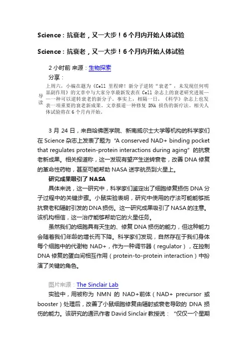
图片来源:The Sinclair Lab实验中,用被称为NMN的NAD+前体(NAD+ precursor或booster)处理后,改善了小鼠细胞修复由辐射或衰老导致的DNA损伤的能力。
该研究的通讯作者David Sinclair教授说:“仅仅一个星期的处理后,衰老小鼠的细胞和年轻小鼠的细胞就无法区分了。
![用于预防和治疗尿失禁的组合物[发明专利]](https://uimg.taocdn.com/0e3acb94f242336c1eb95efc.webp)
专利名称:用于预防和治疗尿失禁的组合物专利类型:发明专利
发明人:渔津,朴明焕
申请号:CN200480043285.1
申请日:20040621
公开号:CN1964729A
公开日:
20070516
专利内容由知识产权出版社提供
摘要:本发明涉及一种组合物,其包括南瓜籽油或南瓜籽提取物、Rubus coreous提取物以及大豆异黄酮。
由于本发明组合物能够减少尿失禁的发病率和日间排尿次数,增加尿道关闭压力(控制尿道括约肌的能力),它可用于治疗或预防尿失禁。
同时,由于该组合物的组分作为安全的物质已经广泛使用,并且本发明组合物不显示急性毒性,所以本发明组合物可用于预防和治疗尿失禁的药物和功能性保健食品中。
申请人:安国药品株式会社
地址:韩国首尔
国籍:KR
代理机构:隆天国际知识产权代理有限公司
代理人:吴小瑛
更多信息请下载全文后查看。

阈值阈值2.4532260序号123456789101112131415161718192021222324252627282930313233343536373839404142434445461区2区3区期刊数期刊数阈值1.5720.96596ISSN 刊名简称分区刊1467-2960FISH FISH 1FISH AN 0065-2113ADV AGRON 1ADVANCES 0928-4249VET RES 1VETERINA 1753-5123REV AQUACULT1Reviews 0168-1923AGR FOREST METEOROL 1AGRICU 0038-0717SOIL BIOL BIOCHEM 1SOIL B 1935-5130FOOD BIOPROCESS TECH1Food an0378-1135VET MICROBIOL 1VETE 1050-4648FISH SHELLFISH IMMUN1FISH & 1939-8425RICE 10363-2415FISHERIES1FIS 0960-3166REV FISH BIOL FISHER 1REVIEW 0021-8561J AGR FOOD CHEM1JOU 0032-079X PLANT SOIL1PLANT 1774-0746AGRON SUSTAIN DEV 1Agronomy 1994-4136MYRMECOL NEWS1Myrmeco 0308-521X AGR SYST 1AGRICULTU1084-2020ILAR J1ILAR 0829-318X TREE PHYSIOL 1TREE P 0022-0302J DAIRY SCI 1JOURNA 0167-1987SOIL TILL RES1SOIL &1090-0233VET J1VETERINA 1161-0301EUR J AGRON 2EUROPEAN 1380-3743MOL BREEDING 2MOLECUL0304-4017VET PARASITOL 2VETE 0021-8812J ANIM SCI 2JOURNA 0926-6690IND CROP PROD 2INDUSTRIA 0378-4290FIELD CROP RES 2FIELD CRO 0925-5214POSTHARVEST BIOL TEC2POSTHARV0016-7061GEODERMA 2GEO 0191-2917PLANT DIS 2PLANT 0929-1393APPL SOIL ECOL 2APPLIED S 1526-498X PEST MANAG SCI 2PEST MA 0931-2250J AGRON CROP SCI 2JOURNAL O 1869-215X AQUACULT ENV INTERAC2Aquacultu 1746-6148BMC VET RES 2BMC Veter1054-6006FISH OCEANOGR 2FIS0891-6640J VET INTERN MED 2JOURNAL O 0378-1127FOREST ECOL MANAG2FOREST E 1351-0754EUR J SOIL SCI 2EUROPEAN 1049-8001INT J WILDLAND FIRE 2INTERNATIO 0706-652X CAN J FISH AQUAT SCI2CANADIAN 0167-5877PREV VET MED 2PREVENTIV 0178-2762BIOL FERT SOILS 2BIOLOGY A0165-2427VET IMMUNOL IMMUNOP 2VETE 1865-1674TRANSBOUND EMERG DIS2Transbo47 48 49 50 51 52 53 54 55 56 57 58 59 60 61 62 63 64 65 66 67 68 69 70 71 72 73 74 75 76 77 78 79 80 81 82 83 84 85 86 87 88 89 90 91 92 93 94 95 96 970093-691X THERIOGENOLOGY2THERIO 1064-1262REV FISH SCI2REVIEWS 0361-5995SOIL SCI SOC AM J2SOIL SCIE 0044-8486AQUACULTURE2AQUA 0269-283X MED VET ENTOMOL2MEDI 0378-3774AGR WATER MANAGE2AGRICULT 1612-4669EUR J FOREST RES2EUROPEAN 1054-3139ICES J MAR SCI2ICES JOURN 1866-7910FOOD ENG REV2Food Engin 0739-7240DOMEST ANIM ENDOCRIN2DOMES 0043-1737WEED RES2WEED 0342-7188IRRIGATION SCI2IRRIGATI 0177-5103DIS AQUAT ORGAN2DISEASES 0011-183X CROP SCI2CROP 0003-1488JAVMA-J AM VET MED A2JAVMA-JOU 0307-9457AVIAN PATHOL2AVIAN P 0168-1591APPL ANIM BEHAV SCI2APPLIE 0140-7775J FISH DIS2JOURN 0377-8401ANIM FEED SCI TECH2ANIMAL F 1936-9751FOOD ANAL METHOD2Food Ana 1436-8730J PLANT NUTR SOIL SC2JOURNA 0266-0032SOIL USE MANAGE2SOIL 0959-4493VET DERMATOL2VETE 0021-8596J AGR SCI2JOU 1385-1314NUTR CYCL AGROECOSYS2NUTRIEN 0425-1644EQUINE VET J2EQUINE 0005-2086AVIAN DIS2AVIAN 1353-5773AQUACULT NUTR2AQUA 0378-4320ANIM REPROD SCI2ANIMAL RE 0002-1962AGRON J2AGRONO 0032-5791POULTRY SCI2POULTR 0929-1873EUR J PLANT PATHOL2EUROPEAN 0165-7836FISH RES2FISHERIE 0931-2668J ANIM BREED GENET2JOURNA 0931-1890TREES-STRUCT FUNCT2TREES-ST 0022-0299J DAIRY RES2JOURNA 0043-1745WEED SCI3WEED 0002-9254AM J ENOL VITICULT3AMERICAN 1751-7311ANIMAL3A 0300-9858VET PATHOL3VETERINAR 1461-9555AGR FOREST ENTOMOL3AGRICU 0936-6768REPROD DOMEST ANIM3REPRO 0014-2336EUPHYTICA3EUP 1286-4560ANN FOREST SCI3ANNALS 0906-6691ECOL FRESHW FISH3ECO 0045-5067CAN J FOREST RES3CANADIAN 1125-7865DENDROCHRONOLOGIA3DENDROC 0002-8487T AM FISH SOC3TRANSAC 0261-1929ATLA-ALTERN LAB ANIM3ATLA-ALTE 0920-1742FISH PHYSIOL BIOCHEM3FISH PHY 1473-5903INT J AGR SUSTAIN3Internatio98 99 100 101 102 103 104 105 106 107 108 109 110 111 112 113 114 115 116 117 118 119 120 121 122 123 124 125 126 127 128 129 130 131 132 133 134 135 136 137 138 139 140 141 142 143 144 145 146 147 1481098-612X J FELINE MED SURG3JOURNA 0925-9864GENET RESOUR CROP EV3GENETIC 0034-5288RES VET SCI3RESE 0161-3499VET SURG3VETERINA 0140-7783J VET PHARMACOL THER3JOURNAL O 1836-0947CROP PASTURE SCI3Crop & Pa 0261-2194CROP PROT3CROP P 0042-4900VET REC3VETERIN 1385-2256PRECIS AGRIC3PRECISION 0015-752X FORESTRY3FOR 1871-1413LIVEST SCI3Livesto 0002-9645AM J VET RES3AMERICAN 0043-9339WORLD POULTRY SCI J3WORLD 1040-6387J VET DIAGN INVEST3JOURNAL O 0921-4488SMALL RUMINANT RES3SMALL 0179-9541PLANT BREEDING3PLANT 1085-3278LAND DEGRAD DEV3LAND DEG 0253-1933REV SCI TECH OIE3REVUE SC 0090-3558J WILDLIFE DIS3JOURNAL 1550-7424RANGELAND ECOL3Rangela 1618-8667URBAN FOR URBAN GREE3URBAN F 0004-9573AUST J SOIL RES3AUSTRALIA 0304-4238SCI HORTIC-AMSTERDAM3SC 1936-5802CHEMOSENS PERCEPT3Chemosen 0889-048X AGR HUM VALUES3AGRICU 0022-4561J SOIL WATER CONSERV3JOURNAL 1537-5110BIOSYST ENG3BIOS 0015-749X FOREST SCI3FORES 0022-1201J FOREST3JOURNAL 1467-2987VET ANAESTH ANALG3VETE 1344-7610BREEDING SCI3BREEDIN 0037-5330SILVA FENN3SILVA 0275-6382VET CLIN PATH3VETERINA 1479-3261J VET EMERG CRIT CAR3JOURNAL O 0167-4366AGROFOREST SYST3AGROF 0044-605X ACTA VET SCAND3ACTA V 1437-4781FOREST PATHOL3FOREST 0830-9000CAN J VET RES3CANADIAN 2042-6496FOOD FUNCT3Food 1389-9341FOREST POLICY ECON3FOREST 0142-5242GRASS FORAGE SCI3GRASS A 0195-5616VET CLIN N AM-SMALL3VETERINA 1476-5810VET COMP ONCOL3Veterinary a 1355-557X AQUAC RES3AQUA 0990-7440AQUAT LIVING RESOUR3AQUA 1463-5216VET OPHTHALMOL3VETE 0969-997X FISHERIES MANAG ECOL3FISHERIES 1212-1819CZECH J ANIM SCI3CZECH J 0144-8609AQUACULT ENG3AQUAC 0962-7286ANIM WELFARE3ANIMAL 1976-1457NUTR RES PRACT3Nutrition4区总计期刊数期刊数2684463年平均IF 5.5744.2153.8013.7493.2713.2413.1723.1523.0863.0032.7592.7572.7032.6742.6512.6442.6392.612.5242.5082.4692.4532.452.4392.3962.3812.362.3472.3262.3192.3192.2962.2512.2232.1882.1862.1622.1462.1432.1342.1162.112.0792.0772.072期刊数刊名全称2011年IF H AND FISHERIES 5.8NCES IN AGRONOMY 5.204RINARY RESEARCH 4.06ews in Aquaculture 4.036RICULTURAL AND 3.389OIL BIOLOGY & 3.504od and Bioprocess 3.703VETERINARY 3.327SH & SHELLFISH3.322Rice 3.105FISHERIES 2.367EVIEWS IN FISH 2.5JOURNAL OF 2.823LANT AND SOIL 2.733nomy for Sustainable 3.33mecological News 2.644ULTURAL SYSTEMS 2.899LAR JOURNAL 2.333EE PHYSIOLOGY 2.876URNAL OF DAIRY 2.564SOIL & TILLAGE 2.425ERINARY JOURNAL 2.239PEAN JOURNAL OF 2.477ECULAR BREEDING 2.852VETERINARY 2.579URNAL OF ANIMAL 2.096STRIAL CROPS AND 2.469 CROPS RESEARCH 2.474HARVEST BIOLOGY 2.411GEODERMA 2.318LANT DISEASE2.449IED SOIL ECOLOGY 2.368ST MANAGEMENT 2.251NAL OF AGRONOMY 2.433culture Environment 2.188Veterinary Research 2FISHERIES2.044NAL OF VETERINARY 1.992EST ECOLOGY AND 2.487PEAN JOURNAL OF 2.34NATIONAL JOURNAL 2.231ADIAN JOURNAL OF 2.213ENTIVE VETERINARY 2.046OGY AND FERTILITY 2.319VETERINARY 2.0762.0272.0162.0082.0031.9661.9321.931.9121.8931.8681.861.8341.821.7991.7881.7771.7681.7671.7591.7581.721.7151.7111.7061.71.6971.6961.6851.6781.6691.6611.641.5911.5781.5771.5721.5711.5551.5541.5481.5451.5231.5191.5181.5061.5021.5011.4841.461.4561.454ERIOGENOLOGY 1.963EWS IN FISHERIES 1.946SCIENCE SOCIETY 1.979AQUACULTURE 2.041MEDICAL AND1.91CULTURAL WATER 1.998PEAN JOURNAL OF 1.982OURNAL OF MARINE2.007Engineering Reviews 1.893OMESTIC ANIMAL 2.056EED RESEARCH 1.924GATION SCIENCE 1.635ASES OF AQUATIC 2.201CROP SCIENCE1.641A-JOURNAL OF THE 1.791IAN PATHOLOGY 1.711PPLIED ANIMAL 1.918OURNAL OF FISH 2MAL FEED SCIENCE 1.691 Analytical Methods 1.943URNAL OF PLANT 1.596SOIL USE AND 1.608VETERINARY 1.943JOURNAL OF2.041RIENT CYCLING IN 1.792UINE VETERINARY 1.456VIAN DISEASES 1.462AQUACULTURE2.179AL REPRODUCTION 1.75ONOMY JOURNAL 1.794ULTRY SCIENCE 1.728PEAN JOURNAL OF 1.413ERIES RESEARCH 1.586URNAL OF ANIMAL 1.455S-STRUCTURE AND 1.685URNAL OF DAIRY 1.566WEED SCIENCE1.733RICAN JOURNAL OF1.826Animal1.744RINARY PATHOLOGY 1.945RICULTURAL AND 1.596PRODUCTION IN 1.356EUPHYTICA1.554NALS OF FOREST 1.788ECOLOGY OF1.573ADIAN JOURNAL OF 1.685DROCHRONOLOGIA 1.525NSACTIONS OF THE 1.592-ALTERNATIVES TO 1.455 PHYSIOLOGY AND1.528rnational Journal of 1.6961.4511.4431.4411.4251.4211.4181.4171.4111.4111.4051.4041.4031.3981.381.3731.3381.3261.3151.2891.2731.271.2581.2561.2451.2391.2351.2321.2291.2281.221.2071.2071.1961.1941.1921.1881.1871.1821.1791.1771.1741.1651.1641.1631.1471.131.1191.0921.091.0841.083URNAL OF FELINE 1.38ETIC RESOURCES 1.554RESEARCH IN1.649ERINARY SURGERY 1.265NAL OF VETERINARY 1.181 & Pasture Science 1.418OP PROTECTION 1.402ERINARY RECORD 1.248SION AGRICULTURE 1.549FORESTRY 1.337vestock Science 1.506RICAN JOURNAL OF 1.269ORLDS POULTRY 1.104NAL OF VETERINARY 1.214MALL RUMINANT 1.295LANT BREEDING1.596D DEGRADATION & 1.402E SCIENTIFIQUE ET 1.099RNAL OF WILDLIFE 1.079ngeland Ecology & 1.461BAN FORESTRY & 1.27RALIAN JOURNAL OF 1.53SCIENTIA1.527osensory Perception 1.643RICULTURE AND 1.54RNAL OF SOIL AND 1.265BIOSYSTEMS 1.354OREST SCIENCE 1.047NAL OF FORESTRY 1.354VETERINARY 0.944EEDING SCIENCE 1.248SILVA FENNICA 1.248ERINARY CLINICAL 1.556NAL OF VETERINARY2.038GROFORESTRY 1.378TA VETERINARIA 1.367REST PATHOLOGY 1.74ADIAN JOURNAL OF 0.939Food & Function 1.179REST POLICY AND 1.482ASS AND FORAGE 1.099RINARY CLINICS OF 1.638nary and Comparative 1.561AQUACULTURE 1.203QUATIC LIVING 1.152VETERINARY0.748RIES MANAGEMENT 1.294ECH JOURNAL OF 1.079QUACULTURAL 1.421NIMAL WELFARE1.135ition Research and 1.0831.0811.0761.0741.0731.0671.0661.0651.0631.0611.0571.0461.0411.0411.0391.0361.0351.0341.0331.0231.0171.0140.9860.9860.9810.9810.9780.9760.970.970.9650.9620.96RINARY RADIOLOGY 1.082NEW ZEALAND0.887DINAVIAN JOURNAL 1.197ORTH AMERICAN 0.943urnal of Veterinary 1.161alian Journal of Crop 1.632and Coastal Fisheries 1.065URNAL OF ANIMAL 0.855RINARY CLINICS OF 1.474CALIFORNIA 1.074URNAL OF SMALL 1RICAN JOURNAL OF 1.234SHERY BULLETIN 1.098CIENCE AND PLANT 1.017RALIAN VETERINARY 0.945AWA JOURNAL 1.042RITISH POULTRY1.005actions of the ASABE 1.033ROPICAL ANIMAL 1.115HIVES OF ANIMAL 0.987SOIL SCIENCE1.144l Production Science 0.986addy and Water 0.986STAN VETERINARY 1.255ADIAN JOURNAL OF 0.821ANTHROZOOS0.86ADIAN VETERINARY 1.063Science & Technology 1.183RNAL OF AQUATIC 0.833RINARY CLINICS OF 0.884ED TECHNOLOGY1.212ee-Ring Research 1.231。
近年来,糖尿病已成为备受人们关注的重大疾病之一。
有资料显示,糖尿病与循环系统表现(包括血管功能障碍)高度相关,而血管功能障碍可能影响各种离子通道[1]。
此外,胰岛素分泌减少或敏感性降低会导致体内代谢紊乱[2],这也是糖尿病产生的重要原因。
胰岛素是机体内唯一降低血糖的激素,由胰岛β细胞分泌[3],可以通过促进肌肉和脂肪组织对葡萄糖的摄取、合成糖原和生成脂肪来增加葡萄糖的消耗[4]。
研究表明,胰岛素分泌是一个极其复杂的过程,在这一过程中,胰岛β细胞膜上的离子通道起着重要作用,其中起主要调控作用的是钠离子通道、钾离子通道和钙离子通道。
1胰岛素的分泌机制胰岛素是维持机体内血糖稳态的一种重要激DOI:10.16605/ki.1007-7847.2020.11.0262离子通道对胰岛β细胞中胰岛素分泌的调控作用收稿日期:2020-11-13;修回日期:2021-01-18;网络首发日期:2021-03-22基金项目:国家自然科学基金资助项目(31960193,31660275);姚丽华江西省“双千计划”科技创新高端人才项目;江西省自然科学基金重点项目(20202ACBL206029)作者简介:孙慧珍(1995—),女,山东菏泽人,硕士研究生;*通信作者:姚丽华(1979—),男,江西玉山人,博士,江西科技师范大学教授,主要从事神经电生理的研究,E-mail:*****************。
孙慧珍1,雷水红2,龚妍春1,姚丽华1*(1.江西科技师范大学生命科学学院,中国江西南昌330013;2.南昌大学第二附属医院内分泌代谢科,中国江西南昌330008)摘要:胰岛β细胞是胰岛细胞的一种,属于内分泌细胞,主要的生理功能是分泌胰岛素以应对葡萄糖水平的升高,其在维持葡萄糖稳态中起着重要作用。
研究表明,胰岛素分泌受到多种机制的调控,其中包括多种离子通道。
近年来,国内外学者越来越关注离子通道调控胰岛素分泌的过程。
本文主要就钠离子通道、钾离子通道、钙离子通道以及3种离子通道之间的相互作用对胰岛素分泌的调控进行简述,同时,简单介绍了离子通道抑制剂在糖尿病临床中的应用,并展望了离子通道研究在未来糖尿病治疗方面的潜在应用价值。
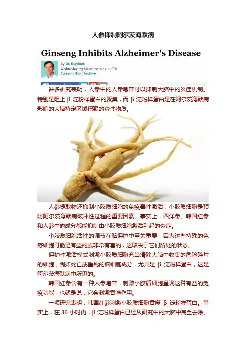
人参抑制阿尔茨海默病许多研究表明,人参中的人参皂苷可以抑制大脑中的炎症机制。
特别是阻止β淀粉样蛋白的聚集,而β淀粉样蛋白是在阿尔茨海默病影响的大脑特定区域积聚的炎性物质。
人参提取物还抑制小胶质细胞的免疫毒性激活,小胶质细胞是预防阿尔茨海默病破坏性过程的重要因素。
事实上,西洋参、韩国红参和人参中的成分都能抑制由小胶质细胞激活引起的炎症。
小胶质细胞活性的调节在脑保护中至关重要,因为这些特殊的免疫细胞可能是有益的或非常有害的,这取决于它们所处的状态。
保护性激活模式刺激小胶质细胞充当清除大脑中收集的危险碎片的细胞,例如死亡或垂死的脑细胞成分,尤其是β淀粉样蛋白,这是阿尔茨海默病中所见的。
韩国红参含有一种人参皂苷,刺激小胶质细胞呈现这种有益的免疫功能:也就是说,它会刺激吞噬作用。
一项研究表明,韩国红参刺激小胶质细胞吞噬β淀粉样蛋白。
事实上,在36小时内,β淀粉样蛋白已经从研究中的大脑中完全去除。
没有已知的药物可以实现这一点如果这还不够,还发现人参中的化合物抑制产生β淀粉样斑块的酶(BASE-1)。
韩国红参似乎特别有效降低兴奋性毒性,保护脑细胞免于细胞凋亡(程序性死亡),刺激大脑修复,减少脑部炎症,减少脑淀粉样蛋白斑块和过度磷酸化的tau,这两种在受阿尔茨海默病影响的大脑中积聚。
人参在保护大脑免受阿尔茨海默病损害方面紧随其后。
西洋参抑制tau蛋白过度磷酸化,这对保护大脑免受阿尔茨海默病的损害至关重要。
西洋参在预防导致阿尔茨海默病痴呆的许多过程方面也显示出巨大的希望。
例如,它已被证明可刺激轴突的生长,轴突是脑细胞通信的连接。
这些在阿尔茨海默病早期就会消失。
由西洋参中的一种成分(称为M1),结肠中的细菌产生的代谢产物已被证明可刺激轴突的生长,即使在对这些轴突进行广泛损伤后,这也会连接神经元。
在西洋参和人参中发现的化合物增加了大脑中抗氧化酶的水平,修复了突触和树突的损伤,刺激了大脑连接的生长,减少了阿尔茨海默病中发现的病理变化(β淀粉样斑块的形成和异常的tau),减少脑炎症,降低大脑中一氧化氮的危险水平。
肿瘤微环境(tumor microenvironment,TME)由肿瘤细胞、成纤维细胞、免疫细胞以及细胞外基质等组成,其中,肿瘤浸润免疫细胞(tumor-infiltratingimmune cell,TIC)在肿瘤发生、发展和治疗中发挥着核心作用[1-2]。
例如,细胞毒性T 淋巴细胞(又名CD8+T 细胞)是抗肿瘤免疫的主要效应细胞,可以特异性地识别和杀死带有新抗原(如突变基因表达产生的肿瘤特异性抗原)的肿瘤细胞[3]。
此外,不同肿瘤类型之间的肿瘤微环境有着巨大的差异,如T 细胞在肝脏肿瘤和结肠肿瘤患者中的比例要显著高于其在肺部肿瘤患者中的比例[4]。
因此,有必要对肿瘤微环境进行深入探究,其中肿瘤浸润免疫细胞的量化分析可揭示免疫细胞在肿瘤控制和治疗反应中的多方面作用。
DOI:10.16605/ki.1007-7847.2022.10.0214量化肿瘤浸润免疫细胞的分析方法研究进展王建平1,2,3,霍子天4,秦钧2,3,白明泽1,2,3*(1.重庆邮电大学大数据生物智能重庆市重点实验室,中国重庆400065;2.北京蛋白质组学研究中心,中国北京102206;3.国家蛋白质科学中心(北京),中国北京102206;4.华中科技大学同济医学院附属同济医院病理科,中国湖北武汉430030)摘要:免疫细胞一直被认为是维持机体平衡的重要调节因子,对肿瘤浸润免疫细胞进行量化分析有望揭示免疫系统在肿瘤中的多方面作用。
本文总结了量化肿瘤浸润免疫细胞的计算方法及相关的分析工具,包括基于标志基因富集分析方法和反卷积分析方法开发的算法模型与工具,列举了它们在揭示疾病发病机制以及药物治疗反应、确定肿瘤潜在生物标志物、辨别肿瘤免疫分型中的应用,提出了它们当前存在的局限性,并针对这些局限性问题进行了分析和展望,以促进该领域的发展。
关键词:肿瘤;肿瘤浸润免疫细胞(TIC);基因集富集分析(GSEA);反卷积中图分类号:Q-332,R730.3文献标志码:A文章编号:1007-7847(2024)02-0143-09Advances in Analytical Methods for Quantifying Tumor-infiltrating Immune CellsWANG Jianping 1,2,3,HUO Zitian 4,QIN Jun 2,3,BAI Mingze 1,2,3*(1.Chongqing Key Laboratory of Big Data for Bio Intelligence ,Chongqing University of Posts and Telecommunications ,Chongqing 400065,China ;2.Proteome Research Center ,Beijing 102206,China ;3.National Center for Protein Sciences(Beijing ),Beijing 102206,China ;4.Department of Pathology ,Tongji Hospital ,Tongji Medical College of Huazhong University ofScience and Technology ,Wuhan 430030,Hubei ,China )Abstract:Immune cells have long been recognized as important regulators for the maintenance of balance in the body system.Quantitative analysis of tumor-infiltrating immune cells is expected to reveal the multi-faceted roles of the immune system in tumors.This review summarized computational methods for quantifying tumor-infiltrating immune cells from tumor tissues,including algorithm models and tools based on marker gene enrichment analysis and deconvolution method,and presented their application in revealing disease pathogenesis and drug response,potential tumor biomarkers,and cancer immunophenotyping.In addition,current limitations of these computational methods were aslo pointed out and analyzed,hoping to promote their development.Key words:tumor;tumor-infiltrating immune cell (TIC);gene set enrichment analysis (GSEA);deconvolution(Life Science Research ,2024,28(2):143-151)收稿日期:2022-10-13;修回日期:2023-04-10;网络首发日期:2023-06-25作者简介:王建平(1995—),女,北京人,硕士研究生;*通信作者:白明泽(1982—),男,重庆人,博士,教授,主要从事生物信息学研究,Tel:************,E-mail:***************.cn 。
2007年,英国牛津大学的刘骥陇等在研究果蝇U 小体和P 小体(U 小体和P 小体是真核生物细胞质中的无膜细胞器)的功能关系时,用4种针对Cup (P 小体中的一种蛋白质)的抗体,对雌性果蝇的卵巢组织进行免疫组织化学染色,染色结果除了预期标记上的P 小体外,还标记出了长条形的丝状结构[1]。
这种结构的形状和数量与纤毛很相似,导致当时以为在果蝇中找到了有纤毛的新细胞类型。
但后来的一系列实验表明,该结构与纤毛没有关系,于是将其命名为“细胞蛇”。
最初是抗Cup 抗体不纯产生假象,意外发现的细胞蛇,而采用亲和层析纯化后的抗Cup 抗体无法再DOI:10.16605/ki.1007-7847.2020.10.0258细胞蛇的研究进展收稿日期:2020-10-22;修回日期:2020-11-19;网络首发日期:2021-07-27基金项目:宁夏自然科学基金项目(2020AAC03179);国家自然科学基金资助项目(31560329)作者简介:李欣玲(1999—),女,广西贵港人,学生;*通信作者:俞晓丽(1984—),女,宁夏银川人,博士,副教授,主要从事干细胞与生殖生物学研究,E-mail:********************。
李欣玲,张樱馨,李进兰,潘文鑫,王彦凤,杨丽蓉,王通,俞晓丽*(宁夏医科大学生育力保持教育部重点实验室临床医学院基础医学院,中国宁夏银川750000)摘要:细胞蛇是近年来细胞生物学研究的热门方向之一,由于其在细胞的增殖、代谢和发育上具有一定的生物学功能,因此,对一些疾病如癌症等的临床诊断或治疗具有一定的指导意义。
细胞蛇是由三磷酸胞苷合成酶(cytidine triphosphate synthetase,CTPS)聚合而成的无膜细胞器,其形成过程及功能在不同类型的细胞中不尽相同。
例如:细胞蛇能促进癌细胞增殖,并使患者病情恶化;过表达的细胞蛇可抑制神经干细胞增殖,影响大脑皮层发育;在卵泡细胞中,细胞蛇相当于CTPS 的存储库,在卵子发生过程起到促进细胞增殖和代谢的作用。
DOI:10.16605/ki.1007-7847.2022.09.0202几丁质降解菌Acinetobacter sp.WML1的生物安全性和发酵优化唐白露1,刘木兰1,旷琦颖1,何雪婷1,刘佳1,唐映红1,何海伦2*(1.湖南文理学院生命与环境科学学院常德市农业生物大分子研究中心环洞庭湖水产健康养殖及加工湖南省重点实验室,中国湖南常德415000;2.中南大学生命科学学院,中国湖南长沙410013)摘要:Acinetobacter sp.WML1分离自淡水湖渔场沉积物,能降解几丁质,具有一定的应用价值。
为了评价菌株WML1的生物安全性,为菌株的安全使用提供参考和保障,采用常规纸片扩散法检测耐药性,用血琼脂平板划线法检测溶血性,用溴甲酚紫显色法检测氨基酸脱羧酶活性,用试剂盒测定硝酸还原酶活性。
在菌株生物安全性分析的基础上,通过单因素试验法和正交试验法进行菌株WML1产几丁质酶发酵条件的优化。
结果表明,菌株WML1对多种抗生素敏感,无溶血性,无硝酸还原酶活性,有精氨酸脱羧酶活性及微弱的赖氨酸和鸟氨酸脱羧酶活性;其最优产酶条件为0.4%黄豆粉、0.20%半乳糖、0.03%KH 2PO 4、0.07%K 2HPO 4、0.002%FeSO 4、0.05%MgSO 4·7H 2O 、0.001%ZnSO 4、0.60%胶体几丁质、pH 9.5、5.0%的接种量、25mL 的装液量(对应250mL 容量瓶)、37℃的培养温度,优化后的几丁质酶活为(2.72±0.09)U/mL 。
综上所述,不动杆菌WML1在使用安全性上危害较小,可防可控,经发酵条件优化后其所产几丁质酶的活性提高了约4.61倍,为产几丁质酶菌株的工业化应用提供了理论基础。
关键词:几丁质降解菌;几丁质酶;不动杆菌WML1;生物安全性;发酵优化中图分类号:Q939.9文献标志码:A文章编号:1007-7847(2023)04-0361-10Biosafety and Fermentation Optimization of Acinetobacter sp.WML1for Chitinase ProductionTANG Bailu 1,LIU Mulan 1,KUANG Qiying 1,HE Xueting 1,LIU Jia 1,TANG Yinghong 1,HE Hailun 2*(1.Hunan Provincial Key Laboratory for Health Aquaculture and Product Processing in Dongting Lake Area ,Changde Research Center for Agricultural Biomacromolecule ,College of Life and Environmental Sciences ,Hunan University of Arts and Science ,Changde 415000,Hunan ,China ;2.School of Life Sciences ,Central South University ,Changsha 410013,Hunan ,China )Abstract:Acinetobacter sp.WML1was isolated from the fishery sediments of freshwater lakes and proved to have application value due to its ability to degrade chitin.Herein,the biosafety of the chitinase-producing strain WML1was first evaluated for application.The drug resistance of the strain was detected with the disk diffusion method,haemolytic activity was analyzed by streaking the blood agar plate,amino acid decarboxy-lase and nitrate reductase activities were measured by the chromogenic assay with bromocresol purple and nitrate reductase activity assay kit,respectively.On the basis of biosafety analysis,the fermentation condi-tions of the strain were then optimized by single-factor and orthogonal tests for chitinase production.The re-sults showed that the strain WML1was sensitive to most antibiotics,and had no haemolytic activity or ni-trate reductase activity,had arginine decarboxylase activity and weak lysine and ornithine decarboxylase ac-收稿日期:2022-09-14;修回日期:2022-12-23;网络首发日期:2023-03-15基金项目:湖南省自然科学基金青年基金项目(2021JJ40377);湖南省自然科学基金面上项目(2021JJ30029);湖南省教育厅科学研究一般项目(20C1270);常德市农业生物大分子研究中心开放课题(2020AB07);湖南省科技创新计划资助项目(2021RC1013)作者简介:唐白露(1991—),女,湖南永州人,博士,讲师,主要从事环境功能微生物及功能产物的研究,E-mail:***************.cn;*通信作者:何海伦(1976—),女,湖南长沙人,博士,教授,博士研究生导师,主要从事微生物酶结构和功能及生物资源酶解利用的相关研究,Tel:*************,E-mail:***************.cn 。

专利名称:与硫氧还蛋白或硫氧还蛋白还原酶基因互补的寡核苷酸序列以及将其用于调节细胞生长的方法
专利类型:发明专利
发明人:J·A·莱特,A·H·杨,Y·S·李
申请号:CN99802489.9
申请日:19990129
公开号:CN1294630A
公开日:
20010509
专利内容由知识产权出版社提供
摘要:本发明涉及互补于硫氧还蛋白和硫氧还蛋白还原酶的寡核苷酸,其在哺乳动物中调节肿瘤细胞生长。
本发明也涉及利用这类化合物抑制哺乳动物细胞中肿瘤细胞生长的方法。
本发明还涉及包括药学可接受的赋形剂以及有效量的本发明化合物的药物组合物。
申请人:吉倪塞思技术公司
地址:加拿大安大略
国籍:CA
代理机构:中国国际贸易促进委员会专利商标事务所
代理人:樊卫民
更多信息请下载全文后查看。
MinireviewRegulation of the cholinergic gene locus by the repressorelement-1silencing transcription factor/neuron restrictivesilencer factor (REST/NRSF)Masahito Shimojo,Louis B.Hersh *Department of Molecular and Cellular Biochemistry,University of Kentucky,Chandler Medical Center,800Rose Street,Lexington,KY 40536-0298,USAReceived 17July 2003;accepted 21August 2003AbstractThe cholinergic gene locus is comprised of two genes,the choline acetyltransferase gene and the vesicular acetylcholine transporter gene.The vesicular acetylcholine transporter gene is located within the first intron of the choline acetyltransferase gene.This arrangement permits coordinate regulation of the locus.Protein kinase A regulates expression of the cholinergic gene locus in PC12cells.This regulation was found to be dependent on the presence of a 21-bp DNA sequence known as the repressor element-1(RE-1)/neuron-restrictive silencer element (NRSE).Repressor element-1silencing transcription factor (REST)/neuron-restrictive silencer factor (NRSF),which binds to the RE-1/NRSE,is a zinc finger containing transcriptional repressor that blocks the expression of many neuronal RE-1/NRSE containing genes in nonneuronal cells.However,REST/NRSF expression has also been observed in neurons as well as the PC12cell line used in these studies.REST/NRSF truncated isoforms were expressed in neuronal cells,suggesting they also function in regulating neuronal gene expression.A study of REST4,one of the REST/NRSF isoforms,suggests that it regulates transcription of the cholinergic gene locus by blocking the repressor activity of REST/NRSF.Protein kinase A regulation of the cholinergic gene locus in PC12cells can thus be attributed,at least in part,to increased synthesis of REST4,which in turn derepresses the repressor activity of REST/NRSF.D 2004Elsevier Inc.All rights reserved.Keywords:Gene transcription;Gene regulation;Cholinergic gene;Transcriptional repression0024-3205/$-see front matter D 2004Elsevier Inc.All rights reserved.doi:10.1016/j.lfs.2003.08.045*Corresponding author.Tel.:+1-859-323-5549;fax:+1-859-323-1727.E-mail address:lhersh@ (L.B.Hersh)./locate/lifescieLife Sciences 74(2004)2213–2225IntroductionThe ability of a cell to regulate its phenotype is dependent upon its ability to carefully and precisely control which genes are expressed and which are not.In the case of neurons there are many genes whose expression is necessary to produce a neuronal phenotype,but whose expression outside the central nervous system is not needed and likely to be detrimental.Recent evidence suggests that one mechanism utilized to prevent expression of neuronal genes outside the central nervous system is that of gene repression.This review focuses on the regulation of one neuron specific gene locus,the cholinergic gene locus,by transcriptional repression.In addition to describing the regulation of the cholinergic gene locus,the complexity of this system,its application to other neuronal genes,and the many questions yet to be answered are noted.Cholinergic neurotransmission in the central nervous system is a key process dependent on the coexpression of proteins involved in the synthesis,storage,and release of the neurotransmitter acetylcholine (ACh).ACh plays an important role in fundamental brain processes such as memory,learning,and sleep (Karczmar,1976;Bartus et al.,1982;Aigner and Mishkin,1986).ACh is used in a cyclic process involving the uptake of choline by a high-affinity uptake system (Jope,1979),the formation of ACh through acetylation of choline catalyzed by the enzyme choline acetyltransferase (ChAT)(EC 2.3.1.6)and the transport of ACh into synaptic vesicles by the vesicular ACh transporter (V AChT).Upon stimulation,synaptic vesicles fuse with the plasma membrane and release ACh,which can then be hydrolyzed by acetylcholinesterase to produce free choline,thereby completing the cycle.The cholinergic gene locus is comprised of both the choline acetyltransferase gene as well as the vesicular acetylcholine transporter gene,the latter being located within the first intron of the ChAT gene.ChAT is primarily if not exclusively a cytosolic protein whereas V AChT has 12transmembrane domains and is integrated in the membrane of synaptic vesicles.In the CNS,ChAT and V AChT are both required for cholinergic neurotransmission.In some neurodegenerative disorders,i.e.Alzheimer’s disease,amyotrophic lateral sclerosis,and schizophrenia,there is a dysfunction of central cholinergic neurons involving a loss of,or abnormality in,basal forebrain ChAT activity (Davies and Maloney,1976;Coyle et al.,1983;Price,1986;Wu and Hersh,1994).Genomic structure and transcription of the cholinergic gene locusThe genes for mouse (Misawa et al.,1992;Pu et al.,1993),rat (Brice et al.,1989;Ishii et al.,1990),and human ChAT (Kong et al.,1989;Hersh et al.,1993)have been isolated and shown to contain three 5V -noncoding exons;exon 1,referred to as the R exon;exon 2,referred to as the N exon;and exon 3,referred to as the M exon (Misawa et al.,1992;Kengaku et al.,1993),Fig.1.The alternative splicing of these three exons results in multiple 5V -mRNA species,which are transcribed from three different promoters upstream of the R,N,and M exons,Fig.1.In the mammalian gene,the M exon resides f 5kb downstream from the R exon and transcription from its promoter appears to represent the major promoter used for generating ChAT mRNA.The V AChT gene in mammalian species (Bejanin et al.,1994;Erickson et al.,1994)as well as C.elegans (Alfonso et al.,1993)was found to lie uniquely within the first intron of the cholinergic gene locus between the first (R)and second (N)non-coding exons.This gene arrangement is conserved across such diverse species such as the nematode,Drosophila ,and mammals.M.Shimojo,L.B.Hersh /Life Sciences 74(2004)2213–22252214There are also multiple mRNA species found for V AChT that differ in their5V-untranslated sequences (Bejanin et al.,1994;Cervini et al.,1995).These mRNA isoforms appear to be generated from either a transcriptional start site upstream of the R exon,which may also be used for ChAT,or from sites within the V AChT exon(Bejanin et al.,1992;Misawa et al.,1992;Kengaku et al.,1993;Alfonso et al.,1994; Cervini et al.,1995),Fig.1.The human ChAT gene(and thus the cholinergic gene locus)has been mapped to chromosome10,region10q11.2–10q22.2(Cervini et al.,1991).Coordinate regulation of ChAT and V AChT gene expressionThe organization of the cholinergic gene locus suggested the possibility of coordinate regulation of the ChAT and V AChT genes at the transcriptional level(Berrard et al.,1995;Berse and Blusztajn,1995;Erickson et al.,1994;Shimojo et al.,1998).Prior to the discovery of the gene structure within the cholinergic gene locus,several studies reported that ChAT activity was regulated by growth factors (Hefti et al.,1986;Gould and Butcher,1989;Saadat et al.,1989;Knusel et al.,1991)and cytokines (Kamegai et al.,1990).Using a PC12mutant cell line,in which protein kinase A (PKA)activity is dramatically reduced by the expression of a defective PKA regulatory subunit,Inoue et al.(1995)reported that PKA could regulate expression of the ChAT gene at the transcriptional level.Transient transfection analysis using a human genomic reporter gene revealed that PKA acts through a site on the human ChAT gene upstream of the R exon.Lonnerberg et al.(1996)reported that this region of the rat ChAT gene contains both enhancer and silencer elements.Misawa et al.(1993)reported that dbcAMP increased ChAT mRNA and activity as well as transcription from the R,N,and M exons,suggesting that the cholinergic gene locus might be regulated by a PKA dependent pathway.Although a number of studies had reported regulation of the ChAT gene (see above),several reports emerged suggesting that the expression of ChAT and V AChT might be regulated coordinately in the CNS (Erickson et al.,1994;Berrard et al.,1995;Berse and Blusztajn,1995;Misawa et al.,1995).Berse and Blusztajn (1995)studied the expression of the ChAT and V AChT genes in a murine septal cell line,SN56,and found that ciliary neurotrophic factor (CNF),retinoic acid,leukemia inhibitory factor (LIF),and dbcAMP increased the mRNA levels for both ChAT and V AChT,although the effects appeared quantitatively different for the two genes.Studies on the regulation of ChAT and V AChT gene transcription in PC12cells using selective inhibitors and two protein kinase A (PKA)mutant PC12cell lines suggested that PKA signaling pathways involving PKAII coordinately regulate both the ChAT and V AChT genes at the transcriptional level (Shimojo et al.,1998).Transcriptional regulation of cholinergic gene locus by REST/NRSFStudies from a number of laboratories suggest that the transcriptional regulation of the ChAT and V AChT genes involves multiple cell specific regulatory elements.The region of the cholinergic gene locus upstream of the R-type promoter contains a consensus repressor element-1(RE-1)/neuron-restrictive silencer element (NRSE)sequence and a cholinergic specific enhancer that presumably operate in a cooperative manner to control cholinergic gene expression in the CNS,Fig.1.The RE-1/NRSE is a 21-basepair sequence originally isolated from the promoters of the type II sodium channel (Maue et al.,1990;Mori et al.,1992;Kraner et al.,1992)and SCG10(Mori et al.,1990,1992),and is implicated in silencing the cholinergic gene locus in non-neuronal cells (Lonnerberg et al.,1996).This sequence is found in a large number of neuron-specific genes including the genes for the N-methyl-D-aspartate receptor (NMDAR)(Bai et al.,1998),synapsin I (Schoch et al.,1996),the m4muscarinic acetylcholine receptor (Wood et al.,1996)and BDNF (Timmusk et al.,1999)as well as choline acetyltransferase (Lonnerberg et al.,1996).The transcription factor that binds to the RE-1/NRSE sequence was cloned independently by two groups;one naming it the RE-1silencing transcription factor or REST (Chong et al.,1995),the other naming it the neuron-restrictive silencing factor or NRSF (Schoenherr and Anderson,1995).The RE-1/NRSE sequence,through its binding of REST/NRSF,leads to the repression of the expression of neuron-specific genes in nonneuronal cells (Chong et al.,1995;Schoenherr and Anderson,1995).REST/NRSF is a member of the Gli-Kruppel family of transcription zinc-finger proteins being a 210-kDa glycoprotein containing nine zinc finger domains (Fig.2).M.Shimojo,L.B.Hersh /Life Sciences 74(2004)2213–22252216It was shown that REST/NRSF acts as a silencer of neuron-specific gene expression in nonneuronal tissues and in undifferentiated neuronal progenitor cells(Chong et al.,1995;Schoenherr and Anderson, 1995).The absence of the RE-1/NRSE sequence(Kallunki et al.,1997)or expression of a dominant negative form of REST/NRSF can produce expression of neuronal genes in both nonneuronal tissues and central nervous system progenitors(Chen et al.,1998).A mouse model in which the REST/NRSF gene was deleted,although embryonic lethal,showed aberrant expression of SCG10and tubulin h III in nonneuronal tissues(Chen et al.,1998).Although it was thought that expression of REST/NRSF was limited to non-neuronal cells where neuron-specific genes are repressed,recent evidence suggests that REST/NRSF may control the expression of neuron-specific genes containing an RE-1/NRSE element in neurons(Kallunki et al.,1997;Palm et al.,1998;Timmusk et al.,1999).To date five REST/NRSF isoforms arising from alternative splicing of the REST/NRSF pre-mRNA have been reported(Palm et al.,1998)and referred to as REST1through REST5.Two of these REST isoforms,REST4and REST5,are expressed specifically in mature neurons of adult brain,albeit at low levels.REST4and REST5contain an insertion of either16nucleotides or28nucleotides respectively,in the region of the gene encoding a spacer between zinc fingers5and6,Fig.2.These insertions in REST4 and REST5,as well as similar insertions in REST2and REST3lead to truncated isoforms that contain only 5of the9zinc finger domains found in REST/NRSF.REST1differs in that it contains only4zinc finger domains,Fig.2.Interestingly,REST4formation is induced by neuronal activity produced by kainate-induced seizure(Palm et al.,1998).REST4exhibits weak binding to the RE-1/NRSE element(Lee et al., 2000),and thus cannot directly regulate neuronal gene expression in RE-1/NRSE containing genes.As noted above the level of transcription of the cholinergic gene locus is greatly reduced in two PC12 cell lines;A123.7(Correll et al.,1989;Ginty et al.,1991)and A126.1B2(Van Buskirk et al.,1985)that have reduced protein kinase A ing reporter gene analysis it was found that the RE-1/NRSEelement was required for repression of the gene in the protein kinase A deficient PC12cells.Introduction of protein kinase A into the PKA deficient cells restored expression from the cholinergic gene locus,that is ChAT and V AChT mRNAs were now detectable.The finding that the concentration of REST/NRSF was essentially the same in both wild type and protein kinase A deficient PC12cells suggests that protein kinase A does not affect the expression levels of REST/NRSF.REST/NRSF in nuclear extracts from the protein kinase A deficient cells bound to the cholinergic RE-1/NRSE element as determined by electrophoretic mobility shift assays.On the other hand nuclear extracts derived from wild type PC12cells exhibited barely detectable binding to the RE-1/NRSE sequence,despite the fact that both nuclear extracts contained the same levels of REST/NRSF immunoreactive protein.The finding of active REST/NRSF in the protein kinase A deficient PC12cells suggests that transcription of the cholinergic locus was repressed in these cells by REST/NRSF,thus accounting for the absence of ChAT and V AChT mRNAs.However,the reason for the absence of repression of the cholinergic locus by REST/NRSF in wild type PC12cells was unclear.The possibility that protein kinase A phosphorylated REST/NRSF and generated an inactivate repressor was considered.However,purified protein kinase A catalytic subunit did not phosphorylate recombinant REST/NRSF.Wild type,but not protein kinase A deficient,PC12cells expressed REST4,the neuron-specific isoform of REST/NRSF described above and in Fig.2.It was shown that REST4forms a heterodimer with REST/NRSF.This heterodimer does not bind to the RE-1/NRSE element from the cholinergic gene locus.Thus even though REST/NRSF is expressed in wild type PC12cells,the presence of REST4inhibits the silencing of the cholinergic gene by blocking the repressor activity of REST/NRSF through heterodimer formation.Treatment of PC12cells with forskolin,which increases intracellular cAMP levels by activating adenylate cyclase,induced REST4expression within 4h and ChAT mRNA expression within 6to 12h.These kinetics are consistent with cAMP activating protein kinase A,which in turn leads to an increase in REST4levels,followed by derepression of the cholinergic locus and increased levels of ChAT mRNA.Thus it appears that protein kinase A regulation of the cholinergic gene locus occurs by PKA increasing the splicing of REST/NRSF pre-mRNA to produce REST4.REST4in turn blocks the repressor activity of REST/NRSF leading to increased gene transcription.Palm et al.(1998)found that a truncated form of REST/NRSF,REST2-5trunc ,also acted as a transcriptional repressor of a REST/NRSF containing reporter gene in Neuro-2A cells,a cell line that does not contain detectable REST/NRSF.On the other hand relatively weak repression of the REST/NRSF containing reporter gene was seen in C6cells,which contain relatively high levels of endogenous REST/NRSF.It was concluded that truncated forms of REST/NRSF produce repression independent of the activity of REST/NRSF and that the repressor activity of the truncated forms resides in an N-terminal repressor domain.It was further suggested that the repressor activity of the REST/NRSF truncated forms could be attributed to the formation of a complex with essential components of the transcriptional machinery.Regulation of REST/NRSF function by its REST4isoformThe finding that REST4can block REST/NRSF repressor activity,coupled with REST4expression in neuronal cells,can account for the presence of REST/NRSF in neurons without neuronal gene expression being repressed.Palm et al.(1998)reported the presence of REST/NRSF in adult neurons,M.Shimojo,L.B.Hersh /Life Sciences 74(2004)2213–22252218albeit at rather low levels relative to its expression in non-neuronal cells.It was suggested that the inability of REST/NRSF to repress gene expression in neuronal cells could be explained by its low concentration.However,as noted above,it appears that the relative levels of REST/NRSF and REST4 determine the extent of transcriptional repression.Although REST/NRSF levels appear to be constant in PC12cells,the levels of REST4can be regulated by cAMP through protein kinase A activity(Shimojo et al.,1999).In the PC12cell line we studied,the cholinergic gene,although transcriptionally active,was not maximally active since there was an excess of REST/NRSF relative to REST4.Thus modulation of transcriptional repression occurs through modulation of REST4,not REST/NRSF.This mechanism, rather than cAMP acting on a cAMP response element within the cholinergic locus,can explain at leasta Fig.3.Schematic showing the possible mechanisms for the de-repression activity of REST4.A.The formation of heterodimers between REST4and REST/NRSF prevents the binding of REST/NRSF to the cholinergic RE-1/NRSE and leads to de-repression of the cholinergic gene locus.Zinc finger domains are represented by numbered black circles.B.One of two repressor domains is located at the N-terminal domain and binds Sin3and histone deacetylase(HDAC).The other,located at the C-terminal domain is the CoREST binding site.All of the REST truncated forms retain the Sin3/HDAC binding site,but not the CoREST binding site.The truncated REST isoforms could form complexes with Sin3/cofactor complexes,resulting in the supression of available Sin3.C.Regulation of RNA splicing.The various truncated forms of REST shown in Fig.2are produced by RNA splicing as illustrated.Exons are shown as boxes and introns are shown as lines.The ORF of each transcript is indicated as a black box.The neuron-specific exon(N)located between exons V and VI has a stop codon thus when inserted produces truncated proteins.The relative expression levels for REST/NRSF and the splice variant REST4is illustrated for neurons and non-neuronal cells.M.Shimojo,L.B.Hersh/Life Sciences74(2004)2213–22252219part of the effects of cAMP on cholinergic gene expression in PC12cells.Whether this mechanism can be extended to the cholinergic gene locus in vivo remains to be determined.Supporting this hypothesis is the report of Tabuchi et al.(2002)who found that REST4affects the activity-dependent activation of the BDNF gene promoter I (BDNFp-I)in cultured rat cortical neurons.As we observed in PC12cells,REST4antagonized silencing of the BDNF gene by REST/NRSF,suggesting a role for REST4in preventing the neuron-specific BDNF gene from being transcriptionally repressed by REST/NRSF.In this case REST4activity varied in response to a variety of neuronal stimuli.Recently Magin et al.(2002)reported that humanized REST4had no discernable effect in NS20Y neuroblastoma cells.REST4did not impair transcriptional repression of the human synapsin I promoter by REST nor did it have a direct effect on the synapsin I promoter.The reason for this discrepancy with our studies and those of Tabuchi et al.(2002)remain unclear,but could be related to the cell type studied.As Tabuchi et al.(2002)suggested,the mechanism of regulation of gene transcription by REST/NRSF isoforms is complex in neurons.There are several mechanisms whereby REST isoforms can function to regulate transcription in the neuron.As shown in Fig.3A ,one mechanism is by heterodimer formation between REST/NRSF and REST4.Since REST4and other REST isoforms retain the N-terminal repressor domain,but do not bind efficiently to DNA,an alternative,or possibly additional mechanism,is that REST isoforms regulate gene expression by competing for binding of the transcriptional repressor complex made up of Sin3and HDAC as illustrated in Fig.3B (Palm et al.,1998;Roopra et al.,2001).The mRNA levels of REST/NRSF and REST isoforms are determined by the extent of alternative splicing of REST/NRSF pre-mRNA (Fig.3C).Our studies suggest that protein kinase A regulates REST/NRSF pre-mRNA splicing.The ability of a cell signaling pathway to regulate neuronal gene transcription through the REST/NRSF -REST isoform system provides a mechanism for a neuron to regulate its plasticity in response to neuronal activity,neuronal stimuli,or neuronal development.Role of zinc finger domains of REST/NRSFREST/NRSF contains nine Cys 2-His 2type zinc finger domains,the first of these is near the N-terminus and is followed by a cluster of seven zinc finger domains,with the last zinc finger domain being near the C-terminus of the molecule,Fig.2.REST4,being a truncated form of REST/NRSF contains only zinc finger domains 1–5,Fig.2.It binds weakly to the RE-1/NRSE with the strength of binding dependent on the number of zinc finger domains present (Lee et al.,2000).These findings suggest that zinc finger domains 2–5are required for DNA binding,while zinc finger domains 6–8contribute to the strength of the binding interaction (Shimojo et al.,2001;Chong et al.,1995).In order to study the effect of zinc finger conformation on REST/NRSF activity,mutational analysis was conducted in which zinc finger domains 6–8were studied.Conversion of a Cys to Arg within a zinc finger domain was shown to disrupt the conformation and function of the Cys 2-His 2type zinc finger domain (Tapia-Ramirez et al.,1997).The results of analyzing zinc finger mutations in REST/NRSF suggest that a combination of three zinc finger domains (zinc finger domains 6–8)contribute to DNA binding,however two adjacent zinc fingers,either zinc finger 6plus7or zinc finger 7plus 8,are required to produce maximal DNA binding.Analysis of the REST4sequence reveals that it lacks the putative nuclear localization signal found in the C-terminal region of REST/NRSF (Chong et al.,1995;Grimes et al.,2000).However,REST4isM.Shimojo,L.B.Hersh /Life Sciences 74(2004)2213–22252220M.Shimojo,L.B.Hersh/Life Sciences74(2004)2213–22252221efficiently targeted to the nucleus.Thus the translocation of REST4and REST/NRSF to the nucleus appears to be regulated by different mechanisms.Either REST/NRSF contains an additional N-terminally located nuclear localization signal,or a different nuclear localization signal in REST4is functional.Deletion mutagenesis indicated that the nuclear targeting signal of REST4resides within the C-terminal region containing zinc finger domains2–5(Lee et al.,2000;Shimojo et al.,2001).Fusion protein analysis showed that zinc finger domains2through5,but not2through4,could target h-galactosidase to the nucleus(Shimojo et al.,2001).Mutation of zinc finger domain5of REST4did not prevent its nuclear localization.However,the REST isoform REST1,which is similar to REST4but lacks zinc finger domain5,was not targeted to the nucleus.These data indicate the importance of the region around zinc finger5and suggest that there is a nuclear targeting signal,other than the zinc finger domain,located within the region preceding and going through zinc finger domain5.An alternative explanation is that deletion of the region around zinc finger5changes the structure of REST4such that a critical conformation needed for nuclear targeting is disrupted.To further study whether the zinc finger domain structure is important for nuclear targeting,REST4 with combinations of Cys to Arg point mutations in the zinc finger domains was fused to GFP.None of the combinations of point mutations altered the localization of REST4from nuclear to cytoplasmic.These data suggest that a functional structure of the zinc finger domains is not required to target REST4to the nucleus.However,certain combinations of zinc finger domain mutations produced abnormal nuclear localization.In these cases,REST4-GFP constructs appeared to localize to the edge of the nucleus,as if they were attached to the nuclear envelope(Shimojo et al.,2001).These data are consistent with previous findings that suggest that zinc finger domains not only function in DNA recognition(Wolfe et al.,2000), but can also be involved in protein-protein interactions(Lee et al.,1993;Milne and Segall,1993). REST/NRSF Repressor DomainsREST/NRSF is a modular protein that represses neuronal gene expression via two distinct repressor domains.Repression through an amino terminal domain is mediated by a Sin3-histone deacetylase (HDAC)complex,which affects chromosomal structure by promoting histone deacetylation(Tapia-Ramirez et al.,1997;Grimes et al.,2000;Huang et al.,1999;Roopra et al.,2001;Naruse et al.,1999). The other repressor domain is found in the carboxyl-terminal domain and binds a novel protein, CoREST and HDAC(Ballas et al.,2001;Andres et al.,1999).In situ hybridization studies of CoREST and Sin3A expression as well as REST/NRSF expression suggest that Sin3is expressed more ubiquitously during early embryogenesis(embryonic day8.5),but at latter stages of embryogenesis (day11.5)CoREST and Sin3A are both expressed fairly ubiquitously throughout the embryo.Evidence has recently been obtained that gene silencing by REST through these domains involves both histone deacetylation and ATP-dependent chromatin remodeling(Battaglioli et al.,2002).The future for RESTIt is evident that the regulation of neuronal gene expression by REST/NRSF and its truncated isoforms is complex and far from understood.As noted above REST mediated gene repression involves two distinct repressor domains,one binding Sin3A and the other binding CoREST.Why two distinctrepressor domains are present in REST/NRSF is puzzling.It is unclear whether both domains are utilized simultaneously or represent different modes of gene regulation that are utilized differentially.The need for a complex system whereby REST4modulates REST/NRSF activity (Fig.3)is also not clear,and whether REST4and the other REST isoforms have additional functions yet to be elucidated is an intriguing question.To date a function has only been attributed to REST4,a function for REST1,REST2,REST3,and REST5remain to be described.It has been shown that a signaling molecule,cAMP,acting through protein kinase A and REST/NRSF pre-mRNA splicing,can regulate cholinergic gene expression in PC12cells.Is this a general mechanism in all neurons and does cAMP and other signaling pathways regulate transcription of neuronal genes in response to cellular stimuli?On the surface it would appear that protein kinase A,through it increase in REST4,would lead to derepression of all REST/NRSF responsive genes.Whether this is the case or whether there are additional mechanisms that permit only a subset of REST/NRSF responsive genes to be affected by REST4remains to be established.Clearly there are many unanswered questions and much to be done to understand how the REST/NRSF system regulates the cholinergic gene locus as well as a host of other neuron specific genes.AcknowledgementsThis work was supported in part by grants AG46734and AG05893from the National Institutes of Health.ReferencesAigner,T.G.,Mishkin,M.,1986.The effects of physostigmine and scopolamine on recognition memory in monkeys.Behav-ioral and Neural Biology 45(1),81–87.Alfonso,A.,Grundahl,K.,Duerr,J.S.,Han,H.-P.,Rand,J.B.,1993.The Caenorhabditis elegans unc-17gene:a putative vesicular acetylcholine transporter.Science 261(5121),617–619.Alfonso,A.,Grundahl,K.,McManus,J.R.,Asbury,J.M.,Rand,J.B.,1994.Alternative splicing leads to two cholinergic proteins in Caenorhabditis elegans.Journal of Molecular Biology 241(4),627–630.Andres,M.E.,Burger,C.,Peral-Rubio,M.J.,Battaglioli,E.,Anderson,M.E.,Grimes,J.,Dallman,J.,Ballas,N.,Mandel,G.,1999.CoREST:a functional corepressor required for regulation of neural-specific gene expression.Proceedings of the National Academy of Sciences,USA 96(17),9873–9878.Bai,G.,Norton,D.D.,Prenger,M.S.,Kusiak,J.W.,1998.Single-stranded DNA-binding proteins and neuron-restrictive silencer factor participate in cell-specific transcriptional control of the NMDAR1gene.Journal of Biological Chemistry 273(2),1086–1091.Ballas,N.,Battaglioli,E.,Atouf,F.,Andres,M.E.,Chenoweth,J.,Anderson,M.E.,Burger,C.,Moniwa,M.,Davie,J.R.,Bowers,W.J.,Federoff,H.J.,Rose,D.W.,Rosenfeld,M.G.,Brehm,P.,Mandel,G.,2001.Regulation of neuronal traits by a novel transcriptional complex.Neuron 31(3),353–365.Bartus,R.T.,Dean,R.L.III,Beer,B.,Lippa,A.S.,,1982.The cholinergic hypothesis of geriatric memory dysfunction.Science 217(4558),408–414.Battaglioli,E.,Andres,M.E.,Rose,D.W.,Chenoweth,J.G.,Rosenfeld,M.G.,Anderson,M.E.,Mandel,G.,2002.REST repression of neuronal genes requires components of the hSWI.SNF complex.J.Biol.Chem.277(43),41038–41045.Bejanin,S.,Habert,E.,Berrard,S.,Edwards,J.B.,Loeffler,J.P.,Mallet,J.,1992.Promoter elements of the rat choline acetyltransferase gene allowing nerve growth factor inducibility in transfected primary cultured cells.Journal of Neuro-chemistry 58(4),1580–1583.M.Shimojo,L.B.Hersh /Life Sciences 74(2004)2213–22252222。