Exome Sequencing of47Chinese Families with Cone-Rod Dystrophy:Mutations in25Known Causative Genes Li Huang1.,Qingyan Zhang2.,Shiqiang Li1.,Liping Guan2,Xueshan Xiao1,Jianguo Zhang2,
Xiaoyun Jia1,Wenmin Sun1,Zhihong Zhu2,Yang Gao1,Ye Yin2,Panfeng Wang1,Xiangming Guo1,
Jun Wang2*,Qingjiong Zhang1*
1State Key Laboratory of Ophthalmology,Zhongshan Ophthalmic Center,Sun Yat-sen University,Guangzhou,Guangdong,China,2BGI-Shenzhen,Shenzhen, Guangdong,China
Abstract
Objective:The goal of this study was to identify mutations in25known causative genes in47unrelated Chinese families with cone-rod dystrophy(CORD).
Methods:Forty-seven probands from unrelated families with CORD were recruited.Genomic DNA prepared from leukocytes was analyzed by whole exome sequencing.Variants in the25genes were selected and then validated by Sanger sequencing.
Results:Fourteen potential pathogenic mutations,including nine novel and five known,were identified in10of the47 families(21.28%).Homozygous,compound heterozygous,and hemizygous mutations were detected in three,four,or three families,respectively.The14mutations in the10families were distributed among CNGB3(three families),PDE6C(two families),ABCA4(one family),RPGRIP1(one family),RPGR(two families),and CACNA1F(one family).
Conclusions:This study provides a brief view on mutation spectrum of the25genes in a Chinese cohort with CORD.
Identification of novel mutations enriched our understanding of variations in these genes and their associated phenotypes.
To our knowledge,this is the first systemic exome-sequencing analysis of all of the25CORD-associated genes.
Citation:Huang L,Zhang Q,Li S,Guan L,Xiao X,et al.(2013)Exome Sequencing of47Chinese Families with Cone-Rod Dystrophy:Mutations in25Known Causative Genes.PLoS ONE8(6):e65546.doi:10.1371/journal.pone.0065546
Editor:Andreas R.Janecke,Innsbruck Medical University,Austria
Received January9,2013;Accepted April25,2013;Published June11,2013
Copyright:?2013Huang et al.This is an open-access article distributed under the terms of the Creative Commons Attribution License,which permits unrestricted use,distribution,and reproduction in any medium,provided the original author and source are credited.
Funding:This study was supported by the National Natural Science Foundation of China(81170881,U1201221,https://www.doczj.com/doc/442529682.html,/Portal0/default152.htm), Guangdong Translational Medicine Public Platform,the"985project"of Sun Yat-sen University,and the Fundamental Research Funds of the State Key Laboratory of Ophthalmology.The funders had no role in study design,data collection and analysis,decision to publish,or preparation of the manuscript.
Competing Interests:The authors have declared that no competing interests exist.
*E-mail:zhangqji@https://www.doczj.com/doc/442529682.html,(QJZ);wangj@https://www.doczj.com/doc/442529682.html,(JW)
.These authors contributed equally to this work.
Introduction
Cone-rod dystrophy(CORD)refers to a series of hereditary retinal disorders with a predominantly cone involvement[1].Rod impairment may occur at the same time as the cone impairment or appear later.Patients with CORD usually have reduced visual acuity,photophobia,and color vision defects.
CORD may be transmitted as an autosomal dominant (adCORD),autosomal recessive(arCORD),or X-linked trait (xlCORD).To date,mutations in at least25genes have been reported to be associated with different forms of CORD,including the following:aryl hydrocarbon receptor interacting protein-like1 (AIPL1)[1];the cone-rod homeobox containing gene(CRX)[2]; guanylate cyclase activator1A(GUCA1A)[3];guanylate cyclase 2D(GUCY2D)[4];PITPNM family member3(PITPNM3)[5]; prominin1(PROM1)[6];peripherin2(PRPH2)[7];regulating synaptic membrane exocytosis1(RIMS1)[8];sema domain, immunoglobulin domain(Ig),transmembrane domain(TM)and short cytoplasmic domain,(semaphorin)4A(SEMA4A)[9];unc-119homolog(UNC119)[10];ATP-binding cassette,sub-family A (ABC1)member4(ABCA4)[11];ADAM metallopeptidase domain 9(ADAM9)[12];chromosome8open-reading frame37 (C8ORF37)[13];calcium channel voltage-dependent alpha2/ delta subunit4(CACNA2D4)[14];cadherin-related family member 1(CDHR1)[15];ceramide kinase-like(CERKL)[16];cyclic nucleotide gated channel beta3(CNGB3)[17];cyclin M4 (CNNM4)[18];potassium channel subfamily V member2(KCNV2) [19];phosphodiesterase6C,cGMP-specific,cone,alpha prime (PDE6C)[20];retina and anterior neural fold homeobox2(RAX2) [21];retinol dehydrogenase5(RDH5)[22];retinitis pigmentosa GTPase regulator interacting protein1(RPGRIP1)[23];calcium channel voltage-dependent L type alpha1F subunit(CACNA1F) [24];and retinitis pigmentosa GTPase regulator(RPGR)[25] (RetNet:https://https://www.doczj.com/doc/442529682.html,/Retnet/).Of the25genes,muta-tions in the first10genes are responsible for adCORD,the next13 for arCORD,and the last two for xlCORD.The associated genomic information of the25genes is listed in Table S1.
In our previous study on CORD,mutations were only detected in7of130(5.38%)Chinese families with CORD by using cycle sequencing of all coding exons of five genes(CRX,GUCY2D,
GUCA1A,PRPH2,and KCNV2)as well as of all exons harboring reported mutations in other 17CORD-associated genes [26,27,28,29].Of the seven families,all mutations were identified in genes responsible for adCORD but none in genes for arCORD and xlCORD.The genetic cause for most families (the remaining 123of 130(94.62%)Chinese families)was still unknown.In order to identify the additional cause of most CORD and to disclose further the mutation spectrum and frequency of the 25genes,whole exome sequencing was used to screen for mutations in 47unrelated Chinese families with CORD.
Materials and Methods Patients
Forty seven probands from unrelated families with CORD were recruited from the Eye Hospital of Zhongshan Ophthalmic Center,Sun Yat-sen University.Patients with identified mutation who were included in our previous study were excluded from this one.Written informed consents were obtained from the partici-pants or their guardians before the study,which was conforms to the tenets of the Declaration of Helsinki and follows the Guidance of Sample Collection of Human Genetic Diseases (863-plan)by the Ministry of Public Health of China.This study was approved by the Institute Review Board of the Zhongshan Ophthalmic Center.Genomic DNA was prepared from the blood leukocytes as previous described [30].
Exome Sequencing
Exome sequencing was completed by using a commercial service from BGI Shenzhen (https://www.doczj.com/doc/442529682.html,/index.php).The exome sequencing,genotype calling,and SNP calling were in the same way as the methods reported before [31].In brief,exome capture was carried out by using a NimbleGen SeqCap EZ Exome (44M)array.Exon-enriched DNA fragments were sequenced by the Illumina Genome Analyzer II.The average sequencing depth was set to 60-fold.SOAP aligner was used to set the sequencing reads to UCSC hg19[32,33].The likelihood of
possible genotypes in the target regions was calculated using SOAPsnp [34].Variants in all the 25genes detected by exome sequencing were selected for validation.Exome sequencing dataset of the patients with identified mutations in this study have been deposited to NIH (https://www.doczj.com/doc/442529682.html,/biosample:ac-cession number SAMN01997562to SAMN01997571).
Sanger Sequencing
Sanger sequencing was used to validate variants in the 25genes that resulted from exome sequencing,including heterozygous variants in the adCORD genes,homozygous or compound heterozygous variants in the arCORD genes,or hemizygous variants in the xlCORD genes.Primers (Table S2)used to amplify the regions containing the variants were designed by primer design tool Primer3(https://www.doczj.com/doc/442529682.html,/primer3/)[35].A touch-down polymerase chain reaction (PCR)was used to amplify the fragments with variants,as previously reported [36],and the amplicons were analyzed with an ABI BigDye Terminator cycle sequencing kit v3.1(Applied Biosystems,Foster City,CA)on an ABI3100Genetic Analyzer (Applied Biosystems).Sequencing results from patients and controls were compared using the SeqManII program of the Lasergene package (DNAStar Inc,Madison,WI).Detected variants were further sequenced in the available family members.Novel variants were further evaluated in 192control individuals.The description of mutations was in accordance with the nomenclature for the description of sequence variants [37](HGVS:https://www.doczj.com/doc/442529682.html,/mutnomen/).The conservation of a variation was evaluated by Phastcons_score (https://www.doczj.com/doc/442529682.html,/Annotation/PhastCons)[38],the effect of a missense variation was analyzed by using SIFT [39](https://www.doczj.com/doc/442529682.html,/)and Polyphen-2[40](https://www.doczj.com/doc/442529682.html,/pph2/)online tools,and the effect of splicing site changes was predicted by Berkeley Drosophila Genome Project (BDGP)[41](https://www.doczj.com/doc/442529682.html,/).
Considering that CORD-causing mutations are rare and the presence of the normal carriers of arCORD gene mutations,we assumed that the affected individuals were likely homozygous
or
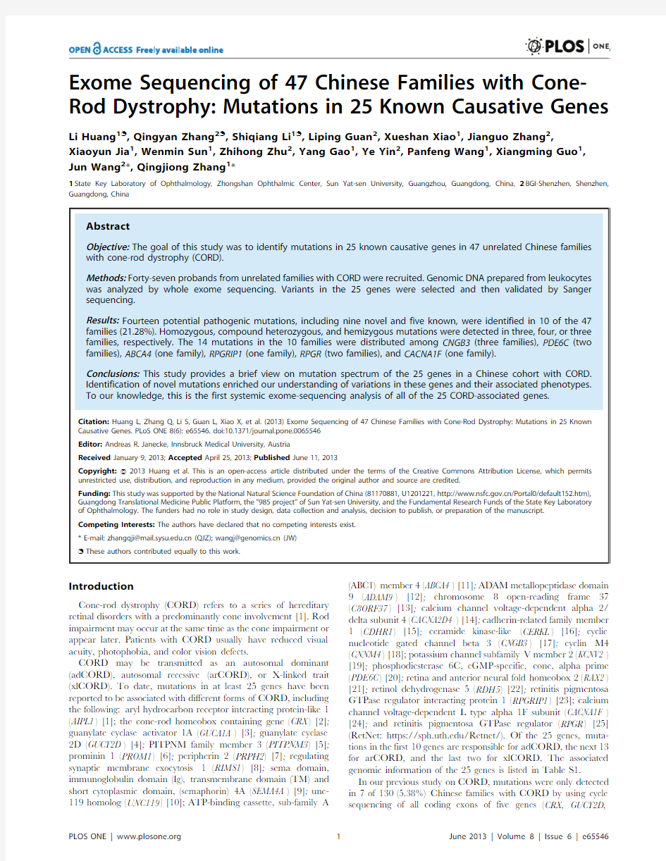
Figure 1.Prevalence of mutations in the investigated genes in our cohort of 47CORD patients.doi:10.1371/journal.pone.0065546.g001
compound heterozygous,so variants absent in the dbSNP134,1000Genome or with allelic frequencies #0.006were considered to be potentially pathogenic (frequency of heterozygote carriers calculated based on a disease incidence of 1:40,000,under the hypothesis that a unique arCORD gene would explain the remaining 40%of cases [42]).
Results
Whole exome sequencing identified 14potential pathogenic mutations in 10of the 47(21.28%)families with CORD (Table 1),including seven homozygous or compound heterozygous muta-tions in four (ABCA4,CNGB3,PDE6C,and RPGRIP1)of the 13genes associated with arCORD,and three hemizygous mutations in the two genes (RPGR and CACNA1F )associated with xlCORD.
Of the 14mutations,nine were novel.The 14mutations in the 10families involved six of the 25CORD-associated genes,including CNGB3(three families),PDE4C (two families),RPGR (two families),ABCA4(one family),RPGRIP1(one family),and CACNA1F (one family)(Figure 1),respectively.Sanger sequencing confirmed the 14mutations in the 10families (Figure S1).Segregation analysis was available for five of the 10families in where the mutations co-segregated with the disease in the family (Figure 2).No potential pathogenic mutation was identified in the other 19genes of the 47families.
Clinical data of the 10probands with potential pathogenic mutations are listed in Table 2.All probands with identified mutations had an early onset severe form of retinal dystrophy with predominantly cone involvement.Fundus changes were mainly
in
Figure 2.Pedigrees of the 10families with mutations.The family numbers and their corresponding mutations were shown just above the pedigrees.
doi:10.1371/journal.pone.0065546.g002
the macular regions,showing mild pigmentary changes and a loss of foveal reflex,as well as attenuated retinal arteries in rare cases. Discussion
Based on an initial screening of exome sequencing and the subsequent confirmation of Sanger sequencing,potential patho-genic mutations were identified in10of the47(21.28%)families with CORD,involving14mutations in six of the25CORD-associated genes.For the47families in this study,the contribu-tions of causative mutations in individual genes are as follows:CNGB3(6.38%),PDE6C(4.26%),RPGR(4.26%),ABCA4(2.13%), RPGRIP1(2.13%),and CACNA1F(2.13%).
The frequency of mutations detected in this study is significantly higher than in our previous study,in which mutations were only detected in seven of130(5.38%)Chinese families with CORD through the process of sequencing the coding exons of five genes and all exons with reported mutations in17other genes [26,27,28,29].In addition,in this study,mutations in seven of the10families were found in arCORD-associated genes but no mutations were detected in the adCORD-associated genes;in contrast,mutations in all seven families from our previous study were found in adCORD-associated genes.This difference may
Table1.Potential pathogenic mutations detected in10of the47families.
Family ID Gene Variations Status Bioinformation analysis Allele frequency in Reference
DNA Protein SIFT Polyphen-2Splice Phastcons
_score patients controls
Family1ABCA4 c.4604dup p.T1537Nfs*18hetero–––0.9971/940/384novel
Family1ABCA4 c.1957C.T p.R653C hetero D PD– 1.0001/94NA[38]
Family2CNGB3 c.1774dup p.E592Gfs*44homo––– 1.0002/940/384novel
Family3CNGB3 c.129+1G.A–homo––DSA 1.0002/940/384novel
Family4CNGB3 c.2415A.C p.E805D hetero D PD– 1.0001/94NA rs186448979# Family4CNGB3 c.1957G.A p.A653T hetero tolerated benign–0.0001/940/384novel
Family5PDE6C c.1935+1del–hetero––DSA 1.0001/940/384novel
Family5PDE6C c.2518+5G.C NA hetero––DSA0.1121/940/384novel
Family6PDE6C c.1004+1G.A–homo––DSA 1.0002/940/384novel
Family7RPGRIP1 c.2592T.G p.Y864*hetero–––0.9941/940/384novel
Family7RPGRIP1 c.799C.T p.R267*hetero––– 1.0001/94NA[36]
Family8CACNA1F c.2542G.A p.G848S hemi tolerated benign– 1.0001/940/384novel
Family9RPGR c.785C.G p.A262G hemi tolerated benign–0.0021/94NA[39]
Family10RPGR c.2447_2461del p.G816_E820del hemi–––NA1/94NA[40]
Note:D=damaging;PD=probably damaging;DSA=donor site abolished.
#The variation was found in1000Genomes database with the Global minor allele frequency(MAF)of G=0.001/3so that the pathogeneity of the variants in this family need to be clarified further.
doi:10.1371/journal.pone.0065546.t001
Table2.Clinical data of the probands with CORD and identified potential pathogenic mutations.
Family ID Gene Nucleotide changes Sex Age
(year)at First
Best
visual
acuity Fundus changes ERG responses from exam onset symptom right left right left rod cone
Family1ABCA4 c.[1957C.T];[4604dup]M7.0 6.0PV,PP0.200.15MA,TPOD,ARA MA,TPOD,ARA Normal Reduced
Family2CNGB3 c.[1774dup];[1774dup]M 6.5EC PV,NYS0.200.20MA,TPOD MA,TPOD Mildly reduced Extinguished Family3CNGB3 c.[129+1G.A];[129+1G.A]F 5.00.3PV,NYS,PP0.200.20MA,TPOD,ARA MA,TPOD,ARA Normal Extinguished Family4CNGB3 c.[1957G.A];[2415A.C]F 4.50.5PP,NYS0.100.20ARA ARA Mildly reduced Severely reduced Family5PDE6C c.[1935+1del];[2518+5G.C]M7.0EC NYS0.050.05High myopic High myopic Normal Severely reduced Family6PDE6C
c.[1004+1G.A];[1004+1G.A]F2.0ECPV,PP,NYSPOPONANAMildly reducedExtinguishedFamily7RPGRIP1c.[799C.T];[2592T.G]M3.6FMBPV,PP, NYSNANAARA,CRDARA,CRDExtinguishedExtinguishedFamily8CACNA1Fc.[2542G.A];[0]M2.3NAPP,NYSNANAMAMASeverely reduced*ExtinguishedFamily
9RPGRc.[785C.G];[0]M28.0ECPV,PP0.100.10MA,TPOD,ARAMA,TPOD,ARAModerately reducedExtinguishedFamily10RPGRc.[2447_2461del];[0]M9.0ECPV,
PP0.200.20MAMANormalExtinguished
Note:F=female;M=male;EC=early childhood;FMB=first few months after birth;PV=poor vision;PP=photophobia;NYS=nystagmus;PO=pursuing object; NA=not available;MA=macular atrophy;TPOD=temporal pallor of optic disc;ARA=attenuated retinal arteries;CRD=carpet-like retinal degeneration.
*This patient did not have the‘‘electronegative’’ERG in the standard combined response.
doi:10.1371/journal.pone.0065546.t002
partly have resulted from1)several genes associated with adCORD having been systemically analyzed before and all patients with mutations in these genes being excluded from this study;and2)selective analysis of exons with reported mutations perhaps failing to identify both mutations in arCORD associated genes if one mutation in the exons is not analyzed.The real proportion of mutations in the25genes should be higher than 21.28%in Chinese patients with CORD if mutations identified in previous study are taken into account[26,27,28,29].Further analysis of the results from exome sequencing for those patients without identified mutations in the25genes may provide useful clues in the identification of new genes responsible for CORD. Previously studies on an individual gene or a set of genes have revealed a different frequency of mutation in CORD-associated genes in different populations.Sanger sequencing of10adCORD-associated genes identified mutations in25of52(48%)German families with adCORD,and mutations were found in GUCY2D (24%),PRPH2(12%),GUCA1A(8%),CRX(4%),and PROM1(2%) [43].In other studies for individual genes,mutations in CRX are responsible for a4.76%proportion of CORD[26],GUCY2D for 11.11%of CORD in a Japanese population[44]and9.09%in a Spanish one[45],GUCA1A for16.67%of CORD in a German one[46],PRPH2for11%of CORD in another German population[43],AIPL1for1.82%of CORD in an American sample[1],PROM1for0.93%of CORD in a Dutch population [16],SEMA4A for8.00%of CORD in a Pakistani group[9],and UNC119for5.00%of CORD in another American population [10].
For genes known to be associated with arCORD,their mutations might be responsible for nearly40%of arCORD [42],whereas mutations in ABCA4are most frequent in European and American populations,ranging from16.13%to65.00% [47,48,49,50].However,ABCA4mutations are very rare in Chinese families with CORD.The mutation frequency of CNGB3 [49],PDE6C[20,49],and PRGRIP1[23]in other populations are relatively rare,as seen in Chinese sample.For other CORD-associated genes without identified mutations in this study, mutations in other populations are also rare,as with RAX2in 0.62%of CORD[21],CERKL in1.85%of Canadian CORD [16],or ADAM9[12],RDH5[22],CDHR1[15],and C8ORF37 [13]in a few reports based on an analysis of a limited number of families.
Supporting Information
Figure S1Sequence chromatography.Forteen sequence changes detected in the probands with CORD are shown(left column)compared with corresponding normal sequences(right column).Some known mutations were not verified in the normal controls,so the normal sequences are absent.
(TIF)
Table S1Genomic information of the25genes referred in this study.This table listed the accession numbers of the genomic DNA,mRNA,and protein for each of the25genes.The information is based on human genome reference GRCh37.p10. (XLS)
Table S2Sequences of primers used in this study.This table listed52primers used to amplify the genomic fragments with variants detected by exome sequencing.
(XLS)
Acknowledgments
The authors are grateful to the patients for their participation.
Author Contributions
Conceived and designed the experiments:QJZ.Performed the experi-ments:LH QYZ SL LG XX JZ XJ WS ZZ YG YY PW XG JW.Analyzed the data:LH QYZ LG JZ WS YY JW QJZ.Contributed reagents/ materials/analysis tools:LH SL XX JZ XJ WS YG PW XG JW QJZ. Wrote the paper:LH QJZ.
References
1.Sohocki MM,Perrault I,Leroy BP,Payne AM,Dharmaraj S,et al.(2000)
Prevalence of AIPL1mutations in inherited retinal degenerative disease.Mol Genet Metab70:142–150.
2.Freund CL,Gregory-Evans CY,Furukawa T,Papaioannou M,Looser J,et al.
(1997)Cone-rod dystrophy due to mutations in a novel photoreceptor-specific homeobox gene(CRX)essential for maintenance of the photoreceptor.Cell91: 543–553.
3.Payne AM,Downes SM,Bessant DA,Taylor R,Holder GE,et al.(1998)A
mutation in guanylate cyclase activator1A(GUCA1A)in an autosomal dominant cone dystrophy pedigree mapping to a new locus on chromosome 6p21.1.Hum Mol Genet7:273–277.
4.Kelsell RE,Gregory-Evans K,Payne AM,Perrault I,Kaplan J,et al.(1998)
Mutations in the retinal guanylate cyclase(RETGC-1)gene in dominant cone-rod dystrophy.Hum Mol Genet7:1179–1184.
5.Kohn L,Kadzhaev K,Burstedt MS,Haraldsson S,Hallberg B,et al.(2007)
Mutation in the PYK2-binding domain of PITPNM3causes autosomal dominant cone dystrophy(CORD5)in two Swedish families.Eur J Hum Genet 15:664–671.
6.Yang Z,Chen Y,Lillo C,Chien J,Yu Z,et al.(2008)Mutant prominin1found
in patients with macular degeneration disrupts photoreceptor disk morphogen-esis in mice.J Clin Invest118:2908–2916.
7.Fishman GA,Stone EM,Alexander KR,Gilbert LD,Derlacki DJ,et al.(1997)
Serine-27-phenylalanine mutation within the peripherin/RDS gene in a family with cone dystrophy.Ophthalmology104:299–306.
8.Kelsell RE,Gregory-Evans K,Gregory-Evans CY,Holder GE,Jay MR,et al.
(1998)Localization of a gene(CORD7)for a dominant cone-rod dystrophy to chromosome6q.Am J Hum Genet63:274–279.
9.Abid A,Ismail M,Mehdi SQ,Khaliq S(2006)Identification of novel mutations
in the SEMA4A gene associated with retinal degenerative diseases.J Med Genet 43:378–381.
10.Kobayashi A,Higashide T,Hamasaki D,Kubota S,Sakuma H,et al.(2000)
HRG4(UNC119)mutation found in cone-rod dystrophy causes retinal degeneration in a transgenic model.Invest Ophthalmol Vis Sci41:3268–3277.11.Cremers FP,van de Pol DJ,van Driel M,den Hollander AI,van Haren FJ,et al.
(1998)Autosomal recessive retinitis pigmentosa and cone-rod dystrophy caused by splice site mutations in the Stargardt’s disease gene ABCR.Hum Mol Genet 7:355–362.
12.Parry DA,Toomes C,Bida L,Danciger M,Towns KV,et al.(2009)Loss of the
metalloprotease ADAM9leads to cone-rod dystrophy in humans and retinal degeneration in mice.Am J Hum Genet84:683–691.
13.Estrada-Cuzcano A,Neveling K,Kohl S,Banin E,Rotenstreich Y,et al.(2012)
Mutations in C8orf37,encoding a ciliary protein,are associated with autosomal-recessive retinal dystrophies with early macular involvement.Am J Hum Genet 90:102–109.
14.Wycisk KA,Budde B,Feil S,Skosyrski S,Buzzi F,et al.(2006)Structural and
functional abnormalities of retinal ribbon synapses due to Cacna2d4mutation.
Invest Ophthalmol Vis Sci47:3523–3530.
15.Ostergaard E,Batbayli M,Duno M,Vilhelmsen K,Rosenberg T(2010)
Mutations in PCDH21cause autosomal recessive cone-rod dystrophy.J Med Genet47:665–669.
16.Littink KW,Koenekoop RK,van den Born LI,Collin RW,Moruz L,et al.
(2010)Homozygosity mapping in patients with cone-rod dystrophy:novel mutations and clinical characterizations.Invest Ophthalmol Vis Sci51:5943–5951.
17.Michaelides M,Aligianis IA,Ainsworth JR,Good P,Mollon JD,et al.(2004)
Progressive cone dystrophy associated with mutation in CNGB3.Invest Ophthalmol Vis Sci45:1975–1982.
18.Polok B,Escher P,Ambresin A,Chouery E,Bolay S,et al.(2009)Mutations in
CNNM4cause recessive cone-rod dystrophy with amelogenesis imperfecta.
Am J Hum Genet84:259–265.
19.Wu H,Cowing JA,Michaelides M,Wilkie SE,Jeffery G,et al.(2006)Mutations
in the gene KCNV2encoding a voltage-gated potassium channel subunit cause "cone dystrophy with supernormal rod electroretinogram"in humans.Am J Hum Genet79:574–579.
20.Thiadens AA,den Hollander AI,Roosing S,Nabuurs SB,Zekveld-Vroon RC,
et al.(2009)Homozygosity mapping reveals PDE6C mutations in patients with early-onset cone photoreceptor disorders.Am J Hum Genet85:240–247. 21.Wang QL,Chen S,Esumi N,Swain PK,Haines HS,et al.(2004)QRX,a novel
homeobox gene,modulates photoreceptor gene expression.Hum Mol Genet13: 1025–1040.
22.Wada Y,Abe T,Sato H,Tamai M(2001)A novel Gly35Ser mutation in the
RDH5gene in a Japanese family with fundus albipunctatus associated with cone dystrophy.Arch Ophthalmol119:1059–1063.
23.Hameed A,Abid A,Aziz A,Ismail M,Mehdi SQ,et al.(2003)Evidence of
RPGRIP1gene mutations associated with recessive cone-rod dystrophy.J Med Genet40:616–619.
24.Jalkanen R,Mantyjarvi M,Tobias R,Isosomppi J,Sankila EM,et al.(2006)X
linked cone-rod dystrophy,CORDX3,is caused by a mutation in the CACNA1F gene.J Med Genet43:699–704.
25.Demirci FY,Rigatti BW,Wen G,Radak AL,Mah TS,et al.(2002)X-linked
cone-rod dystrophy(locus COD1):identification of mutations in RPGR exon ORF15.Am J Hum Genet70:1049–1053.
26.Huang L,Xiao X,Li S,Jia X,Wang P,et al.(2012)CRX variants in cone-rod
dystrophy and mutation overview.Biochem Biophys Res Commun426:498–503.
27.Huang L,Li S,Xiao X,Jia X,Sun W,et al.(2013)Novel GUCA1A mutation
identified in a Chinese family with cone-rod dystrophy.Neurosci Lett541:179–183.
28.Xiao X,Guo X,Jia X,Li S,Wang P,et al.(2011)A recurrent mutation in
GUCY2D associated with autosomal dominant cone dystrophy in a Chinese family.Mol Vis17:3271–3278.
29.Huang L,Li S,Xiao X,Jia X,Wang P,et al.(2013)Screening for variants in20
genes in130unrelated patients with cone-rod dystrophy.Mol Med Rep7: 1779–1785.
30.Wang Q,Wang P,Li S,Xiao X,Jia X,et al.(2010)Mitochondrial DNA
haplogroup distribution in Chaoshanese with and without myopia.Mol Vis16: 303–309.
31.Li Y,Vinckenbosch N,Tian G,Huerta-Sanchez E,Jiang T,et al.(2010)
Resequencing of200human exomes identifies an excess of low-frequency non-synonymous coding variants.Nat Genet42:969–972.
32.Li R,Li Y,Kristiansen K,Wang J(2008)SOAP:short oligonucleotide
alignment program.Bioinformatics24:713–714.
33.Li R,Yu C,Li Y,Lam TW,Yiu SM,et al.(2009)SOAP2:an improved ultrafast
tool for short read alignment.Bioinformatics25:1966–1967.
34.Li R,Li Y,Fang X,Yang H,Wang J,et al.(2009)SNP detection for massively
parallel whole-genome resequencing.Genome Res19:1124–1132.
35.Rozen S,Skaletsky H(2000)Primer3on the WWW for general users and for
biologist programmers.Methods Mol Biol132:365–386.36.Li L,Xiao X,Li S,Jia X,Wang P,et al.(2011)Detection of variants in15genes
in87unrelated Chinese patients with Leber congenital amaurosis.PLoS One6: e19458.
37.den Dunnen JT,Antonarakis SE(2000)Mutation nomenclature extensions and
suggestions to describe complex mutations:a discussion.Hum Mutat15:7–12.
38.Siepel A,Bejerano G,Pedersen JS,Hinrichs AS,Hou M,et al.(2005)
Evolutionarily conserved elements in vertebrate,insect,worm,and yeast genomes.Genome Res15:1034–1050.
39.Kumar P,Henikoff S,Ng PC(2009)Predicting the effects of coding non-
synonymous variants on protein function using the SIFT algorithm.Nat Protoc 4:1073–1081.
40.Adzhubei IA,Schmidt S,Peshkin L,Ramensky VE,Gerasimova A,et al.(2010)
A method and server for predicting damaging missense mutations.Nat Methods
7:248–249.
41.Reese MG,Eeckman FH,Kulp D,Haussler D(1997)Improved splice site
detection in Genie.J Comput Biol4:311–323.
42.den Hollander AI,Black A,Bennett J,Cremers FP(2010)Lighting a candle in
the dark:advances in genetics and gene therapy of recessive retinal dystrophies.
J Clin Invest120:3042–3053.
43.Kohl S,Kitiratschky V,Papke M,Schaich S,Sauer A,et al.(2012)Genes and
mutations in autosomal dominant cone and cone-rod dystrophy.Adv Exp Med Biol723:337–343.
44.Ito S,Nakamura M,Nuno Y,Ohnishi Y,Nishida T,et al.(2004)Novel complex
GUCY2D mutation in Japanese family with cone-rod dystrophy.Invest Ophthalmol Vis Sci45:1480–1485.
45.Garcia-Hoyos M,Auz-Alexandre CL,Almoguera B,Cantalapiedra D,Riveiro-
Alvarez R,et al.(2011)Mutation analysis at codon838of the Guanylate Cyclase 2D gene in Spanish families with autosomal dominant cone,cone-rod,and macular dystrophies.Mol Vis17:1103–1109.
46.Kitiratschky VB,Behnen P,Kellner U,Heckenlively JR,Zrenner E,et al.(2009)
Mutations in the GUCA1A gene involved in hereditary cone dystrophies impair calcium-mediated regulation of guanylate cyclase.Hum Mutat30:E782–796.
47.Ducroq D,Rozet JM,Gerber S,Perrault I,Barbet D,et al.(2002)The ABCA4
gene in autosomal recessive cone-rod dystrophies.Am J Hum Genet71:1480–1482.
48.Fishman GA,Stone EM,Eliason DA,Taylor CM,Lindeman M,et al.(2003)
ABCA4gene sequence variations in patients with autosomal recessive cone-rod dystrophy.Arch Ophthalmol121:851–855.
49.Thiadens AA,Phan TM,Zekveld-Vroon RC,Leroy BP,van den Born LI,et al.
(2012)Clinical course,genetic etiology,and visual outcome in cone and cone-rod dystrophy.Ophthalmology119:819–826.
50.Maugeri A,Klevering BJ,Rohrschneider K,Blankenagel A,Brunner HG,et al.
(2000)Mutations in the ABCA4(ABCR)gene are the major cause of autosomal recessive cone-rod dystrophy.Am J Hum Genet67:960–966.