Indirect flow cytometry protocol
- 格式:pdf
- 大小:65.77 KB
- 文档页数:3
血液透析患者凝血功能变化及因素分析目的探讨血液透析治疗对患者凝血功能的影响及原因分析。
方法选取我院2012年1月~2014年4月共68例进行维持性血液透析患者透析前后的血液样本,采用流式细胞术检测血小板表面P-选择素。
用间接酶链免疫吸附试验检测超敏C反应蛋白、内皮血管性血友病因子和D-二聚体。
并通过对患者的观察、病例资料的整理以及对血透后透析器血容量测量等,回顾性分析血液透析患者发生凝血功能变化的原因及分析。
结果经治疗后患者均痊愈出院,治疗后PT和FIB值明显少于治疗前(P<0.05),APTT和TT值较治疗前差异无统计学意义(P>0.05)。
D-二聚体治疗前阳性率50%,治疗后为0,差异有统计学意义(P<0.05)。
血液透析后患者的血小板表面P-选择素、超敏C反应蛋白、内皮血管性血友病因子、D-二聚体结果明显高于透析前,差异有统计学意义。
影响患者发生凝血功能变化的因素包括自身体质、医生评估不到位、抗凝不当、肝素用量不足及护士操作技术不当。
结论医护人员应加强抗凝知识的学习,提高对血液透析患者凝血状态的评估和检测,并及时合理提供抗凝方案,熟练操作技术,与此同时,虽然在治疗中应用肝素,但患者仍可能处于高凝状态,因此定期进行凝血指标的监测可有效减少血液透析中凝血的发生。
[Abstract] Objective To explore the effects of hemodialysis on patients’ coagulation function and to analyze its reasons. Methods Blood samples of 68 patients before and after hemodialysis who took maintenance hemodialysis in our hospital from January 2012 to April 2014 were selected. Flow cytometry was applied to test P-selectin on platelet surface. Indirect enzyme linked immunosorbent assay was applied to test ultra-sensitive C-reactive protein,endothelial von Willebrand factor and D-dimer. Through the observation on patients,arrangement of patient data,and measurement of dialyzer blood volume after hemodialysis,reasons and analysis for changes of coagulation function for patients taking hemodialysis were retrospectively analyzed. Results All patients were discharged from hospitals after the treatment,and PT and FIB values after the treatment were significantly lower than those before the treatment (P<0.05). APTT and TT values were not statistically different from those before the treatment (P>0.05). Positive rate of D-dimer was 50% before the treatment and 0.0% after the treatment,and the difference compared before and after the treatment was statistically significant (P<0.05). Results of P-selectin on platelet surface,ultra-sensitive C-reactive protein,endothelial von Willebrand factor and D-dimer after hemodialysis were significantly higher than those before hemodialysis,and the differences were statistically significant. Factors affecting changes of patients’coagulation function included individual health quality,doctors’ inaccurate assessment,inappropriate anti-coagulation,insufficient usage of heparin and inappropriate operation techniques by nurses. Conclusion Medical staff should enhance their knowledge of anti-coagulation,improve the assessment and testing of coagulation status for patients taking hemodialysis,provide anti-coagulation protocol timely and properly,and practice operation techniques. Meanwhile,although heparin is used during treatment,patients may still be highly coagulated.Therefore,regular monitoring of coagulation indicators is able to effectively reduce coagulation during hemodialysis.[Key words] Hemodialysis;Coagulation function;Cause目前,我国慢性肾脏病的发病率在各种因素的影响下呈逐年上升趋势,而血液透析则是当前临床上作为终末期肾脏替代治疗的一种主要方式。
AID autoimmune disease 自身免疫病AIDS Acquired immunodeficiency syndrom 获得性免疫缺陷综合征;艾滋病AM adhesion molecule 粘附分子APC antigen-presenting cell抗原提呈细胞AD autoimmune disease 自身免疫病AIDS acquired immuno-deficiency syndrome 获得性免疫缺陷综合症(艾滋病)Ab antibody 抗体Allergen 变应原Allergy 变态反应Allogenic antigen 同种异型抗原Alpha-fetoprotein, AFP 甲种胎儿球蛋白Alternative pathway 旁路途径Antibody-dependent cell-mediated cytotoxicity, ADCC 抗体依赖性细胞介导的细胞毒作用Antigen, Ag 抗原Antigenic determinant 抗原决定簇BB cell receptor, BCR B 细胞(抗原识别)受体CCluster of differentiation, CD 分化群Colony stimulating factor, CSF 集落刺激因子Common variable immunodeficiency 普通变化型免疫缺陷病Complement 补体Complement receptor CR 补体受体Complementarity determining region, CDR 互补性决定区Complete antigen 完全抗原Concanavalin A, Con A 刀豆蛋白AConformational determinant 构象决定簇Constant region 恒定区,C区Co-stimulating signal 协同刺激信号Co-stimulatory molecules, CM 协同刺激分子Cross reaction 交叉反应Cytokine, CK 细胞因子Cytotoxic type 细胞毒型Cytotoxic T lymphocytes, CTL 或Tc 细胞毒性T细胞DDelayed type hypersensitivity, DTH 迟发型超敏反应Delayed type hypersensitivity T cell, T DTH 迟发型超敏反应性T细胞Dendritic cells, DC 树突状细胞DiGeorge syndrome DiGeorge综合征Enzyme linked immunosorbent assay, ELISA 酶联免疫吸附试验Eosinophil chemotactic factor, ECF 嗜酸性粒细胞趋化因子Eosinophil peroxidase, EPO 嗜酸性粒细胞过氧化物酶Epitope 表位Erythropoietin, EPO 红细胞生成素FF Fc receptor, FcR 可结晶片断受体,Fc受体Flow cytometry, FCM 流式细胞术Fragment antigen binding, Fab 抗原结合片断Fragment crystallizable, Fc 结晶片断GGraft versus host reaction, GVHR 移植物抗宿主反应Granulocyte colony stimulating factor, G-CSF 粒细胞集落刺激因子Granulocyte-macrophage colony stimulating factor, GM-CSF 粒细胞-巨噬细胞集落刺激因子HHeat shock protein, HSP 热休克蛋白Heavy chain 重链,H 链High endothelial venule, HEV 高内皮微静脉Hinge region 铰链区Histamin 组胺Histocompatibility antigen-2, H-2 小鼠的组织相容性抗原HLA genotyping HLA 基因分型Homologous restriction factor, HRF 同源限制因子Host versus graft reaction, HVGR 宿主抗移植物反应Human immunodeficiency virus, HIV 人类免疫缺陷病毒Human leukocyte antigen,HLA 人类白细胞抗原Humoral immunity 体液免疫Hypersensitivity 超敏反应Hypervariable region, HVR 高变区IIA immune adherent 免疫粘连IC immune complex免疫复合物ICAM intercellular adhesion molecule细胞间粘附分子ICD immunocomplex disease免疫复合物病IFN interferon 干扰素Ig immunoglobulin免疫球蛋白IHC immunohistochemistry免疫组织化学IIF indirect immunofluorescent method 间接免疫荧光法IL interleukin 白细胞介素Ir immuneresponse 免疫应答Immunogenicity 免疫原性Immunoglobulin superfamily, IgSF 免疫球蛋白超家族Immunological ignorance 免疫忽视Immunological tolerance 免疫耐受Immunology 免疫学Immunoreceptor tyrosine-based activation motifs, ITAM 免疫受体酪氨酸活化基序Immunoreceptor tyrosine-based inhibitory motifs, ITIM 免疫受体酪氨酸抑制基序Immunotherapy 免疫治疗nsulin-dependent diabetes mellitus, IDDM 胰岛素依赖型糖尿病Intercellular adhesion molecular,ICAM 细胞间粘附分子Integrin 整合素Interferon, IFN 干扰素Interleukin, IL 白细胞介素LLangerhans cells, LC 郎格汉斯细胞Lymphocyte 淋巴细胞Lymphocyte function associated antigen, LFA 淋巴细胞功能相关抗原Lymphocyte homing 淋巴细胞归巢Lymphocyte homing receptor, LHR 淋巴细胞归巢受体Lymphoid DC 淋巴系树突状细胞Lymphoid progenitor 淋巴样祖细胞Lymphoid stem cells, LSC 淋巴样干细胞MMacrophages, M φ巨噬细胞Macrophage colony stimulating factor, M-CSF 巨噬细胞集落刺激因子Major histocompatibility complex, MHC 主要组织相容性复合体Mannan-binding lectin, MBL 甘露糖结合凝集素Mast cell, MC 肥大细胞MBL-associated serine protease, MASP MBL 相关的丝氨酸蛋白酶Membrane attack complex, MAC 膜攻击复合物Membrane Ig, mIg 膜表面免疫球蛋白Memory cells 记忆细胞MHC class Ⅰgene MHC Ⅰ类基因MHC class Ⅱgene MHC Ⅱ类基因MHC class Ⅲgene MHC Ⅲ类基因MHC restriction MHC 限制性Monoclonal antibody, McAb 单克隆抗体Monocyte 单核细胞Mononuclear-phagocyte system, MPS 单核-巨噬细胞系统Myeloid stem cell 髓样干细胞NNaive T(B) cells 初始T(B)细胞Nature killer cell, NK cell 自然杀伤细胞Neutrophils 嗜中性粒细胞Non-specific immunity 非特异性免疫Nude mice 裸小鼠,裸鼠OOpsonization 调理作用PPathogen associated molecular pattern, PAMP 病原相关分子模式Pattern recognition receptor, PRR 模式识别受体Peptidoglycan, PGN 肽聚糖Perforin 穿孔素Peripheral lymphoid organ 外周淋巴器官Peyer’s patches 派氏集合淋巴结Phagocyte 吞噬细胞Phagocytosis 吞噬作用Phagolysosome 吞噬溶酶体Phytohemagglutinin, PHA 植物血凝素Placental γglobulin 胎盘丙种球蛋白Plaque forming cell, PFC 空斑形成细胞Plasma cells 浆细胞Platelet activating factor, PAF 血小板活化因子Polymeric Ig receptor, pIgR 多聚免疫球蛋白受体SSecretory IgA,sIgA 分泌型免疫球蛋白A Secretory piece, SP 分泌片Severe combined-immunodeficiency disease, SCID 重症联合免疫缺陷病Superantigen, SAg 超抗原Suppressor T cells, Ts 抑制性T细胞Systemic lupus erythematosus, SLE 系统性红斑狼疮TT cell epitope T细胞表位T cell receptor, TCR T细胞(抗原识别)受体T lymphocyte T淋巴细胞TCR/CD3 complex TCR/CD3 复合物Thymus 胸腺Thymus dependent antigen, TD-Ag 胸腺依赖抗原Thymus epithelial cells, TEC 胸腺上皮细胞Thymus independent antigen, TI-Ag 非胸腺依赖抗原Thyroid stimulating hormone, TSH 促甲状腺激素Tolerogen 耐受原Toll like receptor, TLR Toll样受体Toxoid 类毒素Transforming growth factor, TGF 转化生长因子Tumor-associated antigen, TAA 肿瘤相关抗原Tumor necrosis factor, TNF 肿瘤坏死因子Tumor-specific antigen, TSA 肿瘤特异性抗原VVariable region ( V region ) 可变区,V区X-linked SCID, XSCID 性联重症联合免疫缺陷病。
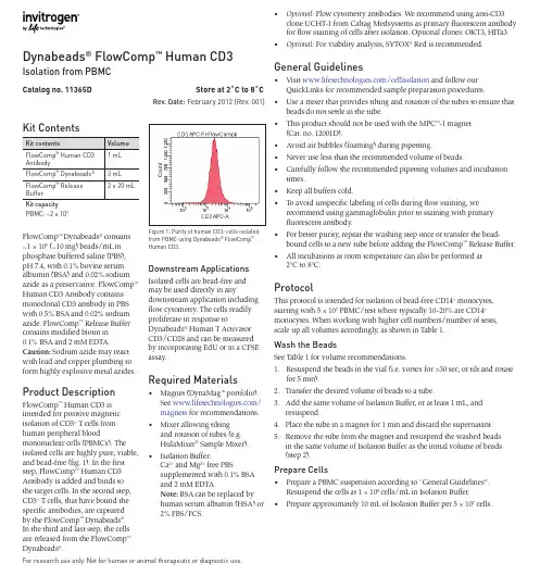
Kit ContentsFlowComp ™ Dynabeads ® contains ~1 × 10⁹ (~10 mg) beads/mL in phosphate buffered saline (PBS), pH 7.4, with 0.1% bovine serum albumin (BSA) and 0.02% sodium azide as a preservative. FlowComp ™ Human CD3 Antibody contains monoclonal CD3 antibody in PBS with 0.5% BSA and 0.02% sodium azide. FlowComp ™ Release Buffer contains modified biotin in 0.1% BSA and 2 mM EDTA.Caution: Sodium azide may react with lead and copper plumbing to form highly explosive metal azides.Product DescriptionFlowComp ™ Human CD3 is intended for positive magnetic isolation of CD3+ T cells from human peripheral bloodmononuclear cells (PBMCs). The isolated cells are highly pure, viable, and bead-free (fig. 1). In the first step, FlowComp ™ Human CD3 Antibody is added and binds to the target cells. In the second step, CD3+ T cells, that have bound the specific antibodies, are captured by the FlowComp ™ Dynabeads ®. In the third and last step, the cells are released from the FlowComp ™ Dynabeads ®.Dynabeads ® FlowComp ™ Human CD3Isolation from PBMCFor research use only. Not for human or animal therapeutic or diagnostic use.Catalog no. 11365D Store at 2˚C to 8˚CRev. Date: February 2012 (Rev. 001)Downstream ApplicationsIsolated cells are bead-free and may be used directly in anydownstream application including flow cytometry. The cells readily proliferate in response toDynabeads ® Human T Activator CD3/CD28 and can be measured by incorporating EdU or in a CFSE assay.Required Materials• Magnet (DynaMag ™ portfolio).See /magnets for recommendations.• Mixer allowing tiltingand rotation of tubes (e.g. HulaMixer ® Sample Mixer).• Isolation Buffer:Ca 2+ and Mg 2+ free PBSsupplemented with 0.1% BSA and 2 mM EDTA.Note: BSA can be replaced by human serum albumin (HSA) or 2% FBS/FCS.• Optional: Flow cytometry antibodies. We recommend using anti-CD3clone UCHT-1 from Caltag Medsystems as primary fluorescent antibody for flow staining of cells after isolation. Optional clones: OKT3, HITa3.• Optional: For viability analysis, SYTOX ® Red is recommended.General Guidelines• Visit /cellisolation and follow ourQuickLinks for recommended sample preparation procedures.• Use a mixer that provides tilting and rotation of the tubes to ensure thatbeads do not settle in the tube.• This product should not be used with the MPC ™-1 magnet(Cat. no. 12001D).• Avoid air bubbles (foaming) during pipetting.• Never use less than the recommended volume of beads.• Carefully follow the recommended pipetting volumes and incubationtimes.• Keep all buffers cold.• To avoid unspecific labeling of cells during flow staining, werecommend using gammaglobulin prior to staining with primary fluorescent antibody.• For better purity, repeat the washing step once or transfer the bead-bound cells to a new tube before adding the FlowComp ™ Release Buffer.• All incubations at room temperature can also be performed at2°C to 8°C.ProtocolThis protocol is intended for isolation of bead-free CD14+ monocytes, starting with 5 × 10⁷ PBMC/test where typically 10–20% are CD14+ monocytes. When working with higher cell numbers/number of tests, scale up all volumes accordingly, as shown in Table 1.Wash the BeadsSee Table 1 for volume recommendations.1. Resuspend the beads in the vial (i.e. vortex for >30 sec, or tilt and rotatefor 5 min).2. Transfer the desired volume of beads to a tube.3. Add the same volume of Isolation Buffer, or at least 1 mL, andresuspend.4. Place the tube in a magnet for 1 min and discard the supernatant.5. Remove the tube from the magnet and resuspend the washed beadsin the same volume of Isolation Buffer as the initial volume of beads (step 2).Prepare Cells• Prepare a PBMC suspension according to “General Guidelines”.Resuspend the cells at 1 × 10⁸ cells/mL in Isolation Buffer. • Prepare approximately 10 mL of Isolation Buffer per 5 × 10⁷cells.PBMC: ~2 × 10⁹Figure 1: Purity of human CD3+ cells isolated from PBMC using Dynabeads ® FlowComp ™Human CD3.For support visit /support or *****************************©2012 Life Technologies Corporation. All rights reserved. The trademarks mentioned herein are the property of Life Technologies Corporation or their respective owners, except where otherwise stated. LIFE TECHNOLOGIES AND/OR ITS AFFILIATE(S) DISCLAIM ALL WARRANTIES WITH RESPECT TO THIS DOCUMENT, EXPRESSED OR IMPLIED, INCLUDING BUT NOT LIMITED TO THOSE OF MERCHANTABILITY OR FITNESS FOR A PARTICULAR PURPOSE. IN NO EVENT SHALL LIFE TECHNOLOGIES AND/OR ITS AFFILIATE(S) BE LIABLE, WHETHER IN CONTRACT, TORT, WARRANTY, OR UNDER ANY STATUTE OR ON ANY OTHER BASIS FOR SPECIAL, INCIDENTAL, INDIRECT, PUNITIVE, MULTIPLE OR CONSEQUENTIAL DAMAGES IN CONNECTION WITH OR ARISING FROM THIS DOCUMENT, INCLUDING BUT NOT LIMITED TO THE USE THEREOF .SPEC-06682on labels is the symbol for catalog number.REF Table 1: Volumes for human CD3+ T cells. This protocol is scalable from 1 × 10⁷ to 5 × 10⁸ PBMC. larger tube than recommended in step 14 to successfully remove the biotin in the sample.** When incubating, tilt and rotate the vial so the cells and beads are kept in the bottom of the tube. Do not perform end-over-end mixing if the volume is small relative to the tube size.Description of MaterialsFlowComp ™ Dynabeads ® are uniform, superparamagnetic polystyrene beads (2.8 μm in diameter) coated with modified streptavidin. FlowComp ™ Human CD3 Antibody contains a DSB-X conjugated monoclonal mouse anti-human CD3. FlowComp ™ Release Buffer contains a modified biotin that displaces the modified biotin on the antibody to release cells from the beads.Limited Use Label LicenseThe purchase of this product conveys to the purchaser the limited, nontransferable right to use the purchased amount of the product only to perform internal research for the sole benefit of the purchaser. No right to resell this product or any of itscomponents is conveyed expressly, by implication, or by estoppel. This product is for internal research purposes only and is not for use in commercial applications of any kind, including, without limitation, quality control and commercial services such as reporting the results of purchaser’s activities for a fee or other form of consideration. For information on obtaining additional rights, please contact outlicensing@ or Out Licensing, Life Technologies, 5791 Van Allen Way, Carlsbad, California 92008.Manufactured by Life Technologies AS, Norway. Life Technologies AS complies with the Quality System Standards ISO 9001:2008 and ISO 13485:2003.Limited Product WarrantyLife Technologies Corporation and/or its affiliate(s) warrant their products as set forth in the Life Technologies' General Terms and Conditions of Sale found on Life Technologies' website at /termsandconditions . If you have any questions, please contact Life Technologies at /support .Isolate CellsThis protocol is based on 5 × 10⁷PBMC, but is directly scalable from 1 × 10⁷to 5 × 10⁸ cells, according to Table 1.1. Transfer 500 μL (5 × 10⁷ cells) prepared cells to a tube and add 25 μLFlowComp ™ Human CD3 antibody. 2. Mix well and incubate for 10 min at 2°C to 8°C.3. Wash by adding 2 mL Isolation Buffer and centrifuge for 8 min at350 × g.4. Remove the supernatant and resuspend in 1 mL Isolation Buffer.5. Add 75 μL washed FlowComp ™ Dynabeads ® and mix well(e.g. vortex 2–3 seconds).6. Incubate for 15 min at room temperature under rolling and tilting.7. Add 1 mL isolation buffer, pipet 2–3 times (or vortex 2–3 seconds) andplace the tube in a magnet for 2 min.8. While the tube is still in the magnet, carefully remove and discard thesupernatant containing the CD3 negative cells.9. Repeat steps 7–8 to wash the bead-bound CD3+ cells. These steps arecritical to obtain a high purity of isolated cells.Release Cells10. Resuspend the bead-bound cells in 1 mL Release Buffer.11. Incubate for 10 min with rolling and tilting at room temperature.12. Pipet 10 times to efficiently release the cells and place in a magnet for2 min. Avoid foaming.13. Transfer the supernatant containing the bead-free CD3+ cells to a newtube, and again place on the magnet for 1 min to remove any residual beads. Transfer again the supernatant containing the bead-free cells to a new tube.14. Add 2 mL Isolation Buffer followed by centrifugation for 8 min at350 × g. Discard the supernatant and resuspend the cell pellet in preferred medium.Keep the cells on 2°C to 8°C until further use in downstream applications.Related Products。
综述·讲座登革病毒感染的实验诊断方法进展徐 华 综述 车小燕 审校南方医科大学珠江医院中心实验室(广州510282) 登革病毒(dengue vir us,DV)是黄病毒科黄病毒属的主要成员,是有包膜的单股正链R NA病毒,基因组长约11kb,整个基因组的编码顺序为:5'Ⅰ型帽子结构、非编码区、C蛋白(核衣壳蛋白)基因、M蛋白(膜蛋白)基因、E蛋白(包膜蛋白)基因、NS1-2A-2B-3-4A-4B-5(7个非结构蛋白)、非编码区3'[1]。
DV分4个血清型,可引起登革热(dengue fever,DF)、登革出血热(dengue hae morr hagic fever,DHF)及登革休克综合征(dengue shock syndro me,DSS),广泛流行于热带和亚热带地区,据WHO估计,目前全球每年约有5千万到1亿人感染登革病毒,24000人死亡[2]。
我国东南沿海属亚热带气候,又是埃及伊蚊和白蚊伊蚊的分布区,存在登革热流行的可能性。
1978年以来,本病在广东、广西、海南、香港发生流行,且各地均出现登革出血热患者。
为了有效控制登革热的传播和流行,需要建立适应不同情况的检测技术,本文将对此类研究进展进行综述。
1 病毒的分离鉴定病毒的分离培养鉴定,是一种传统的检测技术,仍是目前诊断的“金标准”,但有被别的方法逐渐取代的趋势,因为应用病毒特异的或血清型特异的单克隆抗体对分离培养的病毒做间接免疫荧光检测耗时长且灵敏度低。
病毒的分离通常包括乳鼠的脑内接种、蚊虫接种、敏感细胞接种法。
常用的敏感细胞有蚊传代细胞系,如AP-61、Tra-284、C6/36、AP64、CLA-1,哺乳动物细胞系如LL CMK2、Vero和BHK21细胞。
目前常规的登革病毒分离方法是采用患者的血清或血浆接种白蚊伊蚊细胞C6/36,登革病毒可使其形成融合病变。
但是,有少数毒株可能不引起细胞病变,此类病例就不能通过细胞接种的方法检出。
南华大学硕士学位论文dMChR抑制人宫颈癌Hela细胞生长和诱导细胞凋亡作用的实验研究姓名:刘昵申请学位级别:硕士专业:肿瘤学指导教师:陈忠东20070501原创性声明本人声明,所呈交的学位论文是本人在导师指导下进行的研究工作及取得的研究成果。
尽我所知,除了论文中特别加以标注和致谢的地方外,论文中不包含其他人已经发表或撰写过的研究成果,也不包含为获得南华大学或其他单位的学位或证书而使用过的材料。
与我共同工作的同志对本研究所作的贡献均已在论文中作了明确的说明。
作者签名:日期:年月日关于学位论文使用授权说明本人同意南华大学有关保留、使用学位论文的规定,即:学校有权保留学位论文,允许学位论文被查阅和借阅;学校可以公布学位论文的全部或部分内容,可以采用复印、缩印或其它手段保留学位论文;学校可根据国家或湖南省有关部门规定送交学位论文。
作者签名:导师签名:日期:年月日dMChR抑制人宫颈癌Hela细胞生长和诱导细胞凋亡作用的实验研究硕士研究生:刘昵硕士生导师:陈忠东教授中文摘要目的:观察5,7-二甲氧基白杨素(dMChR)对人宫颈癌Hela细胞增殖和凋亡的影响,并初步探讨其作用机制。
方法:体外培养人宫颈癌Hela细胞。
采用MTT比色法检测细胞增殖活性(用人脐静脉内皮ECV-304细胞作为非肿瘤细胞对照);平皿克隆形成实验测定细胞的锚定依赖性生长能力;AO/EB染色荧光显微镜观察凋亡细胞形态,PI染色FCM 测定细胞凋亡率;FITC荧光标记FCM分析细胞的Bcl-2,Bax,Caspase-3蛋白的表达。
结果:1.MTT比色法结果显示:dMChR(0.3µM, 3µM, 30µM, 100µM)孵育细胞48小时,显著抑制Hela细胞增殖,呈浓度依赖性改变,其 IC50值为30.09µM;但相同浓度范围的dMChR对人脐静脉内皮(ECV-304)细胞增殖无明显影响,dMChR作用ECV-304细胞48小时的IC50值为15415.80µM;dMChR对Hela细胞毒性选择指数是512.32。
采集注意事项1、正确的采集标本是保证检验质量的基础。
要注意以下问题:1) 避免干扰物污染,特别是定量分析标本;2) 标本采集部位和方法要正确;3) 标本标识一定要清晰无误;4) 无人为的溶血和混浊因素:如收集标本用力震荡或用玻璃棒及木棍搅拌会导致溶血;餐后采血会出现脂血而致血清、血浆混浊。
5) 合理使用抗凝剂及防腐剂;6) 收集区温度最好不超过20℃;7) 微生物检验标本采集严格无菌概念;8) 标本采集后要尽快送至实验室。
2、标本采集时间:1) 空腹标本:一般指空腹8h后采集的标本。
清晨空腹血液标本常用于临床生化定量测定,受饮食、体力活动、生理活动等的影响较小,易于发现和观察病理情况,而且重复性较好。
2) 随时或急诊标本:指无时间限制或无法规定时间而必须采集的标本,被检者一般无法进行准备。
随时或急诊标本主要用于体内代谢比较稳定以及受体内因素干扰少的物质的检查,或者急诊、抢救病人必须做的检查。
3) 指定时间标本:即指定采集时间的标本,根据不同的检测要求有不同的指定时间,如24h尿蛋白定量、葡萄糖耐量试验、内分泌腺的兴奋或抑制试验、肾脏清除率试验等。
3、采集方法:具体方法见标本采集手册。
患者准备根据所采标本的类型和所分析的物质而定。
标本采集前患者的状态对检测结果有一定的影响,不同检测项目对标本采集前患者的状态有不同的要求。
许多非疾病因素,如是否空腹、精神状况、体力活动、使用药物等都可能影响检验结果。
因此,在标本采集前,要根据需要对患者做好相应的准备。
a)一般要求患者处于安静状态,晨起时的精神、体力、情绪等因素的影响较小,是大部分标本采集的最佳时间;b)患者最好停服干扰检测的药物;c)许多检测对饮食、饮水和药物有特殊要求,如:静脉采血前至少要空腹8小时;d)血脂测定须禁食12-14h后采血;e)情绪紧张会使血糖升高;f)吗啡、可卡因可使淀粉酶(AMY)升高;g)微生物培养最好在使用抗生素之前;h)采血样之前按摩前列腺可引起酸性磷酸酶的升高;i)醛固酮检测要在起床活动前取卧位血,起床后活动2小时取立位血;j)尿液检查按不同项目分别留取晨尿、随机尿、计时尿;k)大便潜血检查前要素食,禁食含动物血的食物等。
医学免疫学英中文词汇Aaccessibility易接近性acetylcholine,Ach乙酰胆碱acquired immune deficiency syndrome,AIDS获得性免疫缺陷综合征(艾滋病)acquired tolerance获得性免疫耐受activation-induc ed cell death,AICD,活化诱导的细胞凋亡active immunotherapy主动免疫疗法acute phase protein,APP急性期蛋白acute rejection 急性排斥反应acute vascular rejection,AVR急性血管性排斥adaptive immune system适应性免疫系统adaptive immunity适应性免疫adenosinedeaminase,ADA腺苷脱氨酶adhesionmolecule,AM黏附分子adjuvant佐剂adoptive immunity过继免疫adult thymectomy,AT成年胸腺切除术affinity亲和力affinitymaturation(抗体)亲和力成熟agglutination凝集反应alkalinephosphatase,AP碱性磷酸酶allergen变应原allergicinflammation,AI变态反应炎症alloantigen同种异型抗原allograft同种异基因移植allotype同种异型alpha-fetoprotein,√廿P甲胎球蛋白alternativepathway旁路(替代)途径anaphylatoxin过敏毒素anaphylaxis过敏反应ankylosingspondylitis,AS强直性脊柱炎antibodydependentcell-mediatedcytotoxicity,ADCC抗体依赖性细胞介导的细胞毒作用antibody,Ab抗体,antigenpresentation抗原提呈antigenpresenting cell,APC抗原提呈细胞,antigen,Ag抗原,antigen-bindingfragment,Fab抗原结合片段,antigen-bindingsite抗原结合部位,antigenicdeterminant,AD抗原决定簇antigenic valence抗原结合价,antigenicity抗原性,anti-idiotype,Aid抗独特型anti—infectionimmunity抗感染免疫antiserum抗血清antitoxin抗毒素apoptosis细胞凋亡artificial activeimmunization人工主动免疫artificialantigen人工抗原artificialpassiveimmunization人工被动免疫ataxiatelangiectasia,AT 毛细血管扩张性共济失调综合征atopy特应性attenuatedvaccine减毒活疫苗autoantigen 自身抗原autograft 自体移植autoimmune disease 自身免疫病autoimmunity自身免疫avidin亲和素(抗生物素蛋白)avidity亲合力azoprotein偶氮蛋白Bp barrel p桶状p lysin乙型溶素B cell receptor,BCR B细胞受体basophil嗜碱粒细胞Bence-Jones protem本周蛋白bbfunction anibody,B{Ab双功能抗体Bioinformaties生物信息学biological response modifier,BRM 生物应答调节剂biotin生物素,biotin-avidinsystem,BAS生物素一亲和素系统bi-specificantibody,BsAb双特异抗体bone marrow骨髓bovine gammaglobulin,BGG牛丙种球蛋白bradykinin缓激肽bursa ofFabricius法氏囊bursa or bonemarfow dependentlymphocyte法氏囊或骨髓依赖的淋巴细胞(B细胞)CC reactionprotein,CRP C反应蛋白C1 inhibitor,C1INH C1抑制物C3b inactivatorC3b灭活因子(I因子)CA bindingprotein,CAbp CA结合蛋白Cadherin钙黏蛋白Calnexin钙联蛋白carcinoembryonicantigen,CEA癌胚抗原carrier载体carrier effect载体效应Caspase半胱天冬蛋白酶CD40 ligand,CD40LCIM0配体cell surfacemarker细胞表面标记cellularrejection细胞性排斥反应central immuneorgan中枢免疫器官central tolerance中枢耐受centroblast生发中心母细胞chemokine趋化因子chemotaxis趋化性chimeric antibody嵌合抗体chronic rejection慢性排斥反应class11-associatedinvariant chainpeptide,CLIP Ⅱ类相关的恒定链肽段classical pathway经典途径clonal anergy克隆无能clonal deletion克隆清除clonal selectiontheory克隆选择学说cluster ofdifferentiation,CD分化群codominance共显性collagen,CA胶原蛋白colony formingunit-culture,CFU-C体外培养集落形成单位colony formingunit-spleen,CFU-S 脾集落形成单位colonystimulating factor,CSF集落刺激因子committed stem cell定向干细胞common antigen共同抗原complement dependent cytotoxicity,CDC 补体依赖的细胞毒complement receptor,CR补体受体complement system 补体系统complement,C补体complementarity determining region,CDR互补决定区complete antigen 完全抗原concanavalin A,ConA刀豆蛋白Aconformational determinant构象决定簇conjugate vaccine 结合疫苗constant region,C区恒定区Coombs test抗球蛋白试验coreceptor共受体cortex皮质区co-stimulatorymoleculereceptor,CMR 协同刺激分子受体co-stimulatorymolecule,CM协同刺激分子cross-reaction交叉反应crystallizablefragment,Fe可结晶片段CTL antigen-4,Cn。
流式细胞内染色英语Flow cytometric staining inside the cellFlow cytometry is a powerful technique used in various fields of research, such as immunology, hematology, and cancer biology, to analyze and characterize cells. One of the essential components of flow cytometry is the ability to stain cells with fluorescent markers to identify specific cell populations or molecules of interest. While traditional flow cytometric staining primarily targets cell surface markers, there is also a need to stain intracellular molecules. This is where flow cytometric staining inside the cell, also known as intracellular staining, becomes crucial.Intracellular staining allows researchers to study the presence and localization of specific molecules within cells. This technique involves permeabilizing the cell membrane to allow the fluorescent marker to enter the cell and bind to the target molecule. The staining can be performed on fixed cells or cells in suspension, depending on the experimental design and objectives.To carry out flow cytometric staining inside the cell, several steps need to be followed. Firstly, the cells of interest are harvested and washed to remove any external debris or contaminants. Following this, the cells can be fixed with a fixative, such as paraformaldehyde, to preserve their morphology and prevent any further biological activity. Fixation is essential to maintain the structural integrity of the cells during subsequent permeabilization and staining steps.Next, the fixed cells are permeabilized to allow the entry of the fluorescent marker. Permeabilization can be achieved using detergents, such as Triton X-100 or saponin, which disrupt the lipid bilayer of the cell membrane. The choice of permeabilization agent depends on the specific requirements of the experiment and the compatibility with the target molecule and the fluorescent marker.Once the cells are permeabilized, the intracellular staining can be performed. A primary antibody, specific to the target molecule, is added to the cells and allowed tobind. The primary antibody can be conjugated to a fluorescent dye directly or indirectly through a secondary antibody. Indirect staining involves adding a secondary antibody that recognizes and binds to the primary antibody, which is then conjugated to a fluorescent dye. This amplifies the signal and enhances the detection of the target molecule.After staining, the cells are washed to remove any unbound antibodies or excess fluorescent dye. Finally, the stained cells are resuspended in a suitable buffer and analyzed using a flow cytometer. The flow cytometer detects the emitted fluorescence from each individual cell as it passes through a laser beam, providing quantitative information about the stained molecule.Flow cytometric staining inside the cell has a wide range of applications. It allows researchers to investigate intracellular signaling pathways, protein expression, and cellular processes. For example, immunologists use intracellular staining to analyze the production of cytokines by immune cells in response to specific stimuli. In cancer research, intracellular staining can be used to study the expression of oncogenes or tumor suppressor genes within cancer cells.In conclusion, flow cytometric staining inside the cell is a valuable technique that enables the study of intracellular molecules and processes. It involves permeabilizing the cell membrane, staining the target molecule with a fluorescent marker, and analyzing the stained cells using a flow cytometer. This technique has widespread applications in various fields of research and contributes to a deeper understanding of cellular biology.。
收获P3代生长状态良好细胞,0.25%胰酶消化,4 ℃离心,1 000 r/min,5 min。
↓用PBS(含1%BSA)清洗细胞3次,计数细胞。
↓各管依次加入单克隆抗体CD29、CD34、CD45、CD90。
↓同时每管样品设立同型阴性对照。
↓避光冰上孵育45 min。
↓用PBS(含1%BSA)洗涤细胞3次,以除去未结合抗体,用500 μL PBS(含1%BSA)重悬细胞,流式细胞仪进行检测分析。
(四个CD抗原分子,四个阴性对照,一个空白对照。
九个EP管)细胞一定要足够量,一般要求1×10⁶个细胞。
对于直接标记单色样本,应该设置空白对照、阴性对照(同型抗体对照)和待测样本。
对于直接标记多色样本,在单色基础上另加补偿对照。
阴性对照的设置:在实验过程中,假设做直接标记法,可将实验组细胞,取一管,加上与实验抗体所标记的荧光颜色相同的同型对照来作为阴性对照。
空白对照:即不进行任何标记的细胞。
但是我们买的是pe和fitc荧光标记的抗体,那一管样品内可以同时加两中不同标记的抗体,那它们的同型对照怎么设置?我们的样品可以放在EP管里面吗?检测时,样品至少需要多少量?取第3代第3~5d BMSCs,胰蛋白酶-EDTA消化成为单细胞悬液;↓PBS(1到2ml)洗三次,离心,1000rpm,5min弃液;↓4%多聚甲醛1ml室温下固定40min,1000r/min,离心5min后弃去上清液↓PBS(1到2ml)洗两次,离心,1000rpm,5min弃液;↓分别加入500ul已经稀释的大鼠CD29、CD34、CD45及CD90单克隆抗体;↓避光孵育30min↓用PBS洗涤细胞两次,以除去未结合抗体;↓用PBS重悬细胞,流式细胞仪进行检测分析。
PBS洗涤细胞的过程中细胞丢失?解决办法有:(1)尽量采用尖底的离心管和水平离心机(2)离心后尽量用吸管吸取上清,不要倾倒;吸上清时最好残留1mm左右的水膜,不要吸完。
采集注意事项1、正确的采集标本是保证检验质量的基础。
要注意以下问题:1) 避免干扰物污染,特别是定量分析标本;2) 标本采集部位和方法要正确;3) 标本标识一定要清晰无误;4) 无人为的溶血和混浊因素:如收集标本用力震荡或用玻璃棒及木棍搅拌会导致溶血;餐后采血会出现脂血而致血清、血浆混浊。
5) 合理使用抗凝剂及防腐剂;6) 收集区温度最好不超过20℃;7) 微生物检验标本采集严格无菌概念;8) 标本采集后要尽快送至实验室。
2、标本采集时间:1) 空腹标本:一般指空腹8h后采集的标本。
清晨空腹血液标本常用于临床生化定量测定,受饮食、体力活动、生理活动等的影响较小,易于发现和观察病理情况,而且重复性较好。
2) 随时或急诊标本:指无时间限制或无法规定时间而必须采集的标本,被检者一般无法进行准备。
随时或急诊标本主要用于体内代谢比较稳定以及受体内因素干扰少的物质的检查,或者急诊、抢救病人必须做的检查。
3) 指定时间标本:即指定采集时间的标本,根据不同的检测要求有不同的指定时间,如24h尿蛋白定量、葡萄糖耐量试验、内分泌腺的兴奋或抑制试验、肾脏清除率试验等。
3、采集方法:具体方法见标本采集手册。
患者准备根据所采标本的类型和所分析的物质而定。
标本采集前患者的状态对检测结果有一定的影响,不同检测项目对标本采集前患者的状态有不同的要求。
许多非疾病因素,如是否空腹、精神状况、体力活动、使用药物等都可能影响检验结果。
因此,在标本采集前,要根据需要对患者做好相应的准备。
a)一般要求患者处于安静状态,晨起时的精神、体力、情绪等因素的影响较小,是大部分标本采集的最佳时间;b)患者最好停服干扰检测的药物;c)许多检测对饮食、饮水和药物有特殊要求,如:静脉采血前至少要空腹8小时;d)血脂测定须禁食12-14h后采血;e)情绪紧张会使血糖升高;f)吗啡、可卡因可使淀粉酶(AMY)升高;g)微生物培养最好在使用抗生素之前;h)采血样之前按摩前列腺可引起酸性磷酸酶的升高;i)醛固酮检测要在起床活动前取卧位血,起床后活动2小时取立位血;j)尿液检查按不同项目分别留取晨尿、随机尿、计时尿;k)大便潜血检查前要素食,禁食含动物血的食物等。
World Journal of Forestry 林业世界, 2014, 3, 61-71Published Online October 2014 in Hans. /journal/wjf/10.12677/wjf.2014.34010Research Progress on IdentificationMethods of Grape Chromosome PloidyZiyi Cao, Baihong Chen1College of Life Science and Technology, Gansu Agricultural University, Lanzhou2Agriculture Science Committee, The Senior Professor Association of Gansu Province, Lanzhou3College of Horticulture, Gansu Agricultural University, LanzhouEmail:caozy@Received: Sep. 23rd, 2014; revised Oct. 15th, 2014; accepted: Oct. 19th, 2014Copyright © 2014 by authors and Hans Publishers Inc.This work is licensed under the Creative Commons Attribution International License (CC BY)./licenses/by/4.0/AbstractIt summarized indirect and direct identification methods of grape chromosome ploidy, and eva-luated that the ploidy identification should be rough. In the early stage, we have to use the simple appraisal method with less destruction. Then, we should use the accurate method to do compre-hensive appraisal, adopting different means for grape ploidy from different sources. Determining the grape ploidy accurately can serve for grape breeding practice.KeywordsGrape, Chromosome, Ploidy Determination, Tabletting Method, Flow Cytometry Instrument葡萄染色体倍性鉴定方法研究进展曹孜义,陈佰鸿1甘肃农业大学,生命科学技术学院,兰州2甘肃省老教授协会农业委员会,兰州3甘肃农业大学,园艺学院,兰州Email:caozy@收稿日期:2014年9月23日;修回日期:2014年10月15日;录用日期:2014年10月19日摘要对葡萄染色体倍性的间接、直接鉴定方法进行了综述,并对各种方法的优越性和不足之处进行了评价。
Cell FCM protocol 2015/3/11Indirect flow cytometry protocolGeneral procedure for flow cytometry using a primary antibody and conjugated secondary antibody.Indirect labelling requires two incubation steps, firstly with a primary antibody then with a compatible secondary antibody. The secondary (and not the primary) antibody has the fluorescent dye (FITC, PE, Cy5®, etc.) conjugated. Please note that this is a general protocol and you may need to adapt it for your applications.General procedure:1.Harvest and wash the cells then determine the total cell number.Cells are usually stained in polystyrene round bottom 12 x 75 mm2 Falcon tubes. However, they can be stained in any container for which you have an appropriate centrifuge e.g. test tubes, eppendorf tubes, and96 well, round-bottomed microtiter plates. In general, cells should be spun down hard enough so that thesupernatant fluid can be removed with little loss of cells, but not so hard that the cells are difficult to resuspend.It is always useful to check the viability of the cells which should be around 95% and not less than 90%.2.Resuspend the cells to approximately 1-5 x 10⁶ cells/ml in ice cold PBS, 10% FCS, 1% sodium azide.Use ice cold reagents/solutions and keep cells at 4°C as low temperature and presence of sodium azide prevent the modulation and internalization of surface antigens which can produce a loss of fluorescence intensity.3.Add 100 μl of cell suspension to each tube.4.Add 0.1-10 μg/ml of the primary antibody. Dilutions, if necessary, should be made in 3% BSA/PBS.5.Incubate for at least 30 min at room temperature or 4°C in the dark.6.Wash the cells 3 times by centrifugation at 400 g for 5 min and resuspend them in ice cold PBS. You mayneed to adjust the conditions of the centrifugation (force and time) for the cell types used.7.Dilute the fluorochrome-labeled secondary antibody in 3% BSA/PBS at the optimal dilution (according tothe manufacturer’s instructions) and then resuspend the cells in this solution.8.Incubate for at least 20-30 min at room temperature of 4°C. This incubation must be done in the dark.9.Wash the cells 3 times by centrifugation at 400 g for 5 min and resuspend them in ice cold PBS, 3% BSA,1% sodium azide.10.Store the cell suspension immediately at 4°C in the dark.11.Analysis: For best results, analyze the cells on the flow cytometer as soon as possible.We recommend analysis on the same day. For extended storage (16 hr) as well as for greater flexibility in planning time on the cytometer, resuspend cells in 1% paraformaldehyde to prevent deterioration.FIXATION:If you need to wait longer than 1 hr before analysis, you may need to fix the cells after step 5. This can preserve them for several days (this will stabilize the light scatter and inactivate most biohazardous agents). Controls will required fixation using the same procedure. Cells should not be fixed if they need to remain viable. There are several methods available. The fixation for different antigens will require optimization by the user.1.Paraformaldehyde 0.01% to 1% for 10-15 min only, 100 µl per sample.2.Acetone or methanol:N/B polystyrene/plastic tubes are not suitable for use with acetoneAdd 1ml ice cold acetone to each sample.Mix gently. Place at -20°C for 5-10 min.Centrifuge, wash twice in PBS 1% BSA.Do not add sodium azide to buffers if you are concerned with recovering cell function e.g. if cells are to be collected for functional assays. It inhibits metabolic activity.Flow cytometry intracellular staining protocolGeneral procedure describing detection of intracellular proteins in flow cytometry.Fixing and permeabilization:For intracellular staining, cells can be fixed first to ensure stability of soluble antigens or antigens with a short half-life (see the special recommendations below for important exceptions). This should retain the target protein in the original cellular location.Detection of intracellular antigens requires a cell permeabilization step prior to staining. Antibodies should be prepared in permeabilization buffer to ensure the cells remain permeable. When gating on cell populations, the light scatter profiles of the cells on the flow cytometer will change considerably after permeabilization.N.B. Cell surface staining should be performed prior to fixation.There are several methods available:1.Formaldehyde followed by detergent:Fixation in 0.01% formaldehyde for 10-15 min (this will stabilize proteins), followed by disruption of membrane by detergent.Detergents:Triton or NP-40 (0.1 to 1% in PBS). These will also partially dissolve the nuclear membrane and are therefore very suitable for nuclear antigen staining. It should be noted that loss of cell membrane and cytoplasm will result in decreased light scattering and also in reduced non-specific fluorescence.Tween 20, Saponin, Digitonin and Leucoperm are mild membrane solubilizers. Use at 0.5% v/v in PBS. These give large enough pores for antibodies to go through without dissolving plasma membrane. Suitable for antigens in the cytoplasm or the cytoplasmic face of the plasma membrane. Also suitable for soluble nuclear antigens.2.Formaldehyde (0.01%) followed by methanol (SEE 3)3.Methanol followed by detergent:Add 1 ml ice cold methanol to each sampleMix gently. Place at -20°C for 10 minCentrifuge, wash twice in PBS 1% BSA4.Acetone fixation and permeabilization:Add 1 ml ice cold acetone to each sampleMix gently. Place at -20°C for 5 to 10 minCentrifuge, wash twice in PBS 1% BSAN.B. Polystyrene/plastic tubes are not suitable for use with acetone.SPECIAL RECOMMENDATIONS:Antigens close to the plasma membrane and soluble cytoplasmic antigens will require mild cell permeabilization without fixation.Cytoskeletal, viral and some enzyme antigens usually give optimal results when fixed with acetone, alcohol or formaldehyde (high concentration).Antigens in cytomplasmic organelles and granules will require a fixation and permeabilization method depending on the antigen. The epitope needs to remain accessible.Intracellular staining procedure:1.Add 100 µl of fixative. Incubate for 10 min at required temperature (see above)2.Add 100 µl detergent-based permeabilizing agent and incubate in the dark at room at room temperaturefor 15 min3.Wash the cells by adding 2 ml of PBS (containing 0.1% triton or other permeabilizing detergent), centrifugeat 300 g (2000 rpm) for 5 min, discard supernatant and re-suspend the pellet in the volume remaining4.Follow antibody staining procedure as indicated in our ‘direct’ and ‘indirect’ protocolsAntibodies should be prepared in permeabilization buffer to ensure the cells remain permeable.Detection of secreted proteins:Detection of secreted proteins is difficult as the protein will be released from the cell before detection, or may degrade rapidly. Brefaldin A and other compounds are often used as a Golgi-Block. Cells are incubated with Brefaldin A which prevents proteins being released from the Golgi apparatus. Any cells expressing the protein can then be detected.HAN。