ANTIOXIDANTS & REDOX SIGNALING Volume 11, Number 2, 2009?Mary Ann Liebert, Inc.DOI: 10.1089/ars.2008.2115
Forum Review Article
Connexins in Vascular Physiology and Pathology
Anne C. Brisset,1,2Brant E. Isakson,3and Brenda R. Kwak 1
Abstract
Cellular interaction in blood vessels is maintained by multiple communication pathways, including gap junc-tions. They consist of intercellular channels ensuring direct interaction between endothelial and smooth mus-cle cells and the synchronization of their behavior along the vascular wall. Gap-junction channels arise from the docking of two hemichannels or connexons, formed by the assembly of six connexins, and achieve direct cellular communication by allowing the transport of small metabolites, second messengers, and ions between two adjacent cells. Physiologic variations in connexin expression are observed along the vascular tree, with most common connexins being Cx37, Cx40, and Cx43. Changes in the level of expression of connexins have been correlated to the development of vascular disease, such as hypertension, atherosclerosis, or restenosis. Re-cent studies on connexin-deficient mice highlighted key roles of these communication pathways in the devel-opment of these pathologies and confirmed the need for targeted pharmacologic approaches for their preven-tion and treatment. The aim of this issue is to review the current knowledge on the implication of gap junctions in vascular function and most common cardiovascular diseases.Antioxid. Redox Signal. 11, 267–282.
267
Introduction
C
OORDINATION
of vascular responses is essential for the
control of normal vascular function. Cell-to-cell coupling via gap junctions forms a vital component in this coordina-tion (4, 23, 30). Gap junctions allow the exchange of metabo-lites, ions, and other messenger molecules between adjacent cells (98). In vascular cells, gap junctions enable changes in membrane potential to be propagated electrotonically via coupling between endothelial cells (ECs), smooth muscle cells (SMCs), or ECs and SMCs, or a combination of these,via the myoendothelial junction (MEJ). Gap junctions may also play a role in slower physiologic processes, such as cell growth, differentiation, and development.
Gap junctions consist of connexins (Cx), a family of pro-teins that form channels linking the cytoplasm of adjacent cells (98). Six radially arranged connexins form single-mem-brane channels, hemichannels or connexons, which align with their counterparts in adjacent cell membranes to form a complete intercellular channel. All connexin molecules have four membrane-spanning domains, two extracellular domains, and a cytoplasmic carboxy-terminal tail of varying length that has an important role in the regulation of the gat-ing properties of the channel. The opening and closing of gap-junction channels can be controlled posttranslationally by various growth factors (64), and the permeabilities of gap-junction channels are unique for each connexin isoform.Hemichannels can contain a single connexin subtype (a ho-momeric connexon) or multiple connexin subtypes (a het-eromeric connexon). H omomeric connexons can form ho-motypic intercellular channels consisting of the same connexon subunits in neighboring cells, or a heterotypic in-tercellular channel that consists of different connexon sub-units. Given that 21 mammalian connexins have been char-acterized (113), a large number of physiologically distinct channel types may be formed, thus providing potential for diversity of gap-junctional intercellular communication (GJIC).
In the vascular wall, only four connexins have been found in ECs and SMCs: Cx37, Cx40, Cx43, and Cx45 (33, 48, 59,73, 119, 132). Before the characterization of connexin sub-types, earlier studies suggested that the size and abundance of gap junctions vary in different regions of the vascular tree and change with disease states such as atherosclerosis and hypertension (106, 107). Interestingly, the distribution of con-nexins has also been shown to vary between individual vas-cular beds of one species, between the same vascular beds of different species, as well as during embryologic develop-ment and the progress of disease (46, 107). In this article, we review the current knowledge on the implication of gap junc-tions in vascular function and the most common cardiovas-cular diseases.
1Division of Cardiology and 2Department of Pediatrics, Geneva University Hospitals, Geneva, Switzerland.
3Department of Molecular Physiology and Biological Physics, Robert M. Berne Cardiovascular Research Center, University of Virginia
School of Medicine, Charlottesville, Virginia.
Gap Junctions in Large Arteries
Transmission and scanning electron microscopy (TEM and SEM), as well as immunohistochemistry, demonstrate that the ECs of the large arteries are particularly well coupled with connexins. In general, Cx40 and Cx37 are abundantly expressed in elastic (aorta) and muscular (coronary) arteries of various species (11, 119, 132), whereas the expression of Cx43 is restricted to the ECs at branch points of these arter-ies (33). Functionally, the large-vessel ECs have been dem-onstrated to be coupled with dyes such as lucifer yellow and carboxyfluoresceine, indicating open gap-junction channels (111). Several studies have detected changes in connexin ex-pression or changes in dye-transfer–mediated intercellular communication in response to different flow patterns across the ECs (26); however, the functional significance of these observations is not yet known.
Within the SMCs of the large arteries, gap-junction link-age of the cells allows the coordination of intracellular cal-cium-mediated contraction along the length of the vessel, a concept that has been extensively demonstrated (28). This is in contrast to the fact that gap junctions between SMCs are usually found between small sections of plasma membrane and do not resemble those of ECs (i.e., large gap-junction plaques between tightly sealed opposing plasma mem-branes). Besides coordination of vessel constriction, other roles for gap junctions in the large arteries may also exist, as suggested by Reidy et al.(93). Their experiments consisted of inserting a catheter at the anterior end of the aorta, which they found to cause an increase in thymidine (a marker for cellular mitosis) uptake in the SMCs down the length of the thoracic and abdominal aorta (93). This suggested that long-distance communication along SMCs in the larger vessels might play a role in regulation of mitosis. To test the idea that the factors inducing SMC proliferation were not paracrine mediated (i.e., carried in the direction of blood flow), they alternatively placed the indwelling catheter through the iliac to the posterior base of the abdominal aorta and again found increased thymidine uptake along the length of the vessel up to the thoracic aorta. Data that ap-pear to correlate this were recently obtained from mice with SMC-targeted deletion of Cx43. In this mouse model, the re-moval of Cx43 in the SMCs caused a significant increase in SMC proliferation (70). Similar to the role for Cx43 in mi-gration of neural crest cells (76), the regulation of SMC pro-liferation by Cx43 might also critically depend on the level of GJIC, because reducing Cx43 expression by half did not induce an increase in SMC proliferation, but reduced SMC proliferation instead (15). Although the mechanism remains to be elucidated, these data suggest that Cx43-based com-munication along the SMCs in the large vessels may be re-sponsible for dictating cell-division rates, and thus also pos-sibly SMC differentiation.
Gap Junctions in Resistance Vessels
Anatomic and immunohistochemistry evidence implicates ECs of the resistance vessels as a highly coupled tissue, sim-ilar to the ECs of large arteries. Some of the initial work to address the functional presence of gap junctions was per-formed by Little et al.(74), in which they demonstrated that biocytin, lucifer yellow, carboxyfluorescein, and ethidium bromide could rapidly move between ECs in hamster cheek pouch arterioles (74). Both Cx40 and Cx43 were found to be the potential mediators of the dye transfer (73). Cx43 appears to be especially important for calcium communication in ECs, as convincingly demonstrated in vivo from mouse lung ECs that used caged second-messenger compounds that were UV flashed to release IP3or Ca2?. They found that re-lease of the caged compounds induced a calcium wave among ECs, but that the calcium communication between the ECs was severely diminished in mice with EC-specific deletion of Cx43 (85). Taken together, extensive data from the literature [for review (23)] indicate that ECs of the resis-tance vessels are a highly coupled tissue.
In arteriolar SMCs, it is thought that GJIC allows coordi-nation of vessel constriction. This has shown to be the case by electrical coupling in multiple experiments (3). However, experiments to show coupling in terms of dye transfer within resistance vessels have given mixed results (74, 105). Al-though, as previously mentioned, gap-junctional plaques be-tween SMCs are not similar to those between ECs, making immunofluorescent punctate detection, and thus verification of traditional gap-junction plaques, very difficult in vivo(77). It is clear that more work on the role of gap junctions in SMCs is required to clarify this issue.
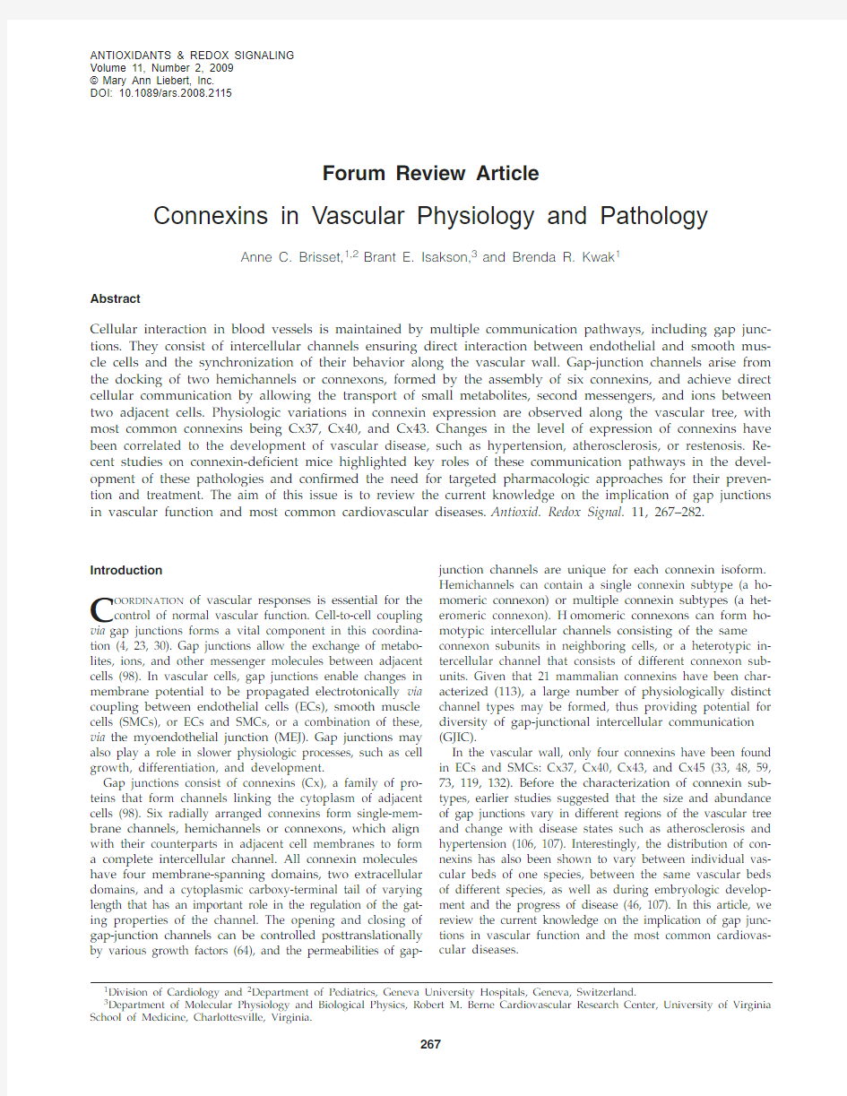
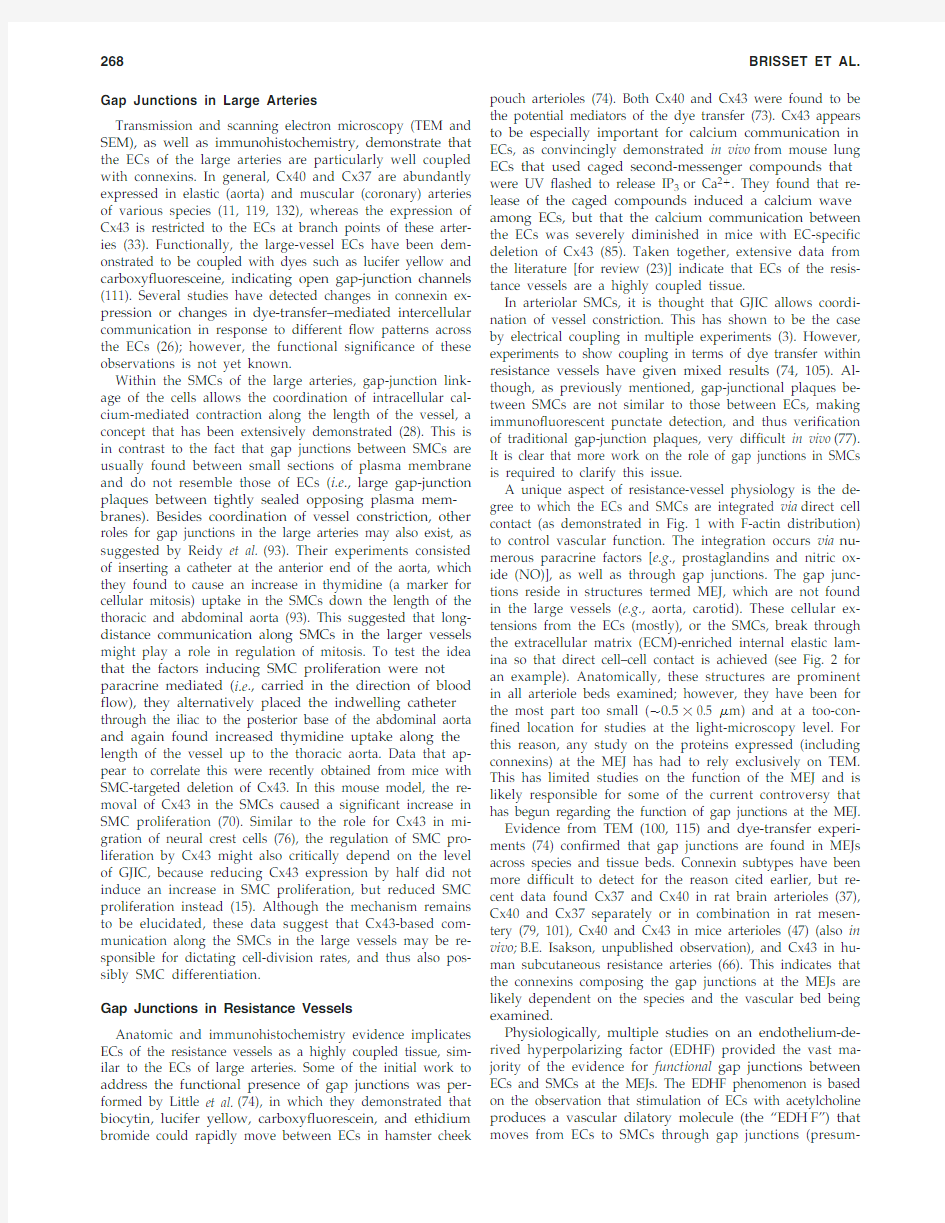
A unique aspect of resistance-vessel physiology is the de-gree to which the ECs and SMCs are integrated via direct cell contact (as demonstrated in Fig. 1 with F-actin distribution) to control vascular function. The integration occurs via nu-merous paracrine factors [e.g., prostaglandins and nitric ox-ide (NO)], as well as through gap junctions. The gap junc-tions reside in structures termed MEJ, which are not found in the large vessels (e.g., aorta, carotid). These cellular ex-tensions from the ECs (mostly), or the SMCs, break through the extracellular matrix (ECM)-enriched internal elastic lam-ina so that direct cell–cell contact is achieved (see Fig. 2 for an example). Anatomically, these structures are prominent in all arteriole beds examined; however, they have been for the most part too small (?0.5?0.5 ?m) and at a too-con-fined location for studies at the light-microscopy level. For this reason, any study on the proteins expressed (including connexins) at the MEJ has had to rely exclusively on TEM. This has limited studies on the function of the MEJ and is likely responsible for some of the current controversy that has begun regarding the function of gap junctions at the MEJ. Evidence from TEM (100, 115) and dye-transfer experi-ments (74) confirmed that gap junctions are found in MEJs across species and tissue beds. Connexin subtypes have been more difficult to detect for the reason cited earlier, but re-cent data found Cx37 and Cx40 in rat brain arterioles (37), Cx40 and Cx37 separately or in combination in rat mesen-tery (79, 101), Cx40 and Cx43 in mice arterioles (47) (also in vivo;B.E. Isakson, unpublished observation), and Cx43 in hu-man subcutaneous resistance arteries (66). This indicates that the connexins composing the gap junctions at the MEJs are likely dependent on the species and the vascular bed being examined.
Physiologically, multiple studies on an endothelium-de-rived hyperpolarizing factor (EDHF) provided the vast ma-jority of the evidence for functional gap junctions between ECs and SMCs at the MEJs. The EDHF phenomenon is based on the observation that stimulation of ECs with acetylcholine produces a vascular dilatory molecule (the “EDH F”) that moves from ECs to SMCs through gap junctions (presum-
BRISSET ET AL.
268
ably), even when inhibitors for prostaglandins and NO are present. This process appears to be dependent on an increase in EC [Ca2?]i, but not necessarily a change in EC membrane potential (82, 81). Surprisingly, as might be expected for gap junction–based communication after an increase in [Ca2?]i (8), the SMCs do not display a corresponding increase in [Ca2?]i(which would cause dilation), but instead respond by dilating. Controversial information still remains as to the nature of the EDH F signal. It has been demonstrated that Ca2?“sparks” can induce SMC hyperpolarization and re-laxation (83), and although a Ca2?spark in SMCs in response to EC has not been demonstrated, this could possibly occur if the current moves from EC through gap junctions to SMCs (81), or possibly Ca2?itself (50), activating ryanodine recep-tors, the characteristic receptors of a Ca2?spark. Multiple factors have also been implicated, including most recently hydrogen peroxide (H2O2) (75). The complexity of the MEJ environment has made this work difficult and limited with standard in vivo techniques and, as such, has required inno-vative new techniques, which have only recently begun to show some interesting results (79).
Although the previously described early EDH F work demonstrated interactions between ECs and SMCs, the di-rect evidence for gap junctions in EDHF comes from a series of articles demonstrating that application of connexin-mimetic peptides selectively blocked SMC response (16, 20, 54). This observation is based on data indicating that pep-tides derived from the extracellular loop of Cx37, Cx40, and Cx43 could specifically and reversibly block gap junctions at the MEJ. With another technique, ECs were loaded in vivo by a pinocytotic method (47) by using hypotonic and hy-pertonic solutions that introduced a Cx40 antibody into the cells (79). The authors demonstrated an EDHF block, possi-bly indicating that the Cx40 antibody in the ECs blocked gap junctions at the MEJ. Both observations are consistent with the idea that gap junctions play a role in EDH F coupling. These data correlate strongly with observations of a dimin-ishing EDHF response as the artery size increases (100, 108), a pattern that is mimicked by the appearance of the MEJs in arteries (4, 94).
Evidence for functional gap junctions at the MEJ is not al-ways readily apparent. Recent experiments have questioned coupling at the MEJ in the conducted response (12, 110). In particular, Siegl et al.(110) demonstrated that microinjection of carboxyfluorescein dye in either ECs or SMCs failed to couple together the opposing cell type (110). It is possible that carboxyfluorescein may be impermeable to the gap junc-tions at the MEJ [due to dye selective permeability through connexin isoforms (27)], as they were also unable to see dye transfer among ECs, a highly coupled cell type (110). How-ever, the authors were also unable to obtain similar mem-brane potentials in ECs and SMCs, again indicating that open gap-junction channels at the MEJ were not readily apparent (110). Because no coupling was found either in ECs, SMCs, or between ECs and SMCs in the vessels tested, and con-
VASCULAR CONNEXINS
269
FIG. 1.Focal planes of in situ m ouse crem aster arteriole. Mouse cremaster arterioles were stained with phalloidin to mark actin in endothelium and smooth muscle and were observed under an Olympus Fluoview confocal microscope in different focal planes. (A) SMCs overlay the arteriole, with the arrow indicating orientation. (B) In the same field of view as A, ECs can be seen running perpendicular to the orientation of SMCs. (C) Another focal plane from the same arteriole in A and B, where the image is viewed transversely. In this orientation, actin can be seen linking the ECs and SMCs, pre-sumably at the MEJ (*). With these different focal planes, one is able to detect with certainty the connexin localization within each cell type. Bar is 15 ?m.
FIG. 2.Myoendothelial junction in mouse cremaster ar-
https://www.doczj.com/doc/2c14077014.html,ually, the use of electron microscopy is required
to identify the MEJ in arterioles. In this electron micrograph,
an EC extension is seen breaking through the internal elas-
tic lamina and making contact with an SMC at the MEJ (*).
Bar is 1 ?m.
nexins are readily expressed in these particular vessels (77), it is very likely these gap-junction channels were present, but closed. This process can occur because connexin proteins are highly regulated to open or close a gap-junction channel [e.g., in response to phosphorylation (63)]; indeed, closed gap junction channels have been hypothesized to be an impor-tant part of regulating cellular communication (10). It is clear that more work on this concept is required.
Last, few data have focused on SMC-to-EC communica-tion through the MEJ, although three different model sys-tems (two in vivo and one in vitro) have demonstrated that an increase in SMC [Ca2?]i causes an increase in EC [Ca2?]i, which is mediated by gap junctions at the MEJ (25, 50, 62). Two of these models independently demonstrated that in-ositol 1,4,5-trisphosphate (IP3) moves from SMCs to ECs through gap junctions to induce the change in EC [Ca2?]i (50, 62), which subsequently induces NO release (25). This may prove to be an important mechanism in which resis-tance vessels are able to control vasoconstriction originating in the SMCs.
Problems in Connexin Research
Studies of the physiologic role of gap junctions in the vas-culature have been difficult to obtain with confidence be-cause the mechanism of action from almost all known phar-macologic gap-junction inhibitors is not known, and they have multiple nonspecific effects [e.g., see discussion in (30)]. Inhibitors like oleamide and halothane are very potent, but they have an inherent problem of closing large numbers of other membrane channels. H owever, even inhibitors that have been described to have, when used at low dose, mini-mal nonspecific effects, like heptanol (114), carbenoxolone (99), glycyrrhetinic acid (9), and connexin mimetic peptides (123), have had problems associated with their use. For ex-ample, after application of carbenoxolone to bovine aortic ECs, Cx43 mRNA expression was reported to be 2 to 3 times higher, corresponding with increased Cx43 protein expres-sion at the plasma membrane, indicative of a negative-feed-back loop with gap junctions (99). Pharmacologically then, it is clear that confirmation of results with multiple gap-junc-tion inhibitors should always be used.
Because connexin mRNA and protein do not normally cor-relate (1), detection of protein expression with connexin an-tibodies becomes vital for experimental purposes. The speci-ficity of connexin antibodies, immunocytochemistry in particular, continues to produce conflicting data. One way to solve this problem has been the use of cell lines that do normally not express connexin proteins in appreciable lev-els. For this reason, use of HeLa cells transfected with con-nexin plasmids has been considered a good method for test-ing antibody specificity; however, the plasmids generated are usually derived against a human sequence, rather than the mouse or rat sequence, which is generally used experi-mentally. Concurrent testing of the antibodies on tissue from connexin-knockout animals (48) should help alleviate some of these problems. Last, issues regarding antibody access or inaccessibility due to different protein confirmation states or interaction with other proteins have rarely been addressed but may be of particular importance (35, 128). Information regarding the role of gap junctions in vascu-lar physiology has evolved from studies on knockout mice,demonstrating the importance of these animals (29, 69). However, connexin-knockout mice have also been reported to have important drawbacks resulting from compensatory effects. In particular, the Cx40?/?mouse has been demon-strated to have decreased expression of Cx37 as well as cel-lular reorganization of Cx43 (48, 111), making physiological interpretation from experiments in these animals very diffi-cult. Therefore, it becomes increasingly clear that we must further develop other methods whereby gap-junction phys-iology could be studied (e.g., antisense or lentiviral shRNA delivery) that may have more minimal nonspecific effects. Gap Junctions in Hypertension
Elevated blood pressure is a common health problem with widespread and sometimes devastating consequences. Al-though hypertension remains often asymptomatic until late in its course, it is one of the most important risk factors for coronary heart disease and cerebrovascular accidents. Fi-nally, it may lead to cardiac hypertrophy with heart failure, aortic dissection, and renal failure. The detrimental effects of elevated blood pressure increase continuously as the pres-sure increases (57).
The magnitude of arterial pressure depends on two he-modynamic variables: cardiac output and total peripheral resistance. Cardiac output is influenced by blood volume; consequently body sodium homeostasis is central to blood-pressure regulation. Total peripheral resistance is predomi-nantly determined at the level of the arterioles and depends effectively on its lumen size. Primary hypertension accounts for ?90–95% of the cases; this essential hypertension is thought to result from an interaction of genetic and envi-ronmental factors that affect cardiac output, peripheral re-sistance, or both (57). The remaining 5–10% of hypertension cases are mostly secondary to renal disease or to atheroscle-rotic narrowing of the renal artery, both affecting the renin–angiotensin system and sodium homeostasis. Hypertension and connexin expression
A large number of studies report changes in Cx expres-sion in spontaneous hypertensive rats (SH Rs) or after in-duction of hypertension in animal models of the disease. Un-fortunately, these studies appear not always consistent, possibly due to intrinsic differences in the experimental models used, and sometimes different results have been ob-tained by using the same animal model. For example, ob-servations on changes in connexins in SHRs are mixed: Cx40 and Cx37 are consistently reduced in ECs, but both increased and decreased arterial Cx43 has been reported in these rats (42, 53, 97, 96, 136). Moreover, in certain arteries (mesenteric), no differences in Cx43 mRNA could be observed between SH Rs and their control Wistar–Kyoto rats, questioning the correlation between gap junctions and hypertension (117). Interestingly and possibly more relevant, normalization of blood pressure in the SH Rs by using an angiotensin-con-verting enzyme inhibitor or candesartan restores endothelial connexin expression to normal in parallel with the normal-ization of blood pressure (53, 97).
In general, the increased expression of Cx40 and Cx43 is observed on induction of renal hypertension, by using the rat DOCA-salt, and the two-kidney, one-clip Goldblatt (2K1C) procedure (38, 39, 127). With an elegant combination
BRISSET ET AL.
270
of different models, it was hypothesized that Cx43 expres-sion might be sensitive to hemodynamic changes such as an increase in intravascular pressure, rather than reacting to renin, because Cx43 is elevated in the SMCs of the aortas in both high-salt and 2K1C models of hypertension, models that display opposite changes in plasma renin (41). In con-trast to the renovascular models, hypertension induced by inhibition of NO synthase is associated with a decreased Cx43 expression (40, 42, 127, 136). In addition, endothelial Cx37, but not Cx40, was reduced in these models (136). In-terestingly, the changes in endothelial Cx43 and Cx37 ex-pression in N(?)-nitro-L-arginine methyl ester–induced hy-pertension were reversed by treatment with carvedilol, an adrenergic blocker (136).
Cx40-deficient mice and hypertension
Recent studies indicate that Cx40 plays an important role in blood-pressure regulation. Deletion of the Cx40 gene in mice results in a marked, sustained hypertension (21, 22). This deletion was associated with segmental constrictions and irregular vasomotion in small arterioles, suggesting a di-rect link between connexins, peripheral resistance, and blood pressure (23, 22). However, a series of recent reports suggest a strong association between renin secretion and Cx40 ex-pression (56, 58, 122). In Cx40-deficient mice, both synthesis and plasma levels of renin secretion are increased. Moreover, the investigators observed increased number of renin-se-creting cells as well as altered distribution of the renin-se-creting cells in the afferent arterioles. Importantly, they showed that the major dysfunction in Cx40-deficient mice appeared to depend on local blood flow–induced signalling in the afferent arteriole, a concept that was elegantly con-firmed in perfused kidney by using a gap-junction blocker (122).
Cx40 genetic polymorphism is associated
with hypertension
Two closely linked polymorphisms in the promoter region of the Cx40 gene have been associated with atria-specific ar-rhythmias (31, 36). Recently, the same research group has in-vestigated whether (a) these polymorphic variants are asso-ciated with hypertension, and (b) they interact with blood pressure in healthy individuals. They found a significant contribution of the minor Cx40 allele or genotype (S44AA/ R71GG) to the risk of hypertension in men. In addition, they observed in the healthy control population a significant ef-fect of Cx40 genotype and sex on systolic blood pressure (32). These findings not only confirm a strong association between Cx40 and hypertension but also identify these polymor-phisms as a risk factor for the disease.
Gap Junctions in Atherosclerosis
Cardiovascular diseases represent the leading cause of death in developed countries and are increasing worryingly in developing countries as the access to food becomes eas-ier. Atherosclerosis is the most important cardiovascular dis-ease, with life-threatening complications such as myocardial infarction, cerebral infarction, and aortic aneurysm. It de-velops silently over years and usually becomes symptomatic after the fourth decade. H ypercholesterolemia, obesity,smoking, hypertension, diabetes, and infection by microor-ganisms are classic risk factors that promote atherosclerosis by triggering inflammation or endothelial injury or both. This chronic immunoinflammatory disease progressively narrows the lumen of medium and large arteries by accu-mulation of lipids, monocyte-derived macrophages, which recruit T lymphocytes, and migration and proliferation of SMCs in the vascular wall (34, 43, 57, 71, 95).
From endothelial injury to atherosclerotic plaque rupture Traditional risk factors of atherosclerosis have been shown to trigger endothelial activation. By cumulating these dele-terious signals, activated endothelium becomes dysfunc-tional, as indicated by the loss of its antiadhesive properties toward leukocytes, and increased permeability to circulating lipids. Antiadhesive and antiinflammatory genes are shut down, and many proinflammatory genes are upregulated in activated endothelium. In particular, new expression of ad-hesion molecules and concomitant release of chemoattrac-tants and cytokines favor the rolling and transmigration of monocytes and T lymphocytes, which accumulate in the subendothelial compartment. Monocytes proliferate and progressively mature into macrophages, which express scavenger receptors to take up lipids. This process becomes unremitting when they turn into foam cells, unable to clear oxidized low-density lipoproteins (LDLs) yielded by free radicals and accumulated in their cytoplasm. Oxidized LDLs are highly antigenic, potent chemoattractants, thus enhanc-ing the inflammatory response. This initial step of athero-sclerotic process, which is called “fatty streak,” occurs more frequently in areas of altered shear stress [i.e.,branch points of arteries (Fig. 3)], where inappropriate flow decreases genes associated with differentiated ECs and SMCs (19). As the inflammatory response progresses, growth factors and cytokines produced by the dysfunctional endothelium and monocytes/macrophages induce the migration of SMCs from the media to the intima. Once they have reached the inner part of the vessel, they proliferate, undergo phenotypic modulation, and produce ECM, thus constituting to the so-called “fibrous cap” that surrounds the atherosclerotic plaque (Fig. 4). Both macrophage foam cells and SMCs are subjected to apoptosis and therefore release their lipidic con-tent in the necrotic core of advanced atherosclerotic plaques (Fig. 5). Decreased numbers of SMCs and further breakdown of the ECM by metalloproteinases, mostly released from macrophage foam cells, weakens the fibrous cap and might induce plaque rupture (121). This will then bring the highly thrombogenic plaque content in direct contact with the bloodstream and will trigger thrombosis at the site of the le-sion, resulting in occlusion of the vessel, impairing blood flow, and causing ischemia of the tissue.
Connexin expression and atherogenesis
The presence of newly recruited inflammatory cells in the vicinity of ECs and SMCs modify the normal vascular envi-ronment; this cellular interplay is tightly influenced by paracrine release of cytokines, chemokines, and growth fac-tors. During the past 15 years, evidence has been seen of the contribution of direct cell–cell communication involving con-nexins in the development of atherosclerosis.
VASCULAR CONNEXINS271
Important changes in the pattern of vascular Cx expres-sion in human vessels and in animal models of atheroscle-rosis have been reported. The expression of Cx evolves as the disease progresses (Figs. 3–5) from a “healthy” vessel, to a dysfunctional endothelium, with early fatty streak to a
“prone-to-rupture” vessel. As the cellularity of vulnerable plaque is very poor, the expression of Cx is reduced, and one can speculate that GJIC will be decreased, with the remain-ing cells being very sparse. Alterations of Cx expression in the different vascular cells might have several consequences
BRISSET ET AL.
272FIG. 4.Expression of connexins in early atheroma.Under the influence of cytokines and growth factors secreted by the dysfunctional endothelium and infiltrated inflammatory cells, medial SMCs migrate to the intima, where they proliferate.Lipids start to accumulate outside the cells and within ECM secreted by intimal SMCs. Cx43 expression is upregulated in intimal SMC (ISMC), especially in the developing fibrous cap. ECs continue to express both Cx37 and Cx40, and macro-
phage foam cells still express Cx37.
FIG. 3.Expression of connexins at the arterial bifurcation.In healthy arteries, under a laminar flow, ECs express both Cx37and Cx40. Medial SMCs (mSMC) display Cx43. Circulating monocytes express low levels of Cx37. Under low or oscillatory shear stress, ECs become dysfunctional and express Cx43. Monocytes and T lymphocytes are recruited by the dysfunctional endothelium and migrate over the EC barrier into the subendothelium. Macrophages (indicated as MO) start to express higher levels of Cx37. The dysfunctional endothelium slowly progresses over years to a fatty streak characterized by accumulation of lipids in the macrophages, which hence transform to macrophages foam cells with continued expression of Cx37.
on the molecular messages that are exchanged between these cells. The loss of one Cx between two adjacent cells would then hamper the passage of a particular type of signaling molecules, crucial to cell function.
Cx43 expression is normally restricted to medial SMCs of healthy vessels. Polacek and Davies (89) were the first to ob-serve a concomitant expression of Cx43 mRNA in macro-phage foam cells from human carotid arteries, even if Cx43 was not expressed by their precursor peripheral blood mono-cytes and the loss of Cx43 expression in intimal SMCs in ath-erosclerotic plaques (90). Later, a coordinated scenario was demonstrated, with a first upregulation of Cx43, followed by the previously described loss of Cx43 by medial SMCs un-derlying atherosclerotic plaques (6). These dynamic changes were further confirmed in a mouse model of atherosclerosis (59). Cx43 appears in ECs of the shoulder regions of the ath-erosclerotic lesion (59).
In a healthy vessel, Cx37 is expressed by ECs and circu-lating monocytes (59, 130). Once monocytes start to infiltrate into the vessel wall over a dysfunctional endothelium and progressively transform to macrophages, they exhibit in-creased levels of Cx37, which is maintained in fatty streaks. In advanced atheromatous plaques, Cx37 is downregulated in ECs and starts to be expressed by medial SMCs beneath the atherosclerotic lesion.
Cx40 expression is restricted to ECs of healthy vessels. Its pattern of expression is not affected until advanced ath-erosclerotic plaque forms; it is then strongly downregu-lated. So far, no Cx expression has been found in ECs cov-ering advanced atherosclerotic lesions. As Cx has been implicated in tissue repair, the understanding of vascular lesions and arterial remodeling might benefit from stud-ies on the expression of Cx in adventitia, fibroblasts, and perivascular tissues.Local factors in favor of atherosclerotic development modify Cx expression
Disturbed hemodynamic forces at particular sites of the vascular tree that are of crucial importance in atherogenesis are known to affect the expression of vascular Cx or GJIC between vascular cells (17, 19, 24, 52, 61). Low shear stress or complex flow disturbances affect both EC and SMC be-havior, directly by modifications of pressures exerted on cells and mechanical forces between cells, or indirectly by affect-ing the local soluble factors. In other words, while stimula-tion of mechanical sensors is disturbed, antiatherogenic tran-scription factors and antiadhesive substances such as NO are shut down. In contrast, proatherogenic transcription factors, adhesive proteins, and proinflammatory cytokines are en-hanced (45). As a result, inflammatory cells more easily in-filtrate these regions. Both mechanical strain and fluid shear stress have been shown to upregulate Cx43 expression in ECs and SMCs (17, 61). Moreover, disturbed flows induce the dis-ruption of GJIC between ECs and upregulation and disor-ganization of Cx43 (24). Recent data demonstrate that flow-induced GJIC between human ECs is primarily mediated by Cx40, with a lesser contribution of Cx37 and Cx43 (26).
In addition, in vitro studies have shown that cytokines, growth factors, or substances expressed in the vicinity of vas-cular cells during development of atherosclerotic lesions modify Cx expression. Thus, TNF-?altered the Cx expres-sion pattern and reduced GJIC in human ECs (120). Treat-ment with thrombin led to upregulation of Cx43 associated with increased synthetic activity, yet also enhanced contrac-tile differentiation of aortic SMCs, and only growth arrest by contact inhibition led to progressive reduction in Cx43 ex-pression (80). Surprisingly, PDGF-BB, known to modulate the SMC phenotype in vitro(44), did not affect Cx43 expres-
VASCULAR CONNEXINS
273
FIG. 5.Expression of connexins in advanced atheroma.The fibrous cap completely covering the lesion area is com-posed of intimal SMCs and ECM. Extracellular lipids including cholesterol crystals (easily recognized in histology) are accumulated within the necrotic core composed of debris of apoptotic cells and degraded ECM. Macrophage foam cells close to the necrotic core express both Cx37 and Cx43. ECs still express Cx37 and Cx40 with two exceptions: ECs cov-ering the lesion stop to express connexins, and those of shoulder regions start to express Cx43. In the fibrous cap, ex-pression of Cx43 in intimal SMCs decreases as the lesion becomes more complicated. Medial SMCs beneath the lesion start to express Cx37.
sion in this study. Cx43 expression was reversibly increased in hypoxic cultures of rat aortic SMCs, which exhibited en-hanced intercellular communication in fluorescence recov-ery after photobleaching (FRAP) experiments (18). Interest-ingly, high glucose levels as observed in diabetes (a pathology associated with high risk of cardiovascular dis-eases) reduce Cx43-mediated microvascular endothelial cell communication (102). Moreover, as it releases high amounts of growth factors and cytokines, perivascular adipose tissue, which expresses Cx43, might contribute to the development and/or progression of atherosclerosis through multiple mechanisms, such as proliferative, proinflammatory, pro-thrombotic, and oxidant effects [for a review, see (55)]. Whether Cx43 is involved in the atherogenic effect of perivascular adipose tissue remains to be determined. Accumulation and oxidative modification of LDLs is a key component to the development and progression of athero-sclerosis (5, 68, 112). The oxidation of LDL gives rise to min-imally oxidized LDL (78). Some of these oxidized phospho-lipids have been demonstrated to have extensive biologic activity, in particular the phospholipid derivatives of oxi-dized 1-palmitoyl-2-arachidonoyl-sn-3-glycero-phosphoryl-choline (OxPAPC). For example, one of the components of OxPAPC, 1-palmitoyl-2-oxovaleroyl-sn-glycero-3-phospho-rylcholine (POVPC) can activate protein kinase A (PKA), whereas 1-palmitoyl-2-glutaroyl-sn-glycero-3-phosphoryl-choline (PGPC) activates PKC (67, 125, 126). This presents a fascinating possibility for differential regulation of proteins (e.g., connexins), especially in terms of phosphorylation. Thus far, studies have focused exclusively on inflammatory effects of oxidized lipids on ECs, SMCs, and leukocyte func-tion (78), with few studies focusing on the effects of oxidized phospholipids on GJIC.
The functional consequences of changes in connexin pro-tein expression during atherosclerosis, coupled with the ex-tensive evidence for OxPAPC involvement in the develop-ment of atherosclerosis, gives rise to the possibility of an interaction between OxPAPC and connexins. Recent work demonstrated that all of the vascular connexins in large ves-sels are altered in response to direct application of OxPAPC to mouse carotid vessels, or cultures of ECs and SMCs (49), mimicking very similar although not exact patterns of con-nexin expression demonstrated in atherosclerotic plaques (59). The key difference in connexin expression is that Cx43 was upregulated in SMCs in response to the OxPAPC, whereas in advanced mouse atherosclerosis, Cx43 is largely absent from the intimal SMCs. Because OxPAPC is a mix-ture of oxidized phospholipids, it is possible that the multi-ple components with biologic activity may be having differ-ential effects, and so experiments using OxPAPC species will be necessary to dissect which is capable of inducing changes in a particular connexin subtype, in either ECs or SMCs. Functionally, it was noted that after OxPAPC application, biocytin dye transfer was no longer capable of passing be-tween cells, indicating that the gap junctions were closed by OxPAPC, a function attributed to Cx43 phosphorylation (49). Application of OxPAPC and its individual species has re-cently been shown to induce SMC differentiation (86). This fits closely to the observation made by several laboratories that a correlation exists between connexins and SMC differ-entiation in atherosclerosis (80, 91). Taken together, these data are strong evidence for a direct interaction between Ox-PAPC and connexin in regulation not only of connexin pro-tein expression, but of gap-junction function as well. Lessons from animal models
Two well-characterized mouse models are used to study the development of atherosclerosis (13): the low-density lipoprotein–receptor knockout mice (LDLR?/?) and the apolipoprotein E (ApoE?/?) knockout mice, which have in common the deletion of genes participating in the transport of lipoproteins. Mutations in the corresponding genes in hu-mans result in familial hypercholesterolemia, a risk factor for atherosclerotic development, which is frequently associated with early myocardial infarction.
Deletion of the LDLR impairs the hepatic uptake of LDL and recirculation of cholesterol. ApoE is also necessary for the uptake of lipoproteins. Thus, both LDLR?/?and ApoE?/?mice have high cholesterol levels, even more pro-nounced in ApoE?/?mice. It is, however, necessary to feed LDLR?/?mice a high-cholesterol diet to induce significant hypercholesterolemia and consecutive atherosclerotic le-sions (51), whereas ApoE?/?mice spontaneously develop atherosclerotic lesions, which become even more important with a high-cholesterol diet (88, 138, 137). The time course and distribution of plaques in the arterial tree are repro-ducible and present a great similarity to the human athero-sclerotic lesions. The main limitation of these mouse models is that they lack the spontaneous rupture of plaques. Other animal models are therefore being used.
The Watanabe heritable hyperlipidemic (WH H L) rabbit produces a mutant receptor for plasma LDL that is not trans-ported to the cell surface at a normal rate. As it partly mim-ics some human atherosclerotic plaque ruptures, it is also a frequently used animal model (2, 109). More recently, ath-erosclerosis and restenosis have been studied in pigs (44), which are closest to humans in terms of size and anatomy, and spontaneously develop atherosclerotic lesions in the ab-dominal aorta. Interestingly, natural pig strains produce mu-tants for lipoproteins.
As they are genetically well defined, mice can be easily in-terbred with other knockout mice lacking genes of the Cx family. We have demonstrated that ApoE?/?mice lacking Cx37 and fed a high-cholesterol diet develop accelerated ath-erosclerotic lesions in both the thoracic–abdominal aortas and aortic sinuses (130). We further showed that Cx37 hemichannels borne by monocytes/macrophages limit their adhesion and recruitment to the subendothelium by an au-tocrine release of ATP, thus explaining the protection this protein confers against atherosclerosis (130).
Cx43 is important in cardiac development and function. Cx43 plays a role in the looping of the ascending limb of the heart tube and the development of the right ventricle and the outflow tract, and Cx43-knockout mice die at birth of se-vere cardiac malformations (92). H eterozygous Cx43-defi-cient mice, expressing 50% of the Cx43 normal level, have thus been crossbred with LDLR?/?mice. Cx43?/?LDLR?/?and Cx43?/?LDLR?/?mice have comparable elevated serum cholesterol and triglyceride profiles, leukocyte counts, body weight, and respond similarly to a high-cholesterol diet. Comparison between these two groups of mice fed a high-cholesterol diet allowed us to observe that atheroscle-rosis progression was reduced by 50% in both the aortic roots
BRISSET ET AL.
274
and the thoracic–abdominal aorta of Cx43?/?LDLR?/?mice. Atherosclerotic lesions were less complex, as attested by a reduction in the number of inflammatory cells and a thicker fibrous cap with a high-collagen content and large numbers of SMCs, which resulted in a more stable plaque phenotype [i.e.,less susceptible to rupture (60, 129)]. These data suggested that, in contrast to Cx37, Cx43 has an athero-genic effect.
As described earlier, Cx40-knockout mice have also been generated, but present characteristics of hypertension (21, 22, 122), an independent risk factor for atherosclerosis. There-fore, we recently established a mouse model with selective deletion of Cx40 in endothelium by using the Cre-LoxP sys-tem under the control of the Tie2 promotor. The endothe-lium-specific deletion of Cx40 did not affect mean arterial pressure or heart rate in mice (14). Preliminary data also in-dicate that the surface of atherosclerotic lesions in ApoE?/?mice lacking endothelial Cx40 is larger as compared with that in control animals fed a high-cholesterol diet, suggest-ing that Cx40 has atheroprotective potential (14). The loss of Cx40 in the endothelium covering early atherosclerotic le-sions might thus further facilitate the development of ath-erosclerosis. The mechanism by which Cx40 is atheropro-tective should be explored.
Cx37 genetic polymorphism influences the risk of cardiovascular disease
Recently, research demonstrated an association of a poly-morphism in the Cx37 gene, a protein normally expressed in ECs but also found in monocytes and macrophages, with ar-terial stenosis and myocardial infarction in humans (7, 72, 130, 131, 134). The Cx37 C1019T polymorphism results in a nonconservative amino acid shift from a proline to a serine at codon 319, in the regulatory intracellular carboxy termi-nus of Cx37 protein. The human Cx37 genotype was then also shown to predict survival after an acute coronary syn-drome (65). In this context, the observation that expression of Cx37-319S or Cx37-319P by transfection of a human mac-rophage cell line revealed differential adhesiveness to sub-strates is of particular importance (130). This may be caused by increased permeability of the Cx37-319P hemichannels for ATP, thus providing a potential mechanism by which the Cx37-1019T variant protects against atherosclerosis.
Drugs influencing the development of
atherosclerotic plaques
The 3-hydroxy-3-methylglutaryl-coenzyme A (H MG-CoA) reductase inhibitors, the so-called statins, have been proven to be highly effective in the management of hyperc-holesterolemia and in the prevention of atherosclerotic vas-cular disease. As inhibitors of HMG-CoA reductase, the pri-mary pharmacodynamic effect of statins is to inhibit the synthesis of cholesterol by the liver. The reduction of cho-lesterol in hepatocytes leads to the compensatory increase of hepatic LDLRs, followed by an increase in cholesterol up-take by the liver and a reduction of circulating lipoproteins. The beneficial effects of statins were considered to depend initially on the reduction of LDL cholesterol, followed by the subsequent regression of atherosclerotic lesions. H owever, the observation that lipid lowering by means other than statins, such as ileal bypass surgery, required markedly more time to manifest clinical benefits pointed to additional effects from this class of drugs. Many data support more direct cho-lesterol-independent beneficial effects of statins on athero-sclerosis or plaque rupture (103). They include increased syn-thesis of NO, inhibition of free radical release, decreased synthesis of endothelin-1, inhibition of LDL cholesterol oxi-dation, increased availability of endothelial progenitor cells, reduced number and activity of inflammatory cells in ather-osclerotic lesions, increased SMC proliferation and differen-tiation, reduced production of metalloproteinases, and inhi-bition of platelet adhesion/aggregation. As more of such pleiotropic effects are discovered, increasing understanding exists on how statins mediate their actions.
Chemically, statins competitively inhibit H MG-CoA re-ductase, thereby reducing the availability of L-mevalonate and consequently the biosynthesis of cholesterol as well as prenylated proteins (Fig. 6A). Prenylated proteins or iso-prenoids, such as farnesylpyrophosphate (FPP) and ger-anylgeranylpyrophosphate (GGPP), are important for the posttranslational modification (prenylation) of proteins in-volved in normal cell functions. More recently, statins were also hypothesized to disrupt cholesterol and sphingolipid membrane microdomains, so-called lipid rafts, which are es-sential platforms for the initiation of signal transduction. Only a few studies have examined the effects of statins on connexins. The expression of Cx43 was examined in vitro in primary human vascular cells isolated from saphenous veins in the presence of three different statins, the lipophilic sim-vastatin and atorvastatin and the hydrophilic pravastatin. Each of the statins effectively reduced the expression of Cx43 in ECs and in SMCs in a dose-dependent manner, and this effect was abolished in the presence of L-mevalonate (60). Cx43 was typically immunolocalized along the cell mem-branes of contacting SMCs. Simvastatin treatment strongly reduced the amount of Cx43 immunolabeling; however, the subcellular localization of Cx43 was not affected by the statin. Mechanistically, the statin-induced reduction in Cx43 in SMCs seems to involve geranylgeranylated proteins. As shown in Fig. 6B, the statin-induced reduction in Cx43 ex-pression was reversed by 400 ?M mevalonate and the iso-prenoid intermediate GGPP (10 ?M), but not by 10 ?M FPP. Furthermore, 10 ?M geranylgeranyl transferase inhibitor type I (GGTI-286) reduced Cx43 expression, an effect simi-lar to the one observed with statins, whereas the farnesyl transferase inhibitor FTI-277 (3 ?M) was without effect (Fig. 6B).
In vivo studies on LDLR?/?mice treated orally with pravastatin displayed a more stable plaque phenotype [i.e., atheromas contained fewer inflammatory cells and dis-played thicker fibrous caps, which had more interstitial col-lagen and concentric layers of SMCs (60)]. As described be-fore, a similar plaque phenotype was observed in atheromas of Cx43?/?LDLR?/?mice. Interestingly, immunostaining on atheromas in pravastatin-treated mice showed reduced Cx43 expression throughout the entire atherosclerotic lesion. This decreased expression of Cx43 in the statin-treated mice, as compared with control animals, was confirmed by West-ern blotting of their aorta protein extracts. In another study, it was shown that mouse aortic endothelial connexins and gap junctions were downregulated during long-term hyper-lipidemia. Short-term treatment with simvastatin led to re-covery of Cx37 expression but not Cx40 expression (135).
VASCULAR CONNEXINS275
FIG. 6.(A) Metabolic pathway of mevalonate and cholesterol.The enzymatic reactions and their pharmacologic inhib-itors are indicated. (B, C) To investigate the metabolic pathway involved in statin-induced modulation of Cx43 expression in primary human saphenous vein SMCs (C), cells were stimulated for 24 h with simvastatin (S) alone or in combination with chemical compounds affecting the downstream signalling pathway of HMG-CoA, such as mevalonate (M; 400 ?M), farnesyl transferase inhibitor (FTI-277; 3 ?M), or geranylgeranyl transferase inhibitor type I (GGTI-286; 10 ?M), or with the isoprenoid intermediates FPP or GGPP (at 10 ?M each). A typical example of a Western blot for Cx43 is shown in (C).Bar
chart illustrates quantification of Cx43 expression (normalized to ?-actin) in five independent experiments.
Based on these two studies, the general hypothesis at pres-ent is that hyperlipidemia, a major risk factor of atheroscle-rosis, reduces Cx37 and Cx40 in ECs but induces Cx43 ex-pression in ECs and SMCs. Interestingly, these connexin expression profiles are reversed by statins (e.g ., increased Cx37 and Cx40 expression in ECs but decreased Cx43 in ECs and SMCs). Besides hyperlipidemia, smoking is another con-ventional risk factor of atherosclerosis, thus providing a ra-tionale for studying the effect of nicotine and/or statins on Cx43 expression and GJIC in human ECs (116). Expression of Cx43 was dose-dependently reduced by nicotine. This ef-fect was due to posttranscriptional modification, involving enhancement of Cx43 proteolysis, and was mediated via ac-tivation of acetylcholine receptors sensitive to nicotine (nAChRs). The effect of nicotine was attenuated by various statins, even in the presence of mevalonate, so thus through mechanisms outside the prenylation pathway. Interestingly,Cx43 has been found in lipid rafts (104). Whether the statin-induced nicotine effects on Cx43 involve lipid rafts remains to be investigated.
Gap Junctions in Restenosis
Development of atherosclerosis in coronary arteries re-sults in ischemic heart disease. In addition to coronary by-pass surgery, two common and effective mechanical treat-ments exist for coronary atherosclerosis: percutaneous transluminal coronary angioplasty (PTCA), which consists of disruption of the developing atherosclerotic plaque by means of balloon-catheter distention, and the implantation of a stent after PTCA. However, renarrowing of the vessels at the site of intervention hampers their long-term efficacy and the so-called “restenosis” occurs in 30–60% of the pa-tients after angioplasty (118), and 5–30% of the patients af-ter stent implantation (84). Restenosis mechanisms are not fully understood and are the subject of many investigations.Both intimal hyperplasia and arterial remodeling contribute to the reduction of arterial caliber. The parietal traumatism induced by the balloon distention triggers platelet aggrega-tion and the formation of a mural thrombus. In addition to the mechanical injury, the balloon-catheter also stretches the artery, thus inducing an exaggerated and accelerated re-sponse to injury, involving the recruitment and rapid infil-tration of inflammatory cells (57). Endothelialization repairs the lining of the damaged vasculature and is crucial to pre-vent thrombosis and restenosis. Both GJIC and purinergic signaling contribute to re-endothelialization of human EC by regulating both intra- and extracellular Ca 2?stores (139). The release of growth factors and proinflammatory cytokines by platelets, leukocytes, and SMCs, in addition to the loss of en-dothelial antiproliferating agents such as NO, modulates the phenotype of SMCs, shifting them from a quiescent and “contractile” phenotype to a “synthetic,” proliferating, and less differentiated one. SMCs start to proliferate 24–48 h after angioplasty and migrate toward the intima on the fourth day. They synthesize abundant ECM, which further contributes to intimal hyperplasia. The maximal intimal thickening is reached after 2–3 months, after which SMCs re-turn to a “contractile” phenotype, the wall is remodeled, and intimal thickening stops.
Stenting has become the treatment of choice because it pro-vides a mechanical support to the vessel and extends its
VASCULAR CONNEXINS
277
FIG. 7.Balloon-injury in m ice with reduced Cx43 ex-pression.Representative Van Gieson-Miller staining of carotid cryosections 14 days after balloon distention injury in Cx43?/?LDLR ?/?mouse (left ) and in a Cx43?/?LDLR ?/?mouse (right ). Neointimal hyperplasia is reduced in the Cx43?/?LDLR ?/?mouse as compared with its Cx43?/?LDLR ?/?mouse littermate control. Bar is 100 ?m.
internal diameter to avoid reocclusion of the vessel after angioplasty. H owever, the combined use of systemic an-tithrombotic drugs or the use of drug-eluting stents does not completely circumvent restenosis. Most important functions that should be fulfilled by “the ideal stent” are to decrease SMC proliferation to prevent in stent restenosis while in-creasing EC migration and proliferation to cover the stent,thus allowing the restoration of endothelial antiadhesive properties.
After ballooning in the rat carotid artery, Cx43 was up-regulated in intimal and medial SMCs concomitant with their activation and phenotypic modulation (133). Enhanced Cx43 expression in SMCs and macrophages of restenotic le-sions has been confirmed in different species (15, 87, 90, 124).Cx43?/?LDLR ?/?mice, expressing reduced levels of Cx43,displayed restricted intimal thickening after balloon-disten-tion injury in the carotid artery (Fig. 7), despite marked en-dothelial denudation and SMC activation, as compared with control littermate Cx43?/?LDLR ?/?mice. The reduction of Cx43 resulted in decreased macrophage infiltration and SMC migration and proliferation, associated with accelerated re-endothelialization, suggesting that Cx43 targeting could be a novel therapeutic strategy to prevent restenosis after PTCA or stent implantation.Concluding Remarks
The previous sections have provided support for an im-portant role of connexins in vascular physiology and a largely multifaceted role in the development of disease. So far, research has mostly concentrated on the roles of the three major vascular connexins; however, this does not preclude interesting expression patterns and regulatory functions for other gap-junction proteins (for example, Cx45 or Cx31.9) in blood vessels. After many years of research on the relation between gap junctions and longitudinal conduction along ar-terioles, we only recently have begun to understand the com-position of the MEJ. This has begun to open up an increas-ing understanding of the signaling from ECs to SMCs, and vice versa . Disruption or alteration of this intercellular sig-naling pathway may have considerable implications for de-velopment of disease as well. After a decade of investiga-
tions of changes in connexin expression in diseased vessels, we have been helped by the availability of connexin-defi-cient mice to recognize the roles of these proteins in disease development. Thus, Cx40 appeared critical in blood-pressure regulation at the level of the control of renin secretion. Cx37 has a protective role against atherosclerosis by controlling ATP-dependent monocyte adhesion. Monocyte adhesion is critical for many other immunoinflammatory disorders as well. In contrast to Cx37, Cx43 seems to possess atherogenic properties. Interestingly, the upregulation of this protein ob-served in most vascular disease states can be downregulated by drugs commonly used in clinics, such as statins and ad-renergic blockers. Last, increasing attention is seen in cardi-ology clinics to a person’s genetic makeup with respect to risk stratification and disease prevention. Polymorphisms in connexin genes are therefore of particular interest. Acknowledgments
We thank Cindy Wong, Christos Chadjichristos, Marc Chanson, Francois Mach, Angela Best, Brian Duling, and Richard K. H enley for helpful discussions. Our work has been supported by grants from the Swiss National Science Foundation (PPOOA-68883 and PPOOA-116897 to B.R.K.), the American Heart Association (0735167N to B.E.I.), and the United States National Institutes of H ealth (H L88554 to B.E.I.).
Abbreviations
ApoE, apolipoprotein E; Cx, connexin; ECM, extracellular matrix; ECs, endothelial cells; EDH F, endothelium-derived hyperpolarizing factor; FPP, farnesylpyrophosphate; FRAP, fluorescence recovery after photobleaching; FTI, farnesyl transferase inhibitor; GGPP, geranylgeranylpyrophosphate; GGTI, geranylgeranyl transferase inhibitor; GJIC, gap-junc-tional intercellular communication; H MG-CoA, 3-hydroxy-3-methylglutaryl-coenzyme A; H2O2, hydrogen peroxide; IP3, inositol triphosphate; LDL, low-density lipoprotein; LDLR, low-density lipoprotein receptor; MEJ, myoendothe-lial junctions; nAChRs, acetylcholine receptors sensitive to nicotine; NO, nitric oxide; OxPAPC, oxidized 1-palmitoyl-2-arachidonoyl-sn-3-glycero-phosphorylcholine; PGPC, 1-palmitoyl-2-glutaroyl-sn-glycero-3-phosphorylcholine; PKA, protein kinase A; PKC, protein kinase C; POVPC, 1-palmitoyl-2-oxovaleroyl-sn-glycero-3-phosphorylcholine; PTCA, percutaneous transluminal coronary angioplasty; SEM, scanning electron microscopy; SHR, spontaneously hy-pertensive rat; SMCs, smooth muscle cells; TEM, transmis-sion electron microscopy; WH H L, Watanabe heritable hy-perlipidemic.
References
1.Abraham V, Chou ML, DeBolt KM, and Koval M. Pheno-
typic control of gap junctional communication by cultured alveolar epithelial cells. Am J Physiol 276: L825–L834, 1999.
2.Aliev G, Castellani RJ, Petersen RB, Burnstock G, Perry G,
and Smith MA. Pathobiology of familial hypercholes-terolemic atherosclerosis. J Submicrosc Cytol Pathol 36: 225–240, 2004.
3.Beny J. Electrical coupling between smooth muscle cells
and endothelial cells in pig coronary arteries. Pflugers Arch 433: 364–367, 1997.
4.Beny JL. Information networks in the arterial wall. News
Physiol Sci 14: 68–73, 1999.
5.Berliner JA, Watson AD. A role for oxidized phospholipids
in atherosclerosis. N Engl J Med 353: 9–11, 2005.
6.Blackburn JP, Peters NS, Yeh HI, Rothery S, Green CR, and
Severs NJ. Upregulation of connexin43 gap junctions dur-ing early stages of human coronary atherosclerosis. Arte-rioscler Thromb Vasc Biol 15: 1219–1228, 1995.
7.Boerma M, Forsberg L, Van Zeijl L, Morgenstern R, De Faire
U, Lemne C, Erlinge D, Thulin T, Hong Y, and Cotgreave IA. A genetic polymorphism in connexin 37 as a prognos-tic marker for atherosclerotic plaque development. J Intern Med 246: 211–218, 1999.
8.Boitano S, Dirksen ER, and Sanderson MJ. Intercellular
propagation of calcium waves mediated by inositol trispho-sphate. Science 258: 292–295, 1992.
9.Boitano S, Evans WH. Connexin mimetic peptides re-
versibly inhibit Ca(2?) signaling through gap junctions in airway cells. Am J Physiol Lung Cell Mol Physiol 279: L623–L630, 2000.
10.Brink PR. Gap junctions in vascular smooth muscle. Acta
Physiol Scand 164: 349–356, 1998.
11.Bruzzone R, Haefliger JA, Gimlich RL, and Paul DL. Con-
nexin40, a component of gap junctions in vascular endo-thelium, is restricted in its ability to interact with other con-nexins. Mol Biol Cell 4: 7–20, 1993.
12.Budel S, Bartlett IS, and Segal SS. Homocellular conduction
along endothelium and smooth muscle of arterioles in ham-ster cheek pouch: unmasking an NO wave. Circ Res 93: 61–68, 2003.
13.Carmeliet P, Moons L, and Collen D. Mouse models of an-
giogenesis, arterial stenosis, atherosclerosis and hemosta-sis. Cardiovasc Res 39: 33, 1998.
14.Chadjichristos CE, Roth I, H oepfl B, van Veen TA,
Deutsch U, Van Kempen MJ, De Wit C, and Kwak BR.
Increased development of atherosclerosis in mice with endothelial-specific deletion of connexin40. Circulation 112: 1–142, 2005.
15.Chadjichristos CE, Matter CM, Roth I, Sutter E, Pelli G,
Luscher TF, Chanson M, and Kwak BR. Reduced con-nexin43 expression limits neointima formation after bal-loon distension injury in hypercholesterolemic mice. Cir-culation 113: 2835–2843, 2006.
16.Chaytor AT, Bakker LM, Edwards DH, and Griffith TM.
Connexin-mimetic peptides dissociate electrotonic EDHF-type signalling via myoendothelial and smooth muscle gap junctions in the rabbit iliac artery. Br J Pharmacol 144: 108–114, 2005.
17.Cowan DB, Lye SJ, and Langille BL. Regulation of vascu-
lar connexin43 gene expression by mechanical loads. Circ Res 82: 786–793, 1998.
18.Cowan DB, Jones M, Garcia LM, Noria S, del Nido PJ, and
McGowan FX Jr. Hypoxia and stretch regulate intercellu-lar communication in vascular smooth muscle cells through reactive oxygen species formation. Arterioscler Thromb Vasc Biol 23: 1754–1760, 2003.
19.Davies PF, Shi C, Depaola N, Helmke BP, and Polacek DC.
H emodynamics and the focal origin of atherosclerosis: a
spatial approach to endothelial structure, gene expression, and function. Ann N Y Acad Sci 947: 16, discussion 16–17, 2001.
20.De Vriese AS, Van de Voorde J, and Lameire NH. Effects
of connexin-mimetic peptides on nitric oxide synthase- and cyclooxygenase-independent renal vasodilation. Kidney Int 61: 177–185, 2002.
BRISSET ET AL.
278
21.de Wit C, Roos F, Bolz SS, Kirchhoff S, Kruger O, Willecke
K, and Pohl U. Impaired conduction of vasodilation along arterioles in connexin40-deficient mice. Circ Res 86: 649–655, 2000.
22.de Wit C, Roos F, Bolz SS, and Pohl U. Lack of vascular
connexin 40 is associated with hypertension and irregular arteriolar vasomotion. Physiol Genomics 13: 169–177, 2003.
23.de Wit C, Hoepfl B, and Wolfle SE. Endothelial mediators
and communication through vascular gap junctions. Biol Chem 387: 3–9, 2006.
24.DePaola N, Davies PF, Pritchard WF Jr, Florez L, Harbeck
N, and Polacek DC. Spatial and temporal regulation of gap junction connexin43 in vascular endothelial cells exposed to controlled disturbed flows in vitro. Proc Natl Acad Sci U S A 96: 3154–3159, 1999.
25.Dora KA, Doyle MP, and Duling BR. Elevation of intracel-
lular calcium in smooth muscle causes endothelial cell gen-eration of NO in arterioles. Proc Natl Acad Sci U S A 94: 6529–6534, 1997.
26.Ebong EE, Kim S, and DePaola N. Flow regulates intercel-
lular communication in H AEC by assembling functional Cx40 and Cx37 gap junctional channels. Am J Physiol Heart Circ Physiol 290: H2015–H2023, 2006.
27.Elfgang C, Eckert R, Lichtenberg-Frate H, Butterweck A,
Traub O, Klein RA, H ulser DF, and Willecke K. Specific permeability and selective formation of gap junction chan-nels in connexin-transfected H eLa cells. J Cell Biol 129: 805–817, 1995.
28.Fanchaouy M, Serir K, Meister JJ, Beny JL, and Bychkov R.
Intercellular communication: role of gap junctions in es-tablishing the pattern of ATP-elicited Ca2?oscillations and Ca2?-dependent currents in freshly isolated aortic smooth muscle cells. Cell Calcium 37: 25–34, 2005.
29.Figueroa XF, Paul DL, Simon AM, Goodenough DA, Day
KH, Damon DN, and Duling BR. Central role of connexin40 in the propagation of electrically activated vasodilation in mouse cremasteric arterioles in vivo. Circ Res 92: 793–800, 2003.
30.Figueroa XF, Isakson BE, and Duling BR. Vascular gap junc-
tions in hypertension. Hypertension 48: 804–811, 2006. 31.Firouzi M, Ramanna H, Kok B, Jongsma HJ, Koeleman BP,
Doevendans PA, Groenewegen WA, and Hauer RN. Asso-ciation of human connexin40 gene polymorphisms with atrial vulnerability as a risk factor for idiopathic atrial fib-rillation. Circ Res 95: e29–e33, 2004.
32.Firouzi M, Kok B, Spiering W, Busjahn A, Bezzina CR,
Ruijter JM, Koeleman BP, Schipper M, Groenewegen WA, Jongsma H J, and de Leeuw PW. Polymorphisms in hu-man connexin40 gene promoter are associated with in-creased risk of hypertension in men. J Hypertens 24: 325–330, 2006.
33.Gabriels JE, Paul DL. Connexin43 is highly localized to sites
of disturbed flow in rat aortic endothelium but connexin37 and connexin40 are more uniformly distributed. Circ Res 83: 636–643, 1998.
34.Glass CK and Witztum JL. Atherosclerosis: the road ahead.
Cell 104: 503–516, 2001.
35.Gown AM. Unmasking the mysteries of antigen or epitope
retrieval and formalin fixation. Am J Clin Pathol 121: 172–174, 2004.
36.Groenewegen WA, Firouzi M, Bezzina CR, Vliex S, van
Langen IM, Sandkuijl L, Smits JP, Hulsbeek M, Rook MB, Jongsma HJ, and Wilde AA. A cardiac sodium channel mu-tation cosegregates with a rare connexin40 genotype in fa-milial atrial standstill. Circ Res 92: 14–22, 2003.37.Haddock RE, Grayson TH, Brackenbury TD, Meaney KR,
Neylon CB, and Sandow SL, Hill CE. Endothelial coordi-nation of cerebral vasomotion via myoendothelial gap junc-tions containing connexins 37 and 40. Am J Physiol Heart Circ Physiol 291: H2047–H2056, 2006.
38.H aefliger JA, Castillo E, Waeber G, Aubert JF, Nicod P,
Waeber B, and Meda P. Hypertension differentially affects the expression of the gap junction protein connexin43 in cardiac myocytes and aortic smooth muscle cells. Adv Exp Med Biol 432: 71–82, 1997.
39.Haefliger JA, Castillo E, Waeber G, Bergonzelli GE, Aubert
JF, Sutter E, Nicod P, Waeber B, and Meda P. H yperten-sion increases connexin43 in a tissue-specific manner. Cir-culation 95: 1007–1014, 1997.
40.Haefliger JA, Meda P, Formenton A, Wiesel P, Zanchi A,
Brunner HR, Nicod P, and Hayoz D. Aortic connexin43 is decreased during hypertension induced by inhibition of ni-tric oxide synthase. Arterioscler Thromb Vasc Biol 19: 1615–1622, 1999.
41.Haefliger JA and Meda P. Chronic hypertension alters the
expression of Cx43 in cardiovascular muscle cells. Braz J Med Biol Res 33: 431–438, 2000.
42.H aefliger JA, Polikar R, Schnyder G, Burdet M, Sutter E,
Pexieder T, Nicod P, and Meda P. Connexin37 in normal and pathological development of mouse heart and great ar-teries. Dev Dyn 218: 331–344, 2000.
43.Hansson GK, Libby P, Schonbeck U, and Yan ZQ. Innate
and adaptive immunity in the pathogenesis of atheroscle-rosis. Circ Res 91: 281–291, 2002.
44.Hao H, Ropraz P, Verin V, Camenzind E, Geinoz A, Pep-
per MS, Gabbiani G, and Bochaton-Piallat ML. H etero-geneity of smooth muscle cell populations cultured from pig coronary artery. Arterioscler Thromb Vasc Biol 22: 1093–1099, 2002.
45.Helderman F, Segers D, de Crom R, Hierck BP, Poelmann
RE, Evans PC, and Krams R. Effect of shear stress on vas-cular inflammation and plaque development. Curr Opin Lipidol 18: 527–533, 2007.
46.Hill CE, Phillips JK, and Sandow SL. Heterogeneous con-
trol of blood flow amongst different vascular beds. Med Res Rev 21: 1–60, 2001.
47.Isakson BE, Duling BR. H eterocellular contact at the my-
oendothelial junction influences gap junction organization.
Circ Res 97: 44–51, 2005.
48.Isakson BE, Damon DN, Day KH, Liao Y, and Duling BR.
Connexin40 and connexin43 in mouse aortic endothelium: evidence for coordinated regulation. Am J Physiol Heart Circ Physiol 290: H1199–H1205, 2006.
49.Isakson BE, Kronke G, Kadl A, Leitinger N, and Duling BR.
Oxidized phospholipids alter vascular connexin expres-sion, phosphorylation, and heterocellular communication.
Arterioscler Thromb Vasc Biol 26: 2216–2221, 2006.
50.Isakson BE, Ramos SI, and Duling BR. Ca2?and inositol
1,4,5-trisphosphate-mediated signaling across the myoen-dothelial junction. Circ Res 100: 246–254, 2007.
51.Ishibashi S, Goldstein JL, Brown MS, H erz J, Burns DK.
Massive xanthomatosis and atherosclerosis in cholesterol-fed low density lipoprotein receptor-negative mice. J Clin Invest 93: 1885–1893, 1994.
52.Johnson TL and Nerem RM. Endothelial connexin 37, con-
nexin 40, and connexin 43 respond uniquely to substrate and shear stress. Endothelium 14: 215–226, 2007.
53.Kansui Y, Fujii K, Nakamura K, Goto K, Oniki H, Abe I,
Shibata Y, and Iida M. Angiotensin II receptor blockade corrects altered expression of gap junctions in vascular en-
VASCULAR CONNEXINS279
dothelial cells from hypertensive rats. Am J Physiol Heart Circ Physiol 287: H216–H224, 2004.
54.Karagiannis J, Rand M, and Li CG. Role of gap junctions
in endothelium-derived hyperpolarizing factor-mediated vasodilatation in rat renal artery. Acta Pharmacol Sin 25: 1031–1037, 2004.
55.Katagiri H, Yamada T, and Oka Y. Adiposity and cardio-
vascular disorders: disturbance of the regulatory system consisting of humoral and neuronal signals. Circ Res 101: 27–39, 2007.
56.Krattinger N, Capponi A, Mazzolai L, Aubert JF, Caille D,
Nicod P, Waeber G, Meda P, and Haefliger JA. Connexin40 regulates renin production and blood pressure. Kidney Int 72: 814–822, 2007.
57.Kumar V, Abbas AK, and Fausto N. Robbins and Cotran
pathologic basis of disease. Philadelphia: Saunders, 2004. 58.Kurtz L, Schweda F, de Wit C, Kriz W, Witzgall R, Warth
R, Sauter A, Kurtz A, and Wagner C. Lack of connexin 40 causes displacement of renin-producing cells from afferent arterioles to the extraglomerular mesangium. J Am Soc Nephrol 18: 1103–1111, 2007.
59.Kwak BR, Mulhaupt F, Veillard N, Gros DB, and Mach F. Al-
tered pattern of vascular connexin expression in atheroscle-rotic plaques. Arterioscler Thromb Vasc Biol 22: 225–230, 2002.
60.Kwak BR, Veillard N, Pelli G, Mulhaupt F, James RW,
Chanson M, and Mach F. Reduced connexin43 expression inhibits atherosclerotic lesion formation in low-density lipoprotein receptor-deficient mice. Circulation 107: 1033–1039, 2003.
61.Kwak BR, Silacci P, Stergiopulos N, H ayoz D, and Meda
P. Shear stress and cyclic circumferential stretch, but not pressure, alter connexin43 expression in endothelial cells.
Cell Commun Adhes 12: 261–270, 2005.
https://www.doczj.com/doc/2c14077014.html,mboley M, Pittet P, Koenigsberger M, Sauser R, Beny JL,
and Meister JJ. Evidence for signaling via gap junctions from smooth muscle to endothelial cells in rat mesenteric arteries: possible implication of a second messenger. Cell Calcium 37: 311–320, 2005.
https://www.doczj.com/doc/2c14077014.html,mpe PD and Lau AF. Regulation of gap junctions by
phosphorylation of connexins. Arch Biochem Biophys 384: 205–215, 2000.
https://www.doczj.com/doc/2c14077014.html,mpe PD and Lau AF. The effects of connexin phospho-
rylation on gap junctional communication. Int J Biochem Cell Biol 36: 1171–1186, 2004.
https://www.doczj.com/doc/2c14077014.html,nfear DE, Jones PG, Marsh S, Cresci S, Spertus JA, and
McLeod H L. Connexin37 (GJA4) genotype predicts sur-vival after an acute coronary syndrome. Am Heart J 154: 561–566, 2007.
https://www.doczj.com/doc/2c14077014.html,ng NN, Luksha L, Newby DE, and Kublickiene K. Con-
nexin 43 mediates endothelium-derived hyperpolarizing factor-induced vasodilatation in subcutaneous resistance arteries from healthy pregnant women. Am J Physiol Heart Circ Physiol 292: H1026–H1032, 2007.
67.Leitinger N, Tyner TR, Oslund L, Rizza C, Subbanagounder
G, Lee H, Shih PT, Mackman N, Tigyi G, Territo MC, Berliner JA, and Vora DK. Structurally similar oxidized phospholipids differentially regulate endothelial binding of monocytes and neutrophils. Proc Natl Acad Sci U S A 96: 12010–12015, 1999.
68.Leitinger N. Oxidized phospholipids as modulators of in-
flammation in atherosclerosis. Curr Opin Lipidol 14: 421–430, 2003.
69.Liao Y, Day KH, Damon DN, and Duling BR. Endothelial
cell-specific knockout of connexin 43 causes hypotension
and bradycardia in mice. Proc Natl Acad Sci U S A 98: 9989–9994, 2001.
70.Liao Y, Regan CP, Manabe I, Owens GK, Day KH, Damon
DN, and Duling BR. Smooth muscle-targeted knockout of connexin43 enhances neointimal formation in response to vascular injury. Arterioscler Thromb Vasc Biol 27: 1037–1042, 2007.
71.Libby P. Inflammation in atherosclerosis. Nature 420:
868–874, 2002.
72.Listi F, Candore G, Lio D, Russo M, Colonna-Romano G,
Caruso M, H offmann E, and Caruso C. Association be-tween C1019T polymorphism of connexin37 and acute myocardial infarction: a study in patients from Sicily. Int J Cardiol 102: 269–271, 2005.
73.Little TL, Beyer EC, and Duling BR. Connexin 43 and con-
nexin 40 gap junctional proteins are present in arteriolar smooth muscle and endothelium in vivo. Am J Physiol 268: H729–H739, 1995.
74.Little TL, Xia J, and Duling BR. Dye tracers define differ-
ential endothelial and smooth muscle coupling patterns within the arteriolar wall. Circ Res 76: 498–504, 1995. 75.Liu C, Ngai CY, Huang Y, Ko WH, Wu M, He GW, Gar-
land CJ, Dora KA, and Yao X. Depletion of intracellular Ca2?stores enhances flow-induced vascular dilatation in rat small mesenteric artery. Br J Pharmacol 147: 506–515, 2006.
76.Lo CW, Waldo KL, and Kirby ML. Gap junction commu-
nication and the modulation of cardiac neural crest cells.
Trends Cardiovasc Med 9: 63–69, 1999.
77.Looft-Wilson RC, Payne GW, and Segal SS. Connexin ex-
pression and conducted vasodilation along arteriolar en-dothelium in mouse skeletal muscle. J Appl Physiol 97: 1152–1158, 2004.
78.Lusis AJ. Atherosclerosis. Nature 407: 233–241, 2000.
79.Mather S, Dora KA, Sandow SL, Winter P, and Garland CJ.
Rapid endothelial cell-selective loading of connexin 40 an-tibody blocks endothelium-derived hyperpolarizing factor dilation in rat small mesenteric arteries. Circ Res 97: 399–407, 2005.
80.Matsushita T, Rama A, Charolidi N, Dupont E, and Severs
NJ. Relationship of connexin43 expression to phenotypic modulation in cultured human aortic smooth muscle cells.
Eur J Cell Biol 86: 617–628, 2007.
81.McSherry IN, Spitaler MM, Takano H, and Dora KA. En-
dothelial cell Ca2?increases are independent of membrane potential in pressurized rat mesenteric arteries. Cell Calcium 38: 23–33, 2005.
82.McSherry IN, Sandow SL, Campbell WB, Falck JR, Hill MA,
and Dora KA. A role for heterocellular coupling and EETs in dilation of rat cremaster arteries. Microcirculation 13: 119–130, 2006.
83.Nelson MT, Cheng H, Rubart M, Santana LF, Bonev AD,
Knot H J, and Lederer WJ. Relaxation of arterial smooth muscle by calcium sparks. Science 270: 633–637, 1995. 84.Neumann FJ, Desmet W, Grube E, Brachmann J, Presbitero
P, Rubartelli P, Mugge A, Di Pede F, Fullgraf D, Aengevaeren W, Spedicato L, and Popma JJ. Effectiveness and safety of sirolimus-eluting stents in the treatment of restenosis after coronary stent placement. Circulation 111: 2107–2111, 2005.
85.Parthasarathi K, Ichimura H, Monma E, Lindert J, Quadri
S, Issekutz A, and Bhattacharya J. Connexin 43 mediates spread of Ca2?-dependent proinflammatory responses in lung capillaries. J Clin Invest 116: 2193–2200, 2006.
BRISSET ET AL.
280
86.Pidkovka NA, Cherepanova OA, Yoshida T, Alexander
MR, Deaton RA, Thomas JA, Leitinger N, and Owens GK.
Oxidized phospholipids induce phenotypic switching of vascular smooth muscle cells in vivo and in vitro. Circ Res 101: 792–801, 2007.
87.Plenz G, Ko YS, Yeh H I, Eschert H, Sindermann JR,
Dorszewski A, Hofnagel O, Robenek H, Breithardt G, and Severs NJ. Upregulation of connexin43 gap junctions be-tween neointimal smooth muscle cells. Eur J Cell Biol 83: 521–530, 2004.
88.Plump AS, Smith JD, H ayek T, Aalto-Setala K, Walsh A,
Verstuyft JG, Rubin EM, and Breslow JL. Severe hyperc-holesterolemia and atherosclerosis in apolipoprotein E-de-ficient mice created by homologous recombination in ES cells. Cell 71: 343–353, 1992.
89.Polacek D, Lal R, Volin MV, and Davies PF. Gap junctional
communication between vascular cells: induction of con-nexin43 messenger RNA in macrophage foam cells of ath-erosclerotic lesions. Am J Pathol 142: 593–606, 1993.
90.Polacek D, Bech F, McKinsey JF, and Davies PF. Con-
nexin43 gene expression in the rabbit arterial wall: effects of hypercholesterolemia, balloon injury and their combi-nation. J Vasc Res 34: 19–30, 1997.
91.Rama A, Matsushita T, Charolidi N, Rothery S, Dupont E,
and Severs NJ. Up-regulation of connexin43 correlates with increased synthetic activity and enhanced contractile dif-ferentiation in TGF-beta-treated human aortic smooth mus-cle cells. Eur J Cell Biol 85: 375–386, 2006.
92.Reaume AG, de Sousa PA, Kulkarni S, Langille BL, Zhu D,
Davies TC, Juneja SC, Kidder GM, and Rossant J. Cardiac malformation in neonatal mice lacking connexin43. Science 267: 1831–1834, 1995.
93.Reidy MA. Proliferation of smooth muscle cells at sites dis-
tant from vascular injury. Arteriosclerosis 10: 298–305, 1990.
94.Rhodin JA. The ultrastructure of mammalian arterioles and
precapillary sphincters. J Ultrastruct Res 18: 181–223, 1967.
95.Ross R. Atherosclerosis: an inflammatory disease. N Engl J
Med 340: 115–126, 1999.
96.Rummery NM, McKenzie KU, Whitworth JA, and Hill CE.
Decreased endothelial size and connexin expression in rat caudal arteries during hypertension. J Hypertens 20: 247–253, 2002.
97.Rummery NM, Grayson TH, and Hill CE. Angiotensin-con-
verting enzyme inhibition restores endothelial but not me-dial connexin expression in hypertensive rats. J Hypertens 23: 317–328, 2005.
98.Saez JC, Berthoud VM, Branes MC, Martinez AD, and
Beyer EC. Plasma membrane channels formed by con-nexins: their regulation and functions. Physiol Rev 83: 1359–1400, 2003.
99.Sagar GD and Larson DM. Carbenoxolone inhibits junc-
tional transfer and upregulates connexin43 expression by
a protein kinase A-dependent pathway. J Cell Biochem 98:
1543–1551, 2006.
100.Sandow SL and Hill CE. Incidence of myoendothelial gap junctions in the proximal and distal mesenteric arteries of the rat is suggestive of a role in endothelium-derived hy-perpolarizing factor-mediated responses. Circ Res 86: 341–346, 2000.
101.Sandow SL, Neylon CB, Chen MX, and Garland CJ. Spatial separation of endothelial small- and intermediate-conduc-tance calcium-activated potassium channels (K(Ca)) and connexins: possible relationship to vasodilator function? J Anat 209: 689–698, 2006.102.Sato T, Haimovici R, Kao R, Li AF, and Roy S. Downregu-lation of connexin 43 expression by high glucose reduces gap junction activity in microvascular endothelial cells. Di-abetes 51: 1565–1571, 2002.
103.Sch?nbeck U and Libby P. Inflammation, immunity, and
H MG-CoA reductase inhibitors: statins as antiinflamma-
tory agents? Circulation 109: II18–II26, 2004.
104.Schubert AL, Schubert W, Spray DC, and Isanti MP. Con-nexin family members target to lipid raft domains and interact with caveolin-1. Biochemistry 41: 5754–5764, 2002.
105.Segal SS and Beny JL. Intracellular recording and dye trans-fer in arterioles during blood flow control. Am J Physiol 263: H1–H7, 1992.
106.Severs NJ. Cardiovascular disease. Novartis Found Symp 219: 188–206, 1999.
107.Severs NJ, Rothery S, Dupont E, Coppen SR, Yeh H I, Ko YS, Matsushita T, Kaba R, and Halliday D. Immunocyto-chemical analysis of connexin expression in the healthy and diseased cardiovascular system. Microsc Res Tech 52: 301–322, 2001.
108.Shimokawa H, Yasutake H, Fujii K, Owada MK, Nakaike R, Fukumoto Y, Takayanagi T, Nagao T, Egashira K, Fu-jishima M, and Takeshita A. The importance of the hyper-polarizing mechanism increases as the vessel size decreases in endothelium-dependent relaxations in rat mesenteric cir-culation. J Cardiovasc Pharmacol 28: 703–711, 1996.
109.Shiomi M, Ito T, Yamada S, Kawashima S, and Fan J. Cor-relation of vulnerable coronary plaques to sudden cardiac events: lessons from a myocardial infarction-prone animal model (the WH H LMI rabbit). J Atheroscler Thromb 11: 184–189, 2004.
110.Siegl D, Koeppen M, Wolfle SE, Pohl U, and de Wit C. My-oendothelial coupling is not prominent in arterioles within the mouse cremaster microcirculation in vivo. Circ Res 97: 781–788, 2005.
111.Simon AM and McWhorter AR. Decreased intercellular dye-transfer and downregulation of non-ablated connexins in aortic endothelium deficient in connexin37 or con-nexin40. J Cell Sci 116: 2223–2236, 2003.
112.Skalen K, Gustafsson M, Rydberg EK, Hulten LM, Wiklund O, Innerarity TL, and Boren J. Subendothelial retention of atherogenic lipoproteins in early atherosclerosis. Nature 417: 750–754, 2002.
113.Sohl G and Willecke K. Gap junctions and the connexin protein family. Cardiovasc Res 62: 228–232, 2004.
114.Takens-Kwak BR, Jongsma H J, Rook MB, and Van Gin-neken AC. Mechanism of heptanol-induced uncoupling of cardiac gap junctions: a perforated patch-clamp study. Am J Physiol 262: C1531–C1538, 1992.
115.Taugner R, Kirchheim H, and Forssmann WG. Myoen-dothelial contacts in glomerular arterioles and in renal in-terlobular arteries of rat, mouse and Tupaia belangeri. Cell Tissue Res 235: 319–325, 1984.
116.Tsai CH, Yeh H I, Tian TY, Lee YN, Lu CS, and Ko YS.
Down-regulating effect of nicotine on connexin43 gap junc-tions in human umbilical vein endothelial cells is attenu-ated by statins. Eur J Cell Biol 82: 589–595, 2004.
117.Tsai ML, Watts SW, Loch-Caruso R, and Webb RC. The role of gap junctional communication in contractile oscillations in arteries from normotensive and hypertensive rats. J Hy-pertens 13: 1123–1133, 1995.
118.van der Hoeven BL, Pires NM, Warda HM, Oemrawsingh PV, van Vlijmen BJ, Quax PH, Schalij MJ, van der Wall EE,
VASCULAR CONNEXINS281
and Jukema JW. Drug-eluting stents: results, promises and
problems. Int J Cardiol 99: 9–17, 2005.
119.van Kempen MJ and Jongsma H J. Distribution of con-nexin37, connexin40 and connexin43 in the aorta and coro-
nary artery of several mammals. Histochem Cell Biol 112:
479–486, 1999.
120.van Rijen HV, van Kempen MJ, Postma S, and Jongsma HJ.
Tumour necrosis factor alpha alters the expression of con-
nexin43, connexin40, and connexin37 in human umbilical
vein endothelial cells. Cytokine 10: 258–264, 1998.
121.Virmani R, Burke AP, Farb A, and Kolodgie FD. Pathology of the vulnerable plaque. J Am Coll Cardiol 47: C13–C18,
2006.
122.Wagner C, de Wit C, Kurtz L, Grunberger C, Kurtz A, and Schweda F. Connexin40 is essential for the pressure con-
trol of renin synthesis and secretion. Circ Res 100: 556–563,
2007.
123.Wang J, Ma M, Locovei S, Keane RW, and Dahl G. Modu-lation of membrane channel currents by gap junction pro-
tein mimetic peptides: size matters. Am J Physiol Cell Phys-
iol 293: C1112–C1119, 2007.
124.Wang L, Chen J, Sun Y, Zhang F, Zhu J, Hu S, and Wang DH. Regulation of connexin expression after balloon injury:
possible mechanisms for antiproliferative effect of statins.
Am J Hypertens 18: 1146–1153, 2005.
125.Watson AD, Leitinger N, Navab M, Faull KF, H orkko S, Witztum JL, Palinski W, Schwenke D, Salomon RG, Sha W,
Subbanagounder G, Fogelman AM, and Berliner JA. Struc-
tural identification by mass spectrometry of oxidized phos-
pholipids in minimally oxidized low density lipoprotein
that induce monocyte/endothelial interactions and evi-
dence for their presence in vivo. J Biol Chem 272:
13597–13607, 1997.
126.Watson AD, Subbanagounder G, Welsbie DS, Faull KF, Navab M, Jung ME, Fogelman AM, and Berliner JA. Struc-
tural identification of a novel pro-inflammatory epoxyiso-
prostane phospholipid in mildly oxidized low density
lipoprotein. J Biol Chem 274: 24787–24798, 1999.
127.Watts SW and Webb RC. Vascular gap junctional commu-nication is increased in mineralocorticoid-salt hyperten-
sion. Hypertension 28: 888–893, 1996.
128.Werner M, Von Wasielewski R, and Komminoth P. Anti-gen retrieval, signal amplification and intensification in im-
munohistochemistry. Histochem Cell Biol 105: 253–260, 1996. 129.Wong CW, Burger F, Pelli G, Mach F, and Kwak BR. Dual benefit of reduced Cx43 on atherosclerosis in LDL recep-
tor-deficient mice. Cell Commun Adhes 10: 395–400, 2003.
130.Wong CW, Christen T, Roth I, Chadjichristos CE, Derou-ette JP, Foglia BF, Chanson M, Goodenough DA, and Kwak
BR. Connexin37 protects against atherosclerosis by regu-lating monocyte adhesion. Nat Med 12: 950–954, 2006. 131.Yamada Y, Izawa H, Ichihara S, Takatsu F, Ishihara H, Hi-rayama H, Sone T, Tanaka M, and Yokota M. Prediction of the risk of myocardial infarction from polymorphisms in candidate genes. N Engl J Med 347: 1916–1923, 2002. 132.Yeh H I, Dupont E, Coppen S, Rothery S, and Severs NJ.
Gap junction localization and connexin expression in cyto-chemically identified endothelial cells of arterial tissue. J Histochem Cytochem 45: 539–550, 1997.
133.Yeh HI, Lupu F, Dupont E, and Severs NJ. Upregulation of connexin43 gap junctions between smooth muscle cells af-ter balloon catheter injury in the rat carotid artery. Arte-rioscler Thromb Vasc Biol 17: 3174–3184, 1997.
134.Yeh H I, Chou Y, Liu H F, Chang SC, and Tsai CH. Con-nexin37 gene polymorphism and coronary artery disease in Taiwan. Int J Cardiol 81: 251–255, 2001.
135.Yeh HI, Lu CS, Wu YJ, Chen CC, Hong RC, Ko YS, Shiao MS, Severs NJ, and Tsai CH. Reduced expression of endo-thelial connexin37 and connexin40 in hyperlipidemic mice: recovery of connexin37 after 7-day simvastatin treatment.
Arterioscler Thromb Vasc Biol 23: 1391–1397, 2003.
136.Yeh HI, Lee PY, Su CH, Tian TY, Ko YS, and Tsai CH. Re-duced expression of endothelial connexins 43 and 37 in hy-pertensive rats is rectified after 7-day carvedilol treatment.
Am J Hypertens 19: 129–135, 2006.
137.Zhang SH, Reddick RL, Piedrahita JA, and Maeda N. Spon-taneous hypercholesterolemia and arterial lesions in mice lacking apolipoprotein E. Science 258: 468–471, 1992. 138.Zhang SH, Reddick RL, Burkey B, and Maeda N. Diet-in-duced atherosclerosis in mice heterozygous and homozy-gous for apolipoprotein E gene disruption. J Clin Invest 94: 937–945, 1994.
139.Zhao Z, Walczysko P, and Zhao M. Intracellular Ca(2?) stores are essential for injury induced Ca(2?) signaling and re-endothelialization. J Cell Physiol2007.
Address reprint requests to:
Brenda R. Kwak, Ph.D.
Division of Cardiology, Department of Medicine, Geneva University Hospitals Foundation for Medical Research, 64
Avenue de la Roseraie
CH-1211 Geneva 4, Switzerland E-mail:Brenda.KwakChanson@medecine.unige.ch Date of first submission to ARS Central, May 12, 2008; date of final revised submission, June 6, 2008; date of acceptance, July 10, 2008.
BRISSET ET AL.
282
10. 大还是小 教学过程 第一课时 【课时目标】 1.会认“时、候”等11个字,会写“自、己、衣”3个字,认识双立人、竖心旁2个偏旁。 2.正确、流利地朗读课文。 【教具准备】 课件、生字卡片 【教学过程】 一、激趣导入,引入课题
1.(课件出示2)出示鸡蛋(一大一小)图片。 同学们,这是什么?(鸡蛋),你发现了什么 呢?(这两个鸡蛋一大一小) 2.师板书“大”和“小”。你认为自己是大还 是小呢?说说原因。 3.有一位小朋友,他自己很矛盾,有时候觉得 自己很大,有时候又觉得自己很小,到底怎么回事 呢? 今天,我们一起学习《大还是小》一课,一起 去了解、感受这位小朋友的想法。 (板书课题:大还是小)齐读课题。 二、初读课文,检查预习 下面就让我们一起先来看看小作者认为自己是大 的还是小的。 1.自由读课文,注意读准字音,读通句子,做到“三 不”:不错字,不添字,不漏字。 2.要想读好课文,就必须先认识这些生字朋友。 (课件出示3) shí hou jué de zì jǐ hěn kuài chuān yī fu 时候觉得自己很快穿衣服 你认识它们吗?自己试着读一下。 指正:“自”是平舌音,“时、穿”是翘舌音。 “时候”中的“候”,“衣服”中的“服”在这里都 读轻声。 (1)谁能来当小老师带领大家读一读?其他同学 (2)这些字去掉拼音你还认识吗? (课件出示4) 时候觉得自己很快穿衣 服 我们来开火车读一读。
(3)识记生字: 本课生字以合体字为主,可以运用多种方法帮助学生识记字形、理解字义。 (课件出示5)出示会意字图片:学习会意字“穿”。教师出示老鼠挖掘洞穴的图片,告诉学生:上面是一个“穴”,表示的是野兽居住的洞穴;下面是“牙”,表示野兽用自己的牙齿来挖掘洞穴,是凿通、凿穿的意思。 用熟字组成新词:时间、感觉、得到、很多、大自然、穿过。 小结:识字的时候,我们不仅可以用加一加、换一换的方法,还可以用猜字谜的方法,但要注意编的字谜要合理。 指名认读,齐读。 3.认识了生字朋友,读课文就更容易了。下面我请几位同学接读课文,其他同学边听边想,本文介绍了“我” 什么时候感觉自己很大,什么时候感觉自己很小? 预设:“我”自己穿衣服和系鞋带的时候感觉自己很大。 预设:4.教师评价学生的朗读。 三、观察生字,指导书写 1.出示生字:自、己、衣(课件出示6) 观察字形,记住它们在田字格中位置。 2.指导写字规律。 自:横平竖直,中间几横之间的间距要均匀。 己:整个字上窄下宽,竖弯钩要圆转。 衣:整个字的重心落在田字格的正中,撇捺舒展,呈三角形;注意笔顺,最后一笔是长捺。
精品教学资料,欢迎老师您参考使用! 《大还是小》教学反思 孩子们都希望自己快快长大,成为一个独立的人。与此同时,他们也离不开父母的呵护。《大还是小》这篇课文通过3个“有时候”和“更多的时候”把文章紧密地串联起来,形成一个有机整体。课文多处运用对比的方式来展现儿童的世界,儿童的内心是矛盾的,又是充满趣味的。第二自然段的“大”,第四自然段的“小”,就是这种矛盾的具体体现。教学重点为认识“时”“候”等11个生字和双人旁、竖心旁两个偏旁;会写“自”“己”等3个生字;正确、流利地朗读课文,结合插图,体会“我”自相矛盾的内心世界。结合生活体验,说说什么时候觉得自己很大,什么时候觉得自己很小。上完课后,教学效果感觉良好,也有许多的感受、体会。回顾整堂课的教学,总结如下: 一、教学效果 本节课围绕着教学目标,我取得了以下效果: 1.为了实现教学目标,我的教学思路主要还是提示学生读准字音。为了激发学生的识字兴趣用图片的方式来学习会意字“穿”。出示老鼠挖掘洞穴的图片,告诉学生,上面是一个穴表示的是野兽居住的洞穴,下面是“牙”表示野兽用自己的牙齿来挖掘洞穴,是凿通、凿穿的意思。因为学生认知事物的方式不同,鼓励学生用自己喜欢的方式进行识字。组内交流汇报识字方法,效率高。在写字教学中,采取对比学习方式“自”和“白”;“己”“衣”引导观察笔画互相衔接的位置。 2.朗读指导。采取男女生对读、同桌之间对读的形式,引导孩子读出内心成长的感受,体会“大”和“小”的情感变化,当自己觉得很大时,读出自豪之感;当自己觉得自己很小时,读出一种儿童依赖大人的感觉。在熟读的基础上,通过指导读好几个“有时候”和“更多的时候”,读出文章的结构的特点。第一个“有时候”要读出内心的自豪感;第二个“有时候”朗读时语调要有变化,相较于第一个“有时候”在语调上稍微短一点,读出“我觉得自己很小”中的“很小”。学生在朗读中体会到“我”内心世界的自相矛盾。 3.理解运用。从题目入手,学生说说对大和小的理解,能否用到一个人身上,激发学生的学习兴趣。 4.说一说。结合生活实例,将学生带入文本,加深对课文内容的理解。借助句式“有时候,我觉得自己()。()的时候,()的时候,我觉得自己()”引导学生说感受,把语言学习和内容理解有机结合。 二、成功之处 《语文课程标准》中提出语文课程是一门学习语言文字运用的综合性、实践性课程。读写不分家,学生初步了解课文的基础上,结合生活实例,将学生带入文本,借助句式“有时候,我觉得自己()。()的时候,()的时候,我觉得自己()”练
13、an en in un ün 第一课时 教学目的: 1.学会前鼻韵母an 、en 2个复韵母,读准字音,认清形,能在四线三格中正确书写。 2.学习声母与an 、en 组成的音节,准确拼读音节,读准三拼音节,复习ü上两点省写规则。 3.学习整体认读音节yuan 。 教学重点: 1.学会韵母an 、en 2个复韵母,读准音,认清形,能正确书写。 2.学会声母与an 、en 组成的音节和整体认读音节。 教学难点: 学会介母是ü的三拼音节,读准音节juan 、quan 、xuan 。 教学过程: 一、谈话导入 我们到目前为止学习了哪些复韵母?能按顺序说说吗? (ai 、ei 、ui 、ao 、ou 、iu 、ie 、üe、er )。今天我们一起来学习第13课,再认识几个韵母朋友,请同学们打开书看看。这课书的内容比较多,有信心学好吗?下面我们先来学习前2个韵母及音节。 板书:13 an en 二、看图学习韵母an 、en 1.学习韵母 an (1)出示an 图,问:图上画的是什么? (2)自己试着发an (安) (3)教师指导发音:把嘴张大,摆好a 的口形,让气 流从前鼻腔里出来,也就是n 的尾音。 (4)学生练习读,体会前鼻韵母的发音方法。 (5)同桌同学互读,纠正发音。 (6)指名读,开火车读。 2.学习韵母 en (1)出示en 图,问:你们看这个人在干什么? (2)借助“摁”的第四声交成第一声学生练习发en 的音。 (3)en 是由哪两个字母组成的?(e 和n )发音时,先发e , 嘴半闭,舌尖抬起抵住上牙床快速读,鼻子出气,一口气读出en 的音。 三、书写韵母an 和en
10 大还是小 教材解读: 《大还是小》是一篇富有儿童情趣的文章,内容浅显易懂,同时富有教育意义。孩子们都希望自己快快长大,成为一个独立的人。与此同时,他们也离不开父母的呵护。课文通过 3 个“有时候”和“更多的时候”把文章紧密地串联起来,形成一个有机整体。课文多处运用对比的方式来展现儿童的内心世界,儿童的内心世界是矛盾的,又是充满趣味的。第二自然段的“大”,第四自然段的“小”,就是这种矛盾的具体体现。课文配有一幅插图,“大”和“小”的行为都在其上,可以借助课文插图来展开教学。 教学目标: 1.认识“时、候”等 11 个生字和双立人、点横头、竖心旁 3 个偏旁;会写“自、己” 等3个字。 2.正确、流利地朗读课文。结合插图,体会“我”自相矛盾的内心世界。 3.结合生活体验,说说什么时候觉得自己很大,什么时候觉得自己很小。 教学重点、难点: 教学重点:正确、流利地朗读课文。 教学难点:体会“我”自相矛盾的内心世界;会写“己、衣”等 字。 第一课时 一、课时目标: 1.认识“时、候”等 11 个生字,能用不同的识字方法进行识记。学习双立人、点横头、竖心旁 3 个偏旁;会写“自、己”两个生字。 2.正确、流利地朗读课文,初读课文学习质疑并能通过自读自悟解读疑问。 二、教学过程 (一)激趣导入,引出课题,启发质疑 1.出示字卡“大”。
大声读这个字。说说和它意思相反的字是什么吗? 2.出示字卡“小”。 小声读这个字。(生读:小) 提醒:上课时,回答问题声音不能太小,否则别人就听不到了。老师要看看这节课谁的表现最棒。 3.同时出示字卡“大小”,一起来读一读。 4.质疑:你认为自己是大还是小呢?能说说为什么吗?(指名回答) 5.过渡:有一个小朋友也遇到了这个问题,他是怎么回答的呢?我们这节课就来学习《大还是小》。(板书课文题目) 6.读了课文题目,你有什么疑问?(师生梳理出主要问题) (二)自主探究学习 1.教师出示自读要求 (1)自由朗读课文,遇到不认识的字,借助拼音多读几遍。把词语读正确,句子读通顺。 (2)拼读课前圈画的生字,要读准字音,想办法记住这些生字。 2.根据自探提示先自主学习,然后在小组长的组织下在小组内交流。 (三)初读课文,学习生字 1.检查自主学习情况。 (1)我会读 课件出示词语: 时候觉得穿衣服自己很小快点儿 ①指名开火车朗读,师生正音。 ②齐读。 ③自主选择一个词语说一句话。 ④去掉拼音指名读,齐读。 (2)我会认 ①这些词中有些生字需要我们记住,瞧,它们已经从词中跳出来了,你还能认出它们吗? 课件出示生字,指名读。
10.大还是小 同学们,你们愿意快快长大,还是永远做一个孩子呢?《大还是小》这篇课文的小作者有时候觉得自己很小,有时候觉得自己很大,怎么回事呢,我们一起走进课文看看吧! 学习目标—要知道 1.能正确流利、有感情地朗读课文。 2.会正确认读“候、穿”等12个生字,学会写“自”等4个字,认识“ㄔ”等个部首。 3.感受小作者要长大心情,能自己的事情自己做。 字词详解—要掌握 2.会认的字
3.多音字 ào (睡觉)de (写得) ? (感觉)d ěi (得学会) 运用:我宁愿睡觉也不看这部电影。我觉得你应该走快点。 他的作文写得很华丽。他们得学会尊重并欣赏这一点。 4.近义词 觉得——感觉 陪——伴 照顾——照看 盼着——希望 5.反义词 大——小 多——少 快——慢 运用:①妈妈买了两个西瓜,一个大一个小。 ②我们班的学生多,二班的学生少。 ③散步时奶奶的脚步总是很慢,而我总是走得飞快。 6.词语听写 自己 妈妈 妹妹 7.一词多义 8.词语拓展 反义词:前——后 左——右 上——下 外——内
表示动作的词语:散步漫步飞跑张望倾听大哭 课文内容详解 导读:每个小朋友都有自己的梦想。有的小朋友希望自己快快长大,成为很厉害的人。但是他们有时候又离不开父母的呵护。课文以简洁、生动、形象的语言写出了一个小朋友心里的真实想法。本文共八个自然段,通过生活中的几件小事写出了一个渴望长大的孩子的心情。 课文详解—要领悟 1.概述内容 《大还是小》这篇课文,以简洁、生动、形象的语言写出了“我”在做不同的事的时候,会有不同的想法和感受,表达了自己想要长大的心情。 2理清层次
教学目标: 1.学会前鼻韵母an、en 2个复韵母,读准字音,认清形,能在四线三格中正确书写。 2.学习声母与an、en组成的音节,准确拼读音节,读准三拼音节,复习ü上两点省写规则。 3.学习整体认读音节yuan。 教学重点: 1.学会韵母an、en 2个复韵母,读准音,认清形,能正确书写。 2.学会声母与an、en组成的音节和整体认读音节。 教学难点:学会介母是ü的三拼音节,读准音节juan、quan、xuan。 教学过程: (一)复习检查。 1、你们好,我是喜羊羊。新的一天又开始了,你们还记得昨天学的字母宝宝吗?让我来考考你们! 看哪个小朋友能读得又准确又响亮。 (卡片认读复韵母:ai ei ui ao ou iu ie üe er .) 教师小结:咱们班的小朋友可真会学习!。 (二)教前鼻韵母an和整体认读音节yuan。 老师呢就奖励你们一幅好看的图画,好不好!? 【出示天安门图片】 1.小朋友看,这是什么? 说说你知道的有关天安门的知识。 (天安门在北京,北京是首都,等等) 2、教学an的发音。 真棒,知道了那么多它的知识,小朋友一定能读准它。跟老师读读看,an (1)讲解发音要领:把a和-n合在一起,先发a,口不宜张得太大,马上用舌尖顶住上腭的前部,使气流从鼻孔出来,要念成一个音。 (2)教师范读、领读、指名读、开火车读、齐读。 3、an的四声练习:ān(天安门)ǎn(俺家)àn(黑暗) 4、教学整体认读音节yuan,【出示画了“圆”的图片】,图上画着什么? 5、教学发音,yuan是整体认读音节,板书yuan。教师范读、领读。 6、yuan的声调标在a上,进行四声练习: yuān(冤家)yuán(原因)yuǎn(遥远)yuàn(庭院)(三)教学前鼻韵母en。 1、读得可真棒,来!把掌声送给自己!好,你们小嘴巴很厉害,那老师考考你们的小眼睛,翻开书,看看第三幅图上画着什么?!在什么情况下摁门铃? 摁门铃可是文明礼貌的行为,但是不能乱摁,应该怎么做呢!? 学生发言,表演示范。 教师总结 2、教学en的发音:启发学生用学习an的方法练习en 的发音,提示,先发e,马上用舌尖顶住上腭的前部,使气流从鼻孔中出来。
活动名称:复韵母an en in 教学目标:1.情感目标:培养幼儿学习拼音的兴趣。 2.态度目标:能够认真的学习拼音。 3.知识目标:学习an en in的认读、四声调及书写格式。 4.能力目标:能掌握声母与an en in拼读。 教学重点:学习an en in的认读、四声调及书写格式。 教学难点:能掌握声母与an en in拼读。 教学准备:课件、拼音卡片。 教学过程: 一、开始部分 1.复习:(课件出示蓝猫形象)小朋友们,你们知道这是谁吗?师:对了,这是小朋友 们最喜欢的蓝猫,蓝猫对小朋友们说:“你们还记得以前学过的拼音吗?它们是谁呢?”出示卡片。 2.小朋友们认得都很棒。今天蓝猫又给你们带来了几位新朋友。请看画面 二、基本部分 (一)认读复韵母an。 (1)出示第一幅图(天安门),“a和n合在一起我们读an,a在前,n在后,我们读,anan an。an在发音时:先发a的音,然后舌尖逐步抬起,顶住上牙床发n的音。 模仿读,个别读,开火车读,齐读。 (2)我们编成了一句识记儿歌便于小朋友记忆“天安门天安门anan an”分组读或请个别幼儿重复读。 (3)an的四声练习。 (4)an的书写格式。(占中格) (二)认读复韵母en 课件出示蓝猫图像:“小朋友们真厉害!认识了一个新朋友,蓝猫送给大家热烈的掌声。现在我们来看看第二个新朋友,出示第二幅图(有关点头的画面)小朋友们看,它们在做什么,仿佛在说什么? (2)它们仿佛在说:“恩,小朋友们真行!”)那么我们今天所学的新朋友就是复韵母“en” “e和n做朋友时,我们读en,e在前,n在后,en在发音时:先发e的音,然后舌面抬高,气流从鼻腔泄出,发n的音。 我们也编成了一句话识记儿歌“点点头enenen” (3)en的四声练习。 (4)en的书写格式。(占中格) (三)认读复韵母in (1)课件出示蓝猫图像:“小朋友们真厉害!又认识了一个新朋友,蓝猫送给大家热烈的掌声。现在我们来认识最后一个新朋友,出示印章的画面。这个印章里in的发音就是我们今天要学习的。 (2)我们读印章in时,i在前,n在后,我们读in”in在发音时:先发i的音,气流从鼻腔泄出,然后发n的音。 in的前面加一个y就是印章的印的读音完整的样子了,变成了“yin”,它是一个整体认读音节,什么叫整体认读音节呢?添加一个声母后读音仍然和原来的韵母一样的音节(也就是指不用拼读可以直接认读的音节,叫整体认读音节。) 这里我们也有一句识记儿歌是“印章印章in in in” (3)in的四声练习。 (4)in的书写格式。(占中上格) (四)an en in与声母的拼读。
新人教版部编小学一年级语文上册教案及反思 10、大还是小 教学目标: 知识与技能 1.会认“时”“候”“觉”等11个生字,会写“自”“己”“衣”3个生字。掌握3种偏旁“彳”“亠”“忄”。 2.正确、流利地朗读课文,读准字音。 情感态度与价值观 引导学生要学会正视自己,知道什么时候自己很大,什么时候自己又很小。 教学重难点: 1.掌握本课所学生字,能够按笔顺准确、规范地书写生字。 2.正确、流利地朗读课文,读准字音。 教学课时:2课时 第一课时 教学过程: 一、谈话导入 1.师:同学们,每天早上爸爸妈妈送你们上学的时候,当你们看到高年级的哥哥姐姐们自己来上学,有没有很羡慕呢?(生答:有) 2.师:那个时候,你心里是怎么想的呢?(要是我也像哥哥姐姐一样大多好啊!) 3.师:为什么呢?(因为我再大一些,爸爸妈妈就不用每天辛苦送我上学了。)
4.师:有一个小朋友啊,他也和你们一样,有时候能自己系鞋带、穿衣服时,他觉得自己很大;但是有时候呢,够不到按钮、害怕打雷时,他又觉得自己很小。这节课我们一起去认识这位小朋友吧! 二、看图读文,整体感知 1.让学生自由朗读课文,画出课文生字词,多读几遍。 2.读一读,标出自然段序号。 3.看图,说说图上画了什么。你能根据课文内容说一说吗?(引导学生结合插图说一说。) 4.不明白的地方用横线画下来,并向老师请教。 三、动动脑筋,学习生字 1.看拼音读词语。(课件出示重点词语) 2.课件出示课文生字(去拼音),指名读,开火车读。 3.认读这些生字,并给这些生字找朋友。(口头扩词练习) 4.巧识字形。 (1)师:你们有什么好办法能很快记住这些字的字形吗? (2)四人小组讨论识记方法。(鼓励学生结合字形和字义巧识巧记。)(3)同桌之间互相说一说你是如何记住的,汇报交流识记方法。 (比一比:己—已自—目) 第二课时 教学过程: 一、创设情境,激发兴趣 1.出示本课生字词卡片,检查学生认读情况。 2.出示课件“图图上小学了”。
部编版一年级上册语文10大还是小 1.认识“时、候”等11个生字和双人旁、竖心旁2个偏旁;会写“自、己”等3个生字。 2.正确、流利地朗读课文。结合插图,体会“我”自相矛盾的内心世界。 3.结合生活体验,说说什么时候觉得自己很大,什么时候觉得自己很小。 4.在仿说中迁移运用文中句式。 重点 1.正确、流利地朗读课文。 2.体会“我”自相矛盾的内心世界。 难点 在仿说中迁移运用文中句式,让学生与主人公的心理产生共鸣。 1.字词教学。 生活识字:在教学“穿”时联系学生生活,说说自己还会穿什么,如“穿鞋子、穿裤子”等,在拓展中认读生字,识记生字。 对比识字:“双人旁”是本课新学的偏旁。教学时可引导学生与“单人旁”进行辨析,将“得、很”组合教学,并给“很”组词,通过对“很大、很小、很多、很少”等词语的对读,体会“很”是表示程度的加深,也为后面的朗读指导打好基础。 字义识字:学习“竖心旁”时结合“心”字说说演变过程,从而理解带有“竖心旁”的字大多跟心情有关。 书写生字:“自、己、衣”在写字板块需重点指导。指导“自”时可采用加一加的办法记住字形 “目+ ”;“己”的书写要点是笔顺及书写最后一笔的位置;“衣”要让学生观察笔画的细节,以及几个笔画相互衔接的位置。 2.朗读教学。 本课的语言兼有散文和诗歌的特点,语句有长有短,长句由结构重复的短句组成,如“______的时候,_____的时候,我觉得自己_____”。在指导朗读时,可以采用范读法,让学生能明显判断停顿的位置,进行标注后再反复练习。如:“我自己/穿衣服的时候,我自己/系鞋带的时候,我觉得/自
己很大。”还可采用对比朗读法,一年级的孩子读书时最容易出现拖音拖气的现象,教师通过示范形成对比,让学生正确朗读。评价法也是指导学生朗读的有效途径。如“我自己穿衣服的时候,我自己系鞋带的时候……”教师评价:“你把‘我自己’读得那么响亮,教师听出了你的自豪。” 3.迁移运用。 课文第1、2自然段与第3、4自然段用了以下句式:“有时候,我觉得自己很_____。我自己的时候,我自己______的时候,我觉得自己很_____。”教学时可引导学生联系自身经历进行仿说。不仅做到句式上的迁移运用,还让学生与课文中的主人公产生共鸣。 教学准备 1.借助拼音读课文,认读本课生字。 2.多媒体课件。 教学课时 2课时 第1课时 1.认识“候、得”等生字,注意读准轻声。 2.认识“双人旁”,会写“衣”字。 3.正确、流利地朗读课文。 一、创设情境,引出课题,启发质疑。 1.教师讲故事:在森林王国里住了小白兔一家,一天小白兔欢欢跑到妈妈跟前对妈妈说:“妈妈,妈 妈,你看我长高啦!我是个大孩子了!”同学们,你们同意欢欢的说法吗?说说你的看法。(生自由讨论) 2.质疑:你认为自己是大还是小呢?能说说为什么吗?(生自由回答) 3.有一个小朋友也遇到了这个问题,他是怎么回答的呢?我们这节课就来学习《大还是小》。 4.给课题加上问号,指导学生读出疑问的语气。
部编新人教版一年级语文上册第10课《大还是小》课堂教学实录 本帖最后由 ljalang 于 XX-10-29 11:31 编辑 10 大还是小 名师教学设计片段 ◆激发识字兴趣,运用多种方法识字(教学难点) 师:看到小朋友们能正确地读出这篇课文,老师心里可高兴了。不仅我高兴,连藏在这篇课文里的十一个生字娃娃也为你们高兴呢。瞧它们出来了!(课件出示:时、候、觉、得、穿、衣、服、自、己、很、快) 师:没有拼音,你们还能叫出它们的名字吗?别急,试着读一读吧。 (学生认读,教师巡视) 师:刚才老师看到有几位小朋友皱起了眉头,看来是碰到了不会认的字,不过没有关系,这很正常。只要我们动动脑筋,想想办法,就一定能认识它们。请你想一想:如果碰到了不会认的字,你会怎么办呢? 生1:我会看书上的拼音。
生2:我会去查字典。 师:对,字典的确是一位好老师。 生3:我会举手问老师,还可以问旁边的同学。师:你们真聪明,一下子想出了这么多好办法,有了这么多好办法,老师相信你们一定都能跟这些生字娃娃交朋友。好,赶快行动,想办法和这些生字娃娃交朋友吧。 (学生自由认读生字,有的大声认读,有的在看书上的拼音,还有的问起了旁边的同学,老师一会儿看看这个小朋友,一会儿问问那个小朋友。) 师:你们学得这么认真,一定都和生字娃娃交了朋友。老师想来检查一下,请把书合上。看,生字娃娃都跑到老师这里来了。(出示生字卡片)能叫出它们名字的请举手。 师:这些生字娃娃的名字会认了,那这些生字娃娃的模样我们又该怎么记住呢? (课件出示“很、得”) 师:你有什么发现吗? 生:它们很像。 师:是呀,这两个生字娃娃长得这么像,怎么把它们区分开呢?
《大还是小》说课稿 今天我说课的内容是小学语文一年级上册第七单元第10课《大还是小》。下面我将从教材、学情、教学目标、教法学法、教学过程、教学板书六个方面作简单的说明。 一、说教材 《大还是小》是一篇富有儿童情趣的文章,内容浅显易懂,同时富有教育意义。课文用简洁平实的语言写了“我”有时候觉得自己很大,有时候又觉得自己很小,并举了一些具体的事例,让孩子们意识到自己的事情应该自己做。 二、说学情 一年级的小朋友对“长大”非常向往,但是有时候又有一些力不从心的事情,让他们意识到自己还没有长大。这篇课文能让他们对“长大”有更确切的认识,能够让他们意识到自己的事情应该自己做。 三、说教学目标 依据教材和学情,我设定以下教学目标: 1.认识11个生字,会写3个生字。 2.能正确、流利、有感情地朗读课文。 3.理解课文内容,体验长大的快乐。 本课的教学重点:学会本课生字词,能正确、流利、有感情地朗读课文。 教学难点:理解课文内容,体验长大的快乐。 四、说教法学法 语文课程标准提出:“努力建设开放而有活力的语文课程。”所以本节课主要采用媒体演示、自主读书,自主识字、合作学习、合作解疑的方法。学生在教师的引导下动脑、动手、动口。通过自己的劳动获取知识,变被动学习为主动学习。体现“以教师为主导,学生为主体,训练为主线”的原则。 五、说教学过程 (一)谈话导入,激发兴趣 “兴趣是最好的老师。”“兴趣是求知获艺的先导。”因此,上课伊始,我先出示“大”和“小”两个字,让孩子们说说觉得自己是大还是小,为什么?顺势引出课题,激发学生探究课文的兴趣。 (二)初读感知,学习生字
1、老师先范读课文。 2、孩子们借助拼音自由读课文,圈出不认识的字,向小组内其他的同学请教后,多读几遍。 3、学习生字词。首先,课件出示词语,再出示生字进行认读,接着让学生先小组合作交流识记方法,再全班交流,认识双人旁,竖心旁。并利用游戏的方式激发学生的识字兴趣。然后,把生字词放到句中读,再放到文中分段朗读课文。这样,一层层的推进,集中识字与随文识字相结合,字不离词,词不离句,只有将汉字及时纳入词中、句中,并在语言环境中会认、会读,才算真正“会认”,这样的识字也才是有意义。 4、指导写字。出示生字,让学生先自己观察,说说书写时要注意什么,强调笔画顺序。然后老师范写,学生在练习。 新课标提出:要让学生初步感受汉字的形体美。一年级学生是训练写字的关键时期,上课时有选择地渗透一些书法知识可为学生以后的书写奠定良好的基础。 【设计意图:在这一环节中,我从学生实际出发,以“扎实、朴实”为目标,利用课件,认读字词,努力在字词上抓落实,为深入学习课文打下坚实的基础。】(三)合作探究,细读体悟 《语文课程标准》明确指出:阅读是学生的个性化行为,不应以教师的分析代替学生的阅读实践,要十分重视培养学生的自学能力。因此在教学中,要特别注重学法的指导和渗透。 1、自由朗读课文,勾画语句,思考:什么时候觉得自己“很大”,用“____”画出来;“我”什么时候觉得自己“很小”,用“﹏”画出来。在小组内交流,想一想为什么。在汇报交流中,课件相机出示句子引导理解,并进行朗读指导。 2、仿照课文的句式说一说:你什么时候觉得自己很大?什么时候觉得自己很小? 【设计意图:授人以鱼,不如授人以渔。在这一环节,让学生自己探究并找到答案。,以“画一画”“说一说”“读一读”的方式培养学生的动口、动手、动脑的学习习惯,充分激发了学生的主动意识和进取精神,加深了学生对文本的理解。同时,合作学习的方式,又培养了学生的合作意识和能力。】 (四)联系生活,拓展升华 《语文课程标准》指出:教学要结合课内外资源,多途径的提高学生的语文素养。 1、说一说,你是盼望长大,还是希望一直这样小小的?为什么。在全班交流中对学生进行情感教育,体验长大的快乐。
10.大还是小
课文10 大还是小 【教学目标】 1. 认识“时、候”等 11 个生字和双立人、点横头、竖心旁 3 个偏旁;会写“自、己”等 3 个字。 2. 正确、流利地朗读课文。结合插图,体会“我”自相矛盾的内心世界。 3. 结合生活体验,说说什么时候觉得自己很大,什么时候觉得自己很小。 【教学重点】 正确、流利地朗读课文。 【教学难点】 体会“我”自相矛盾的内心世界;会写“己、衣”等字。 【课前准备】 1.制作多媒体课件,准备生字词卡片。(教师) 2.借助拼音自主朗读课文,预习课文,标出自然段,圈画生字,拼读生字,记忆生字。(学生) 【课时安排】 2课时 【教学过程】 第一课时 一、激趣导入,引出课题,启发质疑 1.出示字卡“大”。 师:同学们,请大声地读这个字。(生读:大) 师:上课时,回答问题的声音要大。你知道和它意思相反的字是什么吗?(生答:小) 2.出示字卡“小”。 师:请小声地读这个字。(生读:小)上课时,回答问题声音不能太小,否则别人就听不到了。老师要
看看这节课谁的表现最棒。(同时出示字卡“大小”)现在,我们一起来读一读。(生读:大小) 3.质疑:你认为自己是大还是小呢?能说说为什么吗?(指名回答) 师:有一个小朋友也遇到了这个问题,他是怎么回答的呢?我们这节课就来学习《大还是小》。(板书课文题目) 4.读了课文题目,你有什么疑问? (师生梳理出主要问题) 5.自主探究学习。 教师出示自探提示一。 (1)自由朗读课文,遇到不认识的字,借助拼音多读几遍。把词语读正确,句子读通顺。 (2)拼读课前圈画的生字,要读准字音,想办法记住这些生字。 6.根据自探提示先自主学习,然后在小组长的组织下在小组内交流。 二、初读课文,学习生字 检查自主学习情况。 (1)我会读 时候觉得穿衣服 自己很小快点儿 (2)我会认 ①这些词中有些生字需要我们记住,瞧,它们已经从词中跳出来了,你还能认出它们吗? 时候觉得自己很穿衣服快 ②识记生字: ③我来考考大家: “我在洞穴里发现了一颗牙。”(穿) 这是我们的识字办法之一——编谜语,猜谜语。接下来要看你们的本领了,说说你们的识字办法吧!(学生自由选择生字说说自己的识字方法。) ④小结:识字的时候,我们不仅可以用加一加、换一换的方法,还可以用猜字谜的方法,但要注意编的字谜要合理。 ⑤指名认读,齐读。 三、写字指导(自、己) 1.交流谈话。 师:你觉得在这十一个生字中哪个字最简单?(己)组一个词好吗?(自己)现在我们就来写好下面这两
12. an en in un ün 教学目标: 1、学会前鼻韵母an en in un ün,读准音,认清形,能正确书写,学会整体认读音节yuan、yin、yun。 2、掌握关于本节课韵母的简单两拼、三拼音节,能自主拼读 教学重点: 1、对课本导引图片的探索与发现(开发同学们的思维探索能力) 2、掌握本节课韵母 新授课过程: 一、复习回忆 回忆学过的声母、单韵母、复韵母(整体背诵) 二、观察拼音,找到共同点 1.引入:今天,老师请来了五个拼音娃娃,它们是——an、en、in、un、ün,请你们仔细观察它们,你们有什么发现? 2.学生发现都有n。 3.师:同学们观察的很仔细,这后面都有一个相同的字母n,但是这个小尾巴,在这儿不读n,那么这个音该怎么读呢,请小朋友看老师的手势(这是小朋友上面一排牙齿,这是上齿背、舌头这是舌尖)读的时候要把舌尖轻轻地顶上去,顶到上齿背(做两遍)请小朋友听老师的发音,舌尖顶住,请大家跟老师读一读。发这个音的时候气流要从鼻子里出来,对,轻轻地。谁来试试看。(提问并表扬,再一起来)再次强调(舌尖抵住上齿背,气流从鼻子里出来)设计意图:通过学生自己对拼音的观察,从而发现前鼻韵母的规律,这样能激发学生的学习兴趣,并把韵母记得更牢。 像这样有一个小尾巴n的韵母,我们叫它们前鼻韵母,来跟我说。 这节课我们一起来学这五个前鼻韵母,请大家看图,图上画着什么呢。 三、观察图片,学习韵母和整体认读音节 (一)教学an 1.出示图片(天安门)引导发音 师:图上画着什么呢(生:天安门城楼)对了,这个天安门的安,跟我们这节课要学习的第一个前鼻韵母的发音是一样的。大家仔细看,这个前鼻韵母是由a和n组成的。在读的时候,我们先要做好a的口型,先把嘴巴张大,再把舌尖顶住上齿背,请小朋友们听老师是怎么读的。An an an,跟我来学一学。(生:an an an)谁来试试? 2.多种方式练读(多次练读)
12 an en in un ?n 教案设计 设计说明 《语文课程标准》指出:学生是学习和发展的主体,语文课程必须根据学生身心发展和语文学习的特点,关注学生的个体差异和不同的学习需求,爱护学生的好奇心,充分激发学生的主动意识和进取精神,倡导自主、合作、探究的学习方式。因此,围绕重点,教学设计遵循了趣味性、活动性和开放性三个原则。意在以活动和游戏为主,使儿童在愉快的教学环境中学习拼音,在多种多样的儿童喜闻乐见的活动中提高拼读能力,从而充分发挥汉语拼音帮助识字、学习普通话的作用。 课前准备 1.制作关于an、en、in、un、?n的多媒体课件。(教师) 2.制作关于前鼻韵母an、en、in、un、?n,整体认读音节yin、yun、yuan的音节卡片。(教师) 课时安排 2课时。 教学过程 第一课时 一、观察拼音,尝试发音 1.引入:今天,老师请来了五个拼音娃娃,它们是——an、en、in、un、?n,请你们仔细观察它们,你们有什么发现? 2.学生发现都有n,尝试发音。 设计意图:通过学生自己对拼音的观察,从而发现前鼻韵母的规律,这样能激发学生的学习兴趣,并把韵母记得更牢。 二、观察图片,学习韵母和整体认读音节 (一)教学an和整体认读音节yuan。 1.出示图片引导发音。 师:请同学们跟随拼音娃娃一起去它们家里看看吧!看,这一家人都在干什么呢?(生:看电视呢)电视上演的是哪儿啊?(生:天安门)天安门的“安”就是旁边这个韵母的发音。谁来试试?发这个音的时候,先做好ɑ的口形,舌头再慢慢往上抬起,感觉音是从鼻子里面发出来的,请看我的口形……谁看清楚了? 2.多种方式练读。 学生自由练读、指读、齐读、开火车读。 3.探究发音方法。 学生编个顺口溜来记这个韵母的发音,拍着小手一起说说。
复韵母“an en in”的拼读 ān án ǎn àn bān bán bǎn bàn pān pán pǎn pàn mān mán mǎn màn fān fán fǎn fàn dān dán dǎn dàn tān tán tǎn tàn nān nán nǎn nàn lān lán lǎn làn gān gán gǎn gàn kān kán kǎn kàn hān hán hǎn hàn zhān zhán zhǎn zhàn
chān chán chǎn chàn shān shán shǎn shàn zān zán zǎn zàn cān cán cǎn càn sān sán sǎn sàn yān yán yǎn yàn wān wán wǎn wàn rān rán rǎn ràn ēn ?n ěn an bēn b?n běn ban pēn p?n pěn pan mēn m?n měn man fēn f?n fěn fan
dēn d?n děn dan nēn n?n něn nan gēn g?n gěn gan kēn k?n kěn kan zhēn zh?n zhěn zhan chēn ch?n chěn chan shēn sh?n shěn shan zēn z?n zěn zan cēn c?n cěn can sēn s?n sěn san wēn w?n wěn wan rēn r?n rěn ran
īn ín ǐn ìn bīn bín bǐn bìn pīn pín pǐn pìn mīn mín mǐn mìn nīn nín nǐn nìn līn lín lǐn lìn jīn jín jǐn jìn qīn qín qǐn qìn xīn xín xǐn xìn 整体认读音节: yīn yín yǐn yìn
an en in an en in 教学目标 1.学会前鼻韵母an、en、in和整体认读音节yin及其四声,读准音,认清形,正确书写。 2.正确认读由声母和an、en、in组成的音节,会读拼音词和拼音句子。教学准备 情境图,歌曲磁带《我爱北京天安门》,。 课时安排:2课时 第一课时 课前游戏 1、做你指我猜的游戏(ai ui iu ye yue ei ao) 2、背儿歌读韵母 一、看图导入 1、小朋友们可真厉害,为了奖励你们,我们一起来看电视。出示情景图 2、谁会开电视机?(请同学开电视) 每个小朋友都跃跃欲试,想来开电视。 3、说说电视里在放什么? 4、电视里的电视机里在放什么?
基本上能够说完整。 5、听小朋友们在唱什么歌?播放《我爱北京天安门》。 6、学习语境歌:有一首儿歌写的就是这幅图的内容,我们一起来学习。 7、听读儿歌:遥控器,摁一摁,荧屏出现天安门,色彩鲜艳音乐美,小朋友越看越开心。(跟老师读) 8、导出韵母,板贴:an en in 二、指导发音 1.学习韵母an。 (1)教师板书an,过渡叙述:这是天安门中“安”的拼音,去掉声调符号就是我们今天要学习的第一个韵母an。 (2)认清形。(教师指着a。)这个单韵母认识吗?在单韵母a的后面加了个鼻音做尾巴,这样组成的韵母就叫前鼻韵母(跟老师读两遍)。(3)指导读准音。教师告诉学生读这个韵尾要用舌尖抵住上齿龈,不留缝隙,鼻子出气。教师做口形,让学生体会舌尖顶住上齿龈的感觉。教师领读,指名读、学生自由练读,齐读,“开火车”读。 (4)指导读好an的四声。出示ān、án、ǎn、àn,请同学自由读,同桌互读,指名读。 2.学习韵母en。 (1)请学生做一做摁遥控器的动作。 (2)板书en,指导读好音。把“摁”改成第一声会读吗?
《an en in》教学设计 教学要求 1.学会前鼻韵母an、en、in和整体认读音节yin、yuan及其四声,读准音,认清形,正确书写。 2.正确认读由声母和an、en、in组成的音节,会读拼音词。 教学准备 情境图,复韵母卡片、课件 教学过程 第一课时 课前复习: 学过哪些单韵母?( a o e i u ü ) 6个 学过哪些复韵母?( ai ei ui ao ou iu ie ue er ) 8+1个 今天再来学习三个复韵母跟一个整体认读音节。 一、看图导入 1.教师出示情境图,引导学生观察:图画上的小兔正在干什么? 屏幕上出现了什么? 2.学习语境歌。 (1)、同时诵读情境歌。 (遥控器,摁一摁,荧屏映出天安门。色彩鲜艳音乐美,小朋友越看越开心。) (2)、请学生跟着老师念两遍情境歌,引出:天安门、摁、音。 3.教师引导过渡:今天我们就学习an、en、in这几个韵母。 同学们观察这三个复韵母。这些复韵母都是在一个单韵母后面加一个鼻音做尾巴,这样组成的韵母就叫鼻韵母。再说一遍读法。指导读前鼻音。 二、指导发音 1.学习韵母an。 (1)教师领读天安门,过渡叙述:“安”去掉声调要学习的an。领读。 (指导读法:先发a音,舌头迅速抵住上牙龈,鼻子发音) (2)指名读、学生自由练读,齐读,“开火车”读。 (3)老师教an写法。写三个,读三遍。 (4)指导读好an的四声。出示ān、án、ǎn、àn,请同学自由读,同桌互读,指名读。 课件出示简单的拼读。领读,小老师领读,自由读。 2.学习韵母en。 (1)我这里有个遥控器,我请一个同学来摁一下,引出en。 (2)领读:en,指导读好音。(先发e音,舌头迅速抵住上牙龈,鼻子发音) (3) 学生自由练读,齐读,“开火车”读。 (4) 老师教en写法。加声调的方法,加上四个声调。 (5)指导读好en的四声。请同学自由读,同桌互读,指名读。 3.学习韵母in和整体认读音节yin。 (1)同学们看这个韵母(板书in)指导学生自学:你们能运用前面学到的发音方法读这个复韵母吗? (2)检查自读情况,纠正问题。
汉语拼音12 an en in un ?n 教师:龙昌小学张顺英概述: 本课有四部分内容。第一部分是五个前鼻韵母ɑn、en、in、un、?n和三个整体认读音节yuɑn、yin、yun,配有图画。第一幅图是天安门,用“安”提示ɑn的音。第二幅图是圆圈,用“圆”提示yuɑn 的音。第三幅图是一个人在摁门铃,用“摁”提示en的音。第四幅图是小兔在树阴下乘凉,用“阴”提示in和yin的音。第五幅图是蚊子,用“蚊”提示un的音。第六幅图是白云,用“云”提示?n和yun的音。 第二部分是拼音练习。包括两项内容:(1)声母与ɑn、en、in、un、?n的拼音,巩固新学的韵母,复习j、q、x跟?组成音节省写?上两点的规则。(2)看图读音节词语,培养学生认识事物、准确拼音的能力。 第三部分是看图借助汉语拼音认字读韵文。配有一幅山区田园图,图画表现了韵文的意思。韵文中有许多含前鼻韵母的音节,还有要认的五个生字。 第四部分是儿歌,配有图画。儿歌中有多个含前鼻韵母的音节。随儿歌学习“半、云、她”三个字。 学情分析: 针对孩子们的年龄特点,在教学中要力求做到趣味性、富有童趣,让学生在游戏活动中能正确认读五个前鼻韵母和它们组成的音节。另外在学习本课之前,学生已学过9个复韵母,会读声母和韵母组成的音节,能按两相拼的方法拼读音节。对三相拼的拼读方法掌握起来比较困难,老师要加强指导。学生单独拼读一个音节容易,如果连成一句话就困难些,老师可以示范教读,然后叫小朋友们模仿读。 教学目标: 知识与技能: 1.学会前鼻韵母an en in un ?n和整体认读音节yuan yin yun,读准音,记清形,正确书写。 2.学习声母与前鼻韵母组成的音节,准确拼读音节,读准三拼音节,复习?上两点省写规则。 3.能够看图说话,根据音节拼读词语和句子。