Recombinant protein expression in Escherichia coli- advances and challenges
- 格式:pdf
- 大小:764.58 KB
- 文档页数:17
P P A M基因shRNA真核表达载体的构建及其干扰效果鉴定辛婧、沈亚非、邓飞\谢亚争、张日华2,刘云21.漯河市中心医院内分泌科,河南漯河462000;2•南京医科大学第一附属医院老年医学内分泌科摘要:目的构建过氧化物酶体增殖物激活受体y(PPARy)基因RNA干扰的真核表达载体,转染小鼠3T3-L1前体脂肪 细胞,并鉴定其干扰效果。
方法选择设计2条针对小鼠PPA R y基因的干扰耙序列,构建真核表达载体PLKO. 1-PPARy-GFP-shRNAl/2,以PCR鉴定并进行序列分析。
证实质粒构建成功后,转染小鼠3T3-U前体脂肪细胞,荧光显微镜下观察 绿色荧光蛋白(eGFP)的表达,计算转染效率;采用实时定量PC R法和W estern印迹检测载体对PPA R y基因的表达干扰效 果;采用油红染色观察PPA R y在脂肪细胞分化过程中对脂滴形成的作用。
结果成功构建PPA R y干扰质粒PLKO.1- PPARy-GFP-shRNAl/2,PC R和DNA测序证实序列与设计完全一致;荧光显微镜下观察到3T3-L1细胞绿色荧光蛋白的表 达,证实重组质粒成功转人细胞,转染效率(92. 67士1.53)%;实时荧光定量PC R和Western B lot印记试验均显示PLKO. 1-PPARy-GFP-shRNAl/2转染的3T3-L1细胞PPARy基因分别被特异性抑制;耙点1干扰效率[(78. 2士2.1)%]高于耙点2 [(55.8±4.3)%],差异有统计学意义(P C0.05),靶点1初步鉴定为有效靶点。
油红染色显示PLKO. 1-PPARy-GFP-shR-NA1/2转染均可抑制3T3-L1脂肪细胞分化和脂滴聚集。
结论成功构建了耙向干扰PPA R y基因的shRNA真核表达质粒 PLKO.l-PPARy-GFP-shRNAl/2,并筛选出有效抑制靶基因的表达质粒,初步验证PPA R y基因对脂肪细胞分化的作用,为 进一步研究提供了有效的分子生物学工具。
2020年第39卷第9期总第343期-121-中国酿造研究报告不同连接肽在硫酸软骨素酶ABC I 重组表达中的应用研究李晔5,陈振娅2,王瑞丽5,沈荣5,危晴5,陈亮5京电子科技职业学院生物工程学院,"京100176;2."京理工大学生命学院京100081)摘要:硫酸软骨素酶ABC K ChSase ABC I )是一类能够将硫酸软骨素、软骨素、透明质酸等糖胺多糖降解为寡糖及不饱和二糖的裂 解酶。
该研究将硫酸软骨素酶ABC I 基因与麦芽糖结合蛋白(MBP )基因分别用FFFFF 和RRRRR 两种连接肽连接,并克隆到pMAL-c2X 载体上,在大肠杆菌(Escherichia col ) BL21(DE3)高效表达。
结果表明,重组酶MBP-FFFFF-ChSase ABC 啲酶活和比酶活分别为716.5 IU/L 发酵液和1.15 IU/mg 蛋白,重组酶MBP-RRRRR-ChSase ABC I 的酶活和比酶活分别为741.0IU/L 发酵液和1.13 IU/mg 蛋白。
十二烷基硫酸 钠-聚丙烯酰胺凝胶电泳(SDS-PAGE )结果表明,重组酶MBP-FFFFF-ChSase ABC I 和MBP-RRRRR-ChSase ABC 啲分子质量均为130 kDa ,并且都为溶蛋白。
关键词:硫酸软骨素酶ABC I ;普通变形杆菌;连接肽中图分类号:Q8(5文章编号:0254-5071 (2020)09-0121-05doi:10.11882/j.issn.0254-5071.2020.09.023引文格式:李晔,陈振娅, ,.同连接肽在硫酸软骨素酶ABCI 重组表达中的应用[J].中国酿造,2020,39(9): 121-125.Application of different linker peptides in Chondroitinase ABC I recombinant expressionLI Ye 1, CHEN Zhenya 2, WANG Ruili 1, SHEN Rong 1, WEI Qing 1, CHEN Liang 1(l.College of B iotechnology, Beijing Polytechnic, Beijing 100176, China;2.College ofLifZ Science, Beijing Institute ofTechnology, Beijing 100081, China)Abstract : Chondroitinase ABC I (ChSase ABC I) is a kind of lyases that can degrade glyco s aminoglycans such as chondroitin sulfate, chondroitin andhyaluronic acid into oligosaccharides and unsaturated disaccharides. The ChSase ABC I gene was linked to maltose binging protein (MBP) gene usingtwo linker peptides including FFFFF and RRRRR, respectively, cloned into pMAL-c2X vector, and successfully expressed in Escherichia coli BL21 (DE3).The results showed that the enzymatic activity and specific enzyme activity of the recombinase MBP-FFFFF-ChSase ABC I were 716.5 IU/L fermenta tion broth and 1.15 IU/mg protein, respectively, and the recombinase MBP-RRRRR-ChSase ABC I were 741.0 IU/L fermentation broth and 1.13 IU/mgprotein, respectively. Sodium dodecyl sulfate-polyacrylamide gel electrophoresis (SDS-PAGE) results showed that the molecular weights of the recombi nases MBP-FFFFF-ChSase ABC I and MBP-RRRRR-ChSase ABC I were 130 kDa, and they were soluble proteins.Key words : Chondroitinase ABC I; Proteus vulgaris ; linker peptide硫酸软骨素(chondroitin sulfate , CS )是糖胺多糖的一 种,是以D -葡萄糖醛酸和2-乙酰氨基-2-脱氧硫酸-D -半乳糖通过0-1,3糖E 键相结合的双糖为基本单位,聚合而成的一类大分子多糖,双糖单位数目一般在50〜70个,分子质 量约在5〜50 kDa 之间。
里!型!!塑型!!垫!婴!型塑坠!人脂联素基因的原核表达、纯化及活性检测。
刘德敏杨莉丽丁玉静孙颖于德民摘要目的:在大肠杆菌中表达具有活性的重组人脂联素蛋白。
方法:利用PCR技术扩增人脂联素基因完全编码序列,选用Gateway原核表达体系,经过BP反应、LR反应两步反应,将PCR产物克隆人pET—DEST42质粒,构建融合表达载体pET—DEST42,attB—adiponectin,转化大肠杆菌B121(DE3),以异丙基硫代一B—D一半乳糖苷(IYrG)诱导表达脂联索融合蛋白,经镍柱亲和层析纯化,NDSB201高效复性后,使重组蛋白作用于体外培养的肝细胞,RTPCR法检测重组脂联素对肝细胞葡萄糖一6一磷酸酶转录水平的影响。
结果:成功构建了表达载体pET—DEST42/attB—adignnectin,DNA序列测定结果与预期一致。
IPrG诱导表达的融合蛋白经纯化后WesternBlot鉴定具有人脂联素抗原性,并可使培养的肝细胞中葡萄糖-6一磷酸酶转录水平下降。
结论:获得具有生物学活性的重组人脂联素为进一步研究脂联素的生理机制奠定了基础。
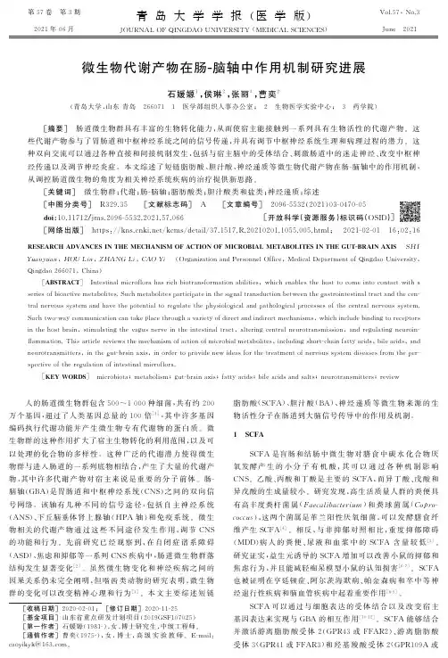
关键词脂联索克隆,分子基因表达肝细胞重组蛋白质类葡糖一6—磷酸酶大肠杆菌ProkaryotiExpression,PurificationandActMtyAnalysisofHumanAdiponectinGeneL1UDentin,YANGLift,DINGY删抽g,SUNYing,YUDeminTianjinMedwdUniversityMetabolicHopitul,Tianjin300070,ChinaAbstractOblective:Toexpressrecombinanthumanadiponeetinwithbiolo咖alaefiviVinEcoli.Methods:TheeDNAcodingfragmentofhumanadiponectingeneⅧamplifiedbyPCRGatewaypmkaryofiexpressionsystemwasusedPCRproductswereclonedintopET-DEST42plasmidtoconstructfusionexpressionvectorpET—DEST42/attB-adiponectinsiterBPreacficmandLRreactionpET-DEST42/anB—adiponeetinWastransformedintoEeoliB12|andthebacteriawⅡeinducedbyIPTGexpressedadiponeetln,fusionproteinW88punfiedbyNi・NTAaⅡinitychromatographyTheeffectofpurifiedrecombmatantadiponeefinonglucose-6-ph08phatonew∞detectedbyRT—PCR.Results:TheDNAsequencingshowed出啦expressionrectorpET—DEsT42f戬啦_adip叫ec血w啦emastructedsuccessfully.purifiedadiponeetinshowedandgenicityandreducedsko”6一ph08phatasetranscriptionallevelConclusion:Wehaveobtainedtherecombinanthamanadiponectinwithbiologicalactivity.whichissignificantforfurtherstudv.Keywordsadiponeetincloning,moleculargeneexpressionhepatocytesrecombinantproteinsglueose一6一phosphataseeschefichiacoli人脂联素(adip(、nectin)基因全长17kb,位于染色体3q27,包含3个外显子和2个内含予。
AAcquired immune deficiency syndrome, AIDS 获得性免疫缺陷综合征(艾滋病) Acquired immunity 获得性免疫Activation induced cell death, AICD 活化诱导的细胞死亡Active immunotherapy 主动免疫治疗Acute phase protein 急性时相蛋白Adapter 转接蛋白Adaptive immunity 适应性免疫Addressin 地址素Adenosine deaminase 腺苷脱氨酶Adjuvant 佐剂Adoptive immunity 过继免疫Adoptive immunotherapy, AIT 过继免疫治疗Affinity 亲和力Affinity maturation 亲和力成熟Agglutination 凝集反应Allelic exclusion 等位排斥Allergen 变应原Allergin 变应素Allergy 变态反应Allogenic antigen 同种异型抗原Allograft 同种异型移植Allotype 同种异型Allorecognition 同种异型识别Alpha-fetoprotein, AFP 甲种胎儿球蛋白Alternative pathway 旁路途径Anamnestic response 回忆应答Anaphylactic shock 过敏性休克Anaphylactogen 过敏原Anaphylaxis 过敏反应Anchor residue 锚定残基Ankylosing spondylitis, AS 强直性脊柱炎Antibacterial immune serum 抗菌免疫血清Antibody, Ab 抗体Antibody-dependent cell-mediated cytotoxicity, ADCC 抗体依赖性细胞介导的细胞毒作用Antiviral immune serum 抗病毒免疫血清Antigen, Ag 抗原Antigenic determinant 抗原决定簇Antigen internal image 抗原内影像Antigen-presenting cells, APC 抗原提呈细胞Antigen specific immune response 抗原特异性免疫应答Antigenicity 抗原性Anti-idiotype 抗独特型Antitoxic serum 抗毒素血清Apoptosis 细胞凋亡Apoptosis cell associated molecular pattern, ACAMP凋亡细胞相关的分子模式 Apoptotic body 凋亡小体Artificial active immunization 人工主动免疫Artificial passive immunization 人工被动免疫Ataxia telangiectasia syndrome, AT 毛细血管扩张共济失调综合征 Atopic dermatitisAutocrineAutograftAutoimmune antibodyAutoimmune disease, AIDAutoimmune hemolytic anemiaAutoimmune thrombocytopenic purpuraAutoimmunityBBacillus Calmette Guerin, BCGBare lymphocyte syndrome, BLSBasophilB cell epitope BB cell hybridoma BB cell linker ptotein, BLNKB cell receptor, BCRBiotin-avidin system, BASBispecific antibody, BsAbBlocking antibodyB lymphocyteBone marrowBone marrow transplantation, BMTBradykininCC1 inhibitor, C1 INHC4 binding protein,C8 binding protein, C8bpC-reactive protein,CRPCadherinCalcineurinCalmodulinCarcinoembryonic antigen, CEACarrierCecropinsCell adhesion molecules, CAM 特应性皮炎自分泌自体移植自身抗体自身免疫性疾病自身免疫性溶血性贫血自身免疫性血小板减少性紫癜自身免疫卡介苗裸淋巴细胞综合征嗜碱性粒细胞细胞表位细胞杂交瘤 B 细胞连接蛋白 B 细胞(抗原识别)受体生物素-亲和素系统双特异性抗体封闭抗体 B淋巴细胞骨髓骨髓移植缓激肽C1抑制分子 C4bp C4 结合蛋 C8 结合蛋白 C-反应蛋白钙粘蛋白钙神经素(钙调磷酸酶)钙调蛋白癌胚抗原载体杀菌肽细胞粘附分子hediak-Higashi syndrome Chediak-Higashi综合征 Chemokine 趋化性细胞因子 Chimeric antibody 嵌合抗体Chronic granulomatous disease, CGD 慢性肉芽肿病 Class Ⅱtransactivator, C Ⅱ TA Ⅱ类反式活化子 Class switch 类别转换Classical pathway 经典途径Clonal anergy 克隆无能Clonal deletion 克隆删除Clonal eliminationClonal expansionCluster of differentiation, CDColony stimulating factor, CSFCommon variable immunodeficiencyComplementComplement deficiencyComplement receptorComplementarity determining region, CDRComplete antigenConcanavalin A, Con AConformational determinantConstant regionCo-receptorCo-stimulating signalCo-stimulatory molecules, CMCo-stimulatory molecule receptor, CMR Cross reactionCryptic determinantCytokine, CKCytolytic typeCytotoxic typeCytotoxic T lymphocytes, CTL 或 Tc Cytotoxic T lymphocytes antigen-4, CTLA-4 DDecay accelerating factor, DAFDefensinsDelayed type hypersensitivity, DTHDelayed type hypersensitivity T cell, T DTH Dendritic cells, DCDiGeorge syndromeDiversity geneDNA vaccineDonorDouble immunodiffusion 克隆消除克隆扩增分化群集落刺激因子普通变化型免疫缺陷病补体补体缺陷补体受体互补性决定区完全抗原刀豆蛋白A 构象决定基恒定区,C区辅助受体协同刺激信号协同刺激分子协同刺激分子受体交叉反应隐蔽决定基细胞因子细胞溶解型细胞毒型细胞毒性T细胞细胞毒性T淋巴细胞(相关)抗原 4 衰变加速因子防御素迟发型超敏反应迟发型超敏反应性T细胞树突状细胞 DiGeorge综合征 D(多样化)基因 DNA疫苗供者双向免疫扩散Double negative cell, DN cell 双阴性细胞Double positive cell, DP cell 双阳性细胞EE rosette test E 花结试验Early phase reaction 早期相反应Endocytosis 胞吞作用Enzyme immunoassay, EIA 酶免疫测定Enzyme linked immunosorbent assay, ELISA 酶联免疫吸附试验Eosinophil, Eos 嗜酸性粒细胞Eosinophil chemotactic factor, ECF 嗜酸性粒细胞趋化因子Eosinophil peroxidase, EPO 嗜酸性粒细胞过氧化物酶 Epitope 表位Erythropoietin, EPO 红细胞生成素Extracellular matrix, ECM 细胞外基质F Fc receptor, FcR 结晶片断受体,Fc受体 Flow cytometry, FCM 流式细胞术Fluorescence-activated cell sorter, FACS 荧光活化细胞分类器 Follicular dendritic cells, FDC 滤泡树突状细胞 Fragment antigen binding, Fab 抗原结合片断Fragment crystallizable, Fc 结晶片断Fragment of variable region Fv 片断Framework region 骨架区Freund complete adjuvant 弗氏完全佐剂GGenetic engineering antibody 基因工程抗体Germ line gene 胚系基因Germinal center 生发中心Glycosylphosphatidylinositol, GPI 糖磷脂酰肌醇Graft versus host reaction, GVHR 移植物抗宿主反应 Granule exocytosis 颗粒胞吐Granulocyte colony stimulating factor, G-CSF 粒细胞集落刺激因子 Granulocyte-macrophage colony stimulating factor, GM-CSF 粒细胞-巨噬细胞集落刺激因子 Granzyme, Gz 颗粒酶Grave disease 毒性弥漫性甲状腺炎 Growth factor 生长因子Growth factor receptor binding protein-2,Grb-2 生长因子受体结合蛋白 2 Guanine nucleotide exchange factor, GEF 鸟苷酸置换因子Gut-associated lymphoid tissue, GALT 肠伴随(相关)淋巴组织 HHaplotype 单元型Hapten 半抗原Hashimoto’s thyroiditis 桥本甲状腺炎Heat shock protein, HSP 热休克蛋白Heavy chain 重链,H 链Helper T cells(lymphocytes), Th 辅助性 T 细胞 Hemolytic plaque assay 溶血空斑试验 Hemopoietic stem cell, HSC 造血干细胞 Heterophil antigen 异嗜性抗原 Hidden antigen 隐蔽抗原High endothelial venule, HEV 高内皮细胞小静脉 Hinge region 铰链区Histamin 组胺Histocompatibility antigen-2, H-2HLA genotypingHomologous restriction factor, HRFHost versus graft reaction, HVGRHuman immunodeficiency virus, HIVHuman leukocyte antigen,HLAHumanized antibodyHumoral immunity Hypersensitivity Hypervariable region, HVRIIdiotype, IdIdiotype networkImmediate hypersensitivity Immune adherent, IAImmune complex, ICImmune function related gene Immune regulationImmune responseImmune response region Immune serumImmune surveillanceImmune systemImmunityImmunocyteImmunodeficiency disease, IDD Immunofluorescence ImmunogenicityImmunoglobulin, IgImmunoglobulin superfamily, IgSFImmunohistochemistry techniqueImmunological competenceImmunological ignorance , ;Immunological toleranceImmunological non-responsiveness 小鼠的组织相容性抗原HLA 基因分型同源限制因子宿主抗移植物反应人类免疫缺陷病毒人类白细胞抗原人源化抗体体液免疫超敏反应高变独特型独特型网络速发型超敏反应免疫粘附免疫复合物免疫功能相关基因免疫调节免疫应答免疫应答区免疫血清免疫监视免疫系统免疫免疫细胞免疫缺陷病免疫荧光法免疫原性免疫球蛋白免疫球蛋白超家族免疫组化技术免疫适能免疫忽视免疫耐受免疫不应答Immunologically privileged sites 免疫隔离部位Immunology 免疫学Immunoreceptor tyrosine-based activation motifs, ITAM 免疫受体酪氨酸活化基序 Immunoreceptor tyrosine-based inhibitory motifs, ITIM 免疫受体酪氨酸抑制基序 Immunotherapy 免疫治疗Inactivated vaccine 灭活疫苗Inducible nitric oxide synthase, iNOS 诱导型一氧化氮合成酶Inflammatory cell 炎症细胞Innate immunity 固有(性)免疫Inositol-1, 4, 5-trisphosphate,IP3Insulin-dependent diabetes mellitus, IDDM Intercellular adhesion molecular,ICAMIntegrinInterferon, IFNInterleukin, ILInternalizationImtraepithelial lymphocytes, IELJJoining chainJoining geneKKiller activatory receptor, KARKiller inhibitory receptor, KIRKiller immunoglobulin like receptor KininogenaseLLangerhans cells, LCLarge granular lymphcytes, LGLsLate phase reactionLectinLectin-like carbohydrate recognition domain, CRDLeucine rich repeat, LRRLeukinLeukocyte adhesion deficiency, LADLeukocyte common antigen, LCALeukocyte differentiation antigen, LDALeukotrienes (Leucotrienes), LTsLigandLight chainLinker for activation of T cell, LATLinear determinantLinkage disequilibriumLipoteichoic acid, LTALipoxygenase pathway 三磷酸肌醇胰岛素依赖型糖尿病细胞间粘附分子整合素干扰素白细胞介素内化上皮细胞间淋巴细胞 J链J基因杀伤细胞活化受体杀伤细胞抑制受体杀伤细胞免疫球蛋白样受体激肽原酶郎格汉斯细胞大颗粒淋巴细胞晚期相反应凝集素凝集素样糖识别结构域富含亮氨酸的重复序列白细胞素白细胞粘附缺陷白细胞共同抗原白细胞分化抗原白三烯配基,配体轻链,L 链 T细胞活化连接蛋白线性决定基连锁不平衡磷壁酸脂氧合酶途径Live-attenuated vaccine 减毒活疫苗Long-acting thyroid stimulator, LATS 长效甲状腺刺激素Low molecular-weight polypeptide, LMP 低分子量多肽LPS binding protein, LBP LPS结合蛋白Lymphocyte 淋巴细胞Lymphocyte function associated antigen, LFA 淋巴细胞功能相关抗原 Lymphocyte homing 淋巴细胞归巢Lymphocyte homing receptor, LHR 淋巴细胞归巢受体Lymphoid DC 淋巴系树突状细胞Lymphoid progenitor 淋巴样祖细胞Lymphoid stem cells, LSC 淋巴样干细胞Lymphokine, LK 淋巴因子Lymphokine activated killer cell, LAK 淋巴因子激活的杀伤细胞 Lymphotoxin, LT 淋巴毒素β -lysin 乙型溶素Lysosome-associated membrane proteins-1,LAMP-1 溶酶体相关膜蛋白1 Lysozyme 溶菌酶MMacrophages, M φ巨噬细胞Macrophage colony stimulating factor, M-CSF 巨噬细胞集落刺激因子 Macropinocytosis 巨吞饮Magainins 爪蟾抗菌肽Major histocompatibility complex, MHC 主要组织相容性复合体 Mannan-binding lectin, MBL 甘露糖结合凝集素Mannose binding protein, MBP 甘露糖结合蛋白Mannose receptor, MR 甘露糖受体Mast cell, MC 肥大细胞MBL-associated serine protease, MASP MBL 伴随的丝氨酸蛋白酶 Membrane attack complex, MAC 膜攻击复合物Membrane cofactor protein, MCP 膜辅助因子蛋白Membrane Ig, mIg 膜表面免疫球蛋白Membrane inhibitor of reactive lysis, MIRL 膜反应性溶解抑制物 Memory cells 记忆细胞MHC class Ⅰ gene MHC Ⅰ类基因MHC class Ⅱ gene MHC Ⅱ类基因MHC class Ⅲ gene MHC Ⅲ类基因MHC restriction MHC 限制性Microfold cell M细胞,微小褶皱细胞β 2-Microglobulin, β2 -m β2 微球蛋白Minor histocompatibility antigen 次要组织相容性抗原 Mitog en 丝裂原Mitogen-activation protein kinase, MAPK 丝裂原激活蛋白激酶 Molecular mimicry 分子模拟Monoclonal antibody, McAb 单克隆抗体第7 / 11页Monocyte 单核细胞Monocyte chemotactic protein, MCP 单核细胞趋化蛋白Monokine, MK 单核因子Mononuclear-phagocyte system, MPS 单核-巨噬细胞系统Mucosal-associated lymphoid tissue, MALT 粘膜伴随(相关)淋巴组织 Mucosal mast cell, MMC 粘膜肥大细胞Multiple hematopoietic stem cells, HSC 多能造血干细胞Multiple sclerosis, MS 多发性硬化症Myasthenia gravis, MG 重症肌无力Myeloid DCMyeloid stem cellMyeloperoxidase, MPONNaive T(B) cellsNatural cytotoxic cellNature killer cell, NK cellNeutrophilsNitric oxide, NONitroblue tetrazolium, NBTNon-classical MHC class Ⅰ geneNon-organ specific autoimmune diseaseNon-specific immunityNude miceOOpsonizationOrgan specific autoimmune diseasePParacrineParoxysmal nocturnal hemoglobineria, PHNPassive immunotherapy ,Passive transfer of lymphocytePathogen associated molecular pattern, PAMPPattern recognition receptor, PRRPeptide antibioticsPeptidoglycan, PGNPerforinPeripheral lymphoid organPeyer’s patchesPhagocytePhagocytosisPhagolysosomePhosphatidylinnosital pathway 髓系树突状细胞髓样干细胞髓过氧化物酶初始T(B)细胞自然细胞毒性细胞自然杀伤细胞嗜中性粒细胞一氧化氮氮蓝四唑非经典性Ⅰ类基因非器官特异性自身免疫病非特异性免疫裸小鼠,裸鼠调理作用器官特异性自身免疫病旁分泌夜间血红蛋白尿被动免疫治疗淋巴细胞被动转移病原相关分子模式模式识别受体肽抗菌肽聚糖穿孔素外周淋巴器官派氏集合淋巴结吞噬细胞吞噬作用吞噬溶酶体 PI途径第8 / 11页Phosphatidylinositol-3-kinase, PI3-K 磷脂酰肌醇3激酶Phosphatidylinositol-4,5-bisphosphate, PIP 2 磷脂酰肌醇4,5二磷酸 Phosphatidylinositol-3,4,5-trisphosphate 磷脂酰肌醇3,4,5三磷酸 Phosphatidylserine, PS 磷脂酰丝氨酸Phosphoinositides 磷酸肌醇Phospholipase 磷脂酶Phospholipid bilineurine 磷脂胆碱Phosphotylinositide-3 kinase 磷酸肌醇-3激酶Phytohemagglutinin, PHA 植物血凝素PinocytosisPinocytotic vesiclePlacental γ globulinPlaque forming cell, PFCPlasma cellsPlatelet activating factor, PAFPolymeric Ig receptor, pIgRPolymorphismPolymorphic genesPolymorphonuclear neutrophils, PMN PrecipitationPrimary immunodeficiency disease, PIDD Primary responsePro-B cellprofessional antigen presenting cells Programmed cell death, PCDProperdin, PProstaglandin, PGProtein kinase C, PKCProtein tyrosine kinase, PTKProtein tyrosine phosphatase, PTP/PTPase Proteolytic enzyme complexProteosomePurine nucleotide phosphorylase, PNPRγδ+T cellRadioimmunoassay, RIAReactive nitrogen intermediates, RNIs Reactive oxygen intermediates, ROIs RearrangementReceptor editingReceptor-mediated endocytosisRecipientRecombinant antigen vaccineRecombinant vector vaccine 胞饮作用吞饮泡胎盘丙种球蛋白空斑形成细胞浆细胞血小板活化因子多聚免疫球蛋白受体多态性多态性基因多形核嗜中性粒细胞沉淀反应原发性免疫缺陷病初次应答祖B细胞专职抗原提呈细胞程序性细胞死亡备解素前列腺素蛋白激酶C 蛋白酪氨酸激酶蛋白酪氨酸磷酸酶蛋白水解酶复合体蛋白酶体嘌呤核苷磷酸化酶γδ+T细胞放射免疫测定法反应性氮中间物反应性氧中间物(基因)重排受体编辑受体介导的胞吞作用受者重组抗原疫苗重组载体疫苗第9 / 11页Recombinase 重组酶Recombination activating genes, RAG 重组活化基因Recombination signal sequences, RSS 重组信号序列Rejection (移植物的)排斥Rheumatoid arthritis, RA 类风湿性关节炎Rheumatoid factor, RF 类风湿因子SScavenger receptor, SR 清杂受体Secondary immunodeficiency disease, SIDD 继发性免疫缺陷病Secondary response 再次应答Secretory component, SC 分泌成分,分泌小体Secretory IgA, sIgA 分泌型免疫球蛋白 ASecretory piece, SP 分泌片Selectin 选择素Selective IgA deficiency 选择性IgA缺陷Serin/threonine phosphatase 丝/苏氨酸磷酸酶Serum amyloid pretein A, SAA 血清淀粉样蛋白 ASevere combined-immunodeficiency disease, SCID 重症联合免疫缺陷病 Signal transduction 信号转导Signal transducers and activator of transcription, STAT 信号转导和活化转录因子 Signalling complex 信号复合体Single immunodiffusion 单向免疫扩散Small G protein 小 G 蛋白Sneaking through 漏逸Soluble TNF receptor, sTNFR 可溶性TNF受体Somatic hypermutation 体细胞高(频)突变Specific immunity 特异性免疫Split tolerance 耐受分离Src family kinase Src家族激酶Staphylococcus enterotoxin, SE 葡萄球菌肠毒素Staphylococcus protein A, SPA 葡萄球菌蛋白 AStem cell factor, SCF 干细胞(生长)因子Subunit vaccine 亚单位疫苗Superantigen, SAg 超抗原Suppressor T cells, Ts 抑制性T细胞Syngraft 同种基因移植,同型移植 Synthetic peptide vaccine 合成肽疫苗Systemic lupus erythematosus, SLE 系统性红斑狼疮TT cell epitope T细胞表位T cell receptor, TCR T细胞(抗原识别)受体 T lymphocyte T 淋巴细胞TCR/CD3 complex TCR/CD3 复合物Terminal deoxynucleotidyl transferase, TdT 末端脱氧核苷酸转移酶第10 / 11页Terminal pathway 末端通路Thrombopoietin 血小板生成素Thymic stromal cells, TSC 胸腺基质细胞Thymocyte 胸腺细胞Thymus 胸腺Thymus dependent antigen, TD-Ag 胸腺依赖抗原Thymus epithelial cells, TEC 胸腺上皮细胞Thymus independent antigen, TI-Ag 非胸腺依赖抗原Thyroid stimulating hormone, TSH 促甲状腺激素Tolerogen 耐受原Toll like receptor, TLR Toll样受体Toxoid &nbs, p; 类毒素Transforming growth factor, TGF 转化生长因子Transporters associated with antigen processing,TAP 抗原处理相关转运蛋白 Tumor-associated antigen, TAA 肿瘤相关抗原Tumor necrosis factor, TNF 肿瘤坏死因子Tumor-specific antigen, TSA 肿瘤特异性抗原VVariable folding 可变折叠Variable geng V基因Variable region ( V region ) 可变区,V区Vitronectin 玻璃连接蛋白,玻连蛋白 Very late appearing ant igen, VLA 迟现抗原WWestern blotting 免疫印迹法Wiskott-Aldrich syndrome, WAS 伴湿疹血小板减少性免疫缺陷病 XX-linked agammaglobulinemia, XLA 性联无丙种球蛋白血症 X-l inked hyperimmunoglobulin M syndrome, HIM 性联高IgM综合征 X-l inked SCID, XSCID 性联重症联合免疫缺陷病。
天津药学TianjinPharmacy2009年第21卷第4期综述重组蛋白在大肠杆菌分泌表达的研究进展’郑海洲,刘晓志,宋欣(华北制药集团新药研究开发有限责任公司,石家庄050015)摘要长期以来,大肠杆菌是表达外源蛋白的首选表达系统,重组蛋白分泌表达与胞内表达相比有很大优越性,在细胞周质腔不仅能促进重组蛋白二硫键的形成及正确折叠,还能促进分泌蛋白的N一端加工。
本文综述了近年来在大肠杆菌中表达可溶性外源蛋白的进展,目的是为了提高外源蛋白的生物活性。
关键词大肠杆菌,分泌表达,重组蛋白中图分类号:Q591.2文献标识码:A文章编号:1006-5687(2009)04-0040-03AdvanceinthesecretoryexpressionofrecombinantproteininescherichiacoilZhengHaizhou,“uXiaozhi,S0ngXin(NCPCNewDrugResearchandDevelopmentCo.,Ltd,Shijiazhuang050015)ABSTRACTEscherichiacoliisoneofthemostwidelyusedhostsfortheproductionofrecombinantproteins.Productionof8ecre—toryproteinsinescherichiacoliprovidesseveraladvantagesoverexpressioninthecytoplasm.Periplasmprovidestheoxidativeen—vironmenttofacilitatecorrectdisulfidebondingandproteinfolding.ItalsoallowscorrectprocessingofN—terminalaminoacidduringsecretion.ThisreviewdiscussesrecentadvancesinsecretoryandextracellularproductionofrecombinantproteinsSOastoimprovethebiologicalactivityofthehetemlogousproteins.KEYWORDSescherichiacoli,secretoryexpression,recombinantprotein大肠杆菌具有遗传背景清楚、繁殖快、成本低、表达量高、表达产物容易纯化等优点,是基因表达技术中发展最早和目前应用最广泛的是经典表达系统…。
基因英语词汇翻译Aactivation domain 活化结构域adapters 连接物adenine 腺嘌呤adenosine 腺ADP (adenosine diphosphate) 腺二磷酸affinity column 亲和柱AFLP (amplified fragment length polymorphisms) 增值性断片长度多态现象agrobacterium 农杆菌属alanine 丙氨酸allele 等位基因amber mutation 琥珀型突变AMP (adenosine monophosphate) 腺一磷酸ampicillin 氨?青霉素anchor primer 锚状引物annealing 退火annealing temperature 退火温度anticodon 反密码子AP-PCR (arbitrarily primed PCR) 任意引物聚合?链反应arbitrary primer 任意引物ATP (adenosine triphosphate) 腺三磷酸autosome 常染色体腺苷脱氨酶缺乏症 adenosine deaminasedeficiency (ADA) 腺病毒 adenovirusAlagille综合征 Alagille syndrome等位基因 allele氨基酸 amino acids动物模型 animal model抗体 antibody凋亡 apoptosis路-巴综合征ataxia-telangiectasia常染色体显性autosomal dominant常染色体 autosomeBbaculovirus 杆状病毒base pair ..基对base sequence ..基顺序beta-galactosidase ..-半乳糖? beta-glucuronidase ..-葡糖醛酸糖? bioluminescence 生物发光bioremediation 生物降解biotechnology 生物技术blotting 印迹法blue-white selection 蓝白斑筛选细菌人工染色体 bacterial artificial chromosome (BAC)碱基对 base pair先天缺陷birth defect骨髓移植bone marrow transplantation blunt end 平(整末)端Ccatalyst 催化剂cDNA library 反向转录DNA库centromere 着丝体centrosome 中心体chemiluminescence 化学发光chiasma 交叉chromomere 染色粒chromoplast 有色体chromosomal aberration 染色体畸变chromosomal duplication 染色体复制chromosomal fibre 染色体牵丝chromosome 染色体chromosome complement 染色体组chromosome map 染色体图chromosome mutation 染色体突变clone 克隆cloning 无性繁殖系化codon 密码子codon degeneracy 密码简并codon usage 密码子选择cohesive end 黏性末端complementary DNA (cDNA) 反向转录DNA complementary gene 互补基因consensus sequence 共有序列construct 组成cosmids 黏性质粒crossing over 互换cyclic AMP (cAMP) 环腺酸cytosine 胞嘧啶癌 cancer后选基因 candidate gene癌 carcinomacDNA文库 cDNA library 细胞cell染色体 chromosome克隆 cloning密码 codon天生的 congenital重叠群 contig囊性纤维化 cystic fibrosis 细胞遗传图 cytogenetic mapDdark band 暗带deamination 脱氨基作用decarboxylation 脱羧基作用degenerate code 简并密码degenerate PCR 退化性聚合?链反应dehydrogenase 脱氢?denaturation 变性deoxyribonucleoside diphospahte 脱氧核糖核一磷酸deoxyribonucleoside monophospahte 脱氧核糖核二磷酸deoxyribonucleoside triphospahte 脱氧核糖核三磷酸deoxyribose 去(脱)氧核糖dicarboxylic acid 二羧酸digoxigenin 洋地黄毒diploid 二倍体DNA (deoxyribonucleic acid) 去(脱)氧核糖核酸DNA binding domain DNA结合性结构域DNA fingerprinting DNA指纹图谱DNA helicase DNA解螺旋?DNA kinase DNA激?DNA ligase DNA连接?DNA polymer DNA聚合物DNA polymerase DNA聚合?double helix 双螺旋double-strand 双链缺失 deletion脱氧核糖核酸 deoxyribonucleic acid (DNA) 糖尿病 diabetes mellitus二倍体 diploidDNA复制 DNA replicationDNA测序 DNA sequencing显性的 dominant双螺旋 double helix复制 duplicationEelectroporation 电穿孔endonuclease 内切核酸? enhancer 增强子enterokinase 肠激? episome 游离基因ethidium bromide 溴乙锭eukaryotic 真核生物的euploid 整倍体exonuclease 外切核酸?expressed-sequence tags 表达的序列标记片段extron 外含子电泳electrophoresis 酶enzyme外显子exonFF factor F因子FAD (flavine adenine dinucleotide) 黄素腺嘌呤二(双)核酸feedback control 反馈控制feedback inhibition 反馈抑制feedback mechanism 反馈机制first filial (F1) generation 第一子代FISH (fluoresence in situ hybridization) 荧光原位杂交forward mutation 正向突变F-pilus F纤毛functional complementation 功能性互补作用fusion protein 融合蛋白家族性地中海热familial Mediterraneanfever 荧光原位杂交fluorescence in situhybridization (FISH) 脆性X染色体综合征Fragile X syndromeGgel electrophoresis 凝胶电泳gene 基因gene cloning 基因克隆gene conversion 基因转变gene duplication 基因复制gene flow 基因流动gene gun 基因枪gene interaction 基因相互作用gene locus 基因位点gene mutation 基因突变gene regulation 基因调节gene segregation 基因分离gene therapy 基因治疗geneome 基因组/ 染色体组genetic map 基因图genetic modified foods (GM foods) 基因食物genetics 遗传学genetypic ratio 基因型比/ 基因型比值genome 基因组/ 染色体组genomic library 基因组文库genotype 基因型giant chromosome 巨染色体globulin 球蛋白glucose-6-phosphate dehydrogenase 6-磷酸葡萄糖脱氢?GP (glycerate phosphate) 磷酸甘油酸脂GTP (guanine triphosphate) 鸟三磷酸guanine 鸟嘌呤基因扩增gene amplification基因表达gene expression基因图谱gene mapping基因库gene pool基因治疗gene therapy基因转移gene transfer遗传密码genetic code (A TGC)遗传咨询genetic counseling遗传图genetic map遗传标记genetic marker遗传病筛查genetic screening基因组genome基因型genotype种系germ lineHhaploid 单倍体haploid generation 单倍世代heredity 遗传heterochromatin 异染色质Hfr strain 高频重组菌株holoenzyme 全?homologous 同源的housekeeping gene 家务基因hybridization 杂交单倍体haploid造血干细胞hematopoietic stem cell 血友病hemophilia 杂合子heterozygous高度保守序列highly conserved sequence Hirschsprung病Hirschsprung's disease纯合子homozygous人工染色体human artificial chromosome (HAC)人类基因组计划Human Genome Project human immunodeficiency virus (HIV)/ 人类免疫缺陷病毒acquired immunodeficiency syndrome (AIDS) 获得性免疫缺陷综合征huntington舞蹈病Huntington's diseaseIimmunoglobulin 免疫球蛋白in vitro 在体外/ 在试管内in vivio 在体内independent assortment 独立分配induced mutation 诱发性突变induction 诱导initiation codon 起始密码子inosine 次黄insert 插入片段insertional inactivation 插入失活interference 干扰intergenic 基因间的interphase 间期intragenic 基因内的intron 内含子inversion 倒位isocaudarner 同尾酸isoschizomer 同切点?Kkanamycin 卡那毒素klenow fragment 克列诺夫片段Llac operon 乳糖操纵子ligase 连接? ligation 连接作用light band 明带linker 连接体liposome 脂质体locus 位点Mmap distance 图距离map unit 图距单位mature transcript 成熟转录物metaphase 中期methylase 甲基化? methylation 甲基化作用microarray 微列microinjection 微注射missense mutation 错差突变molecular genetics 分子遗传学monoploid 单倍体monosome 单染色体messenger RNA (mRNA) 信使RNA multiple alleles 复(多)等位基因mutagen 诱变剂mutagenesis 诱变mutant 突变体mutant gene 突变基因mutant strain 突变株mutation 突变mutation rate 突变率muton 突变子畸形malformation描图mapping标记marker黑色素瘤melanoma孟德尔Mendel, Johann (Gregor)孟德尔遗传Mendelian inheritance信使RNA messenger RNA (mRNA)[分裂]中期metaphase微阵技术microarray technology线立体DNA mitochondrial DNA单体性monosomy小鼠模型mouse model多发性内分泌瘤病multiple endocrine neoplasia, type 1 (MEN1)NNAD (nicotinamide adenine dinucleotide) 烟醯胺腺嘌呤二核酸NADP (nicotinamide adenine dinucleotide phosphate) 烟醯胺腺嘌呤二核酸磷酸nicking activity 切割活性nonsense codon 无意义密码子nonsense mutation 无意义突变Northern blot Northern印迹法nuclear DNA 核DNAnuclear gene 核基因nuclease 核酸?nucleic acid 核酸nucleoside 核nucleoside triphosphate 核三磷酸nucleotidase 核酸?nucleotide 核酸nucleotide sequence 核酸序列神经纤维瘤病neurofibromatosis尼曼-皮克病Niemann-Pick disease, type C (NPC)RNA印记Northern blot核苷酸nucleotide神经核nucleusOoligonucleotide 寡核酸one gene one polypeptide hypothesis 一个基因学说operon 操纵子oxidative decarboxylation 氧化脱羧作用oxidative phosphorylation 氧化磷酸化作用寡核苷酸oligo癌基因oncogenePpeptide ? peptide bond ?键phagemids 噬菌粒phosphorylation 磷酸化作用physical map 物理图谱plasmid 质粒point mutation 点突变poly(A) tail poly(A)尾polymerase 聚合?polyploid 多倍体positional cloning 位置性无性繁殖系化primary transcript 初级转录物primer 引物probe 探针prokaryotic 原核的promoter 启动子protease 蛋白?purine 嘌呤pyrimidine 嘧啶Parkinson病Parkinson's disease血系/谱系pedigree表型phenotype物理图谱physical map多指畸形/多趾畸形polydactyly聚合酶链反应polymerase chain reaction (PCR)多态性polymorphism定位克隆positional cloning原发性免疫缺陷primary immunodeficiency 原核pronucleus前列腺癌prostate cancerRrandom segregation 随机分离RAPD (rapid amplified polymorphic DNA) 快速扩增多态DNAreading frame 阅读码框recessive gene 隐性基因recombinant 重组体recombinant DNA technology 重组DNA技术recombination 重组regulator (gene) 调控基因replica 复制物/ 印模replica plating 复制平皿(板)培养法replication 复制replication origin 复制起点reporter gene 报道基因repression 阻遏repressor 阻遏物repressor gene 阻遏基因resistance strain 抗药性菌株restriction 限制作用restriction enzyme 限制性内切? restriction mapping 限制性内切?图谱retrovirus 反转录病毒reverse transcription 反转录作用RFLP (restricted fragment length polymorphisms) 限制性断片长度多态现象ribonucleotide 核糖核酸ribose 核糖ribosomal RNA (rRNA) 核糖体RNA ribosome 核糖体RNA (ribonucleic acid) 核糖核酸RNA polymerase I RNA聚合?IRNA polymerase II RNA聚合?IIRNA polymerase III RNA聚合?IIIR-plasmid R质粒/ 抗药性质粒隐性recessive逆转录病毒retrovirus核糖核酸ribonucleic acid (RNA)核糖体ribosomeSsecond filial (F2) generation 第二子代self-ligation 自我连接作用shuttle vectors 穿梭载体sigma factor ..因子single nucleotide polymorphism 单核酸多态性single-stranded DNA 单链DNAsister chromatid 姊妹染色单体sister chromosome 姊妹染色体site-directed mutagenesis 定点诱变somatic cell 体细胞Southern blot Southern印迹法splice 拼接star activity 星号活性stationary phase 静止生长期sticky end 黏性末端stop codon 终止密码子structural gene 结构基因supernatant 上层清液supressor 抑制基因序列标记位点sequence-tagged site (STS) 联合免疫缺陷severe combined immunodeficiency (SCID)性染色体sex chromosome伴性的sex-linked体细胞somatic cellsDNA印记Southern blot光谱核型spectral karyotype (SKY)替代substitution自杀基因suicide gene综合征syndromeTtelophase 末期template 模板terminator 终止子tetracycline 四环素thymine 胸腺嘧啶tissue culture 组织培养transcription 转录作用transfer RNA (tRNA) 转移RNA transformation 转化作用transgene 转基因translation 翻译/ 平移transmembrane 跨膜triplet 三联体triplet code 三联体密码triploid 三倍体技术转让technology transfer转基因的transgenic易位translocation三体型trisomy肿瘤抑制基因tumor suppressor geneVvector 载体WWestern blot Western印迹法Wolfram综合征Wolfram syndromeY 酵母人工染色体yeast artificial chromosome (YAC)。
成纤维细胞活化蛋白(FAP)的研究进展?1866?(59—464.[24]KumsawaM,NumazawaS,TaniY,eta1.ERKsignalingmedi. atestheinductionofinflammatoryeytokinesbybufalininhumanmonoeyticceUs[J].AmJPhysiolCellPhysiol,2000,278(3):C50o一508.[25]JingY,WatabeM,HashimotoS,eta1.Cellcyclearrestandpm. teinkinasemodulatingeffectofbufalinonhumanleukemiaML1cells[J].AntlcaneerRes,1994,14(3A):1193—1198.[26]LiuY,Qux,WangP.WT1downregulationduringK562cell MODERNONCOLOGY,Sep.2010.VOI.18,NO.09 differentiationandapoptosisinducedbybufalin[J].ZhonghaaXueYeXueZaZhi,2002,23(7):356—359.[27]AmanoY,ChoY,MatsunawaM,eta1.Increasednuclearexpres—sionandtransaetivafionofvitaminDreceptorbythecardiotonic steroidbufalininhumanmyeloidleukemiacells[J].JSteroidBiochemMolBiol,2009,114(3—5):144—151.[28]LeeDY,YasudaM,YamamotoT,eta1.Bufalininhibitsendo- thelialcellproliferationandangiogenesisinvitro[J].LifeSei,1997,6O(2):127—134.(编校:何蛛)成纤维细胞活化蛋白(FAP)的研究进展张红霞,郭佑民ResearchprogressinfibroblastactivationproteinzHANGHong—xia.GUOYou—minDepartmentofRadiology,BengChaoyangHospital,CapitalMedicalUniversity,Beijing100020,China.【Abstract】Fibroblastactivationprotein(FAP),asurfaceantigenespeciallyexpressedoncarcinomaass ociatedfi—broblasts(CAFs),itisallintegralmembraneserinepeptidase,amemberofthegroupofIIinteg ralserineproteases, whichhasbeenshowntohavedipeptidylpeptidaseandgelatinaseactivity.FAPhasadualfunc tionintumourpro-gression.TheproteolytieactivicyofFAPhasbeenshowntopromotecellinvasivenesstoward stheECMandalsotosupporttumourgrowthandproliferation.OverexpressionofFAPonCAFsanditsspecialend opeptidaseactivityre—presentapotentialtargetfordiagnosisandtreatmentofvariouscarcinomas,makingitfeasible tobeappliedinmolec—ularimaging,oncogenetherapyandimmunotherapyoftargetingtumorceHs.Aseriesofstudi esinvolvingFAPstruc—ture,enzymeactivities,clinicalsignificanceandsoonweresummarized.【Keywords】cancer;fibroblastactivationprotein;carcinomaassociatedfibroblasts ModernOncology2010,18(09):1866—1869【指示性摘要】成纤维细胞活化蛋白(FibroblastActivationProtein,FAP)存在于肿瘤基质成纤维细胞中,在细胞表面发挥作用,是一种膜丝氨酸肽酶,是II型丝氨酸蛋白酶家族成员之一,具有二肽肽酶及胶原酶活性,在肿瘤的生长中具有双重功能.FAP的蛋白水解酶活性可以增强肿瘤细胞对细胞外基质的侵袭力,同样也能促进肿瘤的生长和增殖.故以FAP作为靶点的分子成像以及作为肿瘤基质标志物的病理诊断,肿瘤基因治疗或免疫治疗将成为可能.近年来对FAP的研究进展进行如下综述.【关键词】肿瘤;JE纤维细胞活化蛋白;成纤维细胞【中图分类号】R730.3【文献标识码】ADOI:10.3969/j.issn.1672—4992.2010.09.76 【文章编号】1672—4992一(2010)09—1866一o4【收稿日期】【修回日期】【基金项目】【作者单位1【作者简介】【通讯作者】2010一O1—2620lO—o5—20国家自然科学基金资助项目(编号:30900364);北京市科技计划重大项目(编号:~340]010);北京市教育委员会科技计划面上项目(编号:350010920126)首都医科大学附属北京朝阳医院放射科,北京lOOO20张红霞(1985一),河南濮阳人,住院医师,硕士,主要从事肿瘤分子影像学研究.郭佑民(1955一),男,陕西西安人,主任医师,博士生导师,主要从事肿瘤分子影像学研究.E—mail:cjr.guoyoumin@vip.163.eom1研究背景FAP,曾被认为是F19细胞表面抗原,是一种可诱导的细胞表面糖蛋白,它最初是在1986年用单克隆抗体FI9在培养的成纤维细胞中发现的.1994年,F19细胞表面抗原被命名为成纤维细胞活化蛋白(FAP).1990年,一个分子量为170kDa的胶原酶在人类的恶性黑色素瘤细胞株LOX中被识别.1994年,这个170kDa的胶原酶被命名为Seprase.对FAP和Seprase的分子克隆研究发现它们是同一种细胞表面丝氨酸蛋白酶,基因定位于2q23[1I2].为了使本综述清晰,明了,本文将自始至终用FAP来表示这种蛋白酶.2FAP的结构特征和酶特性2.1结构特征现代肿瘤医学2010年09月第1鲞箍具有活性的FAP是一个170kDa的同型二聚体,包括97kDa的两个N末端糖基化的亚单位,(GenBankGI 1888316)一种Ⅱ型穿膜糖蛋白具有一个大的C末端胞外区域.FAP有5个潜在的N一糖基化位点,l3个半胱氨酸残基,3个高度保守的丝氨酸蛋白酶催化区域,一个疏水的跨膜片段和一个小的胞质尾区(6个氨基酸).另外一些研究发现了FAP的血清形式J,溶解形式抗血纤维蛋白酶(Anti—plasminCleavingEnzyme,APCE)[51,FAP的胞外晶体结构. FAP每个亚单位包括:B一螺旋结构区域(氨基酸序列54—492)和a/t3水解酶区域(氨基酸序列27—53和493—760).FAp的催化区域暴露于细胞外环境中,该区域包括起催化作用的丝氨酸($624),它与Asp702和His734组成了催化三联体.FAP保守的丝氨酸蛋白酶基序是G—w—S—Y—G_1].催化三联体定位在13,螺旋结构和B水解酶区域的交界面,它的排列顺序是亲核一酸一基底,是p水解酶区域的特征.这个催化区域是由基本平行的B片通过各个面上的单环连接,围绕着腹侧轨道呈螺旋放射状排列.具有8片"桨叶"的8螺旋结构位于催化三联体的顶端可能对蛋白底物起着"催化门"的作用』.FAP的晶体结构有5个可能的糖基化位点分别定位在天门冬氨酸49,92,227,314和679.4个位于B螺旋结构区,1个位于水解酶区J.糖基化形式使FAP同时有二肽肽酶活性和胶原酶活性,非糖基化形式则无此活性….FAP的活性由亚单位组成的二聚体形式决定,单体无活性.FAP的基因组在几种物种中发现.比如小鼠类及非洲爪蛙类.对于小鼠类的研究在组织中发现了选择剪接和3个特殊的FAP剪接变异体.一个选择性的FAP剪接体在人类的黑色素瘤细胞株LOX中发现,它编码了一个异常的缩短的变异体.剪接变体编码了一个有239个氨基酸分子量为27kDa的多肽,突变型与野生型的FAP的C末端催化区域重叠.FAP的C末端催化区域在不同物种问是高度同源的.2.2酶特性FAP属于转膜丝氨酸肽酶(serineintegralmembrane peptidases,SIMPs)小家族.FAP与二肽肽酶家族成员DPPIV (DPPIV,dipeptidylpeptidaseIV/CD26)具有52%的同源性,同属于S9b肽酶家族(脯氨酰多肽肽酶家族).FAP因此被认为是DPPIV相似基因家族的一员L9].FAP具有两种蛋白水解酶活性.首先是胶原酶活性,其次是二肽肽酶活性【】.FAP的酶活性被活性位点丝氨酸($624)介导,酶谱法分析酶底物特异性,发现FAP可以降解明胶和热的变性的I型胶原和IV型胶原但不能水解层粘连蛋白,纤维结合蛋白,纤维蛋白单体及酪蛋白….DPPIV不具有胶原酶活性,因此可用是否有胶原酶活性鉴别FAP和DPPIV…J.3FAP的组织分布FAP表达于90%以上的上皮性肿瘤的基质成纤维细胞的胞膜和胞浆中,包括结肠癌,乳腺癌,卵巢癌,膀胱癌,肺癌(原发的和转移的)D23,同时可短暂的停留于某些正常胎儿问质组织中,持续存在于上皮性肿瘤和一些肉瘤的活化基质中,也可表达于胃癌间质,一部分骨和软组织肉瘤细胞,伤口愈合的肉芽组织,特发性肺纤维化的损伤间质,慢性丙型肝炎病人的肝实质细胞,胰腺细胞,前列腺癌,甲状腺髓样癌和乳头状癌,Crohng病组织的狭窄部位,口腔鳞状细胞癌间质.1867?成纤维细胞中.FAP阳性的细胞靠近肿瘤毛细血管的内皮细胞并围绕着肿瘤结节,但在正常成人的组织,良性和癌前病变的上皮性损伤中通常不表达.4FAP的病理作用FAP的活性与许多恶性转化细胞的侵袭行为密切相关,但是它在恶性肿瘤细胞中的作用有不同的观点.4.1肿瘤抑制作用Wesley等¨朝研究显示正常人的黑色素细胞进展为恶性黑色素瘤时丧失了DPPIV的表达.DPPIV的重新表达使小鼠黑色素瘤细胞转化为高分化的正常表型并恢复了依赖外源性生长因子的表达,并发现即使是催化的非活化突变体DPPIV的表达也可导致依赖外源性生长因子的恢复;该研究小组认为内源性FAP的表达对突变体DPPIV的表达起协助诱导作用.Montagut等研究发现FAP的表达降低了小鼠恶性黑色素瘤细胞在动物体内的致肿瘤性,并且修复了接触抑制和生长因子依赖,并发现催化的突变型FAP在缺乏活化的蛋白酶活性的情况下对肿瘤起抑制作用,DPPIV的表达可诱导FAP的表达,反之,FAP并不诱导DPPIV的表达.因此,推论野生型和突变型FAP的肿瘤抑制活性很有可能是FAP的固有本能.4.2肿瘤促进作用更多的研究认为FAP有肿瘤促进作用.Goodman等FAP的人乳腺癌细胞株实验表明,该细胞株(MDA—MB一435和MDA—MB一436)正常的表达FAP,有高水平FAP表达的乳腺癌细胞较少依赖外源性的血清生长因子,并可从正常的生长调控中获得独立.从正常的生长调控中独立出来这是恶性细胞区别于正常细胞的重要特性.Huang等.'研究小组实验证明人类乳腺癌细胞株MDA—MB~231缺乏正常的FAP的表达,FAP高表达的鼠肿瘤模型肿瘤生长的更快更富有血管.还发现当细胞在体外生长时表达FAP的细胞生长率和那些不表达FAP的细胞一样.这表明FAP只有在体内乳房脂肪垫的微环境才表现显着的肿瘤促进作用.这项研究是证明FAP的血管源性功能的首个证据,可以得出结论即,FAP至少部分促进乳腺癌的血管生成,Aimes等ⅢJ 研究证明也印证了这个结论:FAPmRNA表达上调导致了上皮细胞重建或者血管形成.这些发现说明了FAP的表达有利改变乳腺癌细胞的微环境.Wang等也发现当鼠角膜中出现新生血管,角膜基质中的多种生化因子发生变化. FAP(+)的角膜细胞出现在基质中,新出现的这些细胞同时伴随着在新生血管内皮细胞的生长.再次证明了FAP的血管源性功能.小鼠的FAP转染HEK293人胚肾细胞实验显示:将同等数量转染FAP的HEK293细胞和非转染HEK293 细胞同时接种到重症免疫缺陷小鼠体内,前者肿瘤发生率比后者高2—4倍,潜伏期比后者短1O~15天,肿瘤生长率比后者提高10—40倍.用HT229肿瘤中FAP的提取物免疫兔子所产生的多克隆抗FAP抗体,可以有效抑制HT229肿瘤生长,这都说明FAP能够促进肿瘤的发生及生长n.Wang等发现过度表达FAP的人肝星形细胞LX一2细胞系,增加了细胞粘附性,转移性和侵袭性,还发现FAP的蛋白水解活性对于这些功能来说并不重要,这些研究结果进一步支持了FAP的前体纤维生成的作用,即FAP也具有重要的非酶功能.?1868?FAP和DPPIV可以形成一个复合物定位于胶原纤维成纤维细胞的表面,该复合物在细胞侵袭中有溶解明胶与明胶结合的活性,可促进细胞迁移].FAP一尿激酶型纤溶酶活化因子受体(plasminogenactivatorreceptor,uPAR)膜复合物在肿瘤侵袭中有协同作用.在模型系统中,与受体结合的uPA有活性抑制作用并限制了转移灶的形成】.因此, FAP可能是抗转移治疗的潜在靶点.在星形细胞癌中,FAP的表达是与WHO分级相关的,随着FAP表达的增加,在肿瘤组织中所有的与DPPIV相似的酶活性也增加.在结肠癌中,肿瘤的分期及体积与FAP的表达呈正相关,提示FAP的表达在肿瘤早期的发展中具有重要作用,FAP高表达的肿瘤更易扩散,因此FAP被认为是早期肿瘤的潜在治疗靶点J.在化生,非典型的食管组织和食管癌组织的FAP的表达量与食管肿瘤进展相关J. FAP在胰腺癌中高表达,肿瘤发生时达到高值.越高的FAP 表达量预示着越差的预后J.从这些研究发现FAP的活性促进了肿瘤的生长并对肿瘤的侵袭,转移起到了一定作用. FAP是肿瘤生长和转移的一个重要调节因子.4.3其他作用FAP是潜在性的判断伤口时间的有效标志物,一SMA (alpha—smoothmuscleactin)则是判断刀割伤中晚期时间的有效标志物.联合应用FAP和—SMA是潜在的可靠判断伤口时间的指标.存在于类风湿和半恶性肿瘤基质中的FAP介导了和类风湿性慢性滑膜炎中的肿瘤样组织形成.FAP和DPP~4/CD26这些丝氨酸蛋白酶在防止类风湿滑囊液纤维化从而保护关节软骨方面扮演着重要角色驯.5FAP的信号系统的破坏目前用于肿瘤免疫治疗的抗原靶位正在研究之中,该抗原靶位选择性表达于肿瘤间质成纤维细胞或毛细血管上皮细胞.通过靶向作用阻止肿瘤的基质源性及血管源性供养.FAP的缺失间接抑制肿瘤细胞的增殖,加快胶原的积累,减少肌成纤维细胞成分,降低肿瘤内血管密度.因此FAP是肿瘤靶向治疗的重要靶点.Garin—Chesa等指出与毒素结合的单克隆抗体或炎性单克隆抗体同种型显示在FAP阳性的肿瘤间质中,它的作用是诱导细胞损伤,导致肿瘤细胞坏死和炎性细胞浸润.补充添加了FAP阳性的成纤维细胞将会更新目标细胞数量并帮助纤维囊包裹和分离上皮性肿瘤细胞.将抗体定位于肿瘤问质减少了免疫逃逸的发生率.从遗传上讲,肿瘤间质中的细胞更稳定是个有希望作为肿瘤免疫治疗的靶向细胞.Loeffler等发现肿瘤问质抗原FAP能提供新的抗肿瘤疫苗靶位,尤其是联合化疗一起使用,肿瘤相关的成纤维细胞是I型胶原的重要来源,I型胶原可以降低肿瘤对化疗药的吸收并调控肿瘤对化疗药敏感性.该研究小组制作了一种口服的针对FAP靶点的DNA疫苗,在鼠类结肠癌和乳腺癌模型中,这个疫苗成功的抑制了主要肿瘤细胞的增殖和转移灶的多重耐药.而且接种了FAP疫苗鼠类的肿瘤组织显示,I型胶原在肿瘤组织中的表达被抑制, 肿瘤组织吸收的化疗药增长了70%.最重要的是接种了FAP疫苗的鼠类接受化疗治疗后生命延长了30%并显着地抑制了肿瘤的增殖,约50%的动物完全抵抗了肿瘤的侵蚀, 并且这个DNA疫苗没有阻碍创伤的愈合或损伤正常组织. MODERNONCOLOGY,Set).2010.VOI.18.N0.O9在靶向诊治方面,Lebeau『3等发现瘤内注射激活的FAP强亲和毒素,可显着地增加对人乳腺癌及前列腺癌的抑制作用,并在动物体的毒性试验证明其不良作用非常小.Adams 等发现一种小的FAP抑制剂,叫做Val—bom—Pro(PT100),通过一个大样本的大鼠肿瘤模型实验发现其可减弱和抑制肿瘤生长.Pui—Chi等口发现了一种新的FAP触发的光动力学分子探针(FAP—triggeredphotodynamicmolec. ularbeacon,FAP—PPB)由一种荧光敏感剂和一种黑洞猝灭剂3通过连接一种FAP特异性肽序列组成.FAP—PPB可被人和鼠的FAP裂解.用HEK293转染细胞(HEK—In FAP,FAP+,HEK—Vector,FAP一),经过系统的体内外实验分别证明在肿瘤细胞和小鼠的异种移植体中FAP—PPB 的FAP特异性活性.在有FAP表达的细胞可出现荧光修复,没有FAP表达的细胞不会出现这种情况.在HEK—in FAP细胞,FAP—PPB显示了FAP特异的光细胞毒性,然而在对照组HEK—Vector细胞中没有出现细胞毒性.这个实验说明了FAP—PPB是一个潜在的上皮性肿瘤的诊治的工具.6结论与展望自从1986年发现FAP,迄今在定位和表达这个蛋白酶上面做了大量的科学研究.然而FAP的生化特性和功能需要进一步研究.尽管大量的研究报告指出这个酶可能有的功能,但是FAP的生理学作用还是没有解释清楚.FAP在肿瘤细胞中通过降解细胞外物质,在侵袭和转移中扮演了重要的角色.FAP与90%的上皮细胞癌有关系,还是潜在的对疾病有预示作用的重要标志物.因此FAP可以作为肿瘤体内免疫诊治较有前途的靶分子.有效率的标准的在体内和体外都适用的FAP抑制剂的出现为FAP生理作用的研究打开了一扇门.FAP的调节功能也许是可以在不同等级上操纵的,基因的,后转录的和激素的,可是相关机制和调控方法还不知道.未来在这个领域的研究将会提供FAP的全面信息, 这些信息包括调控方式和生理功能.这样FAP作为选择性抗肿瘤诊治的靶点将成为可能.参考文献[1]MLPineim—Sanchez,LAGoldstein,JDodt,eta1.Identificationofthe170一kDamelanomamembrane—boundgelatinasefse—prase)asaserineintegralmembranepmtease[J].BiolChem, 1997,272:7595—7601.[2]SMathew,^iJSeanlan,BKMohanRaj,eta1.Thegeneforfibro? blastactivationproteinalpha(FAP),aputativecellsurfacebound sedneproteaseexpressedincancerstinmaandwoundhealing, mapstochromosomeband2q23[J3.Genomies,1995,25:335—337.【3]WTChen,TKeUy.Seprasecomplexesincenularinvasiveness [J].CancerMetastasisRev,2003,22(2—3):259—269.[4]PJCollins,GMcMahon,POBrien,eta1.Purification,identifiea- tionandcharacterisationofseprasefrombovine8erOlll[J].IntJ BiochemCellBiol,2004,36:2320—2333.[5]KNLee,KWJackson,VJChristiansen,eta1.Antiplasmin—clea- vingenzymeisasolubleformoffibroblastactivationprotein[J]. Blood,2006,107:1397—1404.[6]KAertgeerts,ILevin,LShi,eta1.Structuraland~neticanalysis ofthesubstratespecificityofhumanfibroblastactivationproteinal- pha[J].BiolChem,2005,280(20):19441—19444.医学2010年O909[7]SSun,CFAlbright,BHFish,eta1.Expression,purification,and kineticcharaeterizationoffulllengthhumanfibmblastactivation protein[J3.ProteinExprPurif,2002,24(2):274—281.『8]LAGoldstein,WTChen.Identifcationofanalternativelyspliced seDmsemRNAthatencodesanovelintracellularisoform[J].Biol Chem,2000,275:2554—2559.[9]CAAbbott,MDGorrell,JLangner,eta1.Eetopeptidases:CD13/ AminopeptidaseNandCD26/DipeptidylpeptidaseIVinmedicine andbiology[M].NewYork:KluwerAcademic/PlenumPublish—eFs,2002:171—195.[1O]JDCheng,MValianou,AACanutescu,eta1.Abrogationoffibre- blastactivationproteinenzymaticactivitya~enuatestumorgrowth [J].MolCancerTher,2005,4(3):351—360.[11]MDGorrell,VGysbers,GWMcCanghan.CD26:amultifune—tionalintegralmembraneandsecretedproteinofactivatedlympho- cytes[J].ScandJImmunol,2001,54:249—264.[12]PGarin—Chesa,IJJOld,wJRettig.Cellsurfaceglycopmteinof reactivestromalfibroblastsasapotentialantibodytargetinhuman epithelialcancers[J].ProcNatAcadSciUSA,1990,87:7235—7239.[13]uVWesley,APAlbino,STiwari,eta1.Arolefordipeptidyl peptidaseIVinsuppressingthemalignantphenotypeofmelflfflO—cytiecells[J].ExpMed,1999,190:311—322.[14]TRamirez—Montagut,NEBlachere,EVSviderskaya,eta1.FA- Pa;lpha.asurfacepeptidaseexpressedduringwoundhealing,isa tumorsuppressor[J].Oncogene,2004,23:5435—5446.[15]JDGoodman,TLRozypal,TKeHy.Seprase,amembrane—bound protease,alleviatestheselllmgrowthrequirementofhumanbreast cancercells[J].ClinExpMetastasis,2003,20:459—470.[16]YHuang,SWang,TKelly.Seprasepromotesrapidtumorgrowth andincreasedmicrovesseldensityinamousemodelofhuman breastcancer[J].CancerRes,2004,64(8):2712—2716.[17]RTAimes,AZijlstra,JDHoPper,eta1.Endothelialcellserine proteasesexpressedduringvascularmorphogenesisandangiogene—sis[J].ThrombHaemost,2003,89:561—572.[18]WangT,ShiwY,LiSX,eta1.Relationshipbetweencornealneo- vaseularizationandvariousrelevantbiologicalfactorsinsurround—ingcorneastromaofrats[J].ZhonghHaYanKeZaZhi,2009,45(2):158—163.[19]JDCheng,RLDunbrackJr,MValianou,eta1.Promotionof tumorgrowthbymurinefibroblastactivationprotein,aserine protease,inananimalmodel[J].CancerRes,2002,62(16): 4767—4772.[2O]XMWang,DMYu,ASchnapp,eta1.Theroleoffibroblastacti- vationprotein(FAP)incelladhesion,migrationandliverfibrosis [J].Hepatology,2005,42:738A一738A.[21]VV Artym,ALKindzelskii,WTChen,eta1.Molecularproximi—tyofsepraseandtheurokinasetypeplasm—inogenactivatorre—ceptoronmalignantmelanomacellmembranes:dependenceon beta1integrinsandthecyto—skeleton[J].Carcinogenesis,2002,?1869?23:1593—1601.[22]S1wasa,KOkada,WTChen,eta1.Increasedexpressionofse—prase,amembrane—typeserineprotease,isassociatedwith lymphnodemetastasisinhumaneolorectalcancer[J].Cancer Lett,2005,227:229~236.[23]StremenovaJ,Krepola,EMaresV,eta1.Expressionandenzy—maticactivityofdipeptidylpeptidase—IVinhumanastrocytic tumottrsareassociatedwitIlturnoutgrade[J].IntOncol,2007,31:785—792.[24]HenryLR,LeeH0,Lee,etal,Clinicalimplicationsoffibro—blastactivationproteininpatientswithcoloncancer【J].Clin CancerRes,2o0r7,13,1736—1741.[25]GoseiaskiMA,SuoZ,FlorenesV A,eta1.FAP—alphaanduPA showdifferentexpressionpatternsinpremalignantandmalignant esophaneallesions[J].UhrastruetPathol,2008,32(3):89-96.[26]CohenSJ,AlpanghRK,PalazzoI,eta1.Fibroblastactivationpro—teinanditsrelationshiptoclinicaloutcomeinpancreaticadeno—carcinoma[J].Pancreas,2008,37(2):154—158.[27]GaoY,PengX,JinZF,eta1.ExpressionofFAPandalpha—SMAduringtheincisedwoundhealinginmiceskin[J].FaYi XueZaZhi,2009,25(6):405—408.[28]JakobsM,HauplT,KrennV,eta1.MMP—andFAP—mediated non—-inflammation—-relateddestructionofcartilageandbonein rheumatoidarthritis[J].zRheumatol,2009,68(8):683—694. [29]Ospehc,Me~ensJC,JungelA,eta1.Inhibitionoffibroblastacti—rationproteinanddipeptidylpeptidase——4increasescartilagein?- vasionbyrheumatoidarehritissynovialfibroblasts[J].Arthritis Rheum,加10,62(5):1224—1235.[3O]JLee,MFassnaeht,SNair,eta1.Tumorimmunotherapytarge? tingfibroblastactivationprotein,aproductexpressedintumor—associatedfibroblasts[J].CancerRes,2005,65:11156~11163. [31]SantosAM,JungJ,AzlzN,eta1.TargetingFibroblastactivation proteininhibitstumorstromagenesisandgrowthinmice[J].J ClinInvest,2009,l19(12):3613—3625.[32]MLoeffler,JAKruger,AGNiethanuner,eta1.Targetingtumor associatedfibroblastsimprovescancerchemotherapybyincreasing intratumoraldruguptake[J].ClinInvest,2006,116(7):1955—1962.[33]LebeauAM,BrennenwN,AggarwalS,eta1.Targetingthecancer stromawjtllafibroblastactivationproteinactivatedpromeliain protoxin[J].MolCancerTher,2009,8(5):1378—1386.[34]AdamsS,MillerGT,JessonMI,eta1.PT一100,asmallmolecule dipeptidylpeptidaseinhibitor,haspotentantitumorsffectsand augmentsantibody—mediatedeytotoxieityviaanovelimmunemechanism[J].CancerRes,2004,64:5471—5480.[35]Pui—ChiLo,JuanChen,KlaraStefflova,eta1.Photodynamiemo. 1ecularbeacontriggeredbyfibroblastactivationproteinoncancer—associatedfibroblastsfordiagnosisandtreatmentofepithelial cancers[J].MedChem,2009,52(2):358—368.(编校:何妹)。
生物制品分析事例三——ChP注射重组人促红素的鉴别1.生物制品简介生物制品(biological products)是指用微生物(包括细菌、噬菌体、立克次体、病毒等)、微生物代谢产物、动物毒素、人或动物的血液或组织等经加工制成,作为预防、治疗、诊断特定传染病或其他有关疾病的免疫制剂。
根据用途可将生物制品分为预防类、治疗类和诊断类三大类制品,注射用重组人促红素是一种治疗类的生物制品。
EPO 是促红细胞生成素(Erythropoietin)的英文简称。
人体中的促红细胞生成素是由人体中肾皮质肾小管周围间质细胞和肝脏分泌的一种激素类物质,能够促进红细胞的生成。
由于EPO 在体内的作用非常大而天然来源却十分有限(主要从贫血患者的尿中提取)[1],人们便开始利用基因重组技术获得重组人促红素( Recombinant human erythropoietin, rHuEPO),它是将人的EPO基因转入哺乳动物细胞内高效表达的能刺激红细胞生成的糖蛋白激素[2,3]。
它由不同的糖基化体构成,各种糖基化体的区别主要表现在糖基在整个分子中所占的比例不同,它一方面能够刺激骨髓造血功能,及时有效地增加红细胞的数量;另一方面能够增强机体对氧的结合、运输和供应能力,有利于改善缺氧状态,促进肌肉中氧生成,使肌肉更有力、工作时间更长。
2.免疫印迹法由于注射重组人促红素是一种治疗类的生物制品,生物制品一般均为免疫制剂,故可以利用其生物制品的特点利用免疫学的方法的其进行鉴别,《中国药典2010年版》收载的对其鉴别的方法为免疫印迹法。
免疫印迹法系以供试品与特异性抗体结合后,抗体再与酶标抗体特异性结合,通过酶学反应的显色,对供试品的抗原特异性进行检查[4]。
免疫印迹法(immunobiotting test,IBT)也被称作酶联免疫电转移印斑法(enzyme linked immunoeletrotransfer blot,EITB),是20世纪70年代末,发展起来的一门蛋白质多参数检测技术。
收稿日期:2019-07-11基金项目: 国家重点研发计划项目(2016YFD0500707-7);山东省重点研发计划(2019GNC106074);山东省农业重大应用技术创新项目“生猪生态循环养殖综合技术研究与应用示范”;内蒙古农业大学高层次人才引进项目(NDGCC2016-22、NDYB2018-2)作者简介:张洪亮,男,博士研究生,预防兽医学专业通信作者:单虎,E-mail:****************;温永俊,E-mail:******************·研究论文·伪狂犬病病毒gB 和gE 蛋白重组表达及抗体间接ELISA 检测方法的建立摘 要:本研究利用PCR 扩增伪狂犬病病毒(PRV )Bartha-K61株gB 基因核心抗原区和闵A 株gE 基因核心抗原区,构建了重组质粒pET-32a-gB 和pET-32a-gE ,并转化至大肠杆菌BL21(DE3)感受态细胞中,经IPTG 诱导进行表达。
SDS-PAGE 分析表明,pET32a-gB 蛋白分子量约为44 kDa ,pET32a-gE 蛋白分子量约为41 kDa 。
分别以gB 和gE 重组蛋白作为包被抗原,建立了PRV 抗体间接ELISA 检测方法。
该方法中重组蛋白仅与PRV 阳性血清发生特异性反应,检测敏感性可达到1∶1600,批内重复性和批间重复性变异系数均小于10%。
本研究建立的间接ELISA 方法可用于PRV 抗体的监测,鉴别疫苗免疫抗体和野毒感染抗体,有利于猪伪狂犬病净化工作。
关键词:猪伪狂犬病病毒;gB 基因;gE 基因;蛋白表达;间接ELISA 中图分类号:S852.659.1文献标志码: A文章编号:1674-6422(2022)01-0098-08Expression of Recombinant gB and gE Proteins of Pseudorabies Virus andDevelopment of Indirect ELISA Assay for Antibody DetectionZHANG Hongliang 1,2, HAO Zhanwu 1, XIE Qianqian 1, W ANG Fengxue 2, ZHANG Zhi 3,HAN Xianjie 1, SHAN Hu 1, WEN Yongjun 2(1. Shandong Collaborative Innovation Center for Development of Veterinary Pharmaceuticals, Qingdao Research Center for Veterinary Biological Engineering and Technology, College of Animal Medicine, Qingdao Agricultural University, Qingdao 266109, China; 2. Ministry of Agriculture and Rural Affairs Key Laboratory of Clinical Diagnosis and Treatment Technology in Animal Diseases, College of Veterinary Medicine, Inner Mongolia Agricultural University, Hohhot 010018, China; 3. China Animal Health and Epidemiology Center, Qingdao266032, China)张洪亮1,2,郝占武1,解倩倩1,王凤雪2,张 志3,韩先杰1,单 虎1,温永俊2(1.青岛农业大学动物医学院 山东省新兽药创制协同创新中心 青岛市兽医生物技术工程研究中心,青岛266109;2.内蒙古农业大学兽医学院 农业农村部动物疾病临床诊疗技术重点实验室,呼和浩特010018;3.中国动物卫生与流行病学中心,青岛266032)Abstract: In this study, gB and gE genes of Porcine pseudorabies virus (PRV) Bartha-K61 strain were amplifi ed by PCR and cloned into pET32a vector. The recombinant plasmids pET-32a-gB and pET-32a-gE were then transformed into E. coli BL21 (DE3) for protein expression with IPTG induction. SDS-PAGE analysis showed that the pET32a-gB protein had a molecular weight of about 44 kDa, andChinese Journal of Animal Infectious Diseases中国动物传染病学报2022,30(1): 98-105· 99 ·张洪亮等:伪狂犬病病毒gB 和gE 蛋白重组表达及抗体间接ELISA 检测方法的建立第30卷第1期伪狂犬病病毒(Pseudorabies virus, PRV)属疱疹病毒科中疱疹病毒Ⅰ型,可引起多种动物发病,包括猪、绵羊、狗、牛、猫、狐狸、水貂、鼠、兔等35种,猪是其唯一自然宿主[1-3]。
[28][29][30]CALVO -PINILLA E ,CASTILLO -OLIVARES J ,JAB -BAR T ,et al.Recombinant vaccines against bluetongue virus [J ].Virus Res ,2014,182:78-86.MA G ,ESCHBAUMER M ,SAID A ,et al.An equine her -pesvirus type1(EHV -1)expressing VP2and VP5of serotype8bluetongue virus (BTV-8)induces protectionin a murine infection model [J ].PLoS One ,2012,7(4):e34425.苗海生,李乐,廖德芳,等.蓝舌病多克隆抗体C-ELISA 检测方法的建立和比较研究[J ].云南农业大学学报,2014,29(1):58-62.(责任编辑:钱英红)1例青海藏獒犬巴贝斯虫病的诊疗伊平昌1,王守宁2(1.青海省西宁市畜牧兽医站,青海西宁810003;2.青海省大通县桥头镇家畜病院,青海大通810100)摘要:介绍了1例青海藏獒犬巴贝斯虫病的发病情况、临床症状、实验室检查,提出了使用特效杀虫药物,同时采取对症治疗措施的治疗原则,以期为兽医临床防治巴贝斯虫病提供参考。
关键词:藏獒犬;巴贝斯虫病;诊断;对症治疗中图分类号:S858.292.27文献标识码:A 文章顺序编号:1672-5190(2015)11-0110-02犬巴贝斯虫病是以蜱虫为传播媒介,引起急性溶血性疾病发作的一种季节性血液原虫病,属于人兽共患病,目前呈世界性分布[1]。
依据宿主特异性和形态学将犬的巴贝斯虫分为犬巴贝斯虫(Babesiacanis )和吉氏巴贝斯虫(B.gibsoni )2类[2]。
我国主要以吉氏巴贝斯虫为主,呈地方性流行。
人源JAZF1基因真核表达载体的构建及其对胚胎癌细胞凋亡的影响陈倩,李超,杨诚诚,母彬,龚欢,黄超*(四川农业大学动物医学院,实验动物疾病模型研究室,成都611130)摘要:【目的】构建人源转录因子锌指并列基因1(JAZF1)真核表达载体,并探究过表达JAZF1对胚胎癌细胞存活的影响。
【方法】以胚胎癌细胞NCCIT 的cDNA 为模板扩增JAZF1基因片段,并将其与pRK5-myc 载体连接,构建重组质粒,转染至NCCIT 细胞;通过实时荧光定量PCR 和DAPI 染色法分别检测过表达JAZF1对NCCIT 细胞中炎性因子IL1-β、IL8、TGF-β转录水平的影响及对NCCIT 细胞存活的影响。
【结果】过表达JAZF1基因后,促炎因子IL1-β(P <0.05)、IL8(P <0.01)转录水平明显下降,而抑炎因子TGF-β的mRNA 表达水平无明显变化(P >0.05),同时NCCIT 细胞出现更明显的细胞凋亡(P <0.01)。
【结论】JAZF1可能通过抑制促炎因子IL1-β和IL8的表达来抑制炎症发展,进而抑制畸胎瘤进展。
关键词:锌指并列基因1;胚胎癌;炎性因子;细胞凋亡中图分类号:Q78文献标志码:A文章编号:1000-2650(2021)01-0108-06Construction of Eukaryotic Expression Vector of Human Juxtaposedwith Another Zincfinger Gene 1and Its Influence on EmbryonalCarcinoma Cell ApoptosisCHEN Qian ,LI Chao ,YANG Chengcheng ,MU Bin ,GONG Huan ,HUANG Chao *(College of Veterinary Medicine ,Sichuan Agricultural University ,Chengdu 611130,China )Abstract:【Objective 】The eukaryotic expression vector of human juxtaposed with another zincfinger gene1(JAZF1)was constructedto explore the effect of JAZF1on embryonal carcinoma cell survival.【Method 】The JAZF1gene fragment was amplified using the cDNA of embryonic cancer cell NCCIT as a template ,and wascloned into pRK5-myc vector to construct a recombinant plasmid.The recombinant plasmid wastransfected into NCCIT cells ,while the cell viability and the expression of transcription factorIL1-β,IL8and TGF-βwere following evaluated by DAPI staining and quantitative real-time PCR ,respectively.【Result 】Transfected with JAZF1,the transcriptional levels of proinflammatory factor IL1-βand IL8in NCCIT cells showed a significant suppression ,while enhanced expression of antiinflammatory factor T GF-茁was observed.Meanwhile ,we found increased cellularapoptosis in JAZF1-transfected NCCITcells.【Conclusion 】JAZF1may suppressinflammation by inhibiting the expression of IL1-βand IL8,and then inhibit the progressionofembryonic cancer.Keywords:human juxtaposed with another zinc finger gene 1(JZAF1);embryonal carcinoma ;inflammat-oryfactor ;apoptosis第39卷第1期2021年2月收稿日期:2020-07-25基金项目:四川农业大学学科建设双支计划(2018-2019)。
糖尿病肾病小鼠中前纤维蛋白1的表达和免疫细胞浸润分析*麦丽萍, 黄桂萍, 邓春玉, 郑丹琳, 李晓红, 何国东△[南方医科大学附属广东省人民医院(广东省医学科学院)医学研究部,广东 广州 510080][摘要] 目的:探讨前纤维蛋白1(profilin 1, PFN1)在糖尿病肾病小鼠中的表达及其与免疫细胞浸润的相关性。
方法:利用糖尿病肾病患者肾组织芯片转录组表达数据分析PFN1的表达水平和免疫细胞浸润情况,并通过小鼠动物实验进行验证。
将16只C57BL/6小鼠随机分为正常组和模型组,每组8只。
模型组采用腹腔注射链脲佐菌素法建立糖尿病肾病小鼠模型,造模成功后检测小鼠肾组织中CD11b 、F4/80、CC 趋化因子受体4(CC chemo‐kine receptor 4, CCR4)、白细胞介素1受体I 型(interleukin -1 receptor type I , IL -1R1)、B 细胞淋巴瘤2(B -cell lympho‐ma -2, Bcl -2)、Bcl -2相关X 蛋白(Bcl -2-associated X protein , Bax )和胱天蛋白酶3(caspase -3)蛋白的表达水平。
另外,在糖尿病肾病细胞模型中过表达PFN1,检测单核细胞趋化蛋白1(monocyte chemotactic protein -1, MCP -1)和cas‐pase -3蛋白的表达情况。
结果:糖尿病肾病患者肾组织芯片转录组表达数据集GSE30122中,PFN1基因表达量显著升高(P <0.01),且与巨噬细胞和T 细胞有显著相关性。
糖尿病肾病小鼠模型组肾脏组织可见明显病理改变,肾脏组织中PFN1表达量显著升高(P <0.01),免疫指标CD11b 、F4/80、CCR4、IL -1R1和凋亡相关蛋白Bax 、Bcl -2、cas‐pase -3表达升高(P <0.01)。
专利名称:RECOMBINANT PROTEIN EXPRESSION 发明人:DE MARCO, ARIO,GEERLOF, ARIE,BUKAU, BERND,DEUERLING, ELKE申请号:IB0300299申请日:20030107公开号:WO03057897A3公开日:20031127专利内容由知识产权出版社提供摘要:There are provided methods for the expression of a recombinant protein of interest, said methods comprising, in additional to various additional steps: a) culturing a host cell which expresses: i) one or more genes encoding the recombinant protein(s) of interest; ii) at least two genes encoding proteins selected from the group consisting of the chaperone proteins GroEL, GRoES, Dnak, DnaJ, GRpe, ClpB and their homologs (for example, Hsp104, Ydj1 and Ssal in yeast); under conditions suitable for protein expression; and separating said recombinant protein of interest from the host cell culture. Also provided are methods for increasing the degree of refolding of a recombinant protein of interest by ading a composition containing a chaperone protein to a preparation of the recombinant protein of interest in vitro.申请人:EUROPEAN MOLECULAR BIOLOGY LABORATORY,DE MARCO, ARIO,GEERLOF, ARIE,BUKAU, BERND,DEUERLING, ELKE更多信息请下载全文后查看。
REVIEW ARTICLEpublished:17April2014 doi:10.3389/fmicb.2014.00172 Recombinant protein expression in Escherichia coli: advances and challengesGermán L.Rosano1,2*and Eduardo A.Ceccarelli1,21Instituto de Biología Molecular y Celular de Rosario,Consejo Nacional de Investigaciones Científicas y Técnicas,Rosario,Argentina2Facultad de Ciencias Bioquímicas y Farmacéuticas,Universidad Nacional de Rosario,Rosario,ArgentinaEdited by:Peter Neubauer,Technische Universität Berlin,GermanyReviewed by:Jose M.Bruno-Barcena,North Carolina State University,USA Thomas Schweder,Ernst-Moritz-Arndt-Universität Greifswald,Germany*Correspondence:Germán L.Rosano,Instituto de Biología Molecular y Celular de Rosario,Consejo Nacional de Investigaciones Científicas y Técnicas, Esmeralda y Ocampo,Rosario2000, Argentinae-mail:rosano@.ar Escherichia coli is one of the organisms of choice for the production of recombinant proteins.Its use as a cell factory is well-established and it has become the most popular expression platform.For this reason,there are many molecular tools and protocols at hand for the high-level production of heterologous proteins,such as a vast catalog of expression plasmids,a great number of engineered strains and many cultivation strategies.We review the different approaches for the synthesis of recombinant proteins in E.coli and discuss recent progress in this ever-growingfield.Keywords:recombinant protein expression,Escherichia coli,expression plasmid,inclusion bodies,affinity tags, E.coli expression strainsINTRODUCTIONThere is no doubt that the production of recombinant proteins in microbial systems has revolutionized biochemistry.The days where kilograms of animal and plant tissues or large volumes of biologicalfluids were needed for the purification of small amounts of a given protein are almost gone.Every researcher that embarks on a new project that will need a purified protein immediately thinks of how to obtain it in a recombinant form. The ability to express and purify the desired recombinant protein in a large quantity allows for its biochemical characterization,its use in industrial processes and the development of commercial goods.At the theoretical level,the steps needed for obtaining a recom-binant protein are pretty straightforward.You take your gene of interest,clone it in whatever expression vector you have at your disposal,transform it into the host of choice,induce and then,the protein is ready for purification and characterization.In practice, however,dozens of things can go wrong.Poor growth of the host, inclusion body(IB)formation,protein inactivity,and even not obtaining any protein at all are some of the problems often found down the pipeline.In the past,many reviews have covered this topic with great detail(Makrides,1996;Baneyx,1999;Stevens,2000;Jana and Deb,2005;Sorensen and Mortensen,2005).Collectively,these papers gather more than2000citations.Yet,in thefield of recombi-nant protein expression and purification,progress is continuously being made.For this reason,in this review,we comment on the most recent advances in the topic.But also,for those with modest experience in the production of heterologous proteins, we describe the many options and approaches that have been successful for expressing a great number of proteins over the last couple of decades,by answering the questions needed to be addressed at the beginning of the project.Finally,we provide a troubleshooting guide that will come in handy when dealing with difficult-to-express proteins.FIRST QUESTION:WHICH ORGANISM TO USE?The choice of the host cell whose protein synthesis machinery will produce the precious protein will initiate the outline of the whole process.It defines the technology needed for the project, be it a variety of molecular tools,equipment,or reagents.Among microorganisms,host systems that are available include bacteria, yeast,filamentous fungi,and unicellular algae.All have strengths and weaknesses and their choice may be subject to the protein of interest(Demain and Vaishnav,2009;Adrio and Demain,2010). For example,if eukaryotic post-translational modifications(like protein glycosylation)are needed,a prokaryotic expression sys-tem may not be suitable(Sahdev et al.,2008).In this review, we will focus specifically on Escherichia coli.Other systems are described in excellent detail in accompanying articles of this series.The advantages of using E.coli as the host organism are well known.(i)It has unparalleled fast growth kinetics.In glucose-salts media and given the optimal environmental conditions,its dou-bling time is about20min(Sezonov et al.,2007).This means that a culture inoculated with a1/100dilution of a saturated starter culture may reach stationary phase in a few hours.However,it should be noted that the expression of a recombinant protein may impart a metabolic burden on the microorganism,causing a considerable decrease in generation time(Bentley et al.,1990). (ii)High cell density cultures are easily achieved.The theoretical density limit of an E.coli liquid culture is estimated to be about 200g dry cell weight/l or roughly1×1013viable bacteria/ml(Lee, 1996;Shiloach and Fass,2005).However,exponential growth incomplex media leads to densities nowhere near that number.In the simplest laboratory setup (i.e.,batch cultivation of E.coli at 37◦C,using LB media),<1×10¹⁰cells/ml may be the upper limit (Sezonov et al.,2007),which is less than 0.1%of the theoret-ical limit.For this reason,high cell-density culture methods were designed to boost E.coli growth,even when producing a recom-binant protein (Choi et al.,2006).Being a workhorse organism,these strategies arose thanks to the wealth of knowledge about its physiology.(iii)Rich complex media can be made from read-ily available and inexpensive components.(iv)Transformation with exogenous DNA is fast and easy.Plasmid transformation of E.coli can be performed in as little as 5min (Pope and Kent,1996).SECOND QUESTION:WHICH PLASMID SHOULD BE CHOSEN?The most common expression plasmids in use today are the result of multiple combinations of replicons,promoters,selec-tion markers,multiple cloning sites,and fusion protein/fusion protein removal strategies (Figure 1).For this reason,the catalog of available expression vectors is huge and it is easy to get lost when choosing a suitable one.To make an informed decision,these fea-tures have to be carefully evaluated according to the individual needs.REPLICONGenetic elements that undergo replication as autonomous units,such as plasmids,contain a replicon.It consists of one origin of replication together with its associated cis -acting control elements.An important parameter to have in mind when choosing a suitable vector is copy number.The control of copy number resides in the replicon (del Solar and Espinosa,2000).It is logical to think that high plasmid dosage equals more recombinant protein yield as many expression units reside in the cell.However,a high plasmidnumber may impose a metabolic burden that decreases the bacte-rial growth rate and may produce plasmid instability,and so the number of healthy organisms for protein synthesis falls (Bentley et al.,1990;Birnbaum and Bailey,1991).For this reason,the use of high copy number plasmids for protein expression by no means implies an increase in production yields.Commonly used vectors,such as the pET series,possess the pMB1origin (ColE1-derivative,15–60copies per cell;Bolivar et al.,1977)while a mutated version of the pMB1origin is present in the pUC series (500–700copies per cell;Minton,1984).The wild-type ColE1origin (15–20copies per cell;Lin-Chao and Bremer,1986;Lee et al.,2006)can be found in the pQE vec-tors (Qiagen).They all belong to the same incompatibility group meaning that they cannot be propagated together in the same cell as they compete with each other for the replication machinery (del Solar et al.,1998;Camps,2010).For the dual expression of recombinant proteins using two plasmids,systems with the p15A ori are available (pACYC and pBAD series of plasmids,10–12copies per cell;Chang and Cohen,1978;Guzman et al.,1995).Though rare,triple expression can be achieved by the use of the pSC101plasmid.This plasmid is under a stringent control of repli-cation,thus it is present in a low copy number (<5copies per cell;Nordstrom,2006).The use of plasmids bearing this repli-con can be an advantage in cases where the presence of a high dose of a cloned gene or its product produces a deleterious effect to the cell (Stoker et al.,1982;Wang and Kushner,1991).Alter-natively,the use of the Duet vectors (Novagen)simplifies dual expression by allowing cloning of two genes in the same plas-mid.The Duet plasmids possess two multiple cloning sites,each preceded by a T7promoter,a lac operator and a ribosome bind-ing site.By combining different compatible Duet vectors,up to eight recombinant proteins can be produced from four expressionplasmids.FIGURE 1|Anatomy of an expression vector.The figure depicts the major features present in common expression vectors.All of them are described in the text.The affinity tags and coding sequences for their removal were positioned arbitrarily at the N-terminus for simplicity.MCS,multiple cloning site.Striped patterned box:coding sequence for the desired protein.PROMOTERThe staple in prokaryotic promoter research is undoubtedly the lac promoter,key component of the lac operon(Müller-Hill, 1996).The accumulated knowledge in the functioning of the system allowed for its extended use in expression c-tose causes induction of the system and this sugar can be used for protein production.However,induction is difficult in the presence of readily metabolizable carbon sources(such as glu-cose present in rich media).If lactose and glucose are present, expression from the lac promoter is not fully induced until all the glucose has been utilized.At this point(low glucose),cyclic adenosine monophosphate(cAMP)is produced,which is neces-sary for complete activation of the lac operon(Wanner et al.,1978; Postma and Lengeler,1985).This positive control of expression is known as catabolite repression.In accordance,cAMP levels are low in cells growing in lac operon-repressing sugars,and this cor-relates with lower rates of expression of the lac operon(Epstein et al.,1975).Also,glucose abolishes lactose uptake because lac-tose permease is inactive in the presence of glucose(Winkler and Wilson,1967).To achieve expression in the presence of glu-cose,a mutant that reduces(but does not eliminate)sensitivity to catabolite regulation was introduced,the lac UV5promoter(Sil-verstone et al.,1970;Lanzer and Bujard,1988).However,when present in multicopy plasmids,both promoters suffer from the disadvantage of sometimes having unacceptably high levels of expression in the absence of inducer(a.k.a.“leakiness”)due to titration of the low levels of the lac promoter repressor pro-tein LacI from the single chromosomal copy of its gene(about 10molecules per cell;Müller-Hill et al.,1968).Basal expression control can be achieved by the introduction of a mutated pro-moter of the lacI gene,called lacI Q,that leads to higher levels of expression(almost10-fold)of LacI(Calos,1978).The lac pro-moter and its derivative lac UV5are rather weak and thus not very useful for recombinant protein production(Deuschle et al., 1986;Makoff and Oxer,1991).Synthetic hybrids that combine the strength of other promoters and the advantages of the lac pro-moter are available.For example,the tac promoter consists of the−35region of the trp(tryptophan)promoter and the−10 region of the lac promoter.This promoter is approximately10 times stronger than lac UV5(de Boer et al.,1983).Notable exam-ples of commercial plasmids that use the lac or tac promoters to drive protein expression are the pUC series(lac UV5promoter, Thermo Scientific)and the pMAL series of vectors(tac promoter, NEB).The T7promoter system present in the pET vectors(pMB1 ori,medium copy number,Novagen)is extremely popular for recombinant protein expression.This is not surprising as the target protein can represent50%of the total cell protein in suc-cessful cases(Baneyx,1999;Graumann and Premstaller,2006). In this system,the gene of interest is cloned behind a promoter recognized by the phage T7RNA polymerase(T7RNAP).This highly active polymerase should be provided in another plas-mid or,most commonly,it is placed in the bacterial genome in a prophage(λDE3)encoding for the T7RNAP under the transcriptional control of a lac UV5promoter(Studier and Mof-fatt,1986).Thus,the system can be induced by lactose or its non-hydrolyzable analog isopropylβ-D-1-thiogalactopyranoside (IPTG).Basal expression can be controlled by lac I Q but also by T7 lysozyme co-expression(Moffatt and Studier,1987).T7lysozyme binds to T7RNAP and inhibits transcription initiation from the T7promoter(Stano and Patel,2004).In this way,if small amounts of T7RNAP are produced because of leaky expression of its gene, T7lysozyme will effectively control unintended expression of het-erologous genes placed under the T7promoter.T7lysozyme is provided by a compatible plasmid(pLysS or pLysE).After induc-tion,the amount of T7RNAP produced surpasses the level of polymerase that T7lysozyme can inhibit.The“free”T7RNAP can thus engage in transcription of the recombinant gene.Yet another level of control lies in the insertion of a lacO operator downstream of the T7promoter,making a hybrid T7/lac pro-moter(Dubendorff and Studier,1991).All three mechanisms (tight repression of the lac-inducible T7RNAP gene by lac I Q,T7 RNAP inhibition by T7lysozyme and presence of a lac O operator after the T7promoter)make the system ideal for avoiding basal expression.The problem of leaky expression is a reflection of the nega-tive control of the lac promoter.Promoters that rely on positive control should have lower background expression levels(Siegele and Hu,1997).This is the case of the araP BAD promoter present in the pBAD vectors(Guzman et al.,1995).The AraC protein has the dual role of repressor/activator.In the absence of ara-binose inducer,AraC represses translation by binding to two sites in the bacterial DNA.The protein–DNA complex forms a loop,effectively preventing RNA polymerase from binding to the promoter.Upon addition of the inducer,AraC switches into“acti-vation mode”and promotes transcription from the ara promoter (Schleif,2000,2010).In this way,arabinose is absolutely needed for induction.Another widely used approach is to place a gene under the control of a regulated phage promoter.The strong leftward pro-moter(pL)of phage lambda directs expression of early lytic genes(Dodd et al.,2005).The promoter is tightly repressed by theλcI repressor protein,which sits on the operator sequences during lysogenic growth.When the host SOS response is trig-gered by DNA damage,the expression of the protein RecA is stimulated,which in turn catalyzes the self-cleavage ofλcI,allow-ing transcription of pL-controlled genes(Johnson et al.,1981; Galkin et al.,2009).This mechanism is used in expression vectors containing the pL promoter.The SOS response(and recom-binant protein expression)can be elicited by adding nalidixic acid,a DNA gyrase inhibitor(Lewin et al.,1989;Shatzman et al.,2001).Another way of activating the promoter is to con-trolλcI production by placing its gene under the influence of another promoter.This two-stage control system has already been described for T7promoter/T7RNAP-based vectors.In the pLEX series of vectors(Life Technologies),theλcI repressor gene was integrated into the bacterial chromosome under the control of the trp promoter.In the absence of tryptophan,this pro-moter is always“on”andλcI is continuously produced.Upon addition of tryptophan,a tryptophan-TrpR repressor complex is formed that tightly binds to the trp operator,thereby block-ingλcI repressor synthesis.Subsequently,the expression of the desired gene under the pL promoter ensues(Mieschendahl et al., 1986).Transcription from all promoters discussed so far is initiated by chemical cues.Systems that respond to physical signals(e.g., temperature or pH)are also available(Goldstein and Doi,1995). The pL promoter is one example.A mutantλcI repressor protein (λcI857)is temperature-sensitive and is unstable at temperatures higher than37◦C.E.coli host strains containing theλcI857pro-tein(either integrated in the chromosome or into a vector)are first grown at28–30◦C to the desired density,and then protein expression is induced by a temperature shift to40–42◦C(Menart et al.,2003;Valdez-Cruz et al.,2010).The industrial advantage of this system lies in part in the fact that during fermenta-tion,heat is usually produced and increasing the temperature in high density cultures is easy.On the other hand,genes under the control of the cold-inducible promoter cspA are induced by a downshift in temperature to15◦C(Vasina et al.,1998).This temperature is ideal for expressing difficult proteins as will be explained in another section.The pCold series of plasmids have a pUC118backbone(a pUC18derivative;Vieira and Messing, 1987)with the cspA promoter(Qing et al.,2004;Hayashi and Kojima,2008).In the original paper,successful expression was achieved for more than30recombinant proteins from different sources,reaching levels as high as20–40%of the total expressed proteins(Qing et al.,2004).However,it should be noted that in various cases the target proteins were obtained in an insoluble form.SELECTION MARKERTo deter the growth of plasmid-free cells,a resistance marker is added to the plasmid backbone.In the E.coli system,antibiotic resistance genes are habitually used for this purpose.Resistance to ampicillin is conferred by the bla gene whose product is a periplasmic enzyme that inactivates theβ-lactam ring ofβ-lactam antibiotics.However,as theβ-lactamase is continuously secreted, degradation of the antibiotic ensues and in a couple of hours, ampicillin is almost depleted(Korpimaki et al.,2003).Under this situation,cells not carrying the plasmid are allowed to increase in number during cultivation.Although not experimentally verified, selective agents in which resistance is based on degradation,like chloramphenicol(Shaw,1983)and kanamycin(Umezawa,1979), could also have this problem.For this reason,tetracycline has been shown to be highly stable during cultivation(Korpimaki et al., 2003),because resistance is based on active efflux of the antibiotic from resistant cells(Roberts,1996).The cost of antibiotics and the dissemination of antibiotic resistance are major concerns in projects dealing with large-scale cultures.Much effort has been put in the development of antibiotics-free plasmid systems.These systems are based on the concept of plasmid addiction,a phenomenon that occurs when plasmid-free cells are not able to grow or live(Zielenkiewicz and Ceglowski,2001;Peubez et al.,2010).For example,an essen-tial gene can be deleted from the bacterial genome and then placed on a plasmid.Thus,after cell division,plasmid-free bac-teria die.Different subtypes of plasmid-addiction systems exist according to their principle of function:(i)toxin/antitoxin-based systems,(ii)metabolism-based systems,and(iii)oper-ator repressor titration systems(Kroll et al.,2010).While this promising technology has been proved successful in large-scale fermentors(Voss and Steinbuchel,2006;Peubez et al.,2010), expression systems based on plasmid addiction are still not widely distributed.AFFINITY TAGSWhen devising a project where a purified soluble active recom-binant protein is needed(as is often the case),it is invaluable to have means to(i)detect it along the expression and purifica-tion scheme,(ii)attain maximal solubility,and(iii)easily purify it from the E.coli cellular milieu.The expression of a stretch of amino acids(peptide tag)or a large polypeptide(fusion partner) in tandem with the desired protein to form a chimeric protein may allow these three goals to be straightforwardly reached(Nilsson et al.,1997).Being small,peptide tags are less likely to interfere when fused to the protein.However,in some cases they may provoke nega-tive effects on the tertiary structure or biological activity of the fused chimeric protein(Bucher et al.,2002;Klose et al.,2004; Chant et al.,2005;Khan et al.,2012).Vectors are available that allow positioning of the tag on either the N-terminal or the C-terminal end(the latter option being advantageous when a signal peptide is positioned at the N-terminal end for secretion of the recombinant protein,see below).If the three-dimensional structure of the desired protein is available,it is wise to check which end is buried inside the fold and place the tag in the solvent-accessible mon examples of small peptide tags are the poly-Arg-,FLAG-,poly-His-,c-Myc-,S-,and Strep II-tags(Terpe,2003).Since commercial antibodies are available for all of them,the tagged recombinant protein can be detected by Western blot along expression trials,which is extremely helpful when the levels of the desired proteins are not high enough to be detected by SDS-PAGE.Also,tags allow for one-step affinity purification,as resins that tightly and specifically bind the tags are available.For example,His-tagged proteins can be recovered by immobilized metal ion affinity chromatography using Ni2+ or Co2+-loaded nitrilotriacetic acid-agarose resins(Porath and Olin,1983;Bornhorst and Falke,2000),while anti-FLAG affinity gels(Sigma-Aldrich)are used for capturing FLAG fusion proteins (Hopp et al.,1988).On the other hand,adding a non-peptide fusion partner has the extra advantage of working as solubility enhancers (Hammarstrom et al.,2002).The most popular fusion tags are the maltose-binding protein(MBP;Kapust and Waugh,1999), N-utilization substance protein A(NusA;Davis et al.,1999), thioredoxin(Trx;LaVallie et al.,1993),glutathione S-transferase (GST;Smith and Johnson,1988),ubiquitin(Baker,1996)and SUMO(Butt et al.,2005).The reasons why these fusion partners act as solubility enhancers remain unclear and several hypothe-sis have been proposed(reviewed in Raran-Kurussi and Waugh, 2012).In the case of MBP,it was shown that it possesses an intrinsic chaperone activity(Kapust and Waugh,1999;Raran-Kurussi and Waugh,2012).In comparison studies,GST showed the poorest solubility enhancement capabilities(Hammarstrom et al.,2006;Bird,2011).NusA,MBP,and Trx display the best solubility enhancing properties but their large size may lead to the erroneous assessment of protein solubility(Costa et al.,2013). Indeed,when these tags are removed,thefinal solubility of thedesired product is unpredictable(Esposito and Chatterjee,2006). For these reasons,smaller tags with strong solubility enhancing effects are desirable.Recently,the8-kDa calcium binding protein Fh8from the parasite Fasciola hepatica was shown to be as good as or better than the large tags in terms of solubility enhancement. Moreover,the recombinant proteins maintained their solubility after tag removal(Costa et al.,2013).MBP and GST can be used to purify the fused protein by affinity chromatography,as MBP binds to amylose–agarose and GST to glutathione–agarose.MBP is present in the pMAL series of vectors from NEB and GST in the pGEX series(GE).A peptide tag must be added to the fusion partner-containing protein if an affinity chromatography step is needed in the purification scheme.MBP and GST bind to their substrates non-covalently.On the contrary,the HaloTag7 (Promega)is based on the covalent capture of the tag to the resin,making the system fast and highly specific(Ohana et al., 2009).A different group of fusion tags are stimulus-responsive tags, which reversibly precipitate out of solution when subjected to the proper stimulus.The addition ofβroll tags to a recombi-nant protein allows for its selective precipitation in the presence of calcium.Thefinal products presented a high purity and the precipitation protocol only takes a couple of minutes(Shur et al.,2013).Another protein-based stimulus-responsive purifi-cation tags are elastin-like polypeptides(ELPs),which consist of tandem repeats of the sequence VPGXG,where X is Val, Ala,or Gly in a5:2:3ratio(Meyer and Chilkoti,1999).These tags undergo an inverse phase transition at a given temperature of transition(T t).When the T t is reached,the ELP–protein fusion selectively and reversibly precipitates,allowing for quick enrichment of the recombinant protein by centrifugation(Banki et al.,2005).Precipitation can also be triggered by adjusting the ionic strength of the solution(Ge et al.,2005).These techniques represent an alternative to conventional chromatography-based purification methods and can save production costs,especially in large-scale settings(Fong and Wood,2010).The main char-acteristics of the tags mentioned in this section are outlined on Table1.TAG REMOVALIf structural or biochemical studies on the recombinant protein are needed,then the fusion partner must be eliminated from the recombinant protein.Peptide tags should be removed too because they can interfere with protein activity and structure(Wu and Filutowicz,1999;Perron-Savard et al.,2005),but they can be left in place even for crystallographic studies(Bucher et al.,2002;Carson et al.,2007).Tags can be eliminated by either enzymatic cleavage or chemical cleavage.In the case of tag removal by enzyme digestion,expression vectors possess sequences that encode for protease cleavage sites downstream of the gene coding for the tag.Enterokinase,throm-bin,factor Xa and the tobacco etch virus(TEV)protease have all been successfully used for the removal of peptide tags and fusion partners(Jenny et al.,2003;Blommel and Fox,2007).Choosing among the different proteases is based on specificity,cost,number of amino acids left in the protein after cleavage and ease of removal after digestion(Waugh,2011).Enterokinase and thrombin were popular in the past but the use of His-tagged TEV has become an everyday choice due to its high specificity(Parks et al.,1994),it is easy to produce in large quantities(Tropea et al.,2009)and leaves only a serine or glycine residue(or even the natural N-terminus) after digestion(Kapust et al.,2002).As the name implies,in chemical cleavage the tag is removed by treatment of the fusion protein with a chemical reagent.The advantages of using chemicals for this purpose are that they are easy to eliminate from the reaction mixture and are cheap in com-parison with proteolytic enzymes,which makes them an attractive choice in the large-scale production of recombinant proteins (Rais-Beghdadi et al.,1998).However,the reaction conditions are harsh,so their use is largely restricted to purified recombi-nant proteins obtained from IBs.They also often cause unwanted protein modifications(Hwang et al.,2014).The most common chemical cleavage reagent is cyanogen bromide(CNBr).CNBr cleaves the peptide bond C-terminal to methionine residues,so this amino acid should be present between the tag and the protein of interest(Rais-Beghdadi et al.,1998).Also,the target protein should not contain internal Br cleavage can be performed in common denaturing conditions(6M guanidinium chloride)or70%formic acid or trifluoroacetic acid(Andreev et al., 2010).Other chemical methods for protein cleavage can be found in Hwang et al.(2014).THIRD QUESTION:WHICH IS THE APPROPRIATE HOST?A quick search in the literature for a suitable E.coli strain to use as a host will yield dozens of possible candidates.All of them have advantages and disadvantages.However,something to keep in mind is that many are specialty strains that are used in specific situations.For afirst expression screen,only a couple of E.coli strains are necessary:BL21(DE3)and some derivatives of the K-12 lineage.The history of the BL21and BL21(DE3)strains was beautifully documented in Daegelen et al.(2009)and we recommend this article to the curious.BL21was described by Studier in1986after various modifications of the B line(Studier and Moffatt,1986), which in turn Daegelen et al.(2009)traced back to d’Herelle.A couple of genetic characteristics of BL21are worthy of men-tion.Like other parentalB strains,BL21cells are deficient in the Lon protease,which degrades many foreign proteins(Gottesman, 1996).Another gene missing from the genome of the ancestors of BL21is the one coding for the outer membrane protease OmpT, whose function is to degrade extracellular proteins.The liberated amino acids are then taken up by the cell.This is problematic in the expression of a recombinant protein as,after cell lysis, OmpT may digest it(Grodberg and Dunn,1988).In addition, plasmid loss is prevented thanks to the hsd SB mutation already present in the parental strain(B834)that gave rise to BL21.As a result,DNA methylation and degradation is disrupted.When the gene of interest is placed under a T7promoter,then T7RNAP should be provided.In the popular BL21(DE3)strain,theλDE3 prophage was inserted in the chromosome of BL21and contains the T7RNAP gene under the lac UV5promoter,as was explained earlier.The BL21(DE3)and its derivatives are by far the most used strains for protein expression.Still,there are reports where the。