Magnetically Dominated Strands of Cold Hydrogen in the Riegel-Crutcher Cloud
- 格式:pdf
- 大小:659.78 KB
- 文档页数:26

认知衰弱的研究进展马雅军;刘惠;胡志灏;李晓东;李淑娟【摘要】衰弱已被公认为老年不良事件的危险因素之一.然而目前对于衰弱的定义与筛查多只体现了身体生理因素方面,与衰弱相关的认知及社会心理方面的概念与研究仍需完善.本文介绍了由国际营养与衰老研究所和国际老年医学和老年医学协会于2013年首次提出的认知衰弱的概念,即同时存在身体衰弱和认知损害,并排除老年痴呆或其他类型痴呆,叙述了认知衰弱概念的提出及完善,并简述了认知衰弱可能的机制及可能造成认知衰弱的危险因素,还归纳了涉及认知衰弱的一些流行病学研究.分析表明,认知衰弱的提出是衰弱研究进程中重要的一部分,个人和社会应对这一新兴概念予以足够的重视,最终为制定预防老年人认知衰弱的策略做出贡献.此外,根据生物-心理-社会模型的假设,衰弱作为一种综合概念,还应该包括社会心理方面,未来应将与衰弱相关的社会心理因素作为下一步的研究方向.【期刊名称】《中国全科医学》【年(卷),期】2019(022)015【总页数】6页(P1778-1783)【关键词】认知;衰弱;综述【作者】马雅军;刘惠;胡志灏;李晓东;李淑娟【作者单位】100020北京市,首都医科大学附属北京朝阳医院神经内科;100020北京市,首都医科大学附属北京朝阳医院神经内科;100020北京市,首都医科大学附属北京朝阳医院神经内科;100020北京市,首都医科大学附属北京朝阳医院神经内科;100020北京市,首都医科大学附属北京朝阳医院神经内科【正文语种】中文【中图分类】R741当今世界上高龄老年人(80岁及以上)的比例增长快于其他任何年龄段人群,并且高龄老年人的比例预计在2015—2050年增加3倍[1]。
为了预防未来老龄化的危害,各种老年综合征引起了全世界的关注,老年人衰弱也成为近年来的热门话题。
衰弱是老年不良结局的危险因素之一,也是其先兆。
根据病因,衰弱可分为身体衰弱、认知衰弱和社会心理衰弱3种类型[1-2]。
微纳米流动和核磁共振技术英文回答:Microfluidics and nuclear magnetic resonance (NMR) are two important technologies that have revolutionized various fields of science and engineering.Microfluidics refers to the study and manipulation of fluids at the microscale level, typically in channels or chambers with dimensions ranging from micrometers to millimeters. It allows precise control and manipulation of small volumes of fluids, enabling a wide range of applications such as chemical analysis, drug delivery systems, and lab-on-a-chip devices. Microfluidic devices are often fabricated using techniques such as soft lithography, which involve the use of elastomeric materials to create microchannels and chambers.NMR, on the other hand, is a powerful analytical technique that utilizes the magnetic properties of atomicnuclei to study the structure and dynamics of molecules. It is based on the principle of nuclear spin, which is the intrinsic angular momentum possessed by atomic nuclei. By subjecting a sample to a strong magnetic field and applying radiofrequency pulses, NMR can provide information about the chemical composition, molecular structure, and molecular interactions of the sample. NMR has diverse applications in fields such as chemistry, biochemistry, medicine, and materials science.Microfluidics and NMR can be combined to create powerful analytical tools for studying various biological and chemical systems. For example, microfluidic devices can be used to precisely control the flow of samples and reagents, while NMR can provide detailed information about the composition and structure of the samples. This combination has been used in the development ofmicrofluidic NMR systems, which allow rapid and sensitive analysis of small sample volumes. These systems have been applied in areas such as metabolomics, drug discovery, and environmental monitoring.中文回答:微纳米流体力学和核磁共振技术是两种重要的技术,已经在科学和工程的各个领域引起了革命性的变化。
2020年考研英语词汇《疾病类》:查多克征Chaddoch sign查多克征Chamberlain's line硬腭枕大孔(张伯伦)线Charcot's foot夏科氏足(脊髓痨性关节病患者的畸形足)barylalia 言语不清basal plate 基板basiarachnitis 颅底蛛网膜炎basicranial 颅底basilar cistern 基底池basilar fracture 颅底骨折basilar impression 颅底凹陷basilar invagination 颅底陷入症basilar sinus 基底窦basilar suleus 基底沟basinasial 颅底鼻根的basioccipital 枕骨底部的basis pontis 基底部(脑桥)basophil 嗜碱性细胞Bassen-Kornzweig syndrome 棘状红细胞-β-脂蛋白缺乏症Bastian-Bruns Sign 巴斯欣-布伦斯征(从头部到腰膨大部的脊髓有完全横贯性损害,下肢键反射就消失)bathrocephaly 梯头bathyanesthesia 深部感觉缺失bathyesthesia 深部感觉bathyhyperesthesia 深部感觉过敏bathyhypesthesia 深部感觉迟钝Batten-Mayou disease 少年型黑蒙性白痴Bayle's disease 贝尔病(精神错乱者的实行性全身性麻痹) Beale's ganglion cells 比尔神经节细胞(双极细胞)Beard's disease 神经衰弱Behcet syndrome 白塞综合征Bekhterev's layer 别赫捷列夫层(大脑皮层外粒层的纤维层) Bekhterev's nucleus 别赫捷列夫核(前庭神经上核) Bekhterev's reaction 别赫捷列夫反应Abaptiston安全开颅圆锯abarognosis压觉缺失abasia astasia立行不能abasia步行不能abdominal reflex腹壁反射abduction外展abiotrophy生活力缺失。
APT Attached Proton Test 质子连接实验ASIS Aromatic Solvent Induced Shift 芳香溶剂诱导位移BBDR Broad Band Double Resonance 宽带双共振BIRD Bilinear Rotation Decoupling 双线性旋转去偶(脉冲)COLOC Correlated Spectroscopy for Long Range Coupling 远程偶合相关谱COSY ( Homonuclear chemical shift ) COrrelation SpectroscopY (同核化学位移)相关谱CP Cross Polarization 交叉极化CP/MAS Cross Polarization / Magic Angle Spinning 交叉极化魔角自旋CSA Chemical Shift Anisotropy 化学位移各向异性CSCM Chemical Shift Correlation Map 化学位移相关图CW continuous wave 连续波DD Dipole-Dipole 偶极-偶极DECSY Double-quantum Echo Correlated Spectroscopy 双量子回波相关谱DEPT Distortionless Enhancement by Polarization Transfer 无畸变极化转移增强2DFTS two Dimensional FT Spectroscopy 二维傅立叶变换谱DNMR Dynamic NMR 动态NMRDNP Dynamic Nuclear Polarization 动态核极化DQ(C) Double Quantum (Coherence) 双量子(相干)DQD Digital Quadrature Detection 数字正交检测DQF Double Quantum Filter 双量子滤波DQF-COSY Double Quantum Filtered COSY 双量子滤波COSY DRDS Double Resonance Difference Spectroscopy 双共振差谱EXSY Exchange Spectroscopy 交换谱FFT Fast Fourier Transformation 快速傅立叶变换FID Free Induction Decay 自由诱导衰减H,C-COSY 1H,13C chemical-shift COrrelation SpectroscopY 1H,13C 化学位移相关谱H,X-COSY 1H,X-nucleus chemical-shift COrrelation SpectroscopY1H,X-核化学位移相关谱HETCOR Heteronuclear Correlation Spectroscopy 异核相关谱HMBC Heteronuclear Multiple-Bond Correlation 异核多键相关HMQC Heteronuclear Multiple Quantum Coherence异核多量子相干HOESY Heteronuclear Overhauser Effect Spectroscopy 异核Overhause效应谱HOHAHA Homonuclear Hartmann-Hahn spectroscopy 同核Hartmann-Hahn谱HR High Resolution 高分辨HSQCHeteronuclear Single Quantum Coherence 异核单量子相干INADEQUATE Incredible Natural Abundance Double Quantum Transfer Experiment 稀核双量子转移实验(简称双量子实验,或双量子谱)INDOR Internuclear Double Resonance 核间双共振INEPT Insensitive Nuclei Enhanced by Polarization 非灵敏核极化转移增强INVERSE H,X correlation via 1H detection 检测1H的H,X核相关IR Inversion-Recovery 反(翻)转回复JRES J-resolved spectroscopy J-分解谱LIS Lanthanide (chemical shift reagent ) Induced Shift 镧系(化学位移试剂)诱导位移LSR Lanthanide Shift Reagent 镧系位移试剂MAS Magic-Angle Spinning 魔角自旋MQ(C) Multiple-Quantum ( Coherence ) 多量子(相干)MQF Multiple-Quantum Filter 多量子滤波MQMAS Multiple-Quantum Magic-Angle Spinning 多量子魔角自旋MQS Multi Quantum Spectroscopy 多量子谱NMR Nuclear Magnetic Resonance 核磁共振NOE Nuclear Overhauser Effect 核Overhauser效应(NOE)NOESY Nuclear Overhauser Effect Spectroscopy 二维NOE谱NQR Nuclear Quadrupole Resonance 核四极共振PFG Pulsed Gradient Field 脉冲梯度场PGSE Pulsed Gradient Spin Echo 脉冲梯度自旋回波PRFT Partially Relaxed Fourier Transform 部分弛豫傅立叶变换PSD Phase-sensitive Detection 相敏检测PW Pulse Width 脉宽RCT Relayed Coherence Transfer 接力相干转移RECSY Multistep Relayed Coherence Spectroscopy 多步接力相干谱REDOR Rotational Echo Double Resonance 旋转回波双共振RELAY Relayed Correlation Spectroscopy 接力相关谱RF Radio Frequency 射频ROESY Rotating Frame Overhauser Effect Spectroscopy 旋转坐标系NOE谱ROTO ROESY-TOCSY Relay ROESY-TOCSY 接力谱SC Scalar Coupling 标量偶合SDDS Spin Decoupling Difference Spectroscopy 自旋去偶差谱SE Spin Echo 自旋回波SECSY Spin-Echo Correlated Spectroscopy自旋回波相关谱SEDOR Spin Echo Double Resonance 自旋回波双共振SEFT Spin-Echo Fourier Transform Spectroscopy (with J modulation)(J-调制)自旋回波傅立叶变换谱SELINCOR Selective Inverse Correlation 选择性反相关SELINQUATE Selective INADEQUATE 选择性双量子(实验)SFORD Single Frequency Off-Resonance Decoupling 单频偏共振去偶SNR or S/N Signal-to-noise Ratio 信 / 燥比SQF Single-Quantum Filter 单量子滤波SR Saturation-Recovery 饱和恢复TCF Time Correlation Function 时间相关涵数TOCSY Total Correlation Spectroscopy 全(总)相关谱TORO TOCSY-ROESY Relay TOCSY-ROESY接力TQF Triple-Quantum Filter 三量子滤波WALTZ-16 A broadband decoupling sequence 宽带去偶序列WATERGATE Water suppression pulse sequence 水峰压制脉冲序列WEFT Water Eliminated Fourier Transform 水峰消除傅立叶变换ZQ(C) Zero-Quantum (Coherence) 零量子相干ZQF Zero-Quantum Filter 零量子滤波T1 Longitudinal (spin-lattice) relaxation time for MZ 纵向(自旋-晶格)弛豫时间T2 Transverse (spin-spin) relaxation time for Mxy 横向(自旋-自旋)弛豫时间tm mixing time 混合时间τc rotational correlation time 旋转相关时间。

极端酶研究进展及其在食品工业中的应用现状刘欣;魏雪;王凤忠;辛凤姣【摘要】酶是一种高效的生物催化剂,因其反应条件温和、能耗低、特异性强、绿色环保等优势而广泛应用于食品、纺织、饲料、医药、能源等各个领域中.随着食品工业的迅猛发展,食品制造绿色化的需求急剧增加,食品酶在整个酶制剂市场中所占的份额日益越大.大部分食品加工工艺都涉及高温、高压等较为严苛的条件,因此极端酶在食品工业中显示出了极大优势.综述了嗜热酶、嗜冷酶等极端酶的研究进展,主要从结构生物学的角度阐明了其耐热、耐冷等适应机制,并对其在食品工业中的应用现状进行了详细阐述.【期刊名称】《生物产业技术》【年(卷),期】2017(000)004【总页数】8页(P62-69)【关键词】食品酶;极端酶;食品加工;晶体结构【作者】刘欣;魏雪;王凤忠;辛凤姣【作者单位】中国农业科学院农产品加工研究所,北京 100193;中国农业科学院农产品加工研究所,北京 100193;中国农业科学院农产品加工研究所,北京 100193;中国农业科学院农产品加工研究所,北京 100193【正文语种】中文辛凤姣,博士,博士生导师,研究员,中国农业科学院农产品加工研究所食品酶研究与应用创新团队首席科学家,中国农业科学院青年英才,中国农学会食物与营养专业委员会副秘书长。
长期从事农产品加工领域关键酶的生化性质及晶体结构研究,微生物代谢调控研究,酶制剂等生物制品开发等。
主持创新工程、自然科学基金面上项目等项目5项;发表高水平论文10余篇,申请国家发明专利6项。
E-mail:*******************2017年初,国家发展和改革委员会、工业和信息化部关于促进食品工业健康发展的指导意见(发改产业[2017]19号)中多次强调要“绿色制造”、“提升科技创新能力”、“促进食品工业发生质的转变”。
酶作为“绿色制造”的核心工具,在推动食品工业发展中扮演着至关重要的角色。
酶是一类具有高催化效率、高专一性、高多样性、反应条件温和的生物催化剂,除了具有催化活性的RNA(核酶)之外,几乎均为蛋白质。
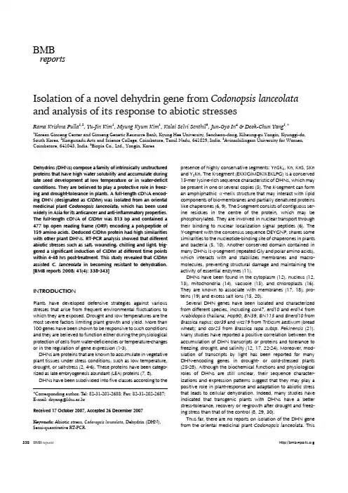
BMBreports338BMB reports*Corresponding author. T el: 82-31-201-2688; Fax: 82-31-202-2687;E-mail: dcyang@khu.ac.krReceived 17 October 2007, Accepted 26 December 2007K eywords: Abiotic stress, Codonopsis lanceolata , Dehydrin (DHN), Semi-quantitative RT-PCRIsolation of a novel dehydrin gene from Codonopsis lanceolata and analysis of its response to abiotic stressesRama Krishna Pulla 1,2, Yu-Jin Kim 1, Myung Kyum Kim 1, Kalai Selvi Senthil 3, Jun-Gyo In 4 & Deok-Chun Yang 1,*1Korean Ginseng Center and Ginseng Genetic Resource Bank, Kyung Hee University, Seocheon-dong, Kiheung-gu Yongin, Kyunggi-do, South Korea, 2Kongunadu Arts and Science College, Coimbatore, Tamil Nadu, 641029, India. 3Avinashilingam University for Women, Coimbatore, 641043, India. 4Biopia Co., Ltd., Yongin, KoreaDehydrins (DHNs) compose a family of intrinsically unstructured proteins that have high water solubility and accumulate during late seed development at low temperature or in water-deficit conditions. They are believed to play a protective role in freez-ing and drought-tolerance in plants. A full-length cDNA encod-ing DHN (designated as ClDhn ) was isolated from an oriental medicinal plant Codonopsis lanceolata , which has been used widely in Asia for its anticancer and anti-inflammatory properties. The full-length cDNA of ClDhn was 813 bp and contained a 477 bp open reading frame (ORF) encoding a polypeptide of 159 amino acids. Deduced ClDhn protein had high similarities with other plant DHNs. RT-PCR analysis showed that different abiotic stresses such as salt, wounding, chilling and light, trig-gered a significant induction of ClDhn at different time points within 4-48 hrs post-treatment. This study revealed that ClDhn assisted C. lanceolata in becoming resistant to dehydration. [BMB reports 2008; 41(4): 338-343]INTRODUCTIONPlants have developed defensive strategies against various stresses that arise from frequent environmental fluctuations to which they are exposed. Drought and low temperatures are the most severe factors limiting plant growth and yield. More than 100 genes have been shown to be responsive to such conditions and they are believed to function either during the physiological protection of cells from water-deficiencies or temperature-changes or in the regulation of gene expression (1-3).DHNs are proteins that are known to accumulate in vegetative plant tissues under stress conditions, such as low temperature, drought, or salt-stress (2, 4-6). These proteins have been catego-rized as late embryogenesis abundant (LEA) proteins (7, 8).DHNs have been subdivided into five classes according to thepresence of highly conservative segments: YnSK 2, Kn, KnS, SKn and Y 2Kn. The K-segment (EKKIGIMDKIKEKLPG) is a conserved 15-mer lysine-rich sequence characteristic of DHNs, which may be present in one or several copies (5). The K-segment can form an amphiphathic α-helix structure that may interact with lipid components of bio-membranes and partially denatured proteins like chaperones (6, 9). The S-segment consists of contiguous ser-ine residues in the centre of the protein, which may be phosphorylated. They are involved in nuclear transport through their binding to nuclear localization signal peptides (6). The Y-segment with the consensus sequence DEYGNP, shares some similarities to the nucleotide-binding site of chaperones in plants and bacteria (5, 10). Another conserved domain contained in many DHNs is ϕ-segment (repeated Gly and polar amino acids), which interacts with and stabilizes membranes and macro-molecules, preventing structural damage and maintaining the activity of essential enzymes (11).DHNs have been found in the cytoplasm (12), nucleus (12, 13), mitochondria (14), vacuole (15), and chloroplasts (16). They are known to associate with membranes (17, 18), pro-teins (19) and excess salt ions (15, 20). Several DHN genes have been isolated and characterized from different species, including cor47, erd10 and erd14 from Arabidopsis thaliana ; Hsp90, BN59, BN115 and Bnerd10 from Brassica napus ; cor39 and wcs19 from Triticum aestivum (bread wheat); and cor25 from Brassica rapa subsp. Pekinensis (21). Many studies have reported a positive correlation between the accumulation of DHN transcripts or proteins and tolerance to freezing, drought, and salinity (12, 17, 22-24). Moreover, mod-ulation of transcripts by light has been reported for many DHN-encoding genes in drought- or cold-stressed plants (25-28). Although the biochemical functions and physiological roles of DHNs are still unclear, their sequence character-izations and expression patterns suggest that they may play a positive role in plant-response and adaptation to abiotic stress that leads to cellular dehydration. Indeed, many studies have indicated that transgenic plants with DHNs have a better stress-tolerance, recovery or re-growth after drought and freez-ing stress than that of the control (8, 29, 30).Thus far, there are no reports on isolation of the DHN gene from the oriental medicinal plant Codonopsis lanceolata . ThisCodonopsis lanceolata dehydrin geneRama Krishna Pulla, et al.339BMBreportsFig. 1. Nucleotide sequence and de-duced amino acid sequence of a ClDhn cDNA isolated from C. lanceolata . Num-bers on the left represent nucleotide positions. The deduced amino acid se-quence is shown in a single-letter code below the nucleotide sequence. The as-terisk denotes the translation stop signal.Amino acids in two double boxes repre-sent the Y-segment and amino acids in a single box the S-segment, respectively.The two underlined sequences represent the K-segments.plant belongs to the family of Campanulaceae (bellflower fam-ily), which contains many famous oriental medicinal plants such as Platycodon grandiflorum (Chinese bellflower or balloon flow-er), Codonopsis pilosula and Adenophora triphylla (nan sha shen). The roots of these plants have been used as herbal drugs to treat bronchitis, cough, spasm, macrophage-mediated immune responses and inflammation, and has also been administered as a tonic (31). C. lanceolata grows in North-eastern china, Korea, and far eastern Siberia. Despite their medicinal importance, little genomic study of this plant has been carried out. In this study, we characterized an Y 2SK 2 type DHN gene from C. lanceolata and analyzed its expression in response to various abiotic stresses.RESULTS AND DISCUSSIONIsolation and characterization of the full length cDNA of the ClDhn geneAs part of a genomic project to identify genes in the medicinal plant C. lanceolata , a cDNA library consisting of about 1,000 cDNAs was previously constructed. A cDNA encoding a dehy-drin (DHN), designated ClDhn was isolated and sequenced. The sequence data of ClDhn has been deposited in GenBank under accession number AB126059. As shown in Fig. 1, ClDhn is 813 bp in length and it has an open reading frame (ORF) of 477 bp nucleotide with an 87-nucleotide upstream sequence and a 248-nucleotide downstream sequence. The ORF of ClDhn starts at nucleotide position 88 and ends at position 565. ClDhn encodes a precursor protein of 159 amino acids resi-dues with no predicted signal peptide at the N-terminal. The calculated molecular mass of the protein is approximately 16.7kDa with a predicated isoelectric point of 6.87. In the deduced amino acid sequence of ClDhn protein, the total number of neg-atively charged residues (Asp +Glu) amounted to 21 while the total number of positively charged residues (Arg +Lys) was 20. In addition, transmembrane helix prediction (TMHMMv2.0) did not identify any transmembrane helices in the deduced protein, implying that the protein did not function in the membrane but might function within the cytosolic or nuclear compartment.Homology analysisA GenBank Blastp search revealed that ClDhn had the highest sequence homology to the carrot (Daucus carota ) DHN (BAD86644) with 51% identity and 61% similarity. ClDhn also shared homology with ginseng (Panax ginseng ) DHN5 (ABF48478, 50% identity and 60% similarity), wild potato (Solanum commersonii ) DHN (CAA75798, 50% identity and 58% similarity), robusta coffee (Coffea canephora ) DHN1α (ABC55670, 47% identity and 55% similarity), grape (Vitis vin-ifera ) DHN (ABN79618, 47% identity and 57% similarity), American beech (Fagus sylvatica ) DHN (CAE54590, 46% iden-tity and 56% similarity), tobacco (Nicotiana tabacum ) DHN (BAD13498, 45% identity and 56% similarity), sunflower (Helianthus annuus ) DHN (CAC80719, 45% identity and 52% similarity), and soybean (Glycine max ) DHN (AAB71225, 44% identity and 52% similarity). The DHNs showing the highest similarities were Y 2SK 2 type DHNs except grape (Vitis vinifera ) DHN (YSK 2 type) (32). Thus ClDhn might belong to Y 2SK 2 type DHNs based on the two Y-segments, one S-segment, and two K-segments present in its amino acid sequence. Phylogenetic analysis of ten of the plant DHNs were carried out using theCodonopsis lanceolata dehydrin gene Rama Krishna Pulla, et al.340BMB reportsFig. 2. A phylogenetic tree based on DHN amino acid sequence, showing the phylogenetic relationship between ClDhn and other plant DHNs . The tree was constructed using the Clustal X method (Neighbor-joining method) and a bar represents 0.1 substitutions peramino acid position.Fig. 3. Alignment of ClDhn with the most closely related DHNs from carrot (Daucus carota , BAD86644), ginseng (Panax ginseng DHN5, ABF48478), com-merson’s wild potato (Solanum commer-sonii , CAA75798), robusta coffee (Coffea canephora , ABC55670), grape (Vitis vin-ifera , ABN79618), American beech (Fagus sylvatica , CAE54590), tobacco (Nicotiana tabacum , BAD13498), sunflower (Helian-thus annuus , CAC80719) and soybean (Glycine max , AAB71225). Gaps are marked with dashes. The conserved ami-no acid residues are shaded and Y-, S-, and K-segments are shown.Clustal X program (Fig. 2). Fig. 3 is a sequence alignment result of ClDhn and other closely related DHNs .The differential expression of ClDhn in different organs of C . lanceolataThe expression patterns of ClDhn in different C . lanceolata or-gans were examined using RT-PCR analysis. Almost similar levels of ClDhn -mRNA expression were observed in leaves and roots, whereas ClDhn was expressed in slightly higher lev-els in the stems. (Data was not shown).Expression of ClDhn in response to various stressesExpression patterns of ClDhn under various conditions were ex-amined using RT-PCR analysis. Fig. 4A showed the accumu-lation of ClDhn -mRNA in response to 100 mM ABA in MS agar. ABA is a hormone secreted when environmental conditions be-come dry. Expression of ClDhn was induced and reached a maximum level after 12 hrs, and then gradually decreased. When plants are submitted to dehydration the endogenous con-tent of ABA increases, with ABA mediating the closure of the stomata. Several studies have identified ABA as a key hormone in the induction pathway of many inducible genes including DHN , in response to drought (33-36). 100 μM of ABA in sprayCodonopsis lanceolata dehydrin geneRama Krishna Pulla, et al.341BMBreportsFig. 4. RT-PCR analyses of the expressions of ClDhn gene in the leaves of C. lanceolata at various time points (h) post-treatment with various stresses: A, 100 mM ABA; B, 100 mM NaCl; C, wounding; D, chilling and E, light treatment. Actin was used as an internal control.induced DHN-levels in Brassica napus and increased its ex-pression up to 48 hrs after treatment with ABA (37). 100 μM of ABA in MS agar induced DHN -levels in rice and cause a max-imum expression level at 1 hr post-treatment (10).Fig. 4B shows the accumulation of ClDhn mRNA in re-sponse to salt stress (100 mM NaCl). ClDhn expression was in-duced at 4 hrs post-treatment and gradually increased until 48 hrs. In Brassica napus , 250 mM NaCl added in the nutrient medium induced DHN-expression and reached a maximum at 48 hrs post-treatment (37). The application of NaCl to soil brought on a progressive decrease of the pre-dawn leaf water potential, a decrease of stomatal-conductance and a growth- reduction. Osmotic potential increase during salt treatmentcould result from Na + or Cl −absorption and from the synthesis of compatible compounds (38).Under wounding stress, ClDhn gene transcription was in-duced at 4 hrs post-treatment and gradually increased until 48 hrs (Fig. 4C). Richard et al . (39) discussed that the cumulative effect of wounding on transcript accumulation could also be associated with greater water-loss through more open surfaces arising from the wounding treatment.Under cold treatment, increase of ClDhn transcripts was ob-served at 4 hrs post-treatment and gradually increased until 48 hrs (Fig. 4D). Induction of DHN by low temperatures has been observed in numerous plants (17, 38). Overexpression of citrus DHN improved the cold tolerance in tobacco (18). Overexpre-ssion of multiple DHN genes in Arabidopsis resulted in accu-mulation of the corresponding DHNs to levels similar or higher than in cold-acclimated wild-type plants (24). Another example showed that overexpression of the acidic DHN WCOR410 could improve freezing tolerance in transgenic strawberry leaves (29). Fig. 4E shows that ClDhn gene expression was induced bylight stress and increased continuously until 48 hrs post-treat-ment. Natali et al . (40) showed that the G-box (CACGTGGC), a motif found in the promoter region of many light regulated genes, was found in the DHN gene promoter of helianthus and that DHN was responsive to light stress (41).In conclusion, we isolated a new dehydrin gene (ClDhn ) from C. lanceolata and characterized its expression in response to various stresses. ClDhn was induced by various stresses related to wa-ter-deficiency (ABA, salt, wounding and cold) and was induced by light, similar to other DHN genes isolated from other plants.MATERIALS AND METHODSPlant materialsCodonopsis lanceolata were grown in vitro on MS medium supplemented with 3% sucrose and 0.8% agar under the 16 hrs light and 8 hrs dark period. Its growth was maintained by regular subculture every 4 weeks. Abiotic stress studies were carried out on plants that were subcultured for one month. To analyze gene expression in different organs, samples were col-lected from leaves, roots and stem of C. lanceolata plants.Sequence analysesThe full-length ClDhn gene was analyzed using the softwares BioEdit, Clustal X, Mega 3 and other databases listed below; NCBI (http://www.ncbi.nlm.nih), SOPMA (http://npsa-pbil.ibcp /npsaautomat.pl?page=npsopma.html).Stress assaysTo investigate the response of the ClDhn gene to various stress-es, the third leaves with petioles from C. lanceolata were used. For treatment with ABA (100 mM) and NaCl (100 mM), leaf samples were incubated in media containing each compound at 25o C for 48 hrs. For mechanical wounding stress, excised leaves were wounded with a needle puncher (42). Chilling stress was applied by exposing the leaves to a temperature of 4o C (43). To investigate the ClDhn gene-expressions in light, leaves were incubated under an electrical lamp with a light in-tensity of 24 mol m-2 s-1 for 48 hrs. All treatments were carried out on MS media with or without the treatment solution (ABA, NaCl). All treated plant materials were immediately frozen in liquid nitrogen and stored at -70o C until further analysis.Semi-quantitative RT-PCR analysisTotal RNA was extracted from various whole plant tissues (leaves, stem, roots) of C. lancolata using the Rneasy mini kit (Qiagen, Valencia, CA, USA). For RT-PCR (reverse tran-scriptase-PCR), 800 ng of total RNA was used as a template for reverse transcription using oligo (dT) primer (0.2 mM)(INTRON Biotechnology, Inc., South Korea) for 5 mins at 75oC. The reaction mixture was then incubated with AMV Reverse Transcriptase (10 U/μl) (INTRON Biotechology, Inc., SouthKorea) for 60 mins at 42oC. The reaction was inactivated byheating the mixture at 94oC for 5 mins. PCR was then per-Codonopsis lanceolata dehydrin gene Rama Krishna Pulla, et al.342BMB reportsformed using a 1 μl aliquot of the first stand cDNA in a final volume of 25 μl containing 5 pmol of specific primers for cod-ing of ClDhn gene (forward, 5'-AAA GAG AGA GAA AAT GGC AGG TTA C-3'; reverse, 5'-GGA GTA GTT GTT GAA GTT CTC TGC T-3') were used. As a control, the primers spe-cific to the C. lanceolata actin gene were used (forward, 5'-CAA GAA GAG CTA CGA GCT ACC CGA TGG-3'; reverse, 5'-CTC GGT GCT AGG GCA GTG ATC TCT TTG CT-3'). PCR was carried out using 1 μl of taq DNA polymerase (Solgent Co., South Korea) in a thermal cycler programmed as follows:an initial denaturation for 5 mins at 95oC, 30 amplification cy-cles [30 s at 95o C (denaturation), 30 s at 53o C (annealing), and90 s at 72oC (polymerization)], followed by a final elongation for 10 mins at 72o C. Actin was used as an internal control to normalize each sample for variations in the amount of RNA used.AcknowledgementsThis work was supported by the Korea Science and Engineering Foundation (KOSEF) grant funded by the Korea government (MOST) (No. R01-2006-000-11178-0).REFERENCES1.Bray, E. A. (2002) Classification of the genes differentially expressed during water-deficit stress in Arabidopsis thali-ana : An analysis using micro array and differential ex-pression data. Ann. Bot. 89, 803-811.2.Ingram, J. and Bartels, D. (1996) The molecular basis of dehydration tolerance in plants. Annu. Rev. Plant Physiol. Plant Mol. Biol. 47, 377-403.3.Kim, S. J., Jeong, D. H., An, G. and Kim, S. R. (2005) Characterization of a drought-responsive gene, OsTPS1, identified by the T-DNA gene-trap system in rice. J. Plant Biol. 48, 371-379.4.Allagulova, Ch. R., Gimalor, F. R., Shakirova, F. M. and Vakhitov, V. A. (2003) The plant dehydrins: Structure and putative functions. Biochemistry (Mosc .) 68, 945-951.5.Close, T. J. (1996) Dehydrins: emergence of a biochemical role of a family of plant dehydration proteins. Physiol. Plant. 97, 795-803.6.Close, T. J. (1997) Dehydrins: a commonalty in the re-sponse of plants to dehydration and low temperature. Physiol. Plant. 100, 291-296.7.Dure, L., Crouch, M., Harada, J., Ho, T-HD., Mundy, J. and Quatrano, R. (1989) Common amino acid sequence domains among the LEA proteins of higher plants. Plant. Mol. Biol. 12, 475-486.8.Hara, M., Terashima, S., Fukaya, T. and Kuboi, T. (2003) Enhancement of cold tolerance and inhibition of lipid per-oxidation by citrus dehydrin in transgenic tobacco. Planta. 217, 290-298.9.Ismail, A. M., Hall, A. E. and Close, T. J. (1999) Allelic variation of a dehydrin gene cosegregates with chilling tolerance during seedling emergence. Proc. Natl. Aca. Sci. U.S.A. 96, 13566-13570.10.Lee, S. C., Lee, M. Y., Kim, S. J., Jun, S. H., An, G. andKim, S. R. (2005) Characterization of an abiotic stress-in-ducible dehydrin gene OsDhn1 in rice (Oryza sativa L .). Mol. Cells. 19, 212-218.11.Svensson J, Ismail, A. M., Palva, E. T. and Close, T. J. (2002) Dehydrins. In: Storey KB, Storey JM (eds) Sensing, signaling and cell adaptation. Elsevier Science B. V., Amsterdam, 99, pp. 155-171.12.Houde, M., Daniel, C., Lachapelle, M., Allard, F., Laliberte, S. and Sarhan, F. (1995) Immunolocalization of freezing- tolerance-associated proteins in the cytoplasm and nucleo-plasm of wheat crown tissues. Plant J. 8, 583-593.13.Godoy, J. A., Lunar, R., Torres-Schumann, S., Moreno, J., Rodrigo, R. M. and Pintor-Toro, J. A. (1994) Expression tis-sue distribution and subcellular localization of dehydrin TAS14 in salt stressed tomato plants. Plant Mol. Biol. 26, 1921-1934.14.Borovskii, G. B., Stupnikova, I. V., Antipina, A. I., Vladimirova, S. V. and Voinikov, V. K. (2002) Accumulation of dehydrin-like proteins in the mitochondria of cereals in re-sponse to cold, freezing, drought and ABA treatment. BMC Plant Biol. [electronic resource] 2, 5.15.Heyen, B. J., Alsheikh, M. K., Smith, E. A., Torvik, C. F., Seals, D. F. and Randall, S. K. (2002) The calcium-binding activity of a vacuole-associated, dehydrin-like protein is regulated by phosphorylation. Plant Physiol. 130, 675-687. 16.Mueller, J. K., Heckathorn, S. A. and Fernando, D. (2003) Identification of a chloroplast dehydrin in leaves of ma-ture plants. Int. J. Plant Sci. 164, 535-542.17.Danyluk, J., Perron, A., Houde, M., Limin, A., Fowler, B., Benhamoun, N. and Sarhan, F. (1998) Accumulation of an acidic dehydrin in the vicinity of plasma membrane dur-ing cold acclimation of wheat. Plant Cell. 10, 623-638.18.Koag, M. C., Fenton, R. D., Wilkens, S. and Close, T. J. (2003) The binding of maize DHN1 to lipid vesicles. Gain of structure and lipid specificity. Plant Physiol. 131, 309-316. 19.Rinne, P. L. H., Kaikuranta, P. L. M., van der Plas, L. H. W. and van der Schoot, C. (1999) Dehydrins in cold-accli-mated apices of birch (Betula pubescens Ehrh.): production, localization and potential role in rescuing enzyme function during dehydration. Planta. 209, 377-388.20.Alsheikh, M. K., Heyen, B. J. and Randall, S. K. (2003) Ion binding properties of the dehydrin ERD14 are dependent upon phosphorylation. J. Biol. Chem. 278, 40882-40889. 21.Fan, Z. and Wang, X. (2006) Isolation and Characterization of a Novel Dehydrin Gene from Capsella bursa-pastoris. J. Molecular Biology 40, 52-60.22.Cheng, Z., Targolli, J., Huang, X. and Wu, R. (2002) Wheat LEA genes, PMA80 and PMA1959, enhance dehydration tolerance of transgenic rice (Oryza sativa L.). Mol. Breed. 10, 71-82.23.Ismail, A. M., Hall, A. E. and Close, T. J. (1999) Purification and partial characterization of a dehydrin involved in chill-ing tolerance during seedling emergence of cowpea. Plant Physiol. 120, 237-244.24.Puhakainen, T., Hess, M. W., Mäkelä, P., Svensson, J., Heino, P. and Palva, E. T. (2004) Over expression of mul-tiple dehydrin genes enhances tolerance to freezing stress in Arabidopsis. Plant Mol. Biol. 54, 743-753.25.Chauvin, L. P., Oude, M. and Sarhan, F. (1993) A leaf-spe-cific gene stimulated by light during wheat acclimation to low temperature. Plant Mol. Biol. 23, 255-265.Codonopsis lanceolata dehydrin geneRama Krishna Pulla, et al.343 BMB reports 26.Crosatti, C., Polverino, D. L. P., Bassi, R. and Cattivelli, L.(1999) The interaction between cold and light controls the expression of the cold-regulated barley gene cor14b and the accumulation of the corresponding protein. Plant Physiol. 119, 671-680.27.Ohno, R., Takumi, S. and Nakamura, C. (2003) Kinetics oftranscript and protein accumulation of a low-molecular weight wheat LEA D-11 dehydrin in response to low temperature. J. Plant Physiol. 160, 193-200.28.Panta, G. R., Rieger, M. W. and Rowland, L. J. (2001)Effect of cold and drought stress on blue berry dehydrin accumulation. J. Hort. Sci. Biotech. 76, 549-556.29.Houde, M., Dallaire, S., N’Dong, D. and Sarhan, F. (2004)Over expression of the acidic dehydrin WCOR410 im-proves freezing tolerance in transgenic strawberry leaves. Plant Biotech. J. 2, 381-388.30.Yin, Z., Pawlowicz, I., Bartoszewski, G., Malinowski, R.,Malepszy, S. and Rorat, T. (2004) Transcritional expression of a Solanum sogarandinum pGT Dhn10 gene fusion in cucumber, and its correlation with chilling tolerance in transgenic seedling. Cell Mol. Biol. Lett. 9, 891-902.31.Lee, K. T., Choi, J., Jung, W. T., Nam, J. H., Jung, H. J. andPark, H. J. (2002) Structure of a New Echinocystic Acid Bisdesmoside Isolated from Codonopsis lanceolata Roots and the Cytotoxic Activity of Prosapogenins. J. Agric. Food Chem. 50, 4190-4193.32.Xiao, H. and Nassuth, A. (2006) Stress- and development-induced expression of spliced and unspliced transcripts from two highly similar dehydrin1 genes in V. riparia and V . vinifera. Plant Cell Rep. 25, 968-977.33.Bray, E. A. (1997) Plant responses to water deficit. TrendsPlant Sci. 2, 48-54.34.Chandler, P. M. and Robertson, M. (1994) Gene expressionregulated by abscisic acid and its relation to stress tolerance. Annu. Rev. Plant Physiol. Plant Mol. Biol. 45, 113-141.35.Rabbani, M. A., Maruyama, K., Abe, H., Kyan, M. A.,Katsura, K., Yoshiwara, K., Shinozaki, K. and Yamaguchi- Shinozaki, K. (2003) Monitoring expression profiles of ricegenes under cold drought and high salinity and abscisic acid application using cDNA micro array and RNA gel-blot analyses. Plant Physio. 133, 1755-1767.36.Shinozaki, K. and Yamaguchi-Shinozaki, K. (1996) Mole-cular responses to drought and cold stress. Curr. Opin. Biotechnol. 7, 161-167.37.Deng, Z. X., Pang, Y. Z., Kong, W. W., Chen, Z. H., Wang, X. L., Liu, X. J., Pi, Y., Sun, X. F. and Tang, K. X. ( 2005) A novel ABA dependent dehydrin ERD10 gene from Brassica napus. DNA seq. 16, 28-35.38.Caruso, A., Morabito, D., Delmotte, F., Kahlem, G. and Carpin, S. (2002) Dehydrin induction during drought and osmotic stress in Populus. Plant Physiol. Biochem. 40, 1033-1042.39.Richard, S., Morency, M. J., Drevet, C., Jouanin, L. and S´eguin, A. (2000) Isolation and characterization of a de-hydrin gene from white spruce induced upon wounding, drought and cold stresses. Plant Mol. Biol. 43, 1-10.40.Natali., Giordani, T., Lercari, B., Maestrini, P., Cozza, R., Pangaro, T., Vernieri, P., Martinelli, F. and Cavallini. (2007) A. Light induces expression of a dehydrin-encoding gene during seedling de-etiolation in sunflower (Helianthus an-nuus L .). J. Plant Physiol. 164, 263-273.41.Menkens, A. E., Schindler, U. and Cashmore, A, R. (1995) The G-box: a ubiquitous regulatory DNA element in plants bound by the GBF family of bZIP proteins. Trends Biochem Sci. 20, 506-510.42.Huh, G. H., Lee, S. J., Bae, Y. S., Liu, J. R. and Kwak, S. S. (1997) Molecular cloning and characterization of cDNAs for anionic and neutral peroxidases from suspension cul-tured cells of sweet potato and their differential expression in response to stress. Mol. Gen. Genet. 255, 382-391.43.Wu, W., Pang, Y., Shen, G., Lu, J., Lin, J., Wang, J., Sun, X. and Tang, K. (2006) Molecular Cloning, Characterization and Expression of a Novel Trehalose-6-phosphate Synthase Homologue from Ginkgo biloba. J. Biochem. Mol. Biol. 39, 158-166.。
核磁共振英语词汇英文回答:Nuclear magnetic resonance (NMR) is a powerfulanalytical tool that utilizes magnetic fields and radio waves to investigate the properties of atoms and molecules. It offers a non-destructive and versatile technique for characterizing materials at the atomic and molecular level. NMR has various applications across multiple scientific disciplines, including chemistry, physics, biology, and medicine.The basic principle of NMR involves the interaction between atomic nuclei with a magnetic field. Certain nuclei, such as 1H (proton), 13C, 15N, and 31P, possess anintrinsic magnetic moment due to their nuclear spin. When placed in a magnetic field, these nuclei align with or against the field, resulting in two distinct energy states. By applying radio waves to the sample at specific frequencies, it is possible to induce transitions betweenthese energy states.The absorption of radio waves by the nuclei leads to the resonance phenomenon, which forms the basis of NMR. The resonant frequency for a particular nucleus depends on its chemical environment, including the electron density and surrounding atoms. By analyzing the resonance frequencies and patterns, NMR provides detailed information about the structure, dynamics, and interactions of molecules.NMR spectroscopy is a widely used technique for identifying and quantifying different atoms and functional groups within molecules. It plays a crucial role in determining the molecular structure of organic and inorganic compounds, as well as studying chemical reactions and reaction mechanisms. NMR also finds applications in drug discovery and development, protein structure determination, and metabolomics.In medical imaging, NMR is employed as a non-invasive tool for obtaining detailed anatomical and functional information about the human body. Magnetic resonanceimaging (MRI) utilizes NMR techniques to create high-resolution images of organs, tissues, and blood vessels. MRI is particularly valuable for diagnosing and monitoring a wide range of medical conditions, including brain disorders, cardiovascular diseases, and musculoskeletal injuries.NMR also has applications in other fields, such as materials science, polymer characterization, and geological studies. It is a versatile technique that provides valuable insights into the structure, dynamics, and properties of various materials and systems.In summary, nuclear magnetic resonance (NMR) is a powerful analytical tool that offers a non-destructive and versatile approach for investigating the properties of atoms and molecules. Its applications span multiple scientific disciplines, including chemistry, physics, biology, and medicine, providing insights into molecular structure, dynamics, and interactions.中文回答:核磁共振(NMR)是一种强大的分析工具,利用磁场和射频波来研究原子和分子的性质。
地球磁场在减弱的证据英文回答:Evidence of Earth's Weakening Magnetic Field.The Earth's magnetic field, also known as the geomagnetic field, is generated by the movement of molten iron in the outer core of the planet. This magnetic field is crucial for protecting the Earth from harmful solar radiation and maintaining a stable climate. However, there is mounting evidence that the Earth's magnetic field is weakening.One piece of evidence is the observation of the South Atlantic Anomaly (SAA). The SAA is an area in the South Atlantic Ocean where the Earth's magnetic field is significantly weaker than in other regions. Satellites passing through this anomaly experience higher levels of radiation, which can affect their electronic systems. This anomaly has been expanding over the past few decades,indicating a weakening of the Earth's magnetic field.Another piece of evidence comes from studies of ancient rocks. Rocks contain tiny magnetic minerals that align with the Earth's magnetic field at the time of their formation. By analyzing these rocks, scientists can determine the strength and direction of the Earth's magnetic field in the past. These studies have revealed that the Earth's magnetic field has been weakening over the past few centuries.Furthermore, researchers have found that the rate of decline in the Earth's magnetic field has been accelerating in recent years. This rapid decline suggests that the weakening of the magnetic field is not a gradual process but rather a more significant and concerning phenomenon. If this trend continues, it could have significantimplications for our planet.The weakening of Earth's magnetic field has several potential consequences. One of the most significant is the increased exposure to solar radiation. The magnetic field acts as a shield, deflecting charged particles from the Sunaway from the Earth. Without a strong magnetic field, more solar radiation would reach the Earth's surface, increasing the risk of skin cancer and other health issues.Additionally, a weakened magnetic field could have implications for navigation systems that rely on magnetic compasses. The accuracy of compasses could be compromised, leading to errors in navigation. This could be particularly problematic for ships and aircraft that heavily rely on magnetic compasses for direction.In conclusion, there is compelling evidence that the Earth's magnetic field is weakening. The South Atlantic Anomaly, studies of ancient rocks, and the accelerating rate of decline all point to this concerning phenomenon. The implications of a weakened magnetic field range from increased exposure to solar radiation to potential navigation issues. It is crucial for scientists to continue monitoring and studying this phenomenon to better understand its implications for our planet.中文回答:地球磁场减弱的证据。

Contents01.02.03.04.05.06.07.08.09.10.11.12.13.14.15.16.17.18.19.20.21.22.23.24.25.26.27.28.29.30.31.32.33.34.35.36.37.38.39.40. THE ELEMENTS AND THE PERIODIC TABLE...................................................... - 3 - THE NONMETAL ELEMENTS .................................................................................. - 5 - GROUPS IB AND IIB ELEMENTS ............................................................................ - 7 - GROUPS IIIB—VIIIB ELEMENTS ............................................................................ - 9 - INTERHALOGEN AND NOBLE GAS COMPOUNDS ........................................... - 11 - THE CLASSIFICATION OF INORGANIC COMPOUNDS .................................... - 13 - THE NOMENCLATURE OF INORGANIC COMPOUNDS.................................... - 15 - BRONSTED'S AND LEWIS' ACID-BASE CONCEPTS.......................................... - 19 - THE COORDINATION COMPLEX.......................................................................... - 22 - ALKANES .................................................................................................................. - 25 - UNSATURATED COMPOUNDS ............................................................................. - 28 - THE NOMENCLATURE OF CYCLIC HYDROCARBONS ................................... - 30 - SUBSTITUTIVE NOMENCLATURE....................................................................... - 33 - THE COMPOUNDS CONTAINING OXYGEN ....................................................... - 37 - PREPARATION OF A CARBOXYLiC ACID BY THE GRIGNARD METHOD .. - 39 - THE STRUCTURES OF COVALENT COMPOUNDS ............................................ - 41 - OXIDATION AND REDUCTION IN ORGANIC CHEMISTRY ............................ - 44 - SYNTHESIS OF ALCOHOLS AND DESIGN OF ORGANIC SYNTHESIS .......... - 47 - ORGANOMETALLICS—METAL π COMPLEXES ................................................ - 49 - THE ROLE OF PROTECTIVE GROUPS IN ORGANIC SYNTHESIS................... - 52 - ELECTROPHILIC REACTIONS OF AROMATIC COMPOUNDS ........................ - 54 - POLYMERS................................................................................................................ - 57 - ANALYTICAL CHEMISTRY AND PROBLEMS IN SOCIETY ............................ - 61 - VOLUMETRIC ANALYSIS ...................................................................................... - 63 - QUALITATIVE ORGANIC ANALYSIS .................................................................. - 65 - VAPOR-PHASE CHROMATOGRAPHY ................................................................. - 67 - INFRARED SPECTROSCOPY.................................................................................. - 70 - NUCLEAR MAGNETIC RESONANCE (I) .............................................................. - 72 - NUCLEAR MAGNETIC RESONANCE(II).............................................................. - 75 - A MAP OF PHYSICAL CHEMISTRY...................................................................... - 77 - THE CHEMICAL THERMODYNAMICS ................................................................ - 79 - CHEMICAL EQUILIBRIUM AND KINETICS ........................................................ - 82 - THE RATES OF CHEMICAL REACTIONS ............................................................ - 85 - NATURE OF THE COLLOIDAL STATE................................................................. - 88 - ELECTROCHEMICAL CELLS ................................................................................. - 90 - BOILING POINTS AND DISTILLATION ............................................................... - 93 - EXTRACTIVE AND AZEOTROPIC DISTILLATION ............................................ - 96 - CRYSTALLIZATION ................................................................................................ - 98 - MATERIAL ACCOUNTING ................................................................................... - 100 - THE LITERATURE MATRIX OF CHEMISTRY................................................... - 102 -01. THE ELEMENTS AND THE PERIODIC TABLEThe number of protons in the nucleus of an atom is referred to as the atomic number, or proton number, Z. The number of electrons in an electrically neutral atom is also equal to the atomic number, Z. The total mass of an atom is determined very nearly by the total number of protons and neutrons in its nucleus. This total is called the mass number, A. The number of neutrons in an atom, the neutron number, is given by the quantity A-Z.The term element refers to, a pure substance with atoms all of a single kind. To the chemist the "kind" of atom is specified by its atomic number, since this is the property that determines its chemical behavior. At present all the atoms from Z = 1 to Z = 107 are known; there are 107 chemical elements. Each chemical element has been given a name and a distinctive symbol. For most elements the symbol is simply the abbreviated form of the English name consisting of one or two letters, for example:oxygen==O nitrogen == N neon==Ne magnesium == MgA complete listing of the elements may be found in Table 1.Beginning in the late seventeenth century with the work of Robert Boyle, who proposed the presently accepted concept of an element, numerous investigations produced a considerable knowledge of the properties of elements and their compounds1. In 1869, D.Mendeleev and L. Meyer, working independently, proposed the periodic law. In modern form, the law states that the properties of the elements are periodic functions of their atomic numbers. In other words, when the elements are listed in order of increasing atomic number, elements having closely similar properties will fall at definite intervals along the list. Thus it is possible to arrange the list of elements in tabular form with elements having similar properties placed in vertical columns2. Such an arrangement is called a periodic Each horizontal row of elements constitutes a period. It should be noted that the lengths of the periods vary. There is a very short period containing only 2 elements, followed by two short periods of 8 elements each, and then two long periods of 18 elements each. The next period includes 32 elements, and the last period is apparently incomplete. With this arrangement, elements in the same vertical column have similar characteristics. These columns constitute the chemical families or groups. The groups headed by the members of the two 8-element periods are designated as main group elements, and the members of the other groups are called transition or inner transition elements.In the periodic table, a heavy stepped line divides the elements into metals and nonmetals. Elements to the left of this line (with the exception of hydrogen) are metals, while those to the right are nonmetals. This division is for convenience only; elements bordering the line—the metalloids-have properties characteristic of - both metals and nonmetals. It may be seen that most of the elements, including all the transition and inner transition elements, are metals.Except for hydrogen, a gas, the elements of group IA make up the alkali metal family. They are very reactive metals, and they are never found in the elemental state in nature. However, their compounds are widespread. All the members of the alkali metal family, form ions having a charge of 1+ only. In contrast, the elements of group IB —copper, silver, and gold—are comparatively inert. They are similar to the alkali metals in that they exist as 1+ ions in many of their compounds. However, as is characteristic of most transition elements, they form ions having other charges as well.The elements of group IIA are known as the alkaline earth metals. Their characteristic ionic charge is 2+. These metals, particularly the last two members of the group, are almost as reactive as the alkali metals. The group IIB elements—zinc, cadmium, and mercury are less reactive than are those of group II A5, but are more reactive than the neighboring elements of group IB. The characteristic charge on their ions is also 2+.With the exception of boron, group IIIA elements are also fairly reactive metals. Aluminum appears to be inert toward reaction with air, but this behavior stems from the fact that the metal forms a thin, invisible film of aluminum oxide on the surface, which protects the bulk of the metal from further oxidation. The metals of group IIIA form ions of 3+ charge. Group IIIB consists of the metals scandium, yttrium, lanthanum, and actinium.Group IVA consists of a nonmetal, carbon, two metalloids, silicon and germanium, and two metals, tin and lead. Each of these elements forms some compounds with formulas which indicate that four other atoms are present per group IVA atom, as, for example, carbon tetrachloride, GCl4. The group IVB metals —titanium, zirconium, and hafnium —also forms compounds in which each group IVB atom is combined with four other atoms; these compounds are nonelectrolytes when pure.The elements of group V A include three nonmetals —nitrogen, phosphorus, and arsenic—and two metals —antimony and bismuth. Although compounds with the formulas N2O5, PCl5, and AsCl5exist, none of them is ionic. These elements do form compounds-nitrides, phosphides, and arsenides — in which ions having charges of minus three occur. The elements of group VB are all metals. These elements form such a variety of different compounds that their characteristics are not easily generalized.The elements of groups ⅥB, ⅦB, and VIIIB are all metals. They form such a wide Variety of compounds that it is not practical at this point to present any examples as being typical of the behavior of the respective groups.The periodicity of chemical behavior is illustrated by the fact that. excluding the first period, each period begins with a very reactive metal. Successive element along the period show decreasing metallic character, eventually becoming nonmetals, and finally, in group ⅦA, a very reactive nonmetal is found. Each period ends with a member of the noble gas family.02. THE NONMETAL ELEMENTSWe noted earlier. that -nonmetals exhibit properties that are greatly different from those of the metals. As a rule, the nonmetals are poor conductors of electricity (graphitic carbon is an exception) and heat; they are brittle, are often intensely colored, and show an unusually wide range of melting and boiling points. Their molecular structures, usually involving ordinary covalent bonds, vary from the simple diatomic molecules of H2, Cl2, I2, and N2 to the giant molecules of diamond, silicon and boron.The nonmetals that are gases at room temperature are the low-molecular weight diatomic molecules and the noble gases that exert very small intermolecular forces. As the molecular weight increases, we encounter a liquid (Br2) and a solid (I2) whose vapor pressures also indicate small intermolecular forces. Certain properties of a few nonmetals are listed in Table 2.Table 2- Molecular Weights and Melting Points of Certain NonmetalsSimple diatomic molecules are not formed by the heavier members of Groups V and VI at ordinary conditions. This is in direct contrast to the first members of these groups, N2 and O2. The difference arises because of the lower stability of π bonds formed from p orbitals of the third and higher main energy levels as opposed to the second main energy level2. The larger atomic radii and more dense electron clouds of elements of the third period and higher do not allow good parallel overlap of p orbitals necessary for a strong π bond. This is a general phenomenon —strong π bonds are formed only between elements of the second period. Thus, elemental nitrogen and oxygen form stable molecules with both σ and π bonds, but other members of their groups form more stable structures based on σ bonds only at ordinary conditions. Note3 that Group VII elements form diatomic molecules, but π bonds are not required for saturation of valence.Sulfur exhibits allotropic forms. Solid sulfur exists in two crystalline forms and in an amorphous form. Rhombic sulfur is obtained by crystallization from a suitable solution, such as CS2, and it melts at 112°C. Monoclinic sulfur is formed by cooling melted sulfur and it melts at 119°C. Both forms of crystalline sulfur melt into S-gamma, which is composed of S8 molecules. The S8 molecules are puckered rings and survive heating to about 160°C. Above 160°C, the S8 rings break open, and some of these fragments combine with each other to form a highly viscous mixture of irregularly shaped coils. At a range of higher temperatures the liquid sulfur becomes so viscous that it will not pourfrom its container. The color also changes from straw yellow at sulfur's melting point to a deep reddish-brown as it becomes more viscous.As4 the boiling point of 444 °C is approached, the large-coiled molecules of sulfur are partially degraded and the liquid sulfur decreases in viscosity. If the hot liquid sulfur is quenched by pouring it into cold water, the amorphous form of sulfur is produced. The structure of amorphous sulfur consists of large-coiled helices with eight sulfur atoms to each turn of the helix; the overall nature of amorphous sulfur is described as3 rubbery because it stretches much like ordinary rubber. In a few hours the amorphous sulfur reverts to small rhombic crystals and its rubbery property disappears.Sulfur, an important raw material in industrial chemistry, occurs as the free element, as SO2 in volcanic regions, asH2S in mineral waters, and in a variety of sulfide ores such as iron pyrite FeS2, zinc blende ZnS, galena PbS and such, and in common formations of gypsum CaSO4• 2H2O, anhydrite CaSO4, and barytes BaSO4• 2H2O. Sulfur, in one form or another, is used in large quantities for making sulfuric acid, fertilizers, insecticides, and paper.Sulfur in the form of SO2 obtained in the roasting of sulfide ores is recovered and converted to sulfuric acid, although in previous years much of this SO2 was discarded through exceptionally tall smokestacks. Fortunately, it is now economically favorable to recover these gases, thus greatly reducing this type of atmospheric pollution. A typical roasting reaction involves the change:2 ZnS +3 O2—2 ZnO + 2 SO2Phosphorus, below 800℃ consists of tetratomic molecules, P4. Its molecular structure provides for a covalence of three, as may be expected from the three unpaired p electrons in its atomic structure, and each atom is attached to three others6. Instead of a strictly orthogonal orientation, with the three bonds 90° to each other, the bond angles are only 60°. This supposedly strained structure is stabilized by the mutual interaction of the four atoms (each atom is bonded to the other three), but it is chemically the most active form of phosphorus. This form of phosphorus, the white modification, is spontaneously combustible in air. When heated to 260°C it changes to red phosphorus, whose structure is obscure. Red phosphorus is stable in air but, like all forms of phosphorus, it should be handled carefully because of its tendency to migrate to the bones when ingested, resulting in serious physiological damage.Elemental carbon exists in one of two crystalline structures — diamond and graphite. The diamond structure, based on tetrahedral bonding of hybridized sp3orbitals, is encountered among Group IV elements. We may expect that as the bond length increases, the hardness of the diamond-type crystal decreases. Although the tetrahedral structure persists among the elements in this group — carbon, silicon, germanium, and gray tin — the interatomic distances increase from 1.54 A for carbon to 2.80 A for gray tin. Consequently .the bond strengths among the four elements range from very strong to quite weak. In fact, gray tin is so soft that it exists in the form of microcrystals or merely as a powder. Typical of the Group IV diamond-type crystalline elements, it is a nonconductor and shows other nonmetallic properties7.03. GROUPS IB AND IIB ELEMENTSPhysical properties of Group IB and IIBThese elements have a greater bulk use as metals than in compounds, and their physical properties vary widely. Gold is the most malleable and ductile of the metals. It can be hammered into sheets of 0.00001 inch in thickness; one gram of the metal can be drawn into a wire 1.8 mi in length1. Copper and silver are also metals that are mechanically easy to work. Zinc is a little brittle at ordinary temperatures, but may be rolled into sheets at between 120° to 150℃; it becomes brittle again about 200℃-The low-melting temperatures of zinc contribute to the preparation of zinc-coated iron .galvanized iron; clean iron sheet may be dipped into vats of liquid zinc in its preparation. A different procedure is to sprinkle or air blast zinc dust onto hot iron sheeting for a zinc melt and then coating.Cadmium has specific uses because of its low-melting temperature in a number of alloys. Cadmium rods are used in nuclear reactors because the metal is a good neutron absorber.Mercury vapor and its salts are poisonous, though the free metal may be taken internally under certain conditions. Because of its relatively low boiling point and hence volatile nature, free mercury should never be allowed to stand in an open container in the laboratory. Evidence shows that inhalation of its vapors is injurious.The metal alloys readily with most of the metals (except iron and platinum) to form amalgams, the name given to any alloy of mercury.Copper sulfate, or blue vitriol (CuSO4• 5H2O) is the most important and widely used salt of copper. On heating, the salt slowly loses water to form first the trihydrate (CuSO4• 3H z O), then the monohydrate (CuSO4• H2O), and finally the white anhydrous salt. The anhydrous salt is often used to test for the presence of water in organic liquids. For example, some of the anhydrous copper salt added to alcohol (which contains water) will turn blue because of the hydration of the salt.Copper sulfate is used in electroplating. Fishermen dip their nets in copper sulfate solution to inhibit the growth of organisms that would rot the fabric. Paints specifically formulated for use on the bottoms of marine craft contain copper compounds to inhibit the growth of barnacles and other organisms.When dilute ammonium hydroxide is added" to a solution of copper (I) ions, a greenish precipitate of Cu(OH)2 or a basic copper(I) salt is formed. This dissolves as more ammonium hydroxide is added. The excess ammonia forms an ammoniated complex with the copper (I) ion of the composition, Cu(NH3)42+. This ion is only slightly dissociated; hence in an ammoniacal solution very few copper (I) ions are present. Insoluble copper compounds, execpt copper sulfide, are dissolved by ammonium hydroxids. The formation of the copper (I) ammonia ion is often used as a test for Cu2+ because of its deep, intense blue color.Copper (I) ferrocyanide [Cu2Fe(CN)6] is obtained as a reddish-brown precipitate on the addition of a soluble ferrocyanide to a solution of copper ( I )ions. The formation of this salt is also used as a test for the presence of copper (I)ions.3Ag + 4HNO3—3AgNO3 + NO + 2H2OThe salt is the starting material for most of the compounds of silver, including the halides used in photography. It is readily reduced by organic reducing agents, with the formation of a black deposit of finely divided silver; this action is responsible for black spots left on the fingers from the handling of the salt. Indelible marking inks and pencils take advantage of this property of silver nitrate.The halides of silver, except the fluoride, are very insoluble compounds and may be precipitated by the addition of a solution of silver salt to a solution containing chloride, bromide, or iodide ions.The addition of a strong base to a solution of a silver salt precipitates brown silver oxide (Ag2G). One might expect the hydroxide of silver to precipitate, but it seems likely that silver hydroxide is very unstable and breaks down into the oxide and water — if, indeed, it is ever formed at all3. However, since a solution of silver oxide js definitely basic, there must be hydroxide ions present in solution.Ag2O + H2O = 2Ag+ + 2OH-Because of its inactivity, gold forms relatively few compounds. Two series of compounds are known — monovalent and trivalent. Monovalent (aurous) compounds resemble silver compounds (aurous chloride is water insoluble and light sensitive), while the higher valence (auric) compounds tend to form complexes. Gold is resistant to the action of most chemicals —air, oxygen, and water have no effect. The common acids do not attack the metal, but a mixture of hydrochloric and nitric acids (aqua regia) dissolves it to form gold( I ) chloride or chloroauric acid. The action is probably due to free chlorine present in the aqua regia.3HCl + HNO3----→ NOCl+Cl2 + 2H2O2Au + 3Cl2 ----→ 2AuCl3AuCl3+HCl----→ HAuCl4chloroauric acid (HAuCl4-H2O crystallizes from solution).Zinc is fairly high in the activity series. It reacts readily with acids to produce hydrogen and displaces less active metals from their salts. 1 he action of acids on impure zinc is much more rapid than on pure zinc, since bubbles of hydrogen gas collect on the surface of pure zinc and slow down the action. If another metal is present as an impurity, the hydrogen is liberated from the surface of the contaminating metal rather than from the zinc. An electric couple to facilitate the action is probably Set up between the two metals.Zn + 2H+----→ Zn2+ + H2Zinc oxide (ZnO), the most widely used zinc compound, is a white powder at ordinary temperatures, but changes to yellow on heating. When cooled, it again becomes white. Zinc oxide is obtained by burning zinc in air, by heating the basic carbonate, or by roasting the sulfide. The principal use of ZnO is as a filler in rubber manufacture, particularly in automobile tires. As a body for paints it has the advantage over white lead of not darkening on exposure to an atmosphere containing hydrogen sulfide. Its covering power, however, is inferior to that of white lead.04. GROUPS IIIB—VIIIB ELEMENTSThe predominant group oxidation number of the lanthanide series is +3, but some of the elements exhibit variable oxidation states. Cerium forms cerium( III )and cerium ( IV ) sulfates, Ce2 (SO4 )3 and Ce(SO4 )2, which are employed in certain oxidation-reduction titrations. Many rare earth compounds are colored and are paramagnetic, presumably as a result of unpaired electrons in the 4f orbitals.All actinide elements have unstable nuclei and exhibit radioactivity. Those with higher atomic numbers have been obtained only in trace amounts. Actinium (89 Ac), like lanthanum, is a regular Group IIIB element.Group IVB ElementsIn chemical properties these elements resemble silicon, but they become increasingly more metallic from titanium to hafnium. The predominant oxidation state is +4 and, as with silica (SiO2), the oxides of these elements occur naturally in small amounts. The formulas and mineral names of the oxides are TiO2, rutile; ZrO2, zirconia; HfO2, hafnia. Titanium is more abundant than is usually realized. It comprises about 0.44% of the earth's crust. It is over 5.0% in average composition of first analyzed moon rock. Zirconium and titanium oxides occur in small percentages in beach sands.Titanium and zirconium metals are prepared by heating their chlorides with magnesium metal. Both are particularly resistant to corrosion and have high melting points.Pure TiO2 is a very white substance which is taking the place of white lead in many paints. Three-fourths of the TiO2 is used in white paints, varnishes, and lacquers. It has the highest index of refraction (2.76) and the greatest hiding power of all the common white paint materials. TiO2 also is used in the paper, rubber, linoleum, leather, and textile industries.Group VB Elements: Vanadium, Niobium, and TantalumThese are transition elements of Group VB, with a predominant oxidation number of + 5. Their occurrence iscomparatively rare.These metals combine directly with oxygen, chlorine, and nitrogen to form oxides, chlorides, and nitrides, respectively. A small percentage of vanadium alloyed with steel gives a high tensile strength product which is very tough and resistant to shock and vibration. For this reason vanadium alloy steels are used in the manufacture ofhigh-speed tools and heavy machinery. Vanadium oxide is employed as a catalyst in the contact process of manufacturing sulfuric acid. Niobium is a very rare element, with limited use as an alloying element in stainless steel. Tantalum has a very high melting point (2850 C) and is resistant to corrosion by most acids and alkalies.Groups VIB and VIIB ElementsChromium, molybdenum, and tungsten are Group VIB elements. Manganese is the only chemically important element of Group VIIB. All these elements exhibit several oxidation states, acting as metallic elements in lower oxidation states and as nonmetallic elements in higher oxidation states. Both chromium and manganese are widely used in alloys, particularly in alloy steels.Group VIIIB MetalsGroup VIIIB contains the three triads of elements. These triads appear at the middle of long periods of elements in the periodic table, and are members of the transition series. The elements of any given horizontal triad have many similar properties, but there are marked differences between the properties of the triads, particularly between the first triad and the other two. Iron, cobalt, and nickel are much more active than members of the other two triads, and are also much more abundant in the earth's crust. Metals of the second and third triads, with many common properties, are usually grouped together and called the platinum metals.These elements all exhibit variable oxidation states and form numerous coordination compounds.CorrosionIron exposed to the action of moist air rusts rapidly, with the formation of a loose, crumbly deposit of the oxide. The oxide does not adhere to the surface of the metal, as does aluminum oxide and certain other metal oxides, but peelsCorrosion of iron is inhibited by coating it with numerous substances, such as paint, an aluminum powder gilt, tin, or organic tarry substances or by galvanizing iron with zinc. Alloying iron with metals such as nickel or chromium yields a less corrosive steel. "Cathodic protection" of iron for lessened corrosion is also practiced. For some pipelines and standpipes zinc or magnesium rods in the ground with a wire connecting them to an iron object have the following effect: with soil moisture acting as an electrolyte for a Fe — Zn couple the Fe is lessened in its tendency to become Fe2+. It acts as a cathode rather than an anode.2 205. INTERHALOGEN AND NOBLE GAS COMPOUNDSInterhalogen and noble gas compounds comprise a relatively limited family of highly reactive and unstable molecules whose primary importance is their role in testing chemical bonding theory 1. At first it may seem rather strange to treat the chemistry of the halogens and the noble gases, two groups that represent the extremes of chemical activity and inertia, in the same section. The superficial differences between the halogens and the noble gases are much reduced, however, if we focus our attention on the comparison of halide ions (particularly F -) with the isoelectronic noble gas atoms and the noble gas compounds with halogen atoms or the halogens in their higher positive-oxidation states. Noble gases are exceptional in their reluctance to either gain or lose an electron. Halide ions — because of their excess negative charge, relative to the isoelectronic noble gas atoms — have both a lower ionization energy and a lesser electron affinity. On the other hand, noble gas cations have greater electron affinities and greater ionization energies than do isoelectronic halogen atoms. From such considerations, it is obvious that inert gases should be less reactive than are halide ions, but their compounds should show even higher reactivity than the halogens. The big question remaining is : Are there any chemically significant conditions under which noble gases can be persuaded to yield electrons sufficiently to produce stable compounds? The answer is definitely , yes! (The same question can be asked of halogen atoms, which have ionization energies comparable to those of the inert gases.)Another obvious point of similarity between halogen and noble gas compounds is the characteristically large number of electrons that must be accommodated in the valence shell. For a noble gas atom bonded to any number of other atoms, the octet rule must be exceeded; for a halogen atom to be bonded to more than one other atom, the same must be true. It is a curious historical fact that the mythical inertia of a closed shell did much to diminish the energy expended in the search for noble gas compounds, long after numerous examples of superoctet valence shells were known, particularly among interhalogen compounds.We may roughly classify the interhalogen compounds into two categories: those in oxidation state zero (the binary analogs of the elementary diatomics 2) and those in which one of the halogens is in a formally positive oxidation state. Heterodiatomic halogens are generally formed readily on mixing the required pair of halogens in a 1:1 ratio. The bond energies are always higher in the heteropolar molecules than are the average bond energies of the two constituents and in some cases higher than either. It is this factor that drives the reactions. All heterodiatomics 2 are more or less stable under ambient conditions except for BrF, which spontaneously disproportionates to BrF 3 and Br 2. The bonding in the halogen diatomics can be attributed to a single a bond, formed by overlap of p orbitals. In the heterodiatomics, the principal new features are the poorer orbital overlaps that are possible between atoms of widely different principal quantum number (n ), the polarity arising from the difference in electronegativity, the contribution of ionic terms to increase bond energy, and the relief in interelectronic repulsion in the fluorides. relative to difluorine.Dihalogens (except for F 2) usually react by dissociation into atoms or by heterolytic dissociation under the influenceof an attacking reagent. Thus, reaction of Cl with hydroxide may be viewed as displacement of Cl - from Cl by OH -: Cl 2+OH - -----→HOCl + Cl -The tendency to undergo heterolytic fission increases on descending the group, and the I 2 molecule can actually be cleaved to two stable species:I 2 + 2C 5H 5N+AgNO 3-----→ [C 5H 5N)2I]NO 3 + AgI。

http :/ / 中西医结合护理Chinese Journal of Integrative Nursing2023 年第 9 卷第 12 期Vol.9, No.12, 2023OPEN ACCESS雷火灸配合手法穴位按摩改善1例风寒证面瘫患者面神经功能的护理体会李梦儒, 王雪送, 李陈晨, 张晨, 张诗语(北京中医药大学东方医院 脑病二科, 北京, 100078)摘要: 本文总结1例采用雷火灸配合手法穴位按摩改善风寒证面瘫患者面神经功能的护理经验。
在常规护理的基础上,根据患者辨证采取雷火灸配合手法穴位按摩,通过中医外治法精准作用于病灶,增强经络的感知传导,改善面神经活动能力,进而促进患者面瘫的康复。
关键词: 面瘫; 雷火灸; 按摩; 中医护理中图分类号: R 473.5 文献标志码: A 文章编号: 2709-1961(2023)12-0148-03Improvement of facial nerve function by thunder -firemoxibustion combined with acupoint massage in a patientwith facial paralysis of wind -cold patternLI Mengru ,WANG Xuesong ,LI Chenchen ,ZHANG Chen ,ZHANG Shiyu(The Second Department of Encephalopathy , Dongfang Hospital BeijingUniversity of Chinese Medicine , Beijing , 100078)ABSTRACT : This paper summarized the treatment effect of thunder -fire moxibustion combinedwith acupoint massage on the rehabilitation of facial nerve function in a patient with facial paraly⁃sis of wind -cold pattern. Based on the syndrome differentiation , a combination of traditional Chi⁃nese Medicine external techniques including thunder -fire moxibustion and acupoint massage was carried out to relieve the clinical symptoms and improve the recovery of facial nerve function of the patient with facial paralysis of wind -cold pattern.KEY WORDS : facial paralysis ; thunder -fire moxibustion ; massage ; Traditional Chinese Medicine nursing周围性面瘫是面神经核或其下方的神经,受到急性非脓性面神经炎的影响而引发[1]。
a rXiv:077.2783v2[astro-ph]19Mar28Table of contents•Abstract ———1•1.Introduction———–2•2.Observed properties of galactic magnetic fields –4•3.Summary of our present understanding of cosmic magnetic field origins————9•4.Basic equations for magnetic field evolution –12•5.Cowling’s theorem and Parker’s dynamo –15•6.The alpha-Omega disc dynamo —18•7.The magnitudes of αand βin the interstellar medium —23•8.Ferri`e re’s dynamo theory based on supernova and superbubble explosions—26•9.The validity of the vacuum boundary conditions –34•10.Arguments against a primordial origin –37•11.Seed fields —-41•12A protogalactic theory for magnetic field generation —45•13.Generation of small scale magnetic fields by turbulence —48•14.The saturation of the small scale magnetic fields ——–54•15.History of the evolution of a primordial magnetic field ——–58•16.Extragalactic magnetic fields –66•17.Summary and conclusions –67•18.Publications —701On the Origin of Cosmic Magnetic FieldsRussell M.Kulsrud and Ellen G.Zweibel Princeton University and the University ofWisconsin-MadisonAbstractWe review the extensive and controversial literature concerning how the cosmic magneticfields pervading nearly all galaxies and clus-ters of galaxies actually got started.Some observational evidence supports a hypothesis that thefield is already moderately strong at the beginning of the life of a galaxy and its disc.One argument in-volves the chemical abundance of the light elements Be and B,while a second one is based on the detection of strong magneticfields in very young high–red–shift galaxies.Since this problem of initial amplification of cosmic magneticfields involves important plasma problems,it is obvious that one must know the plasma in which the amplification occurs.Most of this review is devoted to this basic problem and,for this,it is necessary to devote ourselves to reviewing studies that take place in environments in which the plasma properties are most clearly understood.For this reason the authors have chosen to restrict themselves almost completely to studies of dynamo action in our Galaxy.It is true that one can get a much better idea of the grand scope of galacticfields in extragalactic systems.However,most mature galaxies share the same dilemma as ours of overcoming important plasma problems.Since the authors are both trained in plasma physics,they may be biased in pursuing this approach,but they feel this restriction it is justified by the above argument.In addition,they feel they can produce a better review by staying close to that which they know best.In addition they have chosen not to consider the saturation prob-lem of the galactic magneticfield since,if the original dynamo ampli-fication fails,the saturation question does not enter.It is generally accepted that seedfields,whose strength is of order 10−20gauss,easily spring up in the era preceding galaxy formation. Several mechanisms have been proposed to amplify these seed mag-neticfields to a coherent structure with the microgauss strengths of the currently observed galactic magneticfields.The standard and most popular mechanism is the alpha-Omega mean–field dynamo theory developed by a number of people in the2late sixties.This theory and its application to galactic magneticfields is discussed in considerable detail in this review.We point out certain difficulties with this theory that make it seem unlikely that this is the whole story.The main difficulty with this as the only such amplifica-tion mechanism,is rooted in the fact that,on galactic scales,flux is constant and is frozen in the interstellar medium.This implies that flux must be removed from the galactic discs,as is well recognized by the standard theory.For our Galaxy this turns out to be a major problem,since unless theflux and the interstellar mass are somehow separated,some inter-stellar mass must also be removed from the deep galactic gravitational well.This is very difficult.It is pointed out that unless thefield has a substantialfield strength,much larger than that of the seedfields, this separation can hardly happen.And of course,the alpha–Omega dynamo must start from the ultra weak seedfield.(It is our philos-ophy,expressed in this review,that if an origin theory is unable to create the magneticfield in our Galaxy it is essentially incomplete.) Thus,it is more reasonable for thefirst and largest amplification to occur before the Galaxy forms and the matter embedded in thefield is gravitationally trapped.Two such mechanisms are discussed for such a pregalactic origin;1)thefields are generated in the turbulence of the protogalaxy and2)thefields come from giant radio jets.Several arguments against a primordial origin are also discussed,as are ways around them.Our conclusion as to the most likely origin of cosmic magnetic fields is that they arefirst produced at moderatefield strengths by primordial mechanisms,and then changed and their strength increased to their present value and structure by a galactic disc dynamo.The primordial mechanisms have not yet been seriously developed as of yet,and this preliminary amplification of the magneticfields is still very open.If a convincing case can be made that these primordial mechanisms are necessary,more effort will of course be devoted to their study.31IntroductionIt is well established that the universe isfilled with magneticfields of very large scale and substantial strength.Thefields exist on all scales, in planets,stars,galaxies,and clusters of galaxies(Parker1979).But with respect to its origin,the magneticfield in stars and planets is secondary,while thefield in galaxies is primary.The situation for clus-ters of galaxies is not very clear(Carilli2002);their magnitude and structure being rather uncertain.Therefore,the best route to under-standing cosmicfields is through discovering their origin in galaxies, and in particular in our Galaxy.(Parker1979,Ruzmaikin et al,1988, Beck et al1996,Zweibel&Heiles1997,Kulsrud1999,Carilli&Taylor 2002,Widrow2002,Vallee2004).The idea embraced in this review is:that one has the clearest idea of what happens in our Galaxy.If one can not understand the origin problem here,then the cosmic origin theory of magneticfields has to be considered incomplete.It must be remarked that this choice of reviewing only the work on dynamos specifically in our Galaxy,is the choice of the authors and represents a certain bias on their part.Generally,reviews of galactic magneticfields discuss the magneticfields in a great variety of extragalactic systems.This in general is justified since,by examining the global shapes and properties offields in external galaxies one can form a much better picture of thesefields,than by restricting ourselves to thefield in our Galaxy,in which we see only its more local parts. Moreover,the display of these magneticfields has aesthetic beauty which alone should justify this approach.However,the authors feel that every one of these extragalactic fields represent a very difficult problem from a plasma physics point of view.If one wants to understand how all thesefield in ours and other galaxies got started from an extremely weak seedfield,one has tofirst deal withfields much weaker than those that can be observed.The problem that needs to be overcome is the problem offlux conservation, basically a plasma problem.Since the authors are trained plasma physicists,they need to know the most basic properties of the plasma in which this happened,so these is no better situation to examine than our own interstellar medium.Therefore,their choice makes it is possible to critically examine the basic plasma physics of galactic dynamos in this weak phase and here at home.In addition,they do not seriously consider the problem of the sat-4uration of the the interstellar magneticfield.In the opinion of the authors this problem is really secondary to the origin of thefield since if thefield cannot be amplified by the large amount required to reach its present value,the saturation problem does not enter.Although it is widely accepted that magneticfields were not pro-duced in the Big Bang,there seems little difficulty with the creation of seedfields in the universe,during the period subsequent to recom-bination,that is the creation offields with strengths of order10−20 gauss.There are a number of mechanisms that can operate during the period of galaxy formation and can generate suchfields.The Biermann battery(Biermann1950)is a simple such mechanism1.On the other hand,the currently observedfield strengths are of the order of10−6gauss.Thus,there is a long way to go betweenfields with these two strengths.Hence,the main problem with the origin of cos-mic magneticfields centers on how the strengths of cosmic magnetic fields were raised from weak values of10−20gauss to the currently observed microgauss strengths.In discussing this subject of magneticfield origins,we distinguish between(1)amplification offields that are already somewhat strong so that the amplification mechanisms can actually by aided by the magneticfields themselves,and(2)amplification of extremely weak fields,those whose strength is so weak that they can play no role in the amplification mechanisms.The latter are passively amplified by mechanisms that are totally unaffected by their presence.There is a second division of the problem of amplification that concerns the nature of the magneticfield itself.As we will see,it is relatively easy to increase the energy of magneticfields if one allows them to be very tangled,changing their direction on very small scales. It is much more difficult to produce coherent magneticfields that change their direction only on very large scales,as is the case for the magneticfield in our Galaxy.Since amplification of the magneticfield energy is relatively easy to understand,whether thefield is very weak or whether it is strong (Batchelor1950,Kazantsev1968,Kraichnan&Nagarajan1967,Kul-srud&Anderson1992,Boldyrev&Cattaneo2004),the real problem of concern for a theory of magneticfield origin is this coherency prob-lem,especially for very weakfields.Why are we interested in the generation and amplification of cos-micfields?There are several reasons.First,until we can be sure we understand this problem,we cannot be sure that we really under-stand the cosmological evolution of the universe.Second,the actual structure of the observed magneticfields is not very well determined because most of the measurements of the magneticfield use tech-niques,such as Faraday rotation,that average magneticfields over large distances,(Zweibel&Heiles1997,Heiles,1998).If we knew theoretically how the magneticfields were generated,this would give extra leverage to determining their local structure.Finally,many of the very mechanisms that produce magneticfields are astronomically interesting,and important in themselves.Why are galactic magneticfields astrophysically important?With-out their universal presence in the interstellar medium its astrophysi-cal properties would be very different.At the present time,magnetic fields play a crucial role in the way stars form(Spitzer1978).They also control the origin and confinement of cosmic rays,which in turn play important astrophysical roles.Further,magneticfields are an important ingredient in the equilibrium balance of the galactic disc.Why is the origin of magneticfields so difficult to understand? First,they are difficult to observe because they are so weak and far away.Second,to understand the physics of their origin one needs to understand astrophysical plasma physics(Kulsrud2005,Cowling 1953),fluid dynamics,and many otherfields of astrophysics.Plasma physics on galactic scales is still not a well developed subject and the details of how it works are hard to observe.More importantly, since the origin of cosmicfields occurs either over the entire life of the Galaxy,(Ruzmaikin et al1988)or possibly in a pregalactic age before galactic discs formed,it is very difficult to gain observational knowledge of the early generation mechanisms.The early stages in other galaxies are observed at large red shift where large distances make these observations difficult to make,(Kronberg,1994,Welter, Perry&Kronberg1984,Watson&Perry1991,Wolfe,Lanzetta,& Oren1992,Oren&Wolfe1995.)For these reasons definitive progress in uncovering magneticfield origins in the universe has been slow.A main goal of this review is to arrive at some sort of conclusion as to whether at the stage when the galactic disc formed the magnetic field was still extremely weak and the amplification occurred during the entire age of the disc,or whether the thefields already were sub-6stantial before the galactic disc started to form.If the former is the case,we will call the origin galactic and the dynamo generating it the galactic dynamo.In the latter case we call the origin pregalactic. (We avoid using primordial which suggest a much earlier origin then occurs,say,during the protogalactic era.)In the next two sections we briefly review the salient observations and present a historical introduction to galactic dynamo theory.The remainder of the review discusses current theories of magneticfield origin and concludes with a critical summary.2Observed properties of galactic mag-neticfieldsOur knowledge of galactic magneticfields rests on four observational pillars.Measurements of Faraday rotation and Zeeman splitting give the magneticfield perpendicular to the plane of the sky,integrated along the line of sight.These effects are direction sensitive,and con-tributions from oppositely directedfields tend to cancel each other. Observations of the polarized synchrotron continuum,and polarized emission and absorption by magnetically aligned dust grains,give the line–of–sight integrated magneticfield components in the plane of the sky.These diagnostics are sensitive to orientation,not direction,but 90◦swings in orientation also cancel.In addition to line–of–sight av-eraging,finite angular resolution of the telescope causes plane–of–sky averaging.According to these observations,the mean orientation of the mag-neticfield is parallel to the Galactic plane and nearly azimuthal;the deviation is consistent with assignment along the spiral arms(Heiles 1996).This orientation is consistent with the effects of induction in a system with strong differential rotation and a spiral density wave pattern(Roberts&Yuan1970).Assuming that the Galactic halo ro-tates more slowly than the disk,induction would act on a verticalfield so as to produce a reversal in the azimuthalfield across the Galactic plane(so-called dipole symmetry).The traditional view has been that thefield does not reverse across the plane(Beck et al1996),although some authors favor asymmetry(Han2003,Han et al1997,Han2001). This important question is still open.The Galacticfield within several kiloparsecs(kpc)of the Sun has both mean and random components(Rand&Kulkarni1989,Han et7al.2006,Beck2007),with the mean component being of order1.4-2µG and the rmsfield about5-6µG.The rms value for the randomfield is derived from the assumption that the randomfield is isotropic and from a measurement of its line–of–sight component.There is some evidence that thefluctuations are anisotropic,with more power paral-lel to the meanfield than perpendicular to it(Zweibel1996,Brown& Taylor2001).There is also evidence that the meanfield reverses with Galactocentric radius(so-called bisymmetric spiral structure),but the locations and frequency of reversal are quite uncertain(see Han& Wielebinski2002for a review,and Weisberg et al.2004,Han et al. 2006,Brown et al2007for more recent studies).The discrepant re-sults of this important and difficult measurement reflect the high level of noise(fluctuations greater than the mean),uncertain distances to the background pulsars against which Faraday rotation is measured, uncertainty in the location of Galactic spiral arms(Vallee2005),and a possible systematic spatial variation in thefield structure which is unaccounted for in the models.There are some complications in the interpretation introduced by the spiral arms perturbing the direction of the magneticfield.Even if the unperturbedfield is toroidal,these spiral arm perturbations give the impression that the globalfield is aligned along the spiral arms and its lines of force are a spiral[Lin,Yuan and Shu,1969,Manchester, 1974(section iv page642)].How far back in time are galactic magneticfields detectable?Young galaxies and their environment have been probed through the absorp-tion lines found in quasar spectra whose redshift is different than that of the quasar.These absorption lines are impressed on the quasar light as it passes through clouds of gas,and these clouds are interpreted as young galaxies.Most of these systems are rather thin and are referred to as part of a Lyman alpha forest.However,some of these systems are much thicker and display both metallic lines(particularly lines of MgII)and very broad damped Lyman alpha lines.The latter are referred to as damped Lyman alpha systems.More important for our purpose,if the quasar emits polarized radio waves,the plane of po-larization of these waves would be Faraday rotated by a significant amount if these systems had coherent magneticfields(Perry1994).To determine this possibility Welter,Kronberg and Perry(1984) searched for Faraday rotation in a number of radio emitting quasars, and found a definite correlation between those which had a rotation measure and those which have metallic absorption lines.The ma-8jor difficulty with these observations is the correct subtraction of the Faraday rotation produced by the magneticfield of our Galaxy,which does not vary smoothly with angular position.Welter et.al.found five unambiguous cases.These results were reaffirmed by Watson& Perry(1991).Since the metallic absorption line systems had red shifts that were fairly large(of order two),these systems probably represent young galaxies in an early state of formation.This data was reana-lyzed by Wolfe and his collaborators(Oren and Wolfe,1995),but in a different manner.They restricted their analysis to61quasars with MgII absorption lines and separated out,as a subclass,11of these that also had damped Lyman alpha lines.They found that the incidence of Faraday rotation in the11damped systems was higher than that in the remaining50undamped systems with a99.8per cent confidence level.In this analysis they concluded that the errors introduced by the Faraday rotation in our Galaxy were larger than those assumed by Welter et al by a factor of three.Thus,they only found two cases in which they were certain that there was Faraday rotation.The detection of only two cases with definite intrinsic rotation measures seems to make a weak case for extragalacticfields in these damped systems.But these cases were those systems with the lowest red shift.For the other systems of larger redshifts z,any intrinsic Faraday rotation produced by them is diluted by a factor of(1+z)2. This is because the frequency of the radio waves passing through them is higher by the factor(1+z)and the amount of Faraday rotation de-creases with the frequency squared.Thus,the other members of the damped class could very well have magneticfields of the same strength as the lower red shift members without producing a detectable signal. This bolsters their correct subtraction of the galactic component of the Faraday rotation.Taking this into account,Oren and Wolfe con-clude with98per cent confidence that all such systems have Faraday rotation and substantial magneticfields.Another important window on the history of the Galactic magnetic fields over cosmic time is provided by analyzing the chemical composi-tion of the atmospheres of the oldest stars in the Galactic halo(Zweibel 2003).As a result of observations from the Hubble space telescope the chemical abundances of these very early stars have now been analyzed. The light elements lithium,beryllium,and boron have been found in even the oldest stars,e.g.those with10−3times solar abundances.In addition,the amount of beryllium and boron in them increases with9the iron abundance and in fact is directly proportional to it.Since the early stars are produced from the interstellar medium their com-position should reflect that of the interstellar medium.(Primas et al. 1999,Duncan et al.1998,Garcia-Lopez et al.1998,1999) Now,it is known that no beryllium was produced in Big Bang nucleosynthesis,and that it is very difficult to make it in stars,since it quickly burns up.The leading theory is that it was made by cosmic ray nucleosynthesis,(Meneguzzi,Audouze&Reeves,1971,Reeves,1994, 2007).The situation for boron is ambiguous,because this element can also be produced by neutrinos during Type II supernova explosions; see Ramaty et al.1997.We will still include boron in the arguments for cosmic rays and magneticfields early in the history of the Galaxy, but this caveat should be kept in mind.The linearity of the Be and B abundances with iron is explained by assuming their creation is by spallation of the low energy(tens of MeV)carbon and oxygen cosmic rays.If it were due to the comple-mentary process,spallation breakup of interstellar carbon and oxygen nuclei,one must take into account that the latter themselves are pro-duced by stellar nucleosynthesis and supernovae.and their abundance should increase proportionally to the amount of iron produced in su-pernovae.Thus,their abundance should increase with that of iron in the interstellar medium,and the abundance of these light elements should be quadratic with iron abundance or in time.However,to get linearity in time with spallation of carbon and oxy-gen cosmic rays,one needs to assume that the composition of cosmic rays must not change with time.This would seem to be a stumbling block since cosmic rays are assumed to be accelerated by shocks from interstellar–medium nuclei.Therefore,they would be expected to also reflect the changing abundances in the interstellar medium and would be expected to increase their abundance in carbon and oxygen with time.Ramaty and others(Ramaty,Lingenfelter,Kozlovsky(2000),Ra-maty,Scully,Lingenfelter,2000a,Lingenfelter,Higdon,Ramaty,2000b, Ramaty,Tatischeffet al.2000)argue that the acceleration of cosmic rays occurs mainly inside superbubbles,and that the material inside these superbubbles is the material that has just emerged from the supernova generating the superbubble.This freshly produced matter has not been diluted with the rest of interstellar medium in the super-bubble region where cosmic ray acceleration occurs.Thus,the relative abundance of different cosmic ray nuclei should be constant in time10and determined by the undiluted material emerging from supernovae. On the other hand,iron in the interstellar material from which stars emerge has been diluted,since it was produced in supernovae.There-fore,its abundance relative its solar abundance increases with time at a constant rate determined by the rate of supernova explosions.This constancy of the chemical abundance of cosmic rays has been supported by detailed numerical simulations of Ramaty and others, and does lead to an explanation of the observations that the abundance of beryllium and boron is directly proportional to the abundance of iron in old stars(Garcia-Lopez et al.1998,1999)Now to explain the numerical value of this ratio,the cosmic ray intensity at tens to hundreds of MeV per nucleon in the early Galaxy had to be the same then as now.Zweibel(2003)has shown that magneticfields several orders of magnitude weaker than now,suffice for cosmic ray acceleration and diffusive propagation at these energies. On the other hand,if thefield is very weak such a cosmic ray intensity might not be confined by a very weakfield.This is because,if most of the mass in the interstellar medium was in the form of discrete, cold clouds,as appears to be the case today,then a high cosmic ray pressure between the clouds,which must be confined by magnetic tension,would inflate thefield lines to infinity(Parker1979).This can be quantified by a simple two dimensional model.Let the cloud distribution be two dimensional and have a scale height H and the clouds be on planes separated by a mean distanceℓ.Let the cosmic ray pressure be proportional to the magneticfield strength squared by a factorβ/α.Then the model shows that the lines bow out above their average height in the clouds by a factor1(β/α+1)ℓ/HThus,if thefield is very weak,β/αis very large and there is no solution,implying thefield lines would bow out to infinity.Although the amount of cold interstellar material in the early Galaxy was doubtless lower than today because of the lower metal-licity,thereby somewhat easing the cosmic ray confinement problem, we are faced with a situation in which primeval galaxies already must have had substantial magneticfields,at a stage too early to have been produced by a conventional dynamo.Finally,the problem of the abundance of the Li6isotope which is not at all linear with that of iron is entirely up in the air.(Reeves112007,Ramaty,Tatischeffet al.2000).Li6also is not produced by Big Bang nucleosynthesis and,because it depletes so easily,can only be produced with great difficulty in stars.Thus,as long as we do not understand the process that produces lithium6,we cannot be sure that this process does not in some way produce beryllium and boron as well,without the aid of cosmic rays.Until the Lithium problem is resolved,we cannot be certain that the beryllium boron argument proves that the origin of Galacticfield is pregalactic.It is interesting that boron has been observed in other galaxies at large red shifts(Prochaska et al2003).Thus,if the ideas concerning the origin of boron hold up,this provides even stronger evidence that magneticfields are already present at the formation time of galactic discs and their origin is pregalactic.3Summary of our present understand-ing of cosmic magneticfield origins Origin theories divide into two further parts:(1)the origin in small bodies such as planets and(2)the origin in larger bodies such as galaxies.This division occurs because,for a large body such as a galaxy,the resistive decay lifetime of its magneticfields is much longer than the age of the universe.For these bodies a so-called fast dynamo is required.On the other hand in small bodies the decay lifetime is much shorter and the required dynamo is called a slow dynamo.For the Earth the problem of where thefield came from,and how it is sustained against resistive decay is easier than the same problem for fields in large bodies(Parker1979,Spitzer1978).If a magneticfield starts to decay,the inductance of the body in which it resides produces a backvoltage(or E.M.F.)that keeps its currentsflowing against its resistance.Thus,one can roughly say that the lifetime of the magnetic field against decay(if no other mechanisms are present)is its total inductance divided by its total resistance.Inductance is proportional to the size of a body,L while resistance is inversely proportional to L so that the lifetime is proportional to L2.Thus,the Earth has a relatively short magnetic decay time of order a few tens of thousands of years,while that of the Galactic disc is many orders of magnitude longer than the age of the universe.Thus,dynamo mechanisms aside, the Earth’sfield would decay away in a time very short compared to its age,and therefore,there must be a mechanism to sustain it,just12as to sustain a current in a laboratory circuit,one needs a battery or dynamo(Parker1955,Elsasser1946,1950.)On the other hand,while one need not worry about the galactic field decaying because of its enormous inductance(Fermi1949),one has to worry about how to get the currents started in the interstellar medium to produce the galacticfield:as the currents rise the back voltage is so large that a very strong generator is needed to balance the back voltage.This turns out to be the essence of the problem behind the origin of galacticfields,(Hoyle,1958).This discussion of magneticfield generation and decay is not quite correct.It treats the bodies as being static and at rest,while in both cases the bodies are eitherfluid or gaseous with motions generated by their dynamics.When afluid moves across a magneticfield,an electricfield E=−v×B/c exists in the frame in which the veloc-ity is measured,and this electricfield results in the dynamo action that is needed to balance the magneticfield against resistive decay in case of the Earth,and balance inductance in the case of the Galaxy. The problem is tofind a reasonablefluid velocity that would properly balance the inductive and resistive effects that must occur during the evolution of thefield(its decay or growth).In1955Parker was able tofind such velocities and to propose a model containing,non axisymmetric motions,that explained how the Earth’sfield could be sustained against decay(Parker1955).To do this,he built on the work of many others(Elsasser,1946,1950). Parker found that a dynamo should exist in the liquid core of the Earth and thefluid motions producing this dynamo action are a combination of differential rotation of the core,and a multitude of rising and falling convective cells,that are twisted by the Coriolis force of the Earth’s rotation.His solution was gradually improved in the next decade(Backus, 1958),untilfinally,in1966,Steenbeck,Krause,and R¨a dler(1966) developed a refined theory for dynamos,the mean–field theory,which consist of such turbulent motionsOnce this theory was accepted as correct,it was applied to the problem of the origin of the Galactic magneticfield.Parker in the United States(Parker,1971a)and Vainshtein and Ruzmaikin in Rus-sia(Vainshtein&Ruzmaikin,1972)in the early seventies showed that motions,similar to the terrestrial motions,exist in the Galaxy,and that they could overcome the inductance problem and generate the Galacticfield.13。
生物化学生物化学是利用化学方法研究细胞和分子生物过程的学科。
他成为一门独立的学科是在20世纪初期,当时科学家结合化学、生理学和生物学来研究生物系统。
从某种意义上来说,生物化学既是一门生命科学,也是一门化学学科。
它利用化学,物理学,分子生物学和免疫学的方法来研究生物与体内复杂分子的结构和行为,以及这些分子如何相互作用以形成细胞,组织,乃至整个有机体。
它涵盖了从基因转录到高分子结构及功能的广泛的细胞功能研究。
生物化学已经成为了解所有生物过程的基础。
它对于人类,动物和植物产生各种疾病的原因做出了解释。
我们的生化知识已经并将继续对人类活动的许多方面产生广泛的影响。
首先,生物化学本身有一个非常美妙神奇的知识内在构造。
我们现在已经知道最根本的生化过程的本质和许多细节,如一个DNA单分子是如何复制到产生两个完全相同的副本,以及一个DNA分子的键继续累是如何决定编码蛋白质中的氨基酸序列的。
我们用详尽的术语描述这些过程的能力为其他生物科学奠定了坚实的化学基础。
此外,我们能够理解重要的生命过程,如遗传信息的传输,化学结构及它们的反应等,认识到这一点具有显著的哲学意义。
从生物化学的意义上来说,人到底是什么?什么是一个人和黑猩猩,老鼠,甚至果蝇之间的生化差异?我们的共同点更多,还是相异性更多?其次,生物化学极大地影响着医药等领域。
造成镰状细胞贫血,囊性纤维化,血友病,和许多其他遗传性疾病的分子病变已经从生化方面得以阐释,一些可能致癌的分子时间也已经确定。
了解这些潜在的根本缺陷为找到有效的治疗方法打开了方便之门。
生物化学使得新药物的科学研发成为可能,肉针对人类免疫缺陷病毒(HIV)复制所必需的酶的抑制剂的研发。
基因工程菌或其他生物可以被用来作为“工厂”,生产宝贵的蛋白质,如胰岛素和血细胞发育雌激素。
生物化学也非常有助于临床诊断。
例如,血液中酶升高就能很准确地揭示病人是否最近有过心肌梗死(心脏病发作)。
DNA探针在遗传性疾病,传染病和癌症的精确诊断方面都开始发挥作用。
成绩:评语:(考试题目及要求):见附页可生物降解的两相共聚物包括:核碱基的合成,在水溶液中的自组装,和药物控制释放的潜在应用匡慧慧,吴苏红,谢志刚,孟泛博,景夏斌,黄玉斌物理与化学高分子国家重点实验室,长春应用化学研究所,中国科学院,长春130022,中国科学院研究生院,北京,100039,中国中国吉林大学中日联谊医院心内科,126号仙台海峡,长春130033,中国摘要:可生物降解的核酸碱基是两相邻甲氧基的共聚物,聚(二醇)-b-聚(llactide —co-2-methyl-2(3 -(2,3-二羟基丙基硫代丙烯羰基)—碳酸丙烯酯/ 1羧甲基胸腺嘧啶-)(MPEG-b P(La Co MPT))来合成的。
核磁共振滴定法和红外光谱表明,氢键可以在mpeg-b-p(La Co—MPT)和9-十六烷基腺嘌呤(a-c16)之间形成。
两亲性嵌段共聚物的疏水性微环境可以在水中保护mpeg-b-p(La Co MPT)和a-c16之间互补的多个氢键。
此外,a-c16不仅可以降低mpeg-b-p(La Co MPT)/ a-c16纳粒子(NPS)在水溶液中的临界聚集浓度(CAC),而且也可以它可以通过透射电子显微镜观察(TEM),诱导不同的形态。
同时,动态光散射(DLS)和比浊法是用来评估温度和pH值对mpeg-b-p(La Co MPT)/ a-c16纳米粒子稳定性的影响。
细胞毒性评价表明mpeg-b-p(La Co MPT)/ a-c16纳米粒子具有良好的生物相容性。
药物在体外释放的曲线表明,随着a-c16含量的增加,其doxorubiucin(DOX)在pH 7.4时释放减少,而观察到pH值5时有更快的释放速率。
重要的是,DOX加载粒子对MDA-MB-231细胞产生了类似的细胞毒性。
这项工作提供了一个通过氢键来稳定NP结构的新方法,它将在药物的控制释放的应用方面有很大的潜力。
介绍共价键由于组件的动态响应,在过去的几十年里,被广泛的应用于各种分子结构的制备。
a rXiv:as tr o-ph/68585v128Aug26Magnetically Dominated Strands of Cold Hydrogen in the Riegel-Crutcher Cloud N.M.McClure-Griffiths,1J.M.Dickey,2B.M.Gaensler,3,4A.J.Green,5and Marijke Haverkorn 6,7ABSTRACT We present new high resolution (100′′)neutral hydrogen (H i )self-absorption images of the Riegel-Crutcher cloud obtained with the Australia Telescope Com-pact Array and the Parkes Radio Telescope.The Riegel-Crutcher cloud lies in the direction of the Galactic center at a distance of 125±25pc.Our observations resolve the very large,nearby sheet of cold hydrogen into a spectacular network of dozens of hair-like filaments.Individual filaments are remarkably elongated,being up to 17pc long with widths of less than ∼0.1pc.The strands are rea-sonably cold,with spin temperatures of ∼40K and in many places appearing to have optical depths larger than paring the H i images with obser-vations of stellar polarization we show that the filaments are very well aligned with the ambient magnetic field.We argue that the structure of the cloud has been determined by its magnetic field.In order for the cloud to be magnetically dominated the magnetic field strength must be >30µG.Subject headings:ISM:structure —ISM:clouds —ISM:magnetic fields —radio lines:ISM1.IntroductionRecent high resolution surveys of neutral hydrogen(H i)in the Galactic Plane show that the structure of the warm atomic medium varies significantly on arcminute scales.The detailed structure of cold H i,however,is much more elusive.Most of our knowledge of cold H i comes from observations of H i absorption towards continuum sources(e.g.Heiles &Troland2003,2005).Although these measurements are effective for determining gas temperature,they have limited applicability to structural studies because they only trace gas along single lines of sight towards an irregularly distributed array of background sources. Another probe of cold H i is H i self-absorption(HISA)towards bright background H i emission.This is not,strictly speaking,self-absorption,but absorption of background H i emission by cold foreground H i.Surveys like the Canadian Galactic Plane Survey(CGPS; Taylor et al.2003)and the Southern Galactic Plane Survey(SGPS;McClure-Griffiths et al. 2005)have revived studies of HISA,showing that it is a good probe of the structure of cold H i on small scales(e.g.Gibson et al.2000;Kavars et al.2005).Although these studies are able to image HISA over large areas,and have better estimates of the background emission than lower resolution surveys,it remains difficult to unravel the structure of the cold foreground H i from variations in the background emission.One of the most famous examples of a large scale HISA feature is the Riegel-Crutcher (hereafter R-C;Riegel&Crutcher1972)cloud in the direction of the Galactic center.The R-C cloud wasfirst discovered by Heeschen(1955)in his survey of H i at the Galactic center and he suggested that the cloud was very extended.Subsequent observations by Riegel&Jennings(1969)showed that the cold cloud extends over∼40degrees of Galactic longitude and∼10degrees of latitude.It has even been suggested that the cloud is part of a much larger structure,Lindblad’s Feature A,spanning the full360deg of Galactic longitude (Lindblad et al.1973).Estimates of the temperature of the cloud show that despite its striking appearance it is not exceptionally cold,with most estimates of spin temperature ranging between35-45K(Montgomery et al.1995;Riegel&Crutcher1972;Riegel& Jennings1969;Heeschen1955).By measuring Ca II lines in the spectra of OB stars Crutcher &Riegel(1974)determined that the cloud is between150and180pc distant.Further Na I observations by Crutcher&Lien(1984)helped constrain the distance to125±25pc,with an estimated thickness of1to5pc.The R-C cloud provides us with one of the best opportunities for imaging the cold neutral medium(CNM)over a large area.The H i background towards the center of the Galaxy is amongst the brightest anywhere in the Galaxy;this direction provides an ideal bright,high column density background against which to image the CNM.Here we present new high resolution(∼100′′)H i images of the R-C cloud,which allow us to study thestructure of the CNM on scales of0.06pc to22pc.In this paper we explore the relationshipof the R-C cloud to the ambient magneticfield and discuss its implications for the structureof the CNM throughout the Milky Way.This paper is structured as follows:in§2we discuss the data taking and reductionstrategy used.In§3we present images of the R-C cloud and in§3.1we derive optical depthand column density maps.We present evidence of an associated magneticfield in§4.In§5.1we discuss the strength of the magneticfield and its effect on the structure of the cloud. In§5.2and5.3we comment on the structure of the R-C cloud,its similarity to molecularclouds and the implications for models for the structure of the CNM.2.Observations and Data AnalysisThe data presented here were obtained as part of an extension to the Southern GalacticPlane Survey(SGPS;McClure-Griffiths et al.2005).Between late2002and early2004the SGPS was extended to cover100deg2around the Galactic Center.The SGPS GalacticCenter(GC)survey,like the main SGPS,is a survey of the21-cm continuum and H i spectralline emission in large regions of the Galactic Plane.The GC survey covers−5◦≤l≤+5◦and−5◦≤b≤+5◦.The main goal of the SGPS GC survey is to study the structure and dynamics of H i in the inner∼1kpc of the Galaxy,a topic which we will defer to a future paper.The GC survey combines data from the Australia Telescope Compact Array(ATCA)and the Parkes Radio Telescope for full sensitivity to angular scales from10degrees downto the100′′resolution of the data.The Parkes data were observed and imaged as part of themain SGPS survey and are fully described in McClure-Griffiths et al.(2005).The ATCAdata were taken in a slightly different manner than the data for the rest of the SGPS.Forcompleteness we describe the full ATCA GC survey parameters here.The ATCA is an interferometer of six22m dishes situated near Narrabri,New SouthWales,Australia.ATCA data were obtained during eight observing sessions between2002December and2004June.An additional day of make-up observations was scheduled in2004November tofill in gaps in the u-v coverage.The ATCA has linear feeds that receive twoorthogonal linear polarizations,X and Y,and can observe at two intermediate frequencies(IFs)simultaneously.As in the SGPS,all data were recorded using a correlator configurationthat records the autocorrelations,XX and Y Y,in1024channels across a4MHz bandwidth,as well as the full polarization products,XX,Y Y,XY,and Y X,in32channels across a128MHz bandwidth.Thefirst IF was centered at1420MHz to observe the H i line andthe second was centered at1384MHz.Here we describe only the narrow-band H i data and reserve discussion of the continuum data for a future paper.Observations were made using six east-west array configurations with maximum base-lines between352m and750m.Most baselines,in multiples of15m,between31m and 750m were sampled by the combination of the configurations EW352,EW367,750A,750B, 750C,and750D.The observing arrays and dates are given in Table1.The100deg2field was covered by a total of967individual pointings;948pointings were newly observed and the remaining19pointings were taken from the SGPS Phase I.All pointings were distributed on a common hexagonal pattern with a separation of19′between adjacent pointings.The ob-servations were made in snap-shot mode with60s integrations per pointing.Each pointing was observed approximately30times for a total integration time of∼30min per pointing.The primaryflux calibrator,PKS B1934-638,was observed once per day for bandpass and absoluteflux calibration,assuming aflux at1420MHz of14.86Jy(Reynolds1994).A secondary calibrator,PKS B1827-360,was observed approximately every45minutes for complex gain and delay calibration.Data editing and calibration were completed within the MIRIAD data reduction package using standard techniques(Sault&Killeen2004).The967pointings were linearly combined and imaged using a standard grid-and-FFT scheme.The mosaicing process uses the joint approach,where dirty images are linearly combined and jointly deconvolved(Sault et al.1996).The joint imaging and deconvolution process improves the sensitivity to large angular scale structures,increasing the maximum scale sampled from∼23′for a single pointing observation to∼30′for the mosaiced image (McClure-Griffiths et al.2005).The deconvolution uses a maximum entropy method,which is very effective at deconvolving large-scale emission.The maximum entropy algorithm in MIRIAD does not deconvolve the dirty beam from areas of“negative emission”,such as continuum sources observed in absorption.The Galactic center region contains many very strongly absorbed continuum sources that would not be deconvolved in standard processing. To reduce the confusing sidelobe levels observed around these sources we modeled the sources and subtracted them from the u-v data prior to imaging.Although we were not able to completely remove the sources,the resultant sidelobes were significantly reduced and do not adversely affect the analysis of the H i cube presented here.Finally,the deconvolved ATCA image cube was combined with the Parkes data cube to recover information on angular scales larger than∼36′.This combination was performed using the MIRIAD task IMMERGE,which Fourier transforms the deconvolved ATCA and Parkes images,reweights them such that the Parkes image is given more weight for large angular scales and the ATCA image more weight for small angular scales.The weighted, Fourier transformed images are linearly combined and then inverse Fourier transformed(Sta-nimirovi´c2002).Thefinal datacube has a resolution of100′′and is sensitive to angular scalesup to the10◦image size.The rms brightness temperature sensitivity in the combined cubeis∼2K.The rms increases to∼3K in the∼1deg2immediately surrounding the Galacticcenter because the broad H i line emission contributes to the ATCA system temperature.Here we present only the part of the H i data cube pertaining to the Riegel-Crutcher cloud,the full SGPS GC dataset will be published in a future paper.3.ResultsAn H i image at an LSR velocity of4.95km s−1from thefinal,combined H i datacube is shown in the left panel of Figure1.The Riegel-Crutcher(R-C)cloud is visible asthe large black swath that extends from upper left to the bottom right of the image.Inorder to estimate the properties of the cloud itself,and to better examine its structure,weneed to separate it from the background H i emission.There are many ways of estimatingthe unabsorbed H i emission,T off,including using averages of spectra observed adjacentto the cloud or interpolating across the absorption with a linear,parabolic or high-orderpolynomialfit.Each technique has its limitations;for very extended features like the R-Ccloud,offcloud spectra are too distant from the on-cloud positions to accurately reflect theunabsorbed emission.High order polynomial interpolations across the absorption profilecan give good estimates for T off,but these are computationally expensive for many spectra.Depending on the line profile a simple linearfit can also give a reasonable approximation tothe off-cloud emission,with minimal computational effort.For simplicity we have chosen tointerpolate across the absorption with a linearfit.An example of an interpolated spectrumand the difference between the absorbed and unabsorbed spectra,∆T≡T on−T off,is shown in Figure2.For each pixel in the GC cube with an absorption depth of more than8Kwe searched for the edges of the absorption feature,interpolated between those edges andreplaced the intermediate channels with the interpolated spectrum.The result is a cube ofunabsorbed emission,T off.The right panel of Fig.1shows the unabsorbed emission for thesame velocity channel as the left panel.Although the interpolation is not perfect,it hasremoved the bulk of the R-C cloud.Velocity channel images of the difference,∆T,between the observed and the unabsorbedcubes are shown in Fig.3.Here we show only velocity channels between3.3km s−1and7.4km s−1,which cover the majority of the R-C cloud at these longitudes.The images have achannel separation of0.82km s−1.The bulk of the cloud is observed in the top-left cornerof the image,from there the cloud extends to higher longitudes and latitudes,not coveredby the SGPS.In this region the cloud appears fairly smooth in structure.Extending fromthe bulk of the cloud towards the lower right of the image is a wispy elongated structure. Unlike the dense top of the cloud,the tail appears to be resolved into a network of narrow filaments,or strands,all roughly aligned.We emphasize that the edges of thesefilaments are real self-absorption features and are not simply places where the background emission drops away.The ensemble offilaments has a distinct curvature to it and resembles a rope of gas made up of individual strands.The individual strands appear to be mostly unresolved or in some cases marginally resolved.They have widths of between2′and5′,which,at a distance of125pc,correspond to physical widths of0.07−0.2pc.Somefilaments can be traced across the majority of the image;the longest continuousfilament is7.◦7,or16.9pc. Many of thefilaments have aspect ratios in the range50-170:1.3.1.Properties of the CloudDeriving the temperature and density of an H i cloud is non-trivial.In theory these can be derived from solutions of the radiative transfer equation.In practice,though,the radiative transfer equation cannot be solved for the complicated H i profiles observed in the Galaxy.To derive the spin temperature,T s,and optical depth,τ,of the R-C cloud we must make some simplifying assumptions.Here we use the four-component model outlined in Gibson et al. (2000)and Kavars et al.(2003).This model assumes that all of the continuum emission,T c, originates behind the HISA cloud,but allows for both foreground and background H i with spin temperature and optical depths of T s,fg,τfg and T s,bg,τbg respectively.In this model,the radiative transfer equation can be solved for the difference between the observed brightness temperature on,T on,and offthe cloud,T off.In the difference between on and offcloud the direct contribution to the brightness temperature from the foreground cloud is the same and cancels out.The on-offdifference is given by:∆T≡T on−T off= T s−T s,bg 1−e−τbg −T c 1−e−τ .(1) All variables,with the exception of T c,are functions of velocity,v.We make a further assumption thatτfg andτbg are small.As in Gibson et al.(2000)we use the variable,p,to describe the fraction of the H i emission originating behind the self-absorption cloud.These assumptions allow us to solve equation(1)for the optical depth,τ=−ln 1−∆Tscales is only10%of the mean continuum emission for a given latitude and not deemed important for the rough temperature analysis presented here.Included in equation(2)are the parameters T offand p,which are not directly observed and must therefore be estimated. For T offwe use the interpolated cube described above.Although the technique of linear in-terpolation across the absorption feature can underestimate the absorption in the line wings, wefind that the errors introduced are small when compared to∆T.As our analysis does not rely heavily on accurate linewidths,this does not significantly affect our results.To estimate p we assume that because the R-C cloud is at a distance of only125pc and lies just beyond the edge of the Local Bubble(Crutcher&Lien1984),there is very little foreground H i emission and the majority of the H i emission will be behind the cloud.We therefore assume that p is constant across the cloud and that it is equal to one,which will give a lower limit for the optical depth.We discuss the implications onτfor non-unity values of p in§3.1.2.It is clear from equation(2)that the optical depth and spin temperature of the cloud are degenerate.Numerous authors have explored methods of constraining either T s orτ, including some efforts applied specifically to the R-C.For example,Montgomery et al.(1995) estimated T s=35K by assuming that all of the measured linewidth,∆v,for the narrowest absorption line can be attributed to random kinetic motions,so that T s=T k=21.6(∆v)2K. This equation must be used with care,however,because it ignores all turbulent motions, which is not always an appropriate assumption.Spin temperature values estimated this way should be considered only as upper limits.This method also demands an accurate measurement of the line-width,which as Levinson&Brown(1980)caution,can be difficult to attain for an H i self-absorption feature.In some circumstances the line shape itself can be used to decouple T s andτ.Assuming a Gaussian optical depth of width∆v,centered at v c,τ(v)=τ0exp[−2ln4(v−v c)2/∆v2],(3)the absorption profile defined by equation(1)willflatten in the core asτ>1.In this√equation∆v is related to the line dispersion,σ,as∆v=are also overlaid for comparison.Fitting all saturated profiles in the GC dataset wefind that the spin temperatures lie in the range30-65K.Because of the errors involved in this method for deriving spin temperatures these are only estimates of the paring the range of spin temperatures that we estimate with those estimated by Montgomery et al. (1995)and Crutcher&Lien(1984)we have chosen to adopt a spin temperature of T s=40 K for the R-C cloud.It is heartening to note that this is the same value that Heeschen (1955)found in his original work on this feature.Thefit to the line profile also gives us an estimate for the observed line width,σobs.We assume that the measured linewidth is related to the thermal and turbulent linewidths by σobs= 8ln2σobs∼3.5km s−1.For40K gas the thermal linewidth isσth∼0.6km s−1,which allows us to approximate the turbulent linewidth as σturb∼1.4km s−1.3.1.1.Density and PressureThe column density of the H i line is related to the spin temperature and optical depth by:N HI=1.83×10¹⁸T s τ(v)dv cm−2,(4) which is valid for smallτ.Using a constant value for T s across the cloud we can solve equation (2)for the optical depth as a function of position and velocity,which may be integrated to produce a column density image for the R-C cloud.We use a constant value of T s=40K across thefield.An image of column density in the R-C cloud is shown in Figure5.The mean column density over the entire structure is2.0±1.4×1020cm−2.The densest regions are in the body of the cloud at positive latitudes and longitudes.There we measure column densities of∼4×1020cm−2,whereas the individualfilaments have lower column densities of∼1×1020cm−2.These estimates for the column density and spin temperature allow us to make some order-of-magnitude estimates for the density and thermal pressure of the cloud,assuming a cloud thickness,∆s.Crutcher&Lien(1984),based on stellar absorption measurements, estimate that the thickness of the R-C cloud is1-5pc.However,our images show that the individualfilaments have plane-of-sky widths of less than∼0.1pc.It is therefore possible that the strands are cylindrical and that the thickness of the strands is similar to the strand width,∆s∼0.1pc.We therefore use∆s=0.1pc to estimate the H i number density,n H, and thermal pressure,P th/k=N HI T s/∆s=n H T s in the individualfilaments.The mean H idensity and pressure of thefilaments are460cm−3and1.8×104K cm−3,respectively.The cylindrical geometry for the strands is more intuitive than a collection of edge-on ribbons. Nevertheless,the data do not exclude an edge-on ribbon scenario.If thefilaments are edge-on sheets of width∆s∼1pc,as suggested from the lower limit of Crutcher&Lien(1984) then the mean H i density and pressure of thefilaments are46cm−3and1.8×103K cm−3, respectively.For the upper left of the cloud we suggest that the smoothness is indicative of manyfilaments integrated along the line of sight.We therefore assume that the width of the base is1−5pc,as estimated by Crutcher&Lien(1984).Assuming∆s=1−5pc,the H i density in the base is∼25−130cm−3,and the pressure is(1−5)×103K cm−3.The thermal pressure of thefilaments in the R-C cloud is almost an order of magnitude larger than that expected for the CNM near the Sun,which is believed to be only P th∼4000K cm−3(Wolfire et al.2003).While it is atfirst disconcerting to estimate pressures that are so far out of thermal equilibrium,these pressures are not exceptional.Jenkins&Tripp (2001)found that as much as25%of the neutral carbon bearing ISM has thermal pressures in the range P/k=103.5−4.0K cm−3and with a few percent having pressures larger than P/k=104.0K cm−3.In most areas of the ISM the thermal pressure is a small fraction of the total pressure.We can estimate the total pressure in the R-C cloud as P=σ2ρincluding both the thermal and turbulent motions,whereρis the mass density assuming10%Helium,ρ=1.4m H n H.Forσ≈1.5km s−1and an average number density of n H∼460cm−4,the total pressure in thefilaments is P/k∼2×105K cm−3.Again the total pressure is about a factor of ten larger than total pressure(thermal plus all non-thermal components)for the general ISM in the midplane(Boulares&Cox1990).3.1.2.Density and Pressure CaveatsIt is worth taking note of some of the caveats to the density and pressure estimates given above.These estimates rely on assumptions made about T s,p,and∆s.Our column density image assumes a constant spin temperature across the cloud.This is almost certainly not true,but in the absence of saturation in the H i profiles it is not possible to solve for T s and τfor all positions.Letting T s vary randomly around the image about a mean40K with a variance of10K we found that the densities measured do not vary significantly from those determined here.The exact value of T s=40K assumed for the spin temperature has an effect on the densities and pressures derived.The cosmic microwave background sets an absolute mini-mum value for T s≥2.73K,which results in column densities about an order of magnitude smaller than those derived for T s=40K.Such a low spin temperature is highly unlikely inalmost all Galactic environments where the UV radiationfield and cosmic ray heating raisethe temperature well above the CMB background.If we assume that T s=30K,which isat the lower end of values estimated by previous authors,then the derived column densitiesare a factor of∼0.6smaller than for T s=40K.Values of T s>40K result in undefinedvalues ofτtowards regions where|∆T|is large.The assumed value of p can significantly effect the derived optical depth and columndensity.By assuming p=1we have minimized the optical depth for any given value of T sand therefore minimized the densities and pressures calculated.In addition,values of p≈1are most likely given the close proximity of the cloud.If we decrease p to an unlikely valueof0.8it has two main effects:first,the optical depth becomes undefined over large areas ofthe map and second,the derived column densities are typically a factor of two larger thanfor p=1.Finally,the assumed cloud thickness has a large impact on the derived densities andpressures.We have assumed a thickness for thefilaments that is an order of magnitudesmaller than previous estimates by Crutcher&Lien(1984).However,Crutcher&Lien(1984)used stellar absorption measurements at distances bracketing the cloud to estimateits thickness.Those measurements did not have subparsec precision and they would nothave been able to exclude a thickness of less than1pc.At the other extreme,if we adoptthe Crutcher&Lien(1984)upper limit of∆s=5pc for thefilaments,the derived pressuresand densities will be a factor offifty smaller than those derived for∆s∼0.1pc.This wouldpresent a very unusual ribbon-like geometry for the R-Cfilaments,which we do not believeis likely.4.The Magnetic Field StructureThefilaments observed here,though curved,have virtually no wiggles.This taughtnesssuggests that the gas structure may be dominated by some process other than turbulence,such as a magneticfield.To explore this suggestion we have searched for the orientation ofthe magneticfield associated with the R-C cloud.We used the Heiles(2000)compilationof optical measurements of stellar polarization to determine the magneticfield direction.From Heiles(2000)we extracted all stars at distances of less than2kpc in the region−5◦≤l≤+5◦,−5◦≤b≤+5◦.To ensure reliable polarization angle measurements we further constrained the selection to stars with polarized intensity greater than1%of thetotal intensity;56stars met the criteria.In Figure6we have overlaid polarization vectorson the H i∆T image of the R-C cloud at v=4.95km s−1.Vectors are shown at the positionof each polarized star,oriented with the polarization angle,1θp.The length of the vectors isproportional to the measured fractional polarized intensity of the star,with a vector of5%fractional polarization shown in the key at the bottom-left.In the regions covered by thecloud there is a remarkable agreement between the orientation of the polarization vectorsand the direction of the cloud’sfilaments.By contrast,in the area l≥−2◦and b≤−3◦there is very little absorption associated with the cloud and these regions show disorderedpolarization vectors.Referring only to the area covered by the cloud(l>−3◦,b>−3◦)themean polarization angle is θp =53◦±11◦.The polarization angle for polarization arising from polarization of background starlight is parallel to the magneticfield direction such thatthe angle of the magneticfield,θB,also equals53±11◦.Stellar polarization measurements probe the magneticfield integrated along the line ofsight,which can lead to a superposition of structures in the observed vectors.Because weinitially included all stars out to2kpc distance it is also possible that the magneticfieldorientation observed is not associated with the R-C cloud,but located behind it.To testthis we examined only stars out to a distance of200pc and found that,although the samplesize is smaller,the mean polarization angle agreed with θp =53◦to within the standard deviation of the full sample.This suggests that the dominant magnetic structure along this line of sight lies within200pc and the very good alignment with the R-C H i strands suggests that they are related.5.Discussion5.1.Magnetic Field StrengthMeasurements of stellar polarization do not directly yield a measurement of the strength of the magneticfield.However,Chandrasekhar&Fermi(1953)suggested that the variance in an ensemble of polarization measurements can be used to estimate the strength of the field.This method is often applied to measurements of starlight polarization due to dust (e.g.Andersson&Potter2006)and has been tested against theoretical MHD models by Heitsch et al.(2001)and Ostriker et al.(2001).The technique assumes that turbulence in the magnetized medium will randomize the magneticfield and that the stronger the regular field is,the less it is disturbed by ing the Chandrasekhar-Fermi(C-F)method, as modified by Heitsch et al.(2001),the strength of the magneticfield in the plane of thesky can be estimated as:B 2=ξ4πρσ2vergs cm−3,(6)∆swhere∆v is in km s−1and∆s is in pc.Considering just thefilaments of the R-C cloud, where N H∼1×1020cm−2,∆v∼3.5km s−1,and∆s≈0.07pc if thefilaments are cylinders, then the kinetic energy density is K=3.7×10⁻¹¹ergs cm−3.If thefilaments are edge-on ribbons instead of cylinders then the kinetic energy density will be smaller.If we approximate thefilaments as cylinders of r=∆s/2and length L,then the total gravitational energy is given by W=−GM2/L(Fiege&Pudritz2000)and the gravitational energy density is:G=Gπ(1.4m H n H r)2,(7) For the R-Cfilaments,the gravitational energy density is G∼3×10⁻¹⁵ergs cm−3.The。