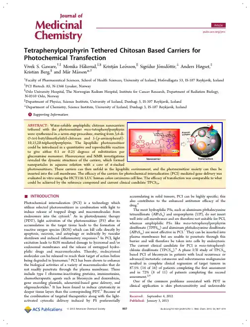
Porphyrin photochemistry in inorganicorganic hybrid materials
- 格式:pdf
- 大小:2.17 MB
- 文档页数:23
第32卷增刊2011年10月材料热处理学报TRANSACTIONS OF MATERIALS AND HEAT TREATMENTVol.32SupplementOctober2011空心玻璃微球表面仿生沉积羟基磷灰石涂层焦燕1,2,吕宇鹏1,2,肖桂勇1,2,朱瑞富1,2,许文花1,2(1.山东大学材料液固结构演变与加工教育部重点实验室,山东济南250061;2.山东大学材料科学与工程学院,山东济南250061)摘要:用NaOH 和生物活性玻璃依次对空心玻璃微球进行预处理。
将处理过的空心玻璃微球浸泡在1.5SBF 溶液中,仿生沉积得到羟基磷灰石涂层。
利用X 射线衍射仪、扫描电镜以及热场发射扫描电镜对空心玻璃微球和涂层进行表征。
结果表明,浸泡15天后在空心玻璃微球表面形成一层均匀致密的羟基磷灰石涂层,随时间延长涂层厚度增加。
关键词:空心玻璃微球;羟基磷灰石;仿生沉积中图分类号:R318.08;TB383文献标志码:A文章编号:1009-6264(2011)增刊-0151-03Biomimetic desposition of hydroxyapatite coating on hollow glass microspheresJIAO Yan 1,2,L Yu-peng 1,2,XIAO Gui-yong 1,2,ZHU Rui-fu 1,2,XU Wen-hua 1,2(1.Key Laboratory for Liquid-Solid Structural Evolution and Processing of Materials ,Ministry of Education ,Shandong University ,Jinan 250061,China ;2.School of Materials Science and Engineering ,Shandong University ,Jinan 250061,China )Abstract :Hollow glass microspheres were pretreated by NaOH and bioactive glass successively ,and then soaked in 1.5SBF at 36.5ħfor 15days.The phase composition and morphology were characterized by X-ray diffractometer (XRD ),scanning electron microscope (SEM )and field emission scanning electron microscope (FESEM ),respectively.The results show that the formed HA coating was uniform and dense after being soaked for 15days with SBF ,and the thickness of the coating increased with the soaking time.Key words :hollow glass microsphere ;hydroxyapatite ;biomimetic desposition收稿日期:2011-06-12;修订日期:2011-08-16基金项目:山东省自然科学基金(ZR2009FM023);山东大学自主创新基金项目(2009TS002)作者简介:焦燕(1984—),女,博士生,E-mail :jiaohiayan_1219@126.com 。
【初中化学】北京大学揭示双酚芴或致小鼠不良妊娠
3月1日发表在《自然?通讯》上的一项研究提出,一种用于生产不含双酚A(BPA)塑料的化学品双酚芴(BHPF)可能在小鼠中导致不良妊娠结果。
研究还表明,商业用水瓶中的BHPF可能会进入饮用水中。
但该研究只在小鼠中进行了试验,没有提供BHPF对人类健康有负面影响的证据。
BPA是一种塑料软化剂,广泛应用于食物和饮水的塑料包装中。
然而,研究显示BPA 会进入食物中,产生类似雌激素的效应,因此科学家在寻找没有雌激素活性的其他化合物进行替代。
北京大学的胡建英及同事报告,在用商业塑料水瓶(包括婴儿奶瓶)盛装的热水中可以检测到BHPF??一种BPA的替代品。
利用培养细胞和10只小鼠进行的实验表明,BHPF具有抗雌激素(阻断雌激素的效应)性,而且会在小鼠中导致低子宫重量、低幼崽体重,有时还会造成妊娠失败。
在100名经常从塑料瓶中喝水的学生中,作者发现其中七人的血液中含有微量BHPF,但并没有检测他们水瓶中的BHPF含量,因此无法确定这些学生接触BHPF的水平是否较高。
这些发现表明,相关部门应该对BHPF和其他被用作BPA替代品的化学物质的安全性进行进一步评估。
虽然一些国家会筛查化学物质的雌激素活性,但抗雌激素性并不在常规筛查项目之列。
(唐凤)
感谢您的阅读,祝您生活愉快。
2011年9月第31卷第9期基础医学与临床Basic &Clinical Medicine September 2011Vol.31No.9收稿日期:2010-08-31修回日期:2010-12-08基金项目:国家大学生创新实验计划(091040303);国家自然科学基金(30860347)*通信作者(corresponding author ):merlynhuang@sohu.com文章编号:1001-6325(2011)09-0970-06研究论文蛇血清分泌型磷脂酶A 2抑制剂防治人炎症的生物信息学分析陈柯,邓欣如,徐曦,李晓,钟立鹏,黄春洪*(南昌大学基础医学院生物化学教研室,江西南昌330006)摘要:目的比较人和蛇毒分泌型磷脂酶A 2(sPLA 2)结构相似性,分析蛇血清磷脂酶A 2抑制剂(PLI )对sPLA 2特异性抑制的结构与功能关系,评价蛇血清PLI 对人炎症的抑制作用。
方法用Clustal W2对人和五步蛇sPLA 2氨基酸序列进行多重序列比对。
用Swiss-model 对各种sPLA 2的空间结构进行同源建模预测,并用Chimera 软件对人和蛇毒sPLA 2空间结构进行三维比对。
结果人和五步蛇sPLA 2一级序列同源性有40%左右,空间结构几乎相似。
各种PLI 具有相同的空间结构特征和保守的sPLA 2结合区。
结论理论上推断蛇血PLI 可有效抑制人sPLA 2活性,减少sPLA 2介导的炎症发生。
关键词:磷脂酶A 2;磷脂酶A 2抑制剂;生物信息学中图分类号:R 318.04文献标志码:ABioinformatics analysis of possible inhibition of human inflammation by secretoryphospholipase A 2inhibitor from snake serumCHEN Ke ,DENG Xin-ru ,XU Xi ,LI Xiao ,ZHONG Li-peng ,HUANG Chun-hong *(Dept of Biochemistry ,College of Basic Medical Sciences ,Nanchang University ,Nanchang 330006,China )Abstract :ObjectiveTo compare the structural similarities of human secretory phospholipase A 2(sPLA 2)withsnake venom sPLA 2,and to analyze the neutralization effect of sPLA 2inhibitors (PLIs )from snake serum against sPLA 2by exploring their relationship between structure and function ,which was meaningful for assessing PLIs'clin-ical application in inflammation protection.MethodsMultiple sequencial alignment of 7human sPLA 2and 3five-pace snake venom sPLA 2was made by Clustal W2.Tertiary structures of the 10sPLA 2were predicted by Swiss-Modelling.Spatial structure similarity between snake venom and human sPLA 2was assessed by UCSF Chimera.Re-sults Human and snake venom sPLA 2shared 40%sequencial homologous and approximately similar spatial struc-tures.All the PLIs (γtype )have similar tertiary charectristics and conserved binding region to sPLA 2.Conclusion Based on the bioinformatical analysis ,snake serum PLIs were found to be an effective inhibitor to snake venom tox-icity and human sPLA 2activity.Thus ,the PLIs were potential blockers to human sPLA 2inflammation pathway and may be used for clinical disease.Key words :phospholipase A 2;phospholipase A 2inhibitor ;bioinformatics陈柯蛇血清分泌型磷脂酶A2抑制剂防治人炎症的生物信息学分析分泌型磷脂酶A2(secretory phospholipase A2,sPLA2)是一类广泛存在于人体各组织的酶,催化磷脂Sn-2脂肪酸水解,产生溶血磷脂和花生四烯酸。
迷迭香酸(罗丹酚酸)对H2O2处理过的皮肤黑色素瘤细胞的抗氧化作用Sun Mi Yoo1 and Jeong Ran Kang2*1.韩国光州500-741号东冈大学美容系2.韩国首尔143-701号建国大学生物工程系2009.2.6收到 2009.4.17接收本学科旨在检测迷迭香酸对人工孵育的皮肤黑色素瘤细胞在ROS下的抗氧化作用。
通过XTT比色法,以细胞毒性和抗氧化作用来分析细胞粘附活性,DPPH自由基清除活性以及H2O2处理1-10h和未经处理的两种情况下乳酸脱氢酶的活性。
用20-110 μM 的H2O2处理皮肤黑色素瘤细胞5-7h后,细胞活性的降低呈剂量和时间依赖性。
通过XTT比色法测得H2O2的半抑制浓度(IC50 )为90μM。
同时H2O2增强了LDH细胞的剂量依赖性。
用50-90μM的H2O2处理8h后测得LDH50为60 μM H2O2。
迷迭香酸能增强细胞活性和DPPH自由基清除活性,降低乳酸盐脱氢酶的活性。
细胞的H2O2处理证实了对人工孵育的皮肤黑色素瘤细胞的强抗氧化作用。
通过H2O2的处理,迷迭香酸能在细胞内能增强细胞活性和DPPH 自由基清除活性,降低乳酸盐脱氢酶的活性。
这被认为是迷迭香酸对ROS(ROS)如H2O2的抗氧化作用。
Key words:DPPH-radical scavenging, LDH, rosmarinic acid, XTT assay关键字:DPPH自由基清除活性,乳酸脱氢酶,迷迭香酸,XTT比色法据研究发现,ROS通过氧化应激对细胞的损伤和一些脑部疾病比如帕金森症或心脏疾病例如心肌梗塞之间有很大的关联[Difazio et al., 1992; Delanty and Dichter, 1998].尤其是研究人员认为ROS是皮肤老化的一个主要的因素后,一直试图从ROS方面研究衰老。
[Yokozawa et al., 1998].据研究表明,ROS的氧化应激会通过萎缩细胞引起各种疾病,例如超氧自由基、H2O2(H2O2)或羟基自由基的巯基蛋白反应中断酶的活性,破坏脱氧RMA(DNA)或RMA(RNA),诱导细胞膜脂质过氧化。
ISSN 058229879生物化学与生物物理学报ACTABIOCHIMICAetBIOPHYSICASINICA2003,35(8):723-727CN 3121300ΠQ集胞蓝藻PCC6803含疏水亚基的NAD(P)H脱氢酶亚复合体的分离邓 勇 叶济宇 米华玲3 沈允钢(中国科学院上海生命科学研究院植物生理生态研究所、植物分子遗传国家重点实验室,上海200032)摘要 利用离子交换与凝胶过滤层析,从n 2dodecyl β2D 2maltoside (DM )处理的集胞蓝藻Synechocystis PCC6803细胞粗提液中,首次分离到两个包含NDH 疏水亚基NdhA 的亚复合体。
酶活性分析表明,分离到的NDH 亚复合体具有NADPH 2氮蓝四唑(NBT )氧化还原酶活性,以NADPH 为电子供体可以还原铁氰化钾、二溴百里香醌(DBMIB )、二氯酚靛酚(DCPIP )、duroquinone 以及UQ 20等质醌类电子受体。
关键词 集胞蓝藻;NAD (P )H 脱氢酶;疏水亚基收稿日期:2003203224 接受日期:2003206202国家重点基础研究发展规划项目(973计划)(No.G1998010100)和国家自然科学基金项目(No.30270123)资助3联系人:Tel,02126404209024513;Fax,021*********;e 2mail,mihl@ 集胞蓝藻Synechocystis PCC6803的基因组包含11个ndh 基因(ndh A ~K )[1],它们都编码一个与线粒体复合体I 高度同源的NAD (P )H 脱氢酶(NDH,EC1.6.99.3)[2]。
在蓝藻中,NDH 是一个多亚基复合体,已知其ndh A ~G 和ndh H ~K 基因分别编码NDH 的疏水亚基和亲水亚基[2,3]。
有证据表明蓝藻NDH 既位于质膜也位于类囊体膜上[2],分别介导呼吸和光合循环电子传递[4,5]。
肿瘤光动力疗法中光敏剂研究新进展-生物研究-生物谷肿瘤光动力疗法中光敏剂研究新进展佚名分享 | 收藏目前大多数批准用于临床的光敏剂都属于卟啉类。
传统的说法, 70年代和80年代早期研发的卟啉等光敏剂称为第一代光敏剂(如:Photofrin)。
80年代后期制成的卟啉衍生物或合成物称为第二代光敏剂(如:ALA)。
第三代光敏剂通常指一些修饰产物,如生物结合物(如:抗体结合物,脂质体结合物)和内含光淬灭或光漂白特性的结合物;这些名称一直还用着,尽管并非毫无争议,这样的光敏剂分类有时也会引起某些混乱。
在不少情况下,所谓新一代光敏剂优于老一代的说法常常是缺乏根据的。
对新型或研究中的光敏剂的过早结论,可能会给研究者或临床医生带来误导信息,以为新一代光敏剂应取代老一代的光敏剂,或暗示患者新一代光敏剂的效果更佳。
临床医生和化学家对理想光敏剂的诠释有所不同。
如化学家可能更注重高消光系数和单态氧的高量子产额,而临床医生则注重低毒性和高选择性。
但是,双方都认为用于临床光动力治疗的理想光敏剂应具备以下条件:市售产品应该是化学纯品,暗毒性低而光毒性高,对目标细胞的选择性高,长波长吸收,体内清除快,可以通过多种途径输入体内。
这些标准为对比提供了一般的指导原则。
尽管一些光敏剂全部或部分符合这些条件,目前在世界范围内,仍只有少数几种光敏剂得到官方批准,这些光敏剂包括、但不仅限于下列制剂:Photofrin(卟吩姆钠 Porfimer sodium;Axcan Pharma, Inc.);Foscan(替莫泊芬temoprfin,四间-羟基苯基二氢卟酚,meta-tetahydroxyphenylchlorin, mTHPC;Biolitec AG);Visudyne(维替泊芬verteporfin, 卟啉苯衍生物的单一酸环Benzoporphyrin derivative monoacidring A, BPD-MA;Novartis Pharmaceuticals);Levulan(盐酸5-氨基乙酰丙酸,5-aminolevulinic acid, ALA;DUSA Pharmaceuticals, Inc.),以及新近批准的 Metvix(甲基-氨基乙酰丙酸酯,methyl aminolevulinate, MLA或M-ALA;PhotoCure ASA.)。
肉桂酸基油凝胶及其乳液的制备及理化性质表征栾慧琳,吴雨卿,郑红霞,柯翔宇,毛立科*(中国农业大学食品科学与营养工程学院,北京100083)摘 要:选择玉米油为溶剂,肉桂酸为凝胶剂制备油凝胶,探究其流变学、质构、持油性、热力学特性及凝胶剂晶体形态;以油凝胶为分散相,制备油凝胶乳液(O /W 型),分析其粒径、流变学、稳定性等物理性质。
结果表明:肉桂酸临界成胶质量分数为4%,4 ℃条件下存放的油凝胶持油性较常温条件下更好;肉桂酸质量分数越大的油凝胶硬度越大,热稳定性越好;肉桂酸在油凝胶中呈现随机分布的长纤维状聚集体;油相质量分数为10%~20%时,油相含量越低,其乳液粒径越小、黏度越低、物理稳定性越好。
油凝胶在常温下稳定性较差,而油凝胶乳液在常温下能保持良好的稳定性,有益于其在食品工业领域的研发与应用。
关键词:肉桂酸;油凝胶;凝胶特性;油凝胶乳液;乳液稳定性Preparation and Physicochemical Characterization of Cinnamic Acid Based Oleogels and Oleogel EmulsionsLUAN Huilin, WU Yuqing, ZHENG Hongxia, KE Xiangyu, MAO Like *(College of Food Science & Nutritional Engineering, China Agricultural University, Beijing100083, China)Abstract: In this study, corn oil and cinnamic acid were respectively used as a solvent and a gelling agent to prepare oleogels. Its rheology, texture, oil-holding capacity, thermodynamic properties and gel crystal morphology were investigated. Further, oil-in-water (O /W) oleogel emulsions were prepared and its physical properties such as particle size, rheology and stability were analyzed. The critical gelling concentration of cinnamic acid was 4%, and the oil-holding capacity of oleogels stored at 4 ℃ was higher than that at room temperature. The hardness and thermal stability of oleogels were higher with increasing concentration of cinnamic acid. Cinnamic acid was randomly distributed in oleogels as long fibrous aggregates. Oleogel emulsions with lower oil phase content in the range from 10% to 20% had smaller particle size, lower viscosity and better physical stability. At room temperature, the oleogels had poorer stability, while the oleogel emulsions could maintain good stability, which will be useful for its application in the food industry.Keywords: cinnamic acid; oleogels; gel properties; oleogel emulsions; emulsion stability DOI:10.7506/spkx1002-6630-20191024-269中图分类号:TS225;TS229 文献标志码:A 文章编号:1002-6630(2021)08-0060-07引文格式:栾慧琳, 吴雨卿, 郑红霞, 等. 肉桂酸基油凝胶及其乳液的制备及理化性质表征[J]. 食品科学, 2021, 42(8): 60-66. DOI:10.7506/spkx1002-6630-20191024-269. LUAN Huilin, WU Yuqing, ZHENG Hongxia, et al. Preparation and physicochemical characterization of cinnamic acid based oleogels and oleogel emulsions [J]. Food Science, 2021, 42(8): 60-66. (in Chinese with English abstract) DOI:10.7506/spkx1002-6630-20191024-269. 收稿日期:2019-10-24基金项目:北京市大学生科学研究与创业行动计划项目(201910019317)第一作者简介:栾慧琳(1998—)(ORCID: 0000-0002-8521-984X ),女,本科生,研究方向为功能配料与功能食品。
·综 述·抗氧化制剂普罗布考的临床研究现状朱冰坡 综述 范利 审校(解放军总医院老年心内科,北京 100853)[关键词] 抗氧化; 氧化型低密度脂蛋白; 动脉粥样硬化; 药物疗法[中图分类号]R972.6 [文献标识码]A [文章编号]1673-1913(2005)04-0252-03收稿日期:2004-06-22作者简介:朱冰坡,男,1978年8月生,河南省孟津市人,住院医师,硕士,从事老年心血管内科专业。
普罗布考(probuco l ),又名丙丁酚,化学名:4,4′-[(1-甲基亚乙基)双(硫)]双[2,6-双(1,1-二甲基乙基)苯酚],于1977年首先在美国上市,最初以降脂药应用于临床,主要降低血清胆固醇。
近几年发现其有抗氧化及抗动脉粥样硬化作用,能够从多个途径降低氧化低密度脂蛋白(o x -LDL )水平,预防或延缓动脉粥样硬化的发生和发展,减少心肌梗死及脑卒中的发病率,同时还可预防P T CA 术后的再狭窄。
本文对其研究现状作一综述。
1 普罗布考的药代动力学普罗布考经胃肠道吸收有限且不规则,如与食物同服可使其吸收达最大。
一次口服后18h 达血药浓度峰值,T 1/2为52~60h 。
每天服可使血药浓度逐渐增高,3~4个月达稳态水平。
动物实验表明,普罗布考主要积聚在脂肪组织,其在脂肪组织的浓度是血清中的100倍,肾上腺的25倍,肝脏的4倍,肌肉和心脏的2倍。
普罗布考可分布于胆汁和哺乳动物的乳汁,但尚不清楚是否分布于人乳中。
普罗布考在体内产生代谢产物。
口服剂量的84%从粪便排出,1%~2%从尿中排出,粪便中以原形为主,尿中以代谢产物为主。
2 血脂调节作用普罗布考对非家族性高胆固醇血症和家族性纯合子及杂合子型高胆固醇血症都有明显的降低作用。
短期用药(<3个月)降低血清总胆固醇(T C )10%~20%,降低低密度脂蛋白胆固醇(LDL -C )10%~20%。
长期用药(>3年)可降低血清T C 20%~25%。
Vol.34高等学校化学学报No.12013年1月 CHEMICAL JOURNAL OF CHINESE UNIVERSITIES 115~122 doi:10.7503/cjcu20120462硒化聚甘露糖醛酸的合成及抗阿尔茨海默症细胞氧化与凋亡朱志杰1,刘 琼1,陈 平1,续 旭1,倪嘉缵1,杨思林2,宋 云2(1.深圳大学生命科学学院,深圳518060;2.华信生物药业股份有限公司,界首236500)摘要 以聚甘露糖醛酸为原料,采用先磺化㊁再硒化的方法合成了硒化聚甘露糖醛酸,产率为54%,产物硒含量为437.25μg /g.在2.5μmol /L 硒浓度下,硒化聚甘露糖醛酸促细胞生长能力达到最适范围,能保护细胞免受过氧化氢损伤,显著提高阿尔茨海默症(AD)模型细胞N2a⁃APP695⁃sw 中的超氧化物歧化酶和谷胱甘肽过氧化物酶的活性,降低细胞内活性氧自由基,增加线粒体膜电位,抑制细胞色素C 的释放,在促进Bcl⁃2表达的同时抑制Bax 的表达,从而具有抑制AD 细胞凋亡的功能.硒化聚甘露糖醛酸也能抑制AD 病理相关蛋白BACE1和APP 的表达.结果表明,硒化聚甘露糖醛酸在抗AD 方面具有潜在的应用前景.关键词 甘露糖醛酸;硒化聚甘露糖醛酸;阿尔茨海默症(AD);活性氧自由基(ROS);细胞凋亡中图分类号 O629;O613.52 文献标志码 A 收稿日期:2012⁃05⁃11.基金项目:国家自然科学基金(批准号:31070731,31000770)㊁广东省自然科学基金(批准号:10151806001000023)和深圳市科技研发项目(批准号:CXB201005240008A)资助.联系人简介:刘 琼,女,博士,教授,主要从事硒的生物化学与分子生物学及其在医药领域应用的研究.E⁃mail:liuqiong@硒是一种生物体必需的微量元素.一些国家和地区土壤中硒含量严重缺乏,而在饮食中补硒对预防某些重大疾病具有良好的效果.在缺硒人群中经常出现的疾病有克山病㊁大骨节病及地方呆小症等[1,2],近年又发现缺硒与癌症㊁心血管病㊁生殖能力降低和神经退行性疾病等也密切相关[2~4].不同形态的硒抵御各种疾病的能力不同[5],因此从天然产物中分离或人工合成新硒化合物,研究其抗病效能成为该领域的重要课题[6~8].阿尔茨海默症(Alzheimer’s Disease,AD)又称老年痴呆症,是一种神经退行性疾病.AD 的神经组织病理学主要特征之一为老年斑(SP),由淀粉样前体蛋白(APP)经分泌酶剪切形成的β淀粉样蛋白(A β)聚集而成[9].褐藻胶是一种来源于褐藻细胞壁的水溶性酸性多糖,主要从海带㊁马尾藻及巨藻等褐藻中提取得到,具有独特的结构和生物活性.它是由α⁃1,4⁃L ⁃古罗糖醛酸(Guluronic acid,简写为G)和β⁃l,4⁃D ⁃甘露糖醛酸(Mannuronic acid,简写为M)通过l⁃4糖苷键连接而成的直链阴离子多糖[10].整个分子由聚甘露糖醛酸片段(PM)㊁聚古罗糖醛酸片段(PG)和聚甘露糖醛酸⁃古罗糖醛酸杂合段(PMG block)3种片段构成.M 与G 在一级结构的C5位上羧基的位置不同,是一对差向异构体,造成其空间结构和理化性质的显著差异.根据M 和G 电离常数的差异,对褐藻酸的部分不完全酸解液进行pH 分级,可使PM 和PG 得以分离[11].大分子褐藻胶具有抗肿瘤[12]㊁抗病毒[13]和增强免疫[14]等活性,而分子中的甘露糖醛酸是增强免疫的主要激发因子[14,15].本文对从褐藻酸钠分离出的聚甘露糖醛酸(PM)进行了磺化和硒化修饰,检测其清除活性氧自由基(ROS)㊁抗氧化损伤和保护神经细胞的能力,并初步研究了该硒多糖对AD 相关指标的作用.为研究硒化褐藻多糖的合成㊁功能及与AD 的关系提供了有意义的数据.611高等学校化学学报 Vol.34 1 实验部分1.1 材料与仪器Neuro⁃2a细胞(N2a)为小鼠神经瘤母细胞,购自中国科学院上海细胞研究所;N2a⁃APP695⁃sw细胞系为转染了人APP695Swedish突变基因的Neuro⁃2a细胞,用作AD模型细胞,由厦门大学许华曦和张云武教授惠赠.聚甘露糖醛酸(PM,分子量约3000)为本实验室自制;二甲基甲酰胺(DMF,优级纯)购自广州化学试剂厂;三氧化硫吡啶复合物(SO3⁃py)购自Sigma公司;Na2SeO3(分析纯)购自天津福晨化学试剂厂;KBH4(优级纯)购自天津科密欧化学试剂厂;硒标准溶液由国家标准物质研究中心提供;硒化卡拉胶(Se⁃Carr,硒质量分数为2%)购自青岛鹏洋科技有限公司;DMEM培养基购自Hyclone公司;OPTI⁃MEM培养基购自Gibco公司;细胞裂解液(RIPA)购自上海博彩生物科技公司;CCK⁃8细胞活力检测㊁活性氧(ROS)检测㊁线粒体膜电位(JC⁃1)检测㊁细胞色素C检测㊁超氧化物歧化酶(SOD)活性检测检测㊁谷胱甘肽过氧化物酶(GPx)活性检测和BCA法蛋白定量等试剂盒均购自碧云天公司;兔源抗BACE1单克隆抗体购自Abcam公司;鼠源抗APP单克隆抗体购自Millipore公司;兔源抗Bcl⁃2抗体购自北京博奥森公司;兔源抗Bax抗体购自北京博奥森公司;鼠源抗α⁃tublin和抗β⁃actin抗体购自Bio⁃science公司.XDS⁃1倒置显微镜(广州光学仪器厂);傅里叶红外光谱仪(美国Thermo公司);AFS⁃920型原子荧光光度计(吉天仪器公司);Spectra Max190型酶标仪(美国Molecular Devices公司);FACS Calibur流式细胞仪(美国BD公司);FV⁃1000型激光扫描共聚焦显微镜(日本OLYMPUS公司);POWER/PAC 300型蛋白电泳仪(Bio⁃RAD公司).1.2 聚甘露糖醛酸的磺化及检测将100mg干燥的PM悬浮于5mL无水DMF中,搅拌下加入用DMF溶解的三氧化硫吡啶(SO3⁃py),于60℃反应4h后,加入体积分数为95%的乙醇,终止反应并醇沉多糖,离心分离反应产物,用乙醇洗涤3次,加适量水溶解,调节pH至中性,透析,过滤,冷冻干燥即得磺化聚甘露糖醛酸(S⁃PM).采用明胶⁃硫酸钡比浊法测硫酸盐含量[16].1.3 聚甘露糖醛酸的硒化及表征将磺化得到的S⁃PM与Na2SeO3按一定质量比加入稀HNO3溶液中,在BaCl2催化下于60℃反应8h,用硫酸盐除去BaCl2,离心取上清液,调节pH至中性,透析至抗坏血酸检测透析袋内液不变红为止,过滤,冷冻干燥即得硒化聚甘露糖醛酸(Se⁃PM).采用原子荧光光度法测定Se⁃PM中硒含量[17].将干燥后的样品与溴化钾混合研磨至粉末,压片后进行红外光谱检测.1.4 CCK⁃8法检测细胞活力N2a细胞的完全培养液为含47.5%(体积分数)OPTI⁃ME和5%(体积分数)胎牛血清的DMEM. N2a⁃APP695⁃sw细胞的完全培养液为含终浓度为200μg/mL G418的N2a细胞完全培养液.取对数生长期细胞消化,制备单细胞悬液,按5×10³/孔的细胞量接种于96孔细胞培养板中,在37℃,含5% (体积分数)CO2培养箱中培养24h.用无血清培养基洗涤3次,加入100μL含不同浓度Se⁃PM的无血清DMEM,每一浓度设6个重复孔(n=6).以不含Se⁃PM的无血清DMEM作为对照组,无细胞组作为空白,继续培养24h.每孔加10μL CCK⁃8试剂,放入培养箱中培养2~4h后,用酶标仪于450nm 波长测定各孔的吸光度值(OD),计算细胞的相对抑制率.1.5 硒化合物及多糖对H2O2损伤的神经细胞的作用按照上节方法接种㊁培养㊁洗涤细胞,加入100μL含不同浓度H2O2的无血清DMEM(n=6).以不含H2O2的无血清DMEM培养基作为对照组,采用CCK⁃8法检测细胞活力,计算半数细胞致死时的H2O2浓度.采用相同方法培养细胞,加入100μL不同硒化合物与多糖的无血清DMEM,使硒的终浓度为2.5μmol/L,多糖质量与对应的硒多糖相同,培养24h后再加入10μL含H2O2的无血清DMEM 培养基,使H2O2的终浓度为60μmol/L(n=6),继续培养,加入CCK⁃8试剂,测各孔的吸光度值.1.6 细胞ROS 、线粒体膜电位和细胞色素C 的检测将N2a 与N2a⁃APP695⁃sw 细胞按5×10⁵/孔数量接种于6孔细胞培养板,培养24h 后洗涤细胞3次,分别加入2mL 不同硒化合物与多糖的无血清DMEM,使硒的终浓度为2.5μmol /L,多糖质量与对应的硒多糖相同(n =3),继续培养24h.胰酶消化后收集细胞.按ROS 检测试剂盒说明书进行染色,采用流式细胞仪于488nm 激发波长㊁525nm 发射波长下检测细胞内ROS.按JC⁃1法测定线粒体膜电位试剂盒说明书进行染色,用激光扫描共聚焦显微镜检测线粒体膜电位.按细胞色素C 检测方法[18],细胞经固定㊁封闭㊁一抗和二抗孵育后,用激光扫描共聚焦显微镜观察和检测细胞中细胞色素C 的释放.一抗为鼠源的细胞色素C 抗体,二抗为抗小鼠的Cy3标记的荧光抗体.1.7 SOD 和GPx 酶活性检测将N2a 与N2a⁃APP⁃sw 细胞按2×10⁶/皿数量接种于7.5cm 培养皿上,采用与上述相同浓度的硒化合物和相同质量的多糖处理细胞,收集作用后的细胞,每106细胞中加入100~200μL 细胞裂解液(RIPA),冰浴裂解细胞,于4℃以14000r /min 转速离心1h.收集上清液,于-80℃冰箱中保存备用.采用BCA 法测定蛋白质浓度.将所提取的细胞总蛋白按SOD 和GPx 酶活性检测试剂盒要求操作,测定和计算酶活力(n =3).1.8 Western Blot 检测样品准备方法同1.7节,具体步骤见文献[19],所用一抗分别为兔源BACE1抗体㊁鼠源APP 抗体㊁兔源Bcl⁃2抗体㊁兔源Bax 抗体㊁鼠源α⁃tublin 抗体和鼠源β⁃actin 抗体,分别检测所对应的蛋白.1.9 统计分析对所有数据进行标准偏差分析,以X ±SD 表示,采用SPSS16.0软件包[20]进行统计处理.2 结果与讨论2.1 硒化聚甘露糖醛酸的合成与鉴定2.1.1 硒化聚甘露糖醛酸的合成 实验对PM 采用先磺化㊁再硒化的方法合成Se⁃PM.硒化方法是对文献[21]方法稍加改进,用亚硒酸根取代硫酸根进行硒化.采用明胶⁃硫酸钡比浊法测得所合成的S⁃PM 的硫取代度(DS)为1.29,磺化平均产率为78%.采用原子荧光法检测所合成的Se⁃PM 的含硒量为437.25μg /g,硒化平均产率为70%,Se⁃PM 合成总产率为54%.Fig.1 FTIR spectra of PM (A ),S⁃PM (B )and Se⁃PM (C )目前,非硫酸酯型多糖的的硒化方法主要有2种:一种是直接进行多糖的酰氯化;另一种是先磺化㊁再硒化,或是直接与多糖上的醇甲基反应生成亚硒酸酯.文献报道的硒化木聚糖通过磺化再硒化方法合成[22];硒化葡聚糖和硒化红薯叶多糖通过酰氯化方法合成[23];硒化甘草多糖㊁硒化百合多糖和硒化枸杞多糖通过与糖环上醇甲基反应合成[24].本实验合成的Se⁃PM 在硒含量上与其它硒多糖相比略偏低,因此实验条件有待进一步优化.然而,所合成的Se⁃PM 在功能上与其它硒多糖相比明显不同,具有明显的抗氧化性,而且对某些AD 相关病理指标也有明显作用.2.1.2 红外光谱分析 在PM 的红外光谱图[图1(A)]中,在939.99,1037.21和1082.53cm -1处为711 No.1 朱志杰等:硒化聚甘露糖醛酸的合成及抗阿尔茨海默症细胞氧化与凋亡811高等学校化学学报 Vol.34 吡喃糖环骨架振动的特征吸收峰;820.40cm-1处为D⁃聚甘露糖醛酸的特征吸收峰.在S⁃PM的红外光图谱[图1(B)]中,856.27和1255.80cm-1处分别为C O S和S O的伸缩振动峰,证明分子中存在硫酸酯基;1418.67和1629.79cm-1处分别为COO-的对称和非对称伸缩振动峰;3442.25cm-1处为羟基的吸收峰.对照PM的红外谱图发现,在S⁃PM中增加了硫酸酯基的吸收峰,表明硫酸酯基已被引入到PM的分子结构中.文献[23]报道亚硒酸钠的红外谱图在788.63和742.29cm-1处有2个特征吸收峰,而PM在此范围内无吸收峰.在Se⁃PM的红外谱图[图1(C)]中,3457.35cm-1处为羟基的吸收峰;1415.33和1614.22cm-1处分别为COO-的对称和非对称伸缩振动峰.各特征吸收峰的位置未发生变化,多糖的基本构型保持不变,其828.76cm-1处的特征吸收峰强度变化不大,且透析后硒多糖中无游离的亚硒酸根.以上结果证明多糖与亚硒酸根以化合键形式结合,而不是硒的单纯吸附.多糖的结构复杂,硒作为硒多糖的特征元素,其存在形式可能有 SeH㊁亚硒酸酯和硒酸酯等[25].目前,有关硒多糖结构精确解析的研究报道较少,也未见硒多糖中硒特征吸收峰的标准红外光谱图.本实验根据PM的结构特点,采用先磺化㊁再硒化的方法合成Se⁃PM,硒化时采用亚硒酸根取代S⁃PM 中的硫酸根,因此硒的存在形式应该为亚硒酸酯.但Se⁃PM中亚硒酸根与多糖的结合位置和方式仍需进一步研究证实.2.2 硒化聚甘露糖醛酸的抗氧化能力2.2.1 不同硒化合物和多糖抗细胞氧化损伤能力比较 采用不同浓度的Se⁃PM(硒终浓度分别为0, 0.025,0.25,2.5和25μmol/L)处理N2a⁃APP695⁃sw细胞24h后,检测细胞活力.结果表明,当加入Se⁃PM的硒浓度为2.5μmol/L时,细胞存活率显著高于对照组(**P<0.01,n=6).因此,实验选定硒浓度为2.5μmol/L的Se⁃PM为促进细胞活力的合适浓度.采用不同浓度的H2O2(终浓度分别为0,25,50和100μmol/L)处理N2a⁃APP695⁃sw细胞24h(n= 6),检测细胞活力.H2O2诱导半数细胞死亡的剂量计算为56μmol/L.因此,采用60μmol/L H2O2处理N2a细胞,用于检测Se⁃PM协助细胞抗H2O2损伤的能力.根据上述实验结果,选用硒浓度为2.5μmol/L的硒化卡拉胶(Se⁃Carr)㊁硒代蛋氨酸(Se⁃Met)和Na2SeO3,以及与Se⁃Carr同等多糖含量的卡拉胶(Carr,10μg/mL),比较它们对N2a细胞抗H2O2氧化损伤的能力,结果见图2(A).可见用H2O2处理N2a细胞,在Se⁃Carr,Se⁃Met和Na2SeO3存在下,细胞的存活率比H2O2对照组明显提高,而Carr对细胞存活率无影响,表明硒化合物能够保护N2a细胞免受H2O2的氧化损伤.Fig.2 Protection of cells from H2O2damage by selenium compounds(A)and polysaccharides(B)(A)a.Control;b.H2O2;c.Se⁃Carr;d.Se⁃Met;e.Carr;f.Na2SeO3;(B)a.control;b.H2O2;c.Se⁃PM;d.S⁃PM;e.PM;f.MOM;g.Na2SeO3.***P<0.001vs.non⁃treated cells(control);###P<0.001vs.H2O2treated cells,n=6.图2(B)示出了硒浓度为2.5μmol/L的Se⁃PM和Na2SeO3㊁糖含量与Se⁃PM相近的S⁃PM和PM (500μg/mL)及不同聚合度的寡糖混合物(MOM,500μg/mL)对N2a⁃APP695⁃sw细胞抗H2O2氧化损伤能力的差异.可见,Se⁃PM,S⁃PM,PM和Na2SeO3均能不同程度地提高H2O2损伤的N2a⁃APP695⁃sw 细胞的存活率,而MOM则对细胞存活率无显著影响.结果表明,Se⁃PM,S⁃PM,PM和Na2SeO3均能保护N2a⁃APP695⁃sw细胞免受H2O2的氧化损伤.2.2.2 硒化合物和多糖对细胞氧化还原水平的影响 图3为对由流式细胞仪检测获得的细胞内活性氧(ROS)数据进行统计学处理所得结果.图3(A)结果表明,N2a⁃APP695⁃sw 细胞与N2a 细胞相比,细胞内ROS 水平明显增加(P <0.01),说明AD 相关基因,即Swedish 突变的APP695的表达,使细胞ROS 水平升高;Se⁃PM 和Se⁃Carr 能使N2a⁃APP695⁃sw 细胞的ROS 水平明显降低.图3(B)结果表明,不仅Se⁃PM 能显著降低N2a⁃APP695⁃sw 细胞内ROS 水平,S⁃PM 和PM 也具有类似作用,但PM 降低ROS 能力小于前两者.Fig.3 Effects of selenium compounds and polysaccharides on intracellular ROS (A ,B )andantioxidative enzymes (C )(A)a.N2a;b.N2a⁃APP695⁃sw;c.Se⁃PM;d.Se⁃Carr;e.Na 2SeO 3;(B )a.control;b.Se⁃PM;c.S⁃PM;d.PM;(C)a.N2a;b.N2a⁃APP695⁃sw;c.Se⁃PM;d.Se⁃Carr;e.Na 2SeO 3;f.S⁃PM;g.PM.ROS was expressed as the ratio of fluo⁃rescence intensity of the treated cells vs.the control cells.*P <0.05,**P <0.01,drug⁃treated N2a⁃APP695⁃sw cells vs.the un⁃treated cells(control);#P <0.05,##P <0.01,N2a⁃APP695⁃sw cells vs.N2a cells.n =3.将N2a 和N2a⁃APP695⁃sw 细胞用Se⁃PM,Se⁃Carr,Na 2SeO 3,S⁃PM 和PM 处理后提取蛋白并定量,检测SOD 和GPx 酶活性.如图3(C)所示,N2a⁃APP695⁃sw 细胞与N2a 细胞相比,SOD 活性无显著变化,而GPx 活性显著降低(P <0.05).经Se⁃PM 和PM 处理的N2a⁃APP695⁃sw 细胞的SOD 活性明显提高(P <0.05),而经Se⁃Carr,Na 2SeO 3和S⁃PM 处理的细胞的SOD 活性无明显变化.经Se⁃PM 和Na 2SeO 3处理的N2a⁃APP695⁃sw 细胞的GPx 活性显著增加(P <0.01),经S⁃PM 和Se⁃Carr 处理的细胞GPx 活性也显著增加(P <0.05),而PM 处理的细胞GPx 活性无明显变化.以上结果表明,细胞中SOD 酶活性与补硒无关,但与补充多糖有关,即多糖能提高细胞的SOD 活性.而GPx 酶活性与细胞补硒紧密相关,Se⁃PM 比Se⁃Carr 和Na 2SeO 3更能有效地提高GPx 酶活性.据文献[26]报道,甘露糖醛酸具有保护神经细胞免受H 2O 2氧化损伤的作用,这与本文结果一致.本文结果还表明,各种含硒化合物均具有保护细胞免受氧化损伤的作用,Se⁃PM 在其中显示了强抗氧化能力.Se⁃PM 能提高SOD 活性主要源于PM 的作用,而提高GPx 的活性主要源于硒的作用,两者结合起来更大程度发挥了Se⁃PM 的抗氧化能力.Fig.4 Effects of selenium compounds (A )and polysaccharides (B )on the expression of BACE1andAPP in N2a⁃APP695⁃sw cellsa.Control;b.Se⁃PM;c.Se⁃Carr;d.Na 2SeO 3;e.S⁃PM;f.PM,***P <0.001,*P <0.05,n =3.BACE1;APP.2.3 硒化聚甘露糖醛酸抑制AD 相关病理指标及其机理2.3.1 AD 相关病理指标 APP 是A β的前体蛋白,BACE1又称β⁃分泌酶1,其对APP 的剪切会产生A β,A β聚集形成老年斑是AD 的重要病理特征之一[27].实验采用Se⁃PM 及其它硒化合物和多糖处理AD 模型细胞N2a⁃APP695⁃sw 24h 后提蛋白,BCA 定量后进行Western blot 检测,结果示于图4.可见,911 No.1 朱志杰等:硒化聚甘露糖醛酸的合成及抗阿尔茨海默症细胞氧化与凋亡Se⁃PM 既可显著降低BACE1蛋白表达(P <0.001),也可显著降低APP 的表达(P <0.05).Se⁃Carr 和Na 2SeO 3也能显著降低BACE1蛋白的表达,但对APP 的表达无明显影响.S⁃PM 和PM 对BACE1蛋白和APP 的表达无明显影响.2.3.2 Se⁃PM 作用机理 当线粒体的膜电位较高时,JC⁃1在线粒体基质中聚集,产生红色荧光;当线粒体膜电位较低时,JC⁃1则不能在线粒体的基质中聚集,产生绿色荧光.因此,可通过检测细胞内荧光颜色,用红绿荧光值的相对比来衡量线粒体膜电位的变化.如图5(A)所示,N2a⁃APP695⁃sw 细胞的红绿荧光比明显低于N2a 细胞(P <0.01),说明N2a⁃APP695⁃sw 细胞的线粒体膜电位比N2a 细胞低,前者线粒体受到的氧化应激强于后者.用硒化合物处理N2a⁃APP695⁃sw 细胞24h 后,经Se⁃PM 作用的细胞的红绿荧光比明显高于对照组(P <0.05,n =5),而经Se⁃Carr 和Na 2SeO 3处理的细胞的红绿荧光比与对照组相比无明显差异.说明Se⁃PM 具有增强N2a⁃APP695⁃sw 细胞膜电位的能力.Fig.5 Effects of selenium compounds on mitochondrial membrane potential (A ),cytochrome Crelease (B )and Bcl⁃2/Bax expression (C )(A)a.N2a;b.N2a⁃APP695⁃sw;c.Se⁃PM;d.Se⁃Carr;e.Na 2SeO 3;(B )a.N2a;b.N2a⁃APP695⁃sw;c.Se⁃PM;d.Se⁃Carr;e.Na 2SeO 3;(C)a.control;b.Se⁃PM;c.Se⁃Carr;d.Na 2SeO 3.##P <0.01,N2a⁃APP695⁃sw cells vs.N2a cells;*P <0.05,***P <0.001,drug⁃treated N2a⁃APP695⁃sw cells vs.the untreatedcells(control).Bax;Bcl⁃2.用硒化合物处理细胞24h 后,进行细胞免疫荧光染色,红色荧光标记细胞色素C,蓝色荧光标记细胞核,通过激光共聚焦显微镜观察㊁拍照和测定荧光值.以细胞核的荧光为参照,测定细胞内细胞色素C 的变化.如图5(B)所示,N2a⁃APP695⁃sw 细胞的红蓝荧光比明显高于N2a 细胞(P <0.01),说明N2a⁃APP695⁃sw 细胞的线粒体膜通透性大,与N2a 细胞相比更多细胞色素C 从线粒体释放到细胞质中.经Se⁃PM 和Se⁃Carr 处理后的N2a⁃APP695⁃sw 细胞的红蓝荧光比明显低于对照组(P <0.05,n =5),而经Na 2SeO 3处理的细胞的红蓝荧光比与对照组相比无明显差异.说明Se⁃PM 能保护线粒体,阻止线粒体膨胀而释放细胞色素C,从而阻断线粒体介导的细胞凋亡通路.Bcl⁃2是细胞凋亡的一种抑制因子,而Bax 的过量表达可加速细胞凋亡.用硒化合物处理N2a⁃APP695⁃sw 细胞24h 后提取蛋白,进行Western Blot 检测,结果如图5(C)所示.可见,经Se⁃PM 处理的细胞的Bc1⁃2蛋白表达明显增加(P <0.001,n =3),而经Se⁃Carr 和Na 2SeO 3处理的细胞的Bc1⁃2蛋白表达无明显变化.但经这3种硒化合物处理的细胞中Bax 蛋白表达均显著降低(P <0.001),因此,经Se⁃PM 处理的细胞中Bc1⁃2与Bax 蛋白的比值明显高于经Se⁃Carr 和Na 2SeO 3处理的细胞.说明Se⁃PM 具有抵抗N2a⁃APP695⁃sw 细胞凋亡的功能.凋亡的过程伴有细胞色素C 从线粒体释放,继而发生Caspases 的激活及其蛋白降解产物引起的线粒体通透性改变.Bcl⁃2属于细胞内膜整合蛋白,它对大多数因素诱导的细胞凋亡具有负调节作用.Bcl⁃2抑制细胞凋亡涉及对ROS 的作用[28].细胞色素C 作为电子呼吸链中重要的电子传递体之一,它从线粒体内膜上的释放会阻断呼吸链上电子向下游的传递,从而危及呼吸链的功能,导致超氧阴离子的加速产生.而Bcl⁃2可以抑制细胞色素C 的释放,从而抑制超氧阴离子的产生.Bcl⁃2可与Bax 形成同源或异源蛋白二聚体,作为细胞在死亡信号通路上的分子开关:如果Bax 的相对量高于Bcl⁃2,则Bax 同源二聚体的数量增多,促进细胞死亡;如果Bcl⁃2的相对量高于Bax,则促进形成Bcl⁃2/Bax 蛋白异源二聚体,并使Bcl⁃2同二聚体的数量增多,进而抑制细胞死亡.本实验AD 模型细胞在Se⁃PM 作用后其线粒体膜电位显著增高,细胞色素C 的释放减少,ROS 水平明显降低,Bcl⁃2蛋白表达明显增加且021高等学校化学学报 Vol.34 Bax 蛋白表达减少,说明Se⁃PM 能通过抗氧化损伤保护线粒体的完整性和生物功能,抵御N2a⁃APP695⁃sw 细胞中由于大量表达突变型APP 和BACE1造成的氧化应激以及由此激活的细胞凋亡通路,阻止或延缓AD 模型细胞的凋亡.3 结 论以聚甘露糖醛酸为原料合成了硒化聚甘露糖醛酸,并研究了其抗氧化和抗AD 功能.结果表明,硒化聚甘露糖醛酸具有保护细胞免受过氧化氢损伤的作用,能显著提高N2a⁃APP695⁃sw 细胞内SOD 和GPx 的酶活性,降低胞内活性氧自由基,增加线粒体膜电位,抑制细胞色素C 释放.硒化聚甘露糖醛酸具有抑制AD 细胞凋亡的功能,能促进Bcl⁃2表达并抑制Bax 表达,也能抑制AD 病理相关蛋白BACE1和APP 的表达,表明硒化聚甘露糖醛酸在抗AD 方面具有潜在的应用前景.致谢:感谢厦门大学许华曦㊁张云武教授惠赠AD 模型细胞系N2a⁃APP695⁃sw.参 考 文 献[1] Bellinger F.P.,Raman A.V.,Reeves M.A.,Berry M.J.,Biochem.J.,2009,422(1),11 22[2] Hatfield D.L.,Berry M.J.,Gladyshev V.N.,Selenium :Its Molecular Biology and Role in Human Health (2nd ed.),Springer Science+Business Media,LLC.,2012,235 248,297 324[3] Gwon A.R.,Park J.S.,Park J.H.,Baik S.H.,Jeong H.Y.,Hyun D.H.,Park K.W.,Jo D.G.,Neurosci.Lett.,2012,469(3),391 395[4] van Eersel J.,Ke Y.D.,Liu X.,Delerue F.,Kril J.J.,Gotz J.,Ittner L.M.,A ,2010,107(31),13888 13893[5] Corcoran N.M.,Martin D.,Hutter⁃Paier B.,Windisch M.,Nguyen T.,Nheu L.,Sundstrom L.E.,Costello A.J.,Hovens C.M.,J.Clin.Neurosci.,2010,17(8),1025 1033[6] Zou X.F.,LüS.W.,Chem.J.Chinese Universities ,2012,33(5),983 987(邹险峰,吕绍武.高等学校化学学报,2012,33(5),983 987)[7] Huang X.C.,Zheng J.S.,Chen T.F.,Zhang Y.B.,Luo Y.,Zheng W.J.,Chem.J.Chinese Universities ,2012,33(5),976982(黄晓纯,郑军生,陈填烽,张逸波,罗懿,郑文杰.高等学校化学学报,2012,33(5),976 982)[8] Yang L.Y.,Zhang S.J.,Zheng X.F.,Chem.J.Chinese Universities ,2008,29(11),2187 2190(杨兰义,张殊佳,郑学仿.高等学校化学学报,2008,29(11),2187 2190)[9] Loef M.,Schrauzer G.N.,Walach H.,J.Alzheimers Dis.,2011,26(1),81 104[10] Haug A.,Larsen B.,Smidsrod O.,Acta Chem.Scand.,1967,21,691 704[11] Anzai H.,Uchida N.,Nishide E.,Nippon Suisan Gakkaishi ,1990,56(1),73 81[12] Fujihara M.,Nagumo T.,Carbohydrate Research ,1993,243,211 216[13] Sano Y.,Carbohydrate Polymers ,1999,38(2),183 186[14] Otterlei M.,Ostgaard K.,Skjak⁃Braek G.,Smidsrød O.,Soon⁃Shiong P.,Espevik T.,Immunother ,1991,10(4),286 291[15] Fujihara M.,Nagumo T.,Carbohydrate Research ,1992,244,343 347[16] Zhao X.,Studies on Preparation ,Structure and Activities of Polyguluronate Sulfate and Its Oligosaccharides ,Qingdao:Ocean University ofChina,2007(赵峡.聚古罗糖醛酸硫酸酯及其寡糖的制备㊁结构与活性研究,2007)[17] Sun L.M.,Modern Agriculture ,2011,2,111 112(孙丽敏.现代农业,2011,2,111 112)[18] Shen L.M.,Liu Q.,Ni J.Z.,Sci.China Ser.B :Chem ,2009,52(11),1814 1820(沈立明,刘琼,倪嘉缵.中国科学B 辑:化学,2009,52(11),1814 1820)[19] Fu L.P.,Liu Q.,Shen L.M.,Wang Y.,J.Trace Elem.Med.Bio.,2011,25(3),130 137[20] Liu R.J.,Zhang Y.W.,Wen C.W.,Computers and Applied Chemistry ,2009,26(3),379 381(刘瑞江,张业旺,闻崇炜.计算机与应用化学,2009,26(3),379 381)[21] Li G.Y.,Miu J.L.,Liu F.Y.,Selenium Polysaccharide Compound and Its Preparation Method ,CN 1288899A,2001⁃3⁃8(李光友,缪锦来,刘发义.硒多糖化合物及其制备方法,CN 1288899A,2001⁃3⁃8)[22] Cao Y.,Isao I.,International Journal of Biological Macromolecules ,2009,45,231 235[23] Gong X.Z.,Ou Y.Z.,Journal of Shenzhen University Science and Engineering ,1998,15(1),70 74(龚晓钟,欧阳政.深圳大学学报理工版,1998,15(1),70 74)[24] Zhang J.,Liu Z.W.,Wang F.X.,Yang Y.P.,Tian Y.R.,Sun W.X.,Wang Y.P.,Chinese Polymer Bulletin ,2009,(10),121 No.1 朱志杰等:硒化聚甘露糖醛酸的合成及抗阿尔茨海默症细胞氧化与凋亡221高等学校化学学报 Vol.34 48 52(张继,刘忠旺,王风霞,杨玉萍,田玉汝,孙文秀,王云普.高分子通报,2009,(10),48 52)[25] Huang Z.,Zheng W.J.,Guo B.J.,Natural Science Journal of Hainan University,2001,19(2),169 175(黄峙,郑文杰,郭宝江.海南大学学报自然科学版,2001,19(2),169 175)[26] Qiu L.,Structure⁃function Relationship Study of Algine⁃derived Saccharides on H2O2⁃induced Cytotoxocity,Qingdao:Ocean University ofChina,2009,16 17(邱琳.不同褐藻酸糖片段及其衍生物对神经细胞氧化损伤保护作用的构效关系研究,2009,16 17)[27] Caito S.W.,Milatovic D.,Hill K.E.,Aschner M.,Burk R.F.,Valentine W.M.,Brain Res.,2011,1398,1 12[28] Cornelli U.,Neurodegener Dis.,2010,7(1 3),193 202Seleno⁃polymannuronate Synthesis and Resistance to Oxidation andApoptosis in Alzheimer’s Disease CellsZHU Zhi⁃Jie1,LIU Qiong1*,CHEN Ping1,XU Xu1,NI Jia⁃Zuan1,YANG Si⁃Lin2,SONG Yun2(1.College of Life Sciences,Shenzhen University,Shenzhen518060,China;2.Huaxin Bio⁃pharmaceutical Co.Ltd.,Jieshou236500,China)Abstract Selenium deficiency is closely relate to multiple diseases.Supplementation with adequate amount of selenium is very important for human health.As various selenium compounds have different biological effects,preparation and functional study of new product are essential for the discovery of selenium⁃containing drug.In this work,polymannuronate(PM)was used as the raw material to synthesize sulfonated polymannur⁃onate(S⁃PM).Seleno⁃polymannuronate(Se⁃PM)was then prepared by the replacement of sulfonated group in S⁃PM with sodium selenite.The yield of Se⁃PM synthesis was54%with a selenium content of437.25μg/g. Purified Se⁃PM was used to study its antioxidative property and effect on Alzheimer’s disease(AD),using N2a⁃APP695⁃sw cells as an AD model.Cell viability was detected by CCK⁃8assay.Intracellular reactive oxy⁃gen species(ROS)was measured by flow cytometry.Mitochondrial membrane potential and cytochrome C re⁃lease were detected by laser scanning confocal microscope.The expression levels of cell apoptosis and AD pa⁃thology relevant proteins were analyzed by Western Blot.The results showed that the optimum selenium con⁃centration was2.5μmol/L for Se⁃PM to significantly increase cell viability.Se⁃PM at this concentration could prevent cells from the oxidative damage of hydrogen peroxide,inhibit intracellular ROS,increase the activity of superoxide dismutase and glutathione peroxidase,promote mitochondrial membrane potential,and suppress cytochrome C release into cytoplasm.Meanwhile,Se⁃PM could also increase Bcl⁃2expression and decrease Bax expression,inhibit the expression of AD pathology relevant proteins BACE1and APP.Those results indi⁃cated that Se⁃PM could resist AD through the prevention of cell apoptosis and amyloid plaque formation,a key pathological feature of AD.It also provides basic data for the development of new anti⁃AD drug. Keywords Mannuronate;Seleno⁃polymannuronate(Se⁃PM);Alzheimer’s disease(AD);Reactive oxygen species(ROS);Cell apoptosis(Ed.:H,Z,K)。
Perampanel通过线粒体自噬缓解过氧化氢诱导神经元氧化应激下铁死亡的作用机制Perampanel通过线粒体自噬缓解过氧化氢诱导神经元氧化应激下铁死亡的作用机制摘要:过氧化氢(H2O2)诱导神经元死亡是许多神经系统疾病的共同特征。
铁死亡是指在细胞内超凡铁离子聚积过多,触发产生自由基反应而造成的细胞死亡。
本研究发现,Perampanel 可以通过线粒体自噬途径缓解H2O2诱导的氧化应激下神经元的铁死亡。
Perampanel通过抑制神经元内铁缩合酶激活而减少了细胞内铁离子的聚积,从而减轻氧化应激导致的铁死亡的程度。
在H2O2处理下,Perampanel还可以激活线粒体自噬途径,通过清除受损线粒体来保护神经元的生存状态。
这些结果表明Perampanel可能是治疗神经系统氧化损伤和铁死亡的有前途的药物。
关键词:Perampanel,过氧化氢,神经元,铁死亡,线粒体自噬引言:过氧化氢是一种强氧化剂,可以直接损伤神经元细胞膜和膜蛋白,同时也能引起细胞内的氧化应激反应。
铁死亡是一种新型的细胞死亡方式,是由于细胞内铁离子过多聚积,导致大量自由基反应而引起的。
许多疾病如中风和帕金森病等都与神经元氧化损伤和铁死亡有关。
因此,治疗神经系统损伤和铁死亡的有效药物十分必要。
Perampanel是一种新型的抗癫痫药,其作用机理是抑制离子型谷氨酸受体并减少神经元内钙离子的流入。
最近的研究表明,Perampanel还可以通过抑制G蛋白耦联受体10的活化,减少神经元内铁缩合酶的激活,从而减轻氧化应激下铁死亡的程度,使神经元得到保护。
结果:我们使用H2O2处理神经元并测量了细胞存活率。
我们发现,Perampanel处理组的细胞存活率明显高于未处理组。
我们还测量了细胞内铁含量,发现Perampanel处理组的铁含量有所降低。
进一步的实验证明,Perampanel可以通过抑制线粒体自噬途径,减少受损线粒体的聚积,从而保护神经元的生存状态。
富血小板血浆在膝关节疾病治疗中的应用刘永辉赵烨王向阳郭马珑崔宏勋【摘要】富血小板血浆(platelet-rich-plasma,PRP)是利用全血各成分沉降系数不同特性离心而得到的高浓度血小板血浆,由于其富含多种促进组织修复的生长因子,且制作、使用便捷而被广泛运用到骨科领域,尤其是近几年在治疗膝关节疾病疗效方面备受关注。
本文就其治疗膝关节骨性关节炎、半月板、交叉韧带损伤及膝关节滑膜炎方面做一综述,为临床治疗膝关节常见疾病提供参考。
【关键词】富血小板血浆;膝关节骨性关节炎;半月板损伤;交叉韧带损伤;膝关节滑膜炎【Abstract】Platelet-rich-plasma is a high-concentration platelet plasma obtained by centrifugation with different characteristics of sedimentation coefficients of all components of the whole blood.It is widely used in the field of orthopedics because it is rich in a variety of growth factors to promote tissue repair and is easy to make and use,especially in the treatment of knee diseases in recent years.In this paper,the treatment of knee osteoarthritis,meniscus,cruciate ligament injury and knee synovitis are reviewed to provide reference for clinical treatment of common knee diseases.【Key words】Platelet-rich-plasma;Knee osteoarthritis;Meniscus injury;Cruciate ligament injury;Knee synovitis富血小板血浆(platelet-rich-plasma,PRP)是自体外周血离心而得到以血小板和白细胞为主的血浆,研究发现[1],PRP中含有转化生长因子β(transforming growth factor-β,TGF-β),成纤维细胞生长因子(fibroblast growth factor,FGF),血小板衍化生长因子(platelet-derived growth factor,PDGF),血管内皮生长因子(vascular endothelial growth factor,VEGF)等多种细胞因子和介质,通过PRP注射到受损组织等方式能够促进损伤组织的修复与再生[2]。
卟啉(英语:Porphyrin)是一类由四个吡咯类亚基的α-碳原子通过次甲基桥(=CH-)互联而形成的大分子杂环化合物。
其母体化合物为卟吩(porphin,C20H14N4),有取代基的卟吩即称为卟啉。
卟啉环有26个π电子,是一个高度共轭的体系,并因此显深色。
“卟啉”一词是对其英文名称porphyrin的音译,其英文名则源于希腊语单词,意为紫色,因此卟啉也被称作紫质。
许多卟啉以与金属离子配合的形式存在于自然界中,如含有二氢卟吩与镁配位结构的叶绿素以及与铁配位的血红素。
人体内卟啉积累过多时会造成卟啉病,也称紫质症。
性质∙卟吩-最简单的卟啉∙卟吩的空间填充模型∙四苯基卟啉的结构∙卟啉环的编号系统卟啉环的编号方式见上图。
习惯命名是将5,10,15,20位称为meso位(间),将1,4,6,9,11,14,16,19位称为alpha位(α),将2,3,7,8,12,13,17,18位称为beta位(β)。
卟啉的大分子环是一个24中心26电子的体系,符合休克尔规则中的4n+2通式,因此具有芳香性。
卟啉自由碱的中心氮原子可以与+2或+3价的金属阳离子配位,两个氮上的氢原子被金属取代,生成金属卟啉。
通常把它们及其衍生物称为金属卟啉化合物。
其反应通式如下:卟啉卟啉四苯并四氮杂卟啉类化合物由苯酐与尿素在氯化亚铜存在下发生缩合制得,呈蓝色,一般称为酞菁。
其分子中四个异吲哚环的氮原子可以与金属离子在中心发生配位,生成金属酞菁。
金属酞菁化合物色泽鲜艳耐晒,耐热性能优良,着色力强,是很常用的颜料和染料。
以卟啉作为结构单元的超分子是目前分子器件研究的主要方向之一。
meso-四苯基卟啉的氯化铁配合物(TPPFeCl)是一个有机合成试剂。
卟啉的衍生物有:咕啉、二氢卟吩(2,3-二氢卟啉)、菌绿素、F430(镍四吡咯)等。
一种以三聚氯氰为桥基的新型小分子功能活性染料的合成张月华;光善仪;郗娇娇;魏刚;郭冰苑;田佳婵;柯福佑;徐洪耀【摘要】Schiff Base(1) was prepared by reduction reaction of salicylal and amino benzoic acid am-ide, which was synthesied by amidation reaction of methyl p-aminobenzoate and hydrazine hydrate . Rhodamine hydrazine ( 2 ) was prepared by ring-closing reaction of Rhodamine B with hydrazine hy-drate.Condensation product ( 3 ) was prepared by condensation reaction of 2 and cyanuric chloride . Condensation product (4) was prepared by substitution react ion of 3 and 1 in THF at 45~50 ℃ for 6 h.The structure was characterized by 1 H NMR, IR, MS and elemental analysis .%以对氨基苯甲酸甲酯和水合肼为原料,经酰胺化反应制得对氨基苯甲酸甲酰胺,进一步与水杨醛通过还原氨化反应生成席夫碱(1);以罗丹明B和水合肼为原料,1经闭环反应制得罗丹明B酰肼(2);2在四氢呋喃溶剂中与三聚氯氰经缩合取代反应生成一缩产物(3);3进一步在四氢呋喃中与1于45~50℃回流6 h,经取代反应生成二缩产物,其结构经1 H NMR,MS,IR和元素分析表征.【期刊名称】《合成化学》【年(卷),期】2016(024)009【总页数】4页(P804-807)【关键词】对氨基苯甲酸甲酯;水合肼;罗丹明B;席夫碱;三聚氯氰;缩合反应;合成【作者】张月华;光善仪;郗娇娇;魏刚;郭冰苑;田佳婵;柯福佑;徐洪耀【作者单位】东华大学化学化工与生物工程学院国家染整工程技术研究中心,上海201620;东华大学化学化工与生物工程学院国家染整工程技术研究中心,上海201620;东华大学材料学院与纤维和高分子改性国家重点实验室,上海 201620;东华大学材料学院与纤维和高分子改性国家重点实验室,上海 201620;东华大学化学化工与生物工程学院国家染整工程技术研究中心,上海 201620;东华大学化学化工与生物工程学院国家染整工程技术研究中心,上海 201620;东华大学材料学院与纤维和高分子改性国家重点实验室,上海 201620;东华大学材料学院与纤维和高分子改性国家重点实验室,上海 201620【正文语种】中文【中图分类】O621.3三聚氯氰是一种重要的精细化工产品,在医药农药工业领域具有广泛的用途,某些反应中可充当催化剂[1-4],而在染料制备中常被用作活性基团。
Journal of Photochemistry and Photobiology C:Photochemistry Reviews7(2006)104–126ReviewPorphyrin photochemistry in inorganic/organic hybrid materials:Clays, layered semiconductors,nanotubes,and mesoporous materials Shinsuke Takagi a,∗,Miharu Eguchi a,b,Donald A.Tryk a,Haruo Inoue a,ca Department of Applied Chemistry,Faculty of Urban Environmental Sciences,Tokyo Metropolitan University,Minami-ohsawa1-1,Hachiohji,Tokyo192-0397,Japanb Japan Society for the Promotion of Science for Young Scientist,Japanc SORST,JST(Japan Science and Technology),JapanReceived24February2006;received in revised form31March2006;accepted10April2006Available online23October2006AbstractPorphyrin derivatives are known as useful functional dyes.Porphyrin derivatives exhibit various properties in complexes with inorganic host materials that are much different from those in homogeneous solutions.In this paper,the structure and photochemical properties of porphyrins in inorganic host materials such as clays,layered semiconductors,nanotubes,and mesoporous materials are described.The photochemical properties, including the absorption properties and excited lifetimes,are much affected by the complex formation with inorganic materials.Aggregation phenomena,structural perturbations,and selected chemical reactions such as metalation and protonation affect the photochemical properties of porphyrins accommodated in inorganic host materials.The combination of porphyrin derivatives and inorganic materials should be promising for the construction of novel hybrid materials.Inorganic materials can act as novel environments for photochemical reactions.The utilization of inorganic materials for photochemical reactions is also described.©2006Elsevier B.V.All rights reserved.Keywords:Porphyrin;Clay minerals;Layered materials;Semiconductor;Nanotube;Mesoporous;Photochemistry;Electron transfer;Energy transfer;AggregationContents1.Introduction (105)2.Porphyrin–clay complexes (105)2.1.Anionic clays (106)2.1.1.Structures and photochemical properties of porphyrin–anionic clay complexes (106)2.1.2.Photochemical reactions in porphyrin–anionic clay complexes (110)2.2.Cationic clays (111)2.2.1.Structure and photochemical properties of porphyrin–cationic clay complexes (111)2.2.2.Photochemical reactions in porphyrin–cationic clay complexes (114)3.Porphyrin-layered metal oxide semiconductor complexes (115)3.1.Structural and photochemical properties of porphyrin-layered metal oxide semiconductor complexes (115)3.2.Photochemical reactions in porphyrin-layered metal oxide semiconductor complexes (117)4.Porphyrin–nanotube complexes (118)5.Other porphyrin–inorganic host material complexes (120)6.Concluding remarks (122)Acknowledgements (122)Appendix A (122)References (124)∗Corresponding author.E-mail addresses:takagi-shinsuke@c.metro-u.ac.jp(S.Takagi),inoue-haruo@c.metro-u.ac.jp(H.Inoue).1389-5567/$20.00©2006Elsevier B.V.All rights reserved.doi:10.1016/j.jphotochemrev.2006.04.002S.Takagi et al./Journal of Photochemistry and Photobiology C:Photochemistry Reviews7(2006)104–126105Shinsuke Takagi received his BS and MS degrees inapplied chemistry from Tokyo Metropolitan Universityin1991and1993.After enrolling in the doctoral pro-gram,he joined the research staff of the Department ofIndustrial Chemistry,Tokyo Metropolitan University,in1995.He was invited as a visiting scientist in Profes-sor V.Ramamurthy’s group(Tulane University,USA)from1999to2000under the auspices of the US-JapanBilateral Program.He received his PhD degree fromTokyo Metropolitan University under the supervision ofProfessor Haruo Inoue.He is currently a Associate Pro-fessor of the Department of Applied Chemistry,Tokyo Metropolitan University. He received the Excellent Lecture Award for Young Scientists from The Chem-ical Society of Japan in2002,the Excellent Lecture Award for Young Scientists from The Clay Science Society of Japan in2003,and an international prize,the APA Prize for Young Scientists from The Asian and Oceanian Photochemistry Association,in2004.His research was selected for a“Frontier in Chemistry”for 2005in the Chemical Society of Japan membership journal Kagaku-to Kogyo (Chemistry and Industry).His research interests include photochemistry of porphyrins,photochemical reactions in chemical reaction micro-environmentsprovided by micelles,reversed micelles,vesicles,zeolites,and clayminerals.Miharu Eguchi received her BS and MS in appliedchemistry from Tokyo Metropolitan University in2002and2004.She was a research fellow of the JSPS(DC1)from2004to2006.She received her PhD degree fromTokyo Metropolitan University under the supervisionof Professor Haruo Inoue in2006.She is currently aresearch fellow of the JSPS in Professor Thomas E.Mallouk’s group(Pennsylvania State University).Shereceived the Excellent Lecture Award from The Chem-ical Society of Japan in2005and an international prize,the Poster Prize at the International Conference on Pho-tochemistry XXII in2005.Her research interests include controlling moleculesby using the structure and photochemical properties of clay–dyecomplexes.Donald A.Tryk graduated from the University ofFlorida in1969with bachelor of science in chemistry.He worked as an environmental chemist for the State ofNew Mexico before returning to graduate school at theUniversity of New Mexico,graduating in1980with adoctorate in chemistry.From there,he went on to CaseWestern Reserve University,working in the ChemistryDepartment as a senior research associate,principallywith the late Professor Ernest Yeager.In1995,he movedto the University of Tokyo,workingfirst as a researchassociate and then as a special associate professor in the group of Professor Akira Fujishima until2001,when he accepted a position as a research associate at Tokyo Metropolitan University in the group of Professor Haruo Inoue.In2003,he took a position as visiting professor in the Chemistry Department at the University of Puerto Rico,working with Professor Carlos Cabrera and returned to Tokyo Metropolitan University in2005,where he is a visiting professor in the Department of Applied Chemistry.His research interests include analytical electrochemistry,electrocatalysis,photoelectrochemistry,and photocatalysis.He is particularly interested in the use of diamond as an electrode material,as well as the development of biomimetic electrocatalysts for redoxreactions such as those involving dioxygen,dihydrogen,and carbondioxide.Haruo Inoue was born in1947in Japan and graduated from the University of Tokyo in1969.Afterfinish-ing his doctoral program at the University of Tokyo, he joined the faculty of the Department of Applied Chemistry at Tokyo Metropolitan University in1972. He received the Japanese Photochemistry Association Award in1997.Currently,he is a full professor of applied chemistry at Tokyo Metropolitan University and serves as a Dean of the Faculty of Urban Envi-ronmental Sciences.He also serves as a Vice President of the Chemical Society of Japan(2004–2006)and as President of the Japanese Photochemistry Association(2006–2007).He has been on the editorial boards of the Journal of Photochemistry and Photobiology A: Chemistry,the Journal of Photochemistry and Photobiology C:Photochemistry Reviews,and Research on Chemical Intermediates.His major research interests include photochemistry,energy coupling among chemical reactions,selective energyflow in solution,anisotropic control of chemical reactions in the excited state,nano-layered compounds,metal complexes,and artificial photosynthesis. He is a project leader of Core Research on Evolutional Science and Technol-ogy(CREST),under the auspices of Japan Science and Technology(JST),on the research subject“Construction of Artificial Photosynthesis with Water as an Electron Source”.1.IntroductionPorphyrin derivatives play highly important roles in vari-ousfields of science,including chemistry,physics,geology, and biology.Researchers around the world have studied por-phyrins intensively from diverse viewpoints[1–4].Their unique properties have attracted great interest.For example,porphyrins generally have intense–*absorption bands in the visible region.They exhibit a wide variety of redox properties.Fur-thermore,since it is possible to control the photochemical and electrochemical properties by modification of the substituents and selection of the central metal,the porphyrin molecule can very likely expand its role in biological,chemical,and physical research.Intrinsically,porphyrins are biogenic compounds and are responsible for various biological processes.In many biological systems,the involvement of a porphyrin is crucial for its func-tionality.For example,porphyrins are beautifully arranged in a circular assembly in the photosynthetic light-harvesting system. The combination of porphyrins and appropriate host materials produces the regulated structure and excellent functionality in biological systems.In enzymes,the structures of the proteins surrounding the porphyrin derivatives are crucial for their func-tions.One important role of the chemist could be to try to mimic the functionality of excellent biological systems,such as enzy-matic and photosynthetic systems.The research on host–guest chemistry involving porphyrins promises to open a window onto a newfield of chemistry. Recently,organic/inorganic hybrids containing porphyrins have been the subject of intensive investigations to explore their novel properties and functionalities.In the present review,porphyrin complexes with inorganic host materials are focused on from the viewpoint of photochemistry.The structures and photochemical properties of porphyrin complexes with inorganic host materi-als,especially for layered materials,are described in the present paper.Two-dimensional interlayer spaces offlexible height have an advantage for incorporating large guest molecules,which cannot be incorporated into rigid cage-like materials with small window size.Several reviews describing porphyrin–inorganic materials hybrids are available[5–14];here,the photochemical aspects of the porphyrin in the inorganic materials are focused upon.2.Porphyrin–clay complexesClay minerals are well known as multi-layered inorganic materials that provide quasi-two-dimensional spaces,which,106S.Takagi et al./Journal of Photochemistry and Photobiology C:Photochemistry Reviews 7(2006)104–126Fig.1.(a)The unit structure of saponite clay,(b)AFM image of clay particles,and (c)AFM image of the surface of clay particle [17].Reprinted with permission.because of their well-defined dimensions,should be interesting from the viewpoint of micro-environments for chemical reac-tions [5–14].The surface of the clay sheet is very flat,even at an atomic level.Thus,the clay will act as an ideal host material to construct regulated structures of guest molecules.Intercala-tion of organic molecules into the clay layer structure can afford very interesting inorganic/organic hybrid compounds.Because of the photochemical interest,complexes between clay minerals and dyes are expected to provide new functions and versatility.By means of the isomorphous-replacement of elements in the layer,the clay surface possesses positive or negative charge.In natural clays,the charge density is widely variable.Thus,vari-ous ionic molecules can be reversibly inserted into the interlayer space through general ion-exchange phenomena.Depending on the purpose,it is possible to choose the best clay minerals based on the exchangeable charge and structure.Furthermore,it is possible to use both natural and chemically synthesized clay minerals.In the case of synthetic clay minerals,the composition is controllable and can be well characterized.Recently,synthetic clay minerals have attracted increasing interest,especially for their applications in photochemical reactions.The characteristic point of clay minerals is the flexibility of the interlayer distance between clay sheets.The interlayer space has a swelling ability with solvents such as water.Thus,it is possible to incorporate many different types of guest molecules,including relatively large molecules such as porphyrins and phthalocyanines.The clay minerals are classified by the structure and exchangeable charge capacity [10].Basically,they are divided into two types,anionic and cationic clays.2.1.Anionic claysThough there are many kinds of anionic clays [5,8–14],smectite group clays are frequently used.Smectite clays possess layers consisting of a 2:1pair of octahedral and tetrahedral sheets.The layer thickness is typically 0.96nm.There are two types of clays,synthetic and natural.Since natural clays sometimes contain iron in the structure,they have a color and can quench the excited state of a guest molecule.The synthetic clays,then,should be appropriate for photochemical research.The clay sheets form large secondary particles in the powder state.Under appropriate conditions,the stacked clay sheets can swell or even exfoliate perfectly in the solution.Though it depends on the dispersion degree and the particle size,solutions containing clay sheets can be quite transparent.Thestructure of saponite,which is one of the typical synthetic smec-tites,is shown in Fig.1(a).The chemical formula is expressed as [(Si 7.20Al 0.80)(Mg 5.97Al 0.03)O 20(OH)4]−0.77(Na 0.49Mg 0.14)+0.77.The isomorphous substitution of Si by Al in the tetrahedral layer produces anionic charge in the structure.The unit structure can be extended widely in two dimensions.In this case,the average inter-charge distance on the clay surface is 1.2nm in the hexagonal array.Since the charge densities of smectite clays are not as high as those in the mica group,it is relatively easy to swell and exfoliate them in aqueous solution [15,16].An atomic force microscopic (AFM)image of clay particles and a highly magnified view of the surface are shown in Fig.1(b and c)[17].In this case,the particle size is 20–50nm.As can be seen in Fig.1(c),the surface of the clay particle is very flat,even at the atomic level over a wide area.This character is suitable for constructing regulated structures of guest molecules on the clay surface.2.1.1.Structures and photochemical properties of porphyrin–anionic clay complexesThe research on the formation of complexes with por-phyrins and clays has been reported since the 1970s by geologists and chemists [18–22].Neutral porphyrins such as tetraphenylporphyrin (TPP)and tetra(4-pyridyl)porphyrin (TPyP)were used in the early stages of these studies.Since typical smectite clays possess anionic charges in the struc-ture,cationic guest molecules can form stable complexes by means of electrostatic interactions.Thus,porphyrin deriva-tives having cationic moieties such as pyridinium or anilin-ium groups can form stable complexes with clays.Recently,cationic porphyrins such as tetrakis(1-methyl-pyridinium-4-yl)porphyrin (TMPyP)and tetrakis(N ,N ,N -trimethyl-anilinium-4-yl)porphyrin (TMAP),which form stable complexes,have been used frequently.It has been reported that the photochemical properties,e.g.,the absorption characteristics,of porphyrins are much affected by complex formation with clays.The absorp-tion spectra of H 2TMPyP and ZnTMPyP without and with clay (synthetic saponite)in aqueous solution are shown in Fig.2[23].Depending on the sample preparation procedure,two types of complexes,exfoliated (b)and intercalated (c)can form.In the exfoliated complex,clay sheets are exfoliated,and the porphyrin molecules adsorb on the clay surface.In the intercalated com-plex,clay sheets are stacked,and porphyrin molecules are inter-calated between the clay sheets.Upon complex formation with the clay,the porphyrin molecule exhibits a relatively large blueS.Takagi et al./Journal of Photochemistry and Photobiology C:Photochemistry Reviews7(2006)104–126107Fig.2.Absorption spectra of cationic porphyrins with clay and without clay in the Soret band region in aqueous solution(H2TMPyP-a(H2TMPyP without clay),H2TMPyP-b(H2TMPyP exfoliated complex),H2TMPyP-c(H2TMPyP intercalated complex),ZnTMPyP-a(ZnTMPyP without clay),ZnTMPyP-b (ZnTMPyP exfoliated complex),and ZnTMPyP-c(ZnTMPyP intercalated com-plex)).The clay used was a synthetic saponite[23].Reprinted with permission. shift in the absorption spectrum.Blue shifts of approximately30 and60nm were observed for exfoliated and intercalated com-plexes,respectively.The free-base and Zn porphyrins exhibit similar spectral shifts.The spectral pattern in the Q-band region is not consistent with protonation of the porphyrin;this indicates that protonation is not the reason for the spectral shift.Cast films,in which porphyrin molecules are intercalated,exhibit absorption spectra similar to those for intercalated complexes in water.According to X-ray diffraction(XRD)measurements, porphyrin molecules should adsorb on the clay surface with par-allel orientation with respect to the clay surface.When TMAP, in which the cationic moiety is bulky,was used as the cationic porphyrin,the spectral shifts were much smaller than those in TMPyP[23,24].A number of researchers explain these spec-tral shifts upon complex formation with clay as being,more or less,due to the structural perturbation of the porphyrin on the clay surface[23–28].Specifically,enhanced-conjugation and the electron-withdrawing effect of the pyridinium group,due to aflattening of the TMPyP on the clay,induce the spectral change.Byflattening,one means that the four cationic tetram-ethylpyridinium moieties become parallel to the porphyrin ring. However,the possibility of ring distortion of the porphyrin in the clay complex was suggested by Raman spectroscopy[29]. Ring distortion could induce the red shift[30–34].A red shift of MnTMPyP bound to montmorillonite was interpreted as the result of a-interaction between the complex aromatic ring and the oxygen-atom planes of the aluminosilicates[35].The rea-sons for the drastic spectral change of porphyrin in the clay complex remain somewhat under discussion.In any case,the adsorption structure and orientation pattern of the porphyrin on the clay surface should be crucial for the photochemicalprop-Fig.3.Arrangement of CoTMPyP in the interlayer space of(a)hectorite and (b)fluorohectorite and of(c)CoPcTs(Co phthalocyanine tetrasulfonate)in the interlayer space of layered double hydroxide[37].Reprinted with permission. erties of the porphyrin,although the electronic interactions with the clay sheet and the conventional polar effect might also be considered to some extent[25,29].It is known that the properties of host materials and guest molecules affect the adsorption structure of porphyrins in the interlayer space.The adsorption structure of the porphyrin molecule on the clay surface or in the interlayer space was investigated by X-ray diffraction[23,36–41],electron spin res-onance(ESR)[38,39],dichroic absorption measurements[42], and dichroic absorption measurements on a waveguide[43]. Generally,the orientation angle of the porphyrin with respect to the clay surface tends to increase as the charge density of the clay increases[37–41].When hectorite clay(charge exchangeable capacity(CEC)=0.7meq g−1)or saponite clay (CEC=0.997meq g−1)were used,the orientation of the por-phyrin was parallel to the clay surface.Whenfluorohectorite (CEC=1.9meq g−1(0.27nm2per charge[39])was adopted, the orientation angle of the porphyrin derivative was estimated to be27◦–35◦[37–39]with respect to the clay surface,as shown in Fig.3.Influorohectorite clay,the aggregation of porphyrin was presumed,according to the absorption spectrum[28].In the case of a layered double hydroxide(LDH,see Section2.2, on cationic clays),which has a much higher ion exchangeable capacity,the orientation of the porphyrin derivative is estimated to be perpendicular.ESR experiments indicate that the orienta-tion can also change in response to atmospheric changes such as humidity[38].The structure of the porphyrin affects the adsorption struc-ture of the porphyrin in the clay complex.In the combination108S.Takagi et al./Journal of Photochemistry and Photobiology C:Photochemistry Reviews7(2006)104–126of TMPyP and synthetic saponite,TMPyP adsorbs on the clay surface with parallel orientation with respect to the clay surface. Meso-tetrakis(5-trimethylammoniopentyl)porphyrin,which has aflexible alkyl chain in the cationic portion,was used as the cationic porphyrin[36].It appears that this porphyrin adsorbs on the clay with a non-parallel orientation with respect to the surface.Since this porphyrin exhibits a blue shift upon complex formation with the clay,the formation of aggregates is presumed.Other chemical reactions occurring in the interlayer space have also been examined.The protonation of porphyrins was observed in specific clays,especially in natural clays[28,29]. It has been reported that the protonated porphyrin can be inter-calated with a parallel orientation[22].The metallation of the free-base porphyrin occurred in specific clay minerals[44].In the case of Sn porphyrins,reversible metallation and demetalla-tion was observed in the clay complex[45].Spectral shifts have also been observed influorescence spec-tra as a result of the complex formation with clay.Thefluo-rescence lifetimes of porphyrins have been observed in clay complexes.The same order of lifetime was observed for syn-thetic saponite–porphyrin complexes compared to porphyrin itself in aqueous solution[23].In the case of methylviologen (MV2+),drastic enhancement offluorescence intensity by the complex formation with clay was reported[46,47].The non-radiative deactivation was suppressed on the clay surface.Thus, the complex formation of dyes with clay minerals could be very interesting from the viewpoint offinding novel properties of dyes and controlling the photochemical properties.The inter-layer spaces include exchangeable metal ions,neutralizing the net negative charges that are generated in the clay structure.The photochemical properties of the adsorbed dye can be controlled by utilizing the effect of exchangeable cations on the clay surface [48].The emission spectrum of naphthalene was observed in a clay complex in which the exchangeable cations were Li+,Rb+, Cs+,or Tl+.When the exchangeable cation was a light atom,flu-orescence was emitted from naphthalene.As the atomic number of the exchangeable cation increased,thefluorescence decreased and the phosphorescence increased.These observations can be explained by the heavy atom effect of the exchangeable cation on the adjacent naphthalene in the clay complex.Although sim-ilar effects have previously been observed in zeolites[49],the latter cannot incorporate large molecules.Because the interlayer space provided by the clay sheet is offlexible height,the utiliza-tion of clays as controllers for the photochemical properties of large molecules is attractive.In the case of natural clays,the excited state of the adsorbed dye is sometimes quenched by the iron contained in the clay structure[50].Regarding the adsorption structure of the porphyrin in the clay complex,an interesting phenomenon was reported.Gen-erally,organic molecules tend to aggregate easily on inorganic surfaces.The absorption spectrum is then affected by dipole transition moment interaction in the aggregates[51].Theflu-orescence also tends to be affected by self-quenching in the aggregate[52–57].In the specific combination of cationic por-phyrins and synthetic clays,the porphyrin molecules do not aggregate on the clay surface up to100%adsorption versus the cation exchange capacity(CEC)of the clay[23,24,26].The Fig.4.Proposed structure of the cationic porphyrin–synthetic clay complex [23].Reprinted with permission.average intermolecular distance was estimated to be2.4nm,as shown in Fig.4,when porphyrin molecules adsorb on the clay surface at100%adsorption versus CEC.The formation of these unique hybrids was rationalized by a size-matching of distances between the charged sites in the porphyrin molecule and those on the clay surface[23,24,26].Thus,this effect has been referred to as the“size-matching rule”.Thefluorescence lifetimes can be analyzed in terms of a single time constant:those of H2TMAP (8.4×10⁻⁶M(6.7%versus CEC))were4.1ns for the exfoliated complex and3.2ns for the intercalated complex,respectively. Since the porphyrin retains a relatively long excited lifetime,it can undergo intermolecular photochemical reactions.The char-acteristic point of this clay–porphyrin complex is the utilization of the host–guest relationship to construct a regulated struc-ture,in contrast to the so-called self-organization phenomenon.A beautiful porphyrin arrangement on the gold surface(111) has been reported[58–65].In these complexes,the guest–guest interactions,for example,van der Waals interactions or hydro-gen bonding interactions,are crucial in determining the arrange-ment of porphyrin on the surface.In clay–porphyrin complexes, there is no interaction between guest molecules.Thus,it is possi-ble to control the porphyrin arrangement by changing the nature of the host clay materials.Further structural control of the porphyrin–clay complex was examined.Specifically,the adsorption orientation angle control of the porphyrin on the clay sheet was reported[66].Cationic porphyrins(TMPyP,cis-DPyP,and trans-DPyP;Fig.5)on the nano-layered compound surface in water were found to be ori-ented with the plane of the rings parallel to the silicate layer. When the solvent was change to an organic solvent,the orienta-tion angle of cis-DPyP was no longer parallel with respect to the clay surface,as shown in Fig.6.Based on dichroic measurements on a waveguide system,the orientation angle of the porphyrin ring with respect to the clay surface was estimated to be approx-imately70◦.The ease of orientational change decreased in the order cis-DPyP>trans-DPyP>TMPyP.When various organic solvents were examined,the solvents with low hydrogen bond-ing ability tended to induce changes in the orientation angle of the porphyrin.Cationic surfactants can be intercalated into the clay inter-layer space easily[67–69].By utilizing the clay–surfactant hybrid,the intercalation of anionic porphyrins and neutral phthalocyanines was reported[42,70–73].In a cationic polyfluo-rinated surfactant–anionic Sb(V)porphyrin complex,the aggre-gation of porphyrin molecules was much enhanced,as shown in Fig.7[42].The absorption and emission measurements sug-S.Takagi et al./Journal of Photochemistry and Photobiology C:Photochemistry Reviews 7(2006)104–126109Fig.5.Structures of TMPyP,cis -DPyP,and trans -DPyP [66].Fig.6.Adsorption angle control of cis -DPyP on the clay surface [66].gest that two types of dimers (J and H dimers)are formed in the polyfluorinated surfactant/clay hybrid interlayers.It was also found that when the amount of adsorbed surfactant decreased,i.e.,when the volume of the polyfluorinated micro-cavities in the interlayer space increased,then the dimerization of Sb(V)TSPP was enhanced.The utilization of micro-cavities formed in the clay–surfactant complex as chemical reaction micro-environments would be interesting.The incorporation of more complicated porphyrins,such as antimony(V)porphyrin,which has two axial ligands,was exam-ined [74,75].Because antimony is a pentavalent element,the net charge of Sb(V)TPP(OR)2(R OH or CH 3)is +1.The anti-mony porphyrin is known to be an interesting sensitizer having strong oxidation power [76–83].The effect of axial ligands on the complex structure was examined.When theseligandsFig.7.Schematic depiction of the aggregation mechanisms of Sb(V)TSPP in polyfluorinated surfactant/clay hybrid compounds [42].Reprinted with permis-sion.are hydroxide anions,the complex exhibits a regulated layered structure,according to the XRD measurement.In contrast,the regulated layer structure disappeared in the case of the methoxy-coordinated porphyrin,as shown in Fig.8[74].The hydroxide group should play an important role in determining the structure of the complex.For antimony porphyrin,with its cationic axial ligands (3-trimethylammoniopropoxo group),aggregation was effectively suppressed [75].Fig.8.Schematic diagram of the layered structure (A)and amorphous structure (B)[74].Reprinted with permission.。