Preparation and Evaluation of peg-pla Micelles as Nanocarriers for Oral Delivery of Cyclosporine A
- 格式:pdf
- 大小:299.12 KB
- 文档页数:9
亲水性小分子药物缓释微球制备方法的研究进展李静涵,魏振平(天津大学化工学院,天津300072)【摘要】摘要:缓释微球可以延长药物的作用时间,从而能够解决亲水性小分子药物由于半衰期较短需要长期频繁给药的问题。
本文综述了亲水性小分子药物缓释微球制备方法的研究现状和发展方向,分别从使用非水溶性载体材料和水溶性载体材料两个方面介绍了亲水性小分子微球制备方法的优缺点。
使用非水溶性载体材料方面,重点阐述了乳化溶剂挥发法和相分离法;使用水溶性载体材料方面,重点阐述了乳化交联法和喷雾干燥法。
并着重分析了层层自组装法以及结合磁性粒子的溶剂挥发法的技术原理和制备过程。
最后指出亲水性小分子药物缓释微球的制备方法将朝着操作简单、安全有效和智能靶向的方向发展。
【期刊名称】化工进展【年(卷),期】2016(000)001【总页数】6【关键词】药物;制备;载体;缓释微球;亲水性小分子;智能靶向第一作者:李静涵(1990—),女,硕士研究生。
联系人:魏振平,副教授。
E-mail zpwei2000@。
微球(microspheres,MS)是一种将药物分子包裹于高分子聚合物中以实现药物长时间平稳释放的缓释药物传递系统[1],具有控制药物释放、提高药物稳定性、掩盖药物不良味道、使药物浓集于靶器官等特点[2],是近年来医药工业和生物化工领域的研究热点。
目前包封于微球中的药物多为大分子亲水性药物或小分子疏水性药物,制备出的微球形态圆整,包封良好,能够达到缓释的目的。
不同于大分子亲水性和小分子疏水性药物微球,将亲水性小分子药物制备成微球存在两方面的挑战:一方面是包封率低,由于微球表面和内部会存在细小的孔隙或通道,因此在制备过程中小分子物质极易穿过通道逃出[3];另一方面是突释,分子量过小的药物在释放过程中经常出现初期大剂量释放的现象[4]。
事实上,大部分的临床用药是小分子化合物,而相对分子质量小于500的分子大都是水溶性的[5],因此制备亲水性小分子药物的缓释微球是一项具有意义的工作。
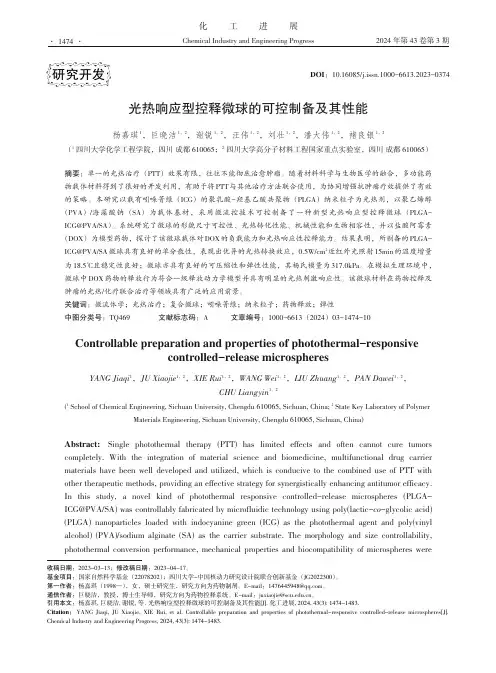
化工进展Chemical Industry and Engineering Progress2024 年第 43 卷第 3 期光热响应型控释微球的可控制备及其性能杨嘉琪1,巨晓洁1,2,谢锐1,2,汪伟1,2,刘壮1,2,潘大伟1,2,褚良银1,2(1 四川大学化学工程学院,四川 成都 610065;2 四川大学高分子材料工程国家重点实验室,四川 成都 610065)摘要:单一的光热治疗(PTT )效果有限,往往不能彻底治愈肿瘤。
随着材料科学与生物医学的融合,多功能药物载体材料得到了很好的开发利用,有助于将PTT 与其他治疗方法联合使用,为协同增强抗肿瘤疗效提供了有效的策略。
本研究以载有吲哚菁绿(ICG )的聚乳酸-羟基乙酸共聚物(PLGA )纳米粒子为光热剂,以聚乙烯醇(PVA )/海藻酸钠(SA )为载体基材,采用微流控技术可控制备了一种新型光热响应型控释微球(PLGA-ICG@PVA/SA )。
系统研究了微球的形貌尺寸可控性、光热转化性能、机械性能和生物相容性,并以盐酸阿霉素(DOX )为模型药物,探讨了该微球载体对DOX 的负载能力和光热响应性控释能力。
结果表明,所制备的PLGA-ICG@PVA/SA 微球具有良好的单分散性,表现出优异的光热转换效应,0.5W/cm 2近红外光照射15min 的温度增量为18.5℃且稳定性良好;微球亦具有良好的可压缩性和弹性性能,其杨氏模量为317.0kPa 。
在模拟生理环境中,微球中DOX 药物的释放行为符合一级释放动力学模型并具有明显的光热刺激响应性。
该微球材料在药物控释及肿瘤的光热/化疗联合治疗等领域具有广泛的应用前景。
关键词:微流体学;光热治疗;复合微球;吲哚菁绿;纳米粒子;药物释放;弹性中图分类号:TQ469 文献标志码:A 文章编号:1000-6613(2024)03-1474-10Controllable preparation and properties of photothermal-responsivecontrolled-release microspheresYANG Jiaqi 1,JU Xiaojie 1,2,XIE Rui 1,2,WANG Wei 1,2,LIU Zhuang 1,2,PAN Dawei 1,2,CHU Liangyin 1,2(1 School of Chemical Engineering, Sichuan University, Chengdu 610065, Sichuan, China; 2 State Key Laboratory of PolymerMaterials Engineering, Sichuan University, Chengdu 610065, Sichuan, China)Abstract: Single photothermal therapy (PTT) has limited effects and often cannot cure tumors completely. With the integration of material science and biomedicine, multifunctional drug carrier materials have been well developed and utilized, which is conducive to the combined use of PTT with other therapeutic methods, providing an effective strategy for synergistically enhancing antitumor efficacy. In this study, a novel kind of photothermal responsive controlled-release microspheres (PLGA-ICG@PVA/SA) was controllably fabricated by microfluidic technology using poly(lactic-co -glycolic acid) (PLGA) nanoparticles loaded with indocyanine green (ICG) as the photothermal agent and poly(vinyl alcohol) (PVA)/sodium alginate (SA) as the carrier substrate. The morphology and size controllability, photothermal conversion performance, mechanical properties and biocompatibility of microspheres were研究开发DOI :10.16085/j.issn.1000-6613.2023-0374收稿日期:2023-03-13;修改稿日期:2023-04-17。
载药纳米微粒PLA(Tet)-NP的制备【摘要】目的制备汉方已甲素(tetrandrine,Tet)聚乳酸纳米微粒,为抑制翼状胬肉术后血管增生研究奠定实验基础。
方法采用复乳法,以天然中药Tet作为试验药物,以聚乳酸(polyactic acid,PLA)为包封材料,制备Tet聚乳酸纳米微粒。
观察其表面形态、结构、粒径及Zeta电位。
结果成功制备出了Tet聚乳酸纳米微粒,微粒的表征:类圆形囊泡,均质,粒径大小约为200-230nm,Zeta电位为-7.7mv。
结论通过聚乳酸为包封材料,可成功制备出载药纳米微粒PLA(Tet)-NP(纳米微粒nanoparticles,NP),观察该纳米微粒作为药物载体的可行性。
为今后进一步开展抑制翼状胬肉术后血管增生的研究奠定了基础。
【关键词】汉方已甲素(Tet);聚乳酸;纳米微粒[Abstract]Objective Preparation of tetrandrine(Tet)polylactic acid nanoparticles,as a basis for study of vascular proliferation after pterygium surgery.Methods Using double emulsion method,with natural Chinese herbal medicine Tet as experimental drug,using polylactic acid(PLA)as embedding materials,preparation of Tet polylactic acid nanoparticles.To observe the surface morphology,structure,particle size and Zeta potential.Results Tet polylactic acid nanoparticles was prepared successfully,particle characterization:round vesicles,homogeneous,the particle size is about 200-230nm,the Zeta potential of -7.7mv.Conclusion The polylactic acid as embedding materials,can be successfully prepared drug loaded PLA(Tet)-NP(nanoparticles,NP),to investigate the feasibility of the nanoparticles as drug carrier.For further inhibit angiogenesis after pterygium surgery research foundation.[keywords]Tetrandrine(Tet);Polylactic Acid;NanoparticlesR779.6翼状胬肉是以睑裂部球结膜血管组织增生并向角膜侵入为特点的眼表疾病之一,是眼科和老年人的常见病和多发病,变性是其突出的病理特征,具有较高的复发率。
耐温抗盐型荧光聚丙烯酰胺纳米微球的制备及性能研究耐温抗盐型荧光聚丙烯酰胺纳米微球的制备及性能研究摘要:本研究旨在探究一种耐温抗盐型荧光聚丙烯酰胺纳米微球的制备方法以及其性能特点。
通过改变实验条件,并对制备得到的纳米微球进行表征和性能分析,得出了一系列结论。
实验结果表明,制备得到的耐温抗盐型荧光聚丙烯酰胺纳米微球具有优异的耐温性和抗盐性能,表现出潜在的应用前景。
关键词:耐温,抗盐,荧光聚丙烯酰胺纳米微球,制备方法,性能研究1. 引言纳米材料作为当今科技领域的前沿研究方向之一,具有许多独特的物理和化学特性,因此在各个领域都有广泛的应用。
荧光纳米微球是一类具有荧光特性的纳米材料,在生物和材料科学领域有重要的应用价值。
然而,传统的荧光纳米微球在高温和高盐环境下容易发生结构破坏,限制了它们在特殊环境中的应用。
因此,开发一种耐温抗盐型荧光纳米微球具有重要的现实意义。
2. 实验方法本研究采用一种改良的溶胶-凝胶法制备耐温抗盐型荧光聚丙烯酰胺纳米微球。
首先,将适量的聚丙烯酰胺溶液与荧光探针掺杂剂混合均匀,并在恒温搅拌条件下制备胶体溶胶。
然后,将得到的溶胶在恒温条件下静置一段时间,使溶胶逐渐发生凝胶化,最终得到耐温抗盐型荧光聚丙烯酰胺纳米微球。
3. 结果与分析通过扫描电子显微镜观察制备得到的纳米微球形貌,结果显示纳米微球呈球形结构且粒径均匀分布在100-300 nm范围内。
荧光光谱分析结果表明,制备得到的荧光聚丙烯酰胺纳米微球在激发波长为λ=365 nm时具有强烈的荧光发射峰。
进一步的热稳定性和盐稳定性测试显示,该纳米微球在高温(>300°C)和高盐(>5 mol/L)条件下仍然保持良好的稳定性,未发生显著的形态和结构变化。
4. 应用前景制备得到的耐温抗盐型荧光聚丙烯酰胺纳米微球具有潜在的应用前景。
首先,在高温环境下的荧光探测和传感领域具有广阔的应用前景。
其次,该纳米微球的优异耐盐性能使其在海洋和盐湖等高盐环境中的应用具有潜力。
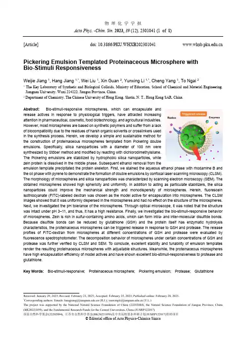
物 理 化 学 学 报Acta Phys. -Chim. Sin. 2023, 39 (12), 2301041 (1 of 8)Received: January 29, 2023; Revised: February 23, 2023; Accepted: February 23, 2023; Published online: February 28, 2023. *Correspondingauthors.Emails:**********************.cn(H.J.);**********************.cn(Y.L.)The project was supported by the National Natural Science Foundation of China (22202084), the Natural Science Foundation of Jiangsu Province, China (BK20221059), and the Fundamental Research Funds for the Central Universities, China (JUSRP122017)国家自然科学基金(22202084), 江苏省自然科学基金(BK20221059)及中央高校基本科研计划(JUSRP122017)资助项目© Editorial office of Acta Physico-Chimica Sinica[Article]doi: 10.3866/PKU.WHXB202301041Pickering Emulsion Templated Proteinaceous Microsphere with Bio-Stimuli ResponsivenessWeijie Jiang 1, Hang Jiang 1,*, Wei Liu 1, Xin Guan 2, Yunxing Li 1,*, Cheng Yang 1, To Ngai 21 The Key Laboratory of Synthetic and Biological Colloids, Ministry of Education, School of Chemical and Material Engineering,Jiangnan University, Wuxi 214122, Jiangsu Province, China.2 Department of Chemistry, The Chinese University of Hong Kong, Shatin, N. T., Hong Kong SAR, China.Abstract: Bio-stimuli-responsive microspheres, which can encapsulate and release actives in response to physiological triggers, have attracted increasing attention in pharmaceutical, cosmetic, food biotechnology, and agricultural industries. However, most microspheres are based on synthetic polymers and suffer from a lack of biocompatibility due to the residues of harsh organic solvents or crosslinkers used in the synthesis process. Herein, we develop a simple and sustainable method for the construction of proteinaceous microspheres templated from Pickering double emulsions. Specifically, silica nanoparticles with a diameter of 100 nm were synthesized by Stöber method and modified by reacting with dichlorodimethylsilane. The Pickering emulsions are stabilized by hydrophobic silica nanoparticles, while zein protein is dissolved in the middle phase. Subsequent ethanol removal from theemulsion template precipitated the protein skeleton. First, we stained the aqueous ethanol phase with rhodamine B and the oil phase with pyrene to demonstrate the formation of double emulsions by confocal laser scanning microscopy (CLSM). The morphology of microspheres and silica nanoparticles was characterized by scanning electron microscopy (SEM). The obtained microspheres showed high sphericity and uniformity. In addition to acting as particulate stabilizers, the silica nanoparticles could improve the mechanical strength and monodispersity of microspheres. Herein, fluorescein isothiocyanate (FITC)-labeled dextran was chosen as the model active for encapsulation into microspheres. The CLSM images showed that it was uniformly dispersed in the microspheres and had no effect on the structure of the microspheres. Next, we investigated the pH tolerance of the microspheres. Through optical microscope, it was noted that the structure was intact under pH 3–11, and thus, it has a high resistance. Finally, we investigated the bio-stimuli-responsive behavior of microspheres. Zein is rich in sulfur-containing amino acids, which can form intra- and inter-molecular disulfide bonds. Because disulfide bonds can be reduced by glutathione (GSH) and the protein itself has enzymatic hydrolysis characteristics, the proteinaceous microspheres can be triggered release in response to GSH and protease. The release profiles of FITC-dextran from microspheres at different concentrations of GSH and protease were evaluated by fluorescence spectrophotometer. The decomposition behavior of microspheres under certain concentrations of GSH and protease was further verified by CLSM and SEM. To conclude, excellent stability and tunability of emulsion templates render the resulting proteinaceous microspheres with adjustable structures. Meanwhile, the proteinaceous microspheres have high encapsulation efficiency of model actives and have shown excellent bio-stimuli-responsiveness to protease and glutathione.Key Words: Bio-stimuli-responsive; Proteinaceous microsphere; Pickering emulsion; Protease; GlutathionePickering乳液模板构建生物刺激响应性蛋白质微球蒋伟杰1,蒋航1,*,刘威1,管鑫2,李云兴1,*,杨成1,魏涛21江南大学化学与材料工程学院,合成与生物胶体教育部重点实验室,江苏无锡2141222香港中文大学化学系,新界沙田香港特别行政区摘要:生物刺激响应性微球可用于封装活性物,并在生理条件刺激下触发释放活性物,在制药,化妆品,食品和农业等领域备受关注。
SPG膜乳化法制备载BSA的PLGA微球的工艺考察万斯斯;杨琪;钟晨;罗宇燕;张永明【摘要】目的采用SPG膜乳化法,制备载蛋白药物的聚乳酸聚乙醇酸(PLGA)微球,并对其形态学性质、载药量、体外释药进行考察.方法以血清白蛋白(BSA)为模型药物,PLGA作为载体材料,采用SPG膜乳化技术结合复乳溶剂挥发法制备微球;分别考察初乳匀浆转速、内水相体积、膜挤出压力、外/内水相加盐等因素对微球质量的影响.结果以优化处方制备的载药微球形态圆整、粒径均一,平均粒径为(55.51±0.24)μm,载药量、包封率分别为7.70%和69.33%,缓释时间达到40 d.结论本研究获得了SPG膜乳化法制备BSA-PLGA缓释微球的新工艺.【期刊名称】《广东药学院学报》【年(卷),期】2016(032)005【总页数】5页(P550-554)【关键词】SPG膜乳化法;PLGA;缓释微球【作者】万斯斯;杨琪;钟晨;罗宇燕;张永明【作者单位】中山大学附属第三医院药剂科,广东广州510630;中山大学附属第三医院药剂科,广东广州510630;中山大学药学院,广东广州510006;中山大学附属第三医院药剂科,广东广州510630;中山大学附属第三医院药剂科,广东广州510630【正文语种】中文【中图分类】R944近年来,随着生物科技的迅猛发展,越来越多疗效显著的蛋白、多肽类药物被开发上市。
由于这类药物稳定性差、口服易降解,冻干粉针类制剂仍是其传统剂型,但难以解决半衰期短,需频繁、长时间注射给药的缺点,导致临床应用受到限制。
利用生物可降解的新型聚合物材料为载体材料[1],将蛋白多肽类药物制成微球注射剂,可达到体内持久缓释,减少给药次数,显著增加患者用药顺应性的目的。
聚乳酸聚乙醇酸(PLGA)以良好的生物相容性受到很多学者的关注,已有多种以PLGA为载体的缓控释药物上市。
SPG膜(shirasu porous glass membrane)是日本SPG公司开发的新型无机膜,膜孔径微小均匀且可控[2],原理是分散相在N2压力的作用下透过微孔膜的膜孔而在膜表面形成液滴,在沿膜表面不断搅拌的外水相溶液的冲洗作用下,液滴的直径达到临界值后,就从膜表面剥离,从而形成乳液,再结合溶剂挥发法固化后即可得到粒径均一可控的微球。
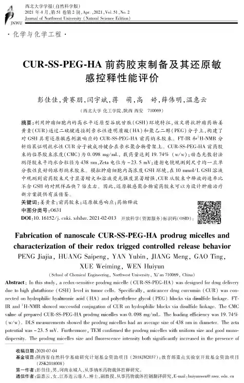
西北大学学报(自然科学版)2021年4月,第51卷第2期,Apr.,2021,VoU51,Nc.2Journal of Northwest University(Natural Science Edition)•化学与化学工程•CUR-SS-PEG-HA前药胶束制备及其还原敏感控释性能评价彭佳佳,黄赛朋,闫宇斌,蒋萌,高婷,薛伟明,温惠云(西北大学化工学院,陕西西安710069)摘要:利用肿瘤细胞内的高水平还原型谷胱甘肽(GSH)环境特征,该文将抗肿瘤药物姜黄素'CUR)通过二硫键连接到亲水性透明质酸(HA)和聚乙二醇(PEG)分子上,构建了对GSH具有还原敏感刺激响应的CUR-SS-PEG-HA前药纳米胶束。
FT-IR和1H-NMR分析结果证明疏水性CUR分子被成功键合在亲水聚合物骨架上。
CUR-SS-PEG-HA前药胶束的临界胶束浓度'CMC)为0.098my/mL,载药量达到19.74%(w/w);动态光散射法测得胶束平均水合粒径为438nm,Zeta电位为-23.5mV;透射电镜观测到尺寸均一且单分散性良好的球形纳米胶束。
模拟肿瘤细胞内高浓度GSH环境,在10mmo/L GSH溶液中观测到前药胶束尺寸显著增大和溶液荧光强度显著增强,CUR从胶束中释放的速率比不含GSH的对照样品快7倍左右。
因此,还原敏感聚合物前药胶束可以为设计肿瘤治疗新方案提供有益借鉴。
关键词:姜黄素;前药胶束;还原敏感响应;药物释放中图分类号:O631DOS:10.16152/j-cnki-edxbzr.2021-02-013开放科学(资源服务)标识码(OSID):Fabrication of nanoscale CUR-SS-PEG-HA prodrug micelles and characterization of their redox trigged controlled release behavior PENG Jiajia,HUANG Saipeng,YAN Yubin,JIQNG Meng,GAO Ting,XUE Weiming,WEN Huiyun(School of Chemical Engineering,Northwest University,Xi'an710069,China)Abstract:In this study,a redox-sensitive podruy micel l a(CUR-SS-PEG-HA)was designed for drug deliveo due to high glutathione(GSH)level in tumor cells-SpeciPcal/,anticancar drug curcumin(CUR)was connected on hydeophooochyaoueonocacod(HA)and pooyethyoenegoycoo(PEG)boock7eoadouooodeoonkage.FT-IR and H-NMR showed successful conjugation of CUR on hydrophilic blocks via dOul/da Unkagg-The CMC eaoueoopeepaeed CUR-SS-PEG-HApeodeugmoce o eswas0.098mg jmL)The ooad ong e o oc oency was19.74% (w/w)-DLS measurements showed the prodrug micelles had an yverage size of438nm in diameter-The zeta potential was-23.5mV-Fuitheoioo,TEM conloied the podruy micelles with unPUm size and good monodispersity-The podruy micelles size and Juorescenco OWns/y both significantly increased in the presence of收稿日期:2020-7-4基金项目:陕西省自然科学基础研究计划基金资助项目(2018JM2037);教育部重点实验室开放基金资助项目(ZSK2018008)第一作者:彭佳佳,男,河南永城人,从事纳米药物载体控释研究&通信作者:温惠云,女,江苏连云港人,博士,副教授,从事药物载体控制翻译研究,E-mail:huiyunwen@第2期彭佳佳等:CUR-S-EGHA前药胶束制备及其还原敏感控释性能评价・297・tumor Alxent GSH(10mmol/C)-Moreover#the prodrug miceXes showed high GSH3X^011/which accelerate in vitro release of CUR from micelles7hmes faster than in absence of GSH-Thus,these redge-sensitive peodeugmoce o esmaypeoeodeauseauoeeaeeenceaoethedesogn oanewtumoeteeatmentpeogeams.Key words:cucumin;prodrug micelle;Adoewens/iw;drug release姜黄素(curcumin,CUR)是从姜科天南星科植物根茎中提取的多酚化合物,具有抗氧化、抗炎、抗感染、抗肿瘤等药理活性(1-)&CUR通过诱导恶性肿瘤细胞分化和凋亡来抑制肿瘤细胞生长,发挥抗肿瘤作用,在肿瘤治疗中具有药理安全性高和抗多药耐药等优点。
Tianjin Biomedical Material KeyLaboratory, Institute of BiomedicalEngineering, Chinese Academy of Medical Sciences & Peking Union Medical College, Tianjin 300192, ChinaWu Li, Chieftechnician, Tianjin Biomedical Material Key Laboratory, Institute of BiomedicalEngineering, Chinese Academy of Medical Sciences & Peking Union Medical College, Tianjin 300192, China wuli.7777@ Yang Jing ★, Master, Associate researcher, Tianjin Biomedical Material KeyLaboratory, Institute of BiomedicalEngineering, Chinese Academy of Medical Sciences & Peking Union Medical College, Tianjin 300192, China Yangjing926@ Wu Li and Yang Jing contributed equally to this paper.Correspondence to: Liu Tian-jun, Doctor, Researcher, Tianjin Biomedical Material Key Laboratory, Institute of BiomedicalEngineering, Chinese Academy of Medical Sciences & Peking Union Medical College, Tianjin 300192, ChinaCorrespondence to: Song Cun-xian,Master, Researcher, Tianjin Biomedical Material KeyLaboratory, Institute of BiomedicalEngineering, Chinese Academy of Medical Sciences & Peking Union Medical College, Tianjin 300192, China武 莉,杨 菁,刘天军,宋存先Acceptability of Taxol-poly (alkyl-cyanoacrylates) micell materialWu Li, Yang Jing, Liu Tian-jun, Song Cun-xianAbstractBACKGROUND: Currently used poly (alkyl-cyanoacrylates) (PACA) produces aldehyde compound in its degradation, which is easily results in toxicity and stimulation to the body. Here, a novel Taxol-PACA micell material was synthesized, which has broad application as a kind of liposolubility drug delivery carrier.OBJECTIVE: To verify the therapeutic efficacy of Paclitaxel-PECA micells for mouse breast cancer.METHODS: Paclitaxel drug delivery micells were prepared by a multi-emulsification technique and were characterized for size, drug loading capacity, and in vitro release. Bablc breast cancer model mice were randomly divided into the physiological saline, vacant control, paclitaxel positive control, and Paclitaxel-PECA micells with low-dose, medium-dose, and high-dose groups. Paclitaxel and Paclitaxel-PECA micells were injected into the location of mouse breast cancer, and then the tumor inhibit rates were detected.RESULTS AND CONCLUSION: The mean diameter of Paclitaxel-PECA micells was 70 nm, with 19.89% loading amount ofPaclitaxel. In vitro , micells maintained sustained release of Paclitaxel for 2 weeks. Compared with the physiological saline group, the Paclitaxel-PECA micells group exhibited superior tumor inhibit effects with doses of 30, 60, and 90 mg/kg (P < 0.001), which was 68.49%, 77.03% and 81.87%, respectively. The results suggested that Paclitaxel-PECA micells material has excellent acceptability as sustained-release preparation for treating mouse breast cancer.Wu L, Yang J, Liu TJ, Song CX.Acceptability of Taxol-poly (alkyl-cyanoacrylates) micell material. Zhongguo Zuzhi Gongcheng Yanjiu yu Linchuang Kangfu. 2010;14(8):1392-1396. [ ]摘要背景:目前研究使用的聚氰基丙烯酸烷基酯,在降解过程中生成了醛类化合物,易导致一定程度的毒性与刺激作用。
聚乙二醇生物降解张成彬(北京化工大学材料科学与工程学院,北京市朝阳区,10029)摘要:聚乙二醇具有良好的生物相容性和两亲性,在生物医药领域中有着广泛的应用。
本文对聚乙二醇的生产、应用以及对生物降解研究进行详细的评述。
聚乙二醇的降解性能取决于分子量的大小,分子量较高的降解不佳。
聚乙二醇的好氧代谢作用机理已较清楚:首先被氧化成乙醛和一元羧酸,进一步进行解聚;但其厌氧代谢作用机理尚不明确,已提出的许多机理还有待进一步研究确证。
关键词:聚乙二醇;生物降解;生产;合成BIODEGRADATION OF POLYETHYLENE GLYCOLS ZHANG Cheng Bin(College of Materials Science and Engineering Beijing University of Chemical Technology,Chaoyang Beijing 100029)Abstract:Polyethylene glycol has good biocompatibility and amphiphilic, has a wide range of applications in the biomedical field .Preparation、application、biodegradation of polyethylene glycols(PEG) is reviewed in this paper .The degradability of PEGs depends on the molecular weight .Higher molecular weight results in poor degradation .Aerobic metabolism of PEGs are clear: first oxidized to an aldehyde and monocarboxylic acid , then followed by depolymerization .On the other hand ,the mechanism of anaerobic metabolism is still not clear, most of the proposed mechanisms need to be firmed .Keywords:Polyethylene glycol, biodegradation, preparation, application1.前言聚乙二醇(PEG)由环氧乙烷开环聚合得到,根据生产工艺路线的不同,也有文献称作聚氧化乙烯(PEO)【1】。
扑尔敏PLGA缓释微球的制备及体外质量评价周荣平;姚雯雯;傅红兴;李慧;叶其纯;夏烽【摘要】目的以扑尔敏为模型药物,开发一种PLGA 缓释微球给药系统.方法采用复乳- 溶剂挥发法制备扑尔敏PLGA缓释微球,紫外分光光度法测定其载药量、药物释放.结果制得的扑尔敏PLGA 缓释微球平均粒径为(112±57)μm,含药量为(798.33±145.00)μg,载药量为(0.32±0.12)%;药物9 d 累积药物释放量可达87%.结论本研究扑尔敏PLGA 缓释微球制备方法简单,药物能达到长期缓慢释放.【期刊名称】《中国医药科学》【年(卷),期】2012(002)009【总页数】2页(P52-53)【关键词】扑尔敏;乳酸/;羟基乙酸共聚物;微球;复乳-;溶剂挥发法【作者】周荣平;姚雯雯;傅红兴;李慧;叶其纯;夏烽【作者单位】温州医学院药学院,浙江温州,325035;温州医学院药学院,浙江温州,325035;温州医学院药学院,浙江温州,325035;温州医学院药学院,浙江温州,325035;温州医学院药学院,浙江温州,325035;温州医学院药学院,浙江温州,325035【正文语种】中文【中图分类】R917微球是指利用天然的或合成的高分子材料将固体微粒或液滴包裹起来的微型球状实体[1]。
微球被用于药物控缓释体系来治疗许多疾病,或者通过靶向作用使药物到达特定的组织和器官后缓慢释放,提高药物的治疗效果[2]。
乳酸-羟基乙酸共聚物(PLGA)是FDA 批准用于临床试验的可生物降解高分子聚合物,体内代谢终产物为CO2和H2O,常用于作为缓释微球的载体[3]。
扑尔敏为水中溶解药物,主要用于抗过敏治疗,临床上有Smith Kline Beecham公司生产的扑尔敏缓释小丸,作为组织胺H1受体拮抗剂,抗组胺作用较持久。
本研究选择扑尔敏做为模型药物,采用复乳(W/O/W)-溶剂挥发法制备扑尔敏缓释微球,探讨了其制备条件对微球的粒径大小及其分布、包封率和载药量的影响。
Colloids and Surfaces B:Biointerfaces 83 (2011) 103–107Contents lists available at ScienceDirectColloids and Surfaces B:Biointerfacesj o u r n a l h o m e p a g e :w w w.e l s e v i e r.c o m /l o c a t e /c o l s u r fbPreparation,characterization of hydrophilic and hydrophobic drug in combine loaded chitosan/cyclodextrin nanoparticles and in vitro release studyJi Jingou ∗,Hao Shilei,Liu Weiqi,Wu Danjun,Wang Tengfei,Xu YiFaculty of Pharmacy,College of Chemistry and Chemical Engineering,University of Chongqing,Chongqing 400030,Chinaa r t i c l e i n f o Article history:Received 19October 2010Received in revised form 30October 2010Accepted 1November 2010Available online 9 November 2010Keywords:Chitosan nanoparticles Cyclodextrin Methotrexate Cross-linkinga b s t r a c tThe compound nanoparticles of chitosan (CS)and cyclodextrin (CD)loading with hydrophilic and hydrophobic drug simultaneously were prepared via the cross-linking method.Methotrexate (MTX)and calcium folinate (CaF)were selected as the model drugs.The prepared nanoparticles were characterized by FT-IR spectroscopy to confirm the cross-linking reaction between CS and cross-linking agent.X-ray diffraction (XRD)was performed to reveal the form of the drug after encapsulation.The average size of nanoparticles ranged from 308.4±15.22to 369.3±30.01nm.The nanoparticles formed were spherical in shape with high zeta potentials (higher than +30mV).In vitro release studies in phosphate buffer saline (pH 7.4)showed an initial burst effect and followed by a slow drug release.Cumulative release data were fitted to an empirical equation to compute diffusional exponent (n ),which indicated the non-Fickian trend for drug release.© 2010 Elsevier B.V. All rights reserved.1.IntroductionCoadministration of two or more drugs in clinical practice is common,such as ezetimibe and simvastatin,sertraline and cis-apride or pimozide,itraconazole and tacrolimus,ketamine and midazolam and so on [1–4],which is useful for improvement of the therapeutic value of medicinal drugs.However,the combined drugs are always taken respectively in practice.The separate administra-tion may make the absorption and distribution of the drugs with poor coordination in vivo ,and the multiple administrations make it inconvenience for the patients.Furthermore,an effective therapy should combine sufficiently high and sustained drug levels at the injury site with minimal systemic and local toxicity.In the recent years,there has been considerable interest in developing biodegradation nanoparticles as effective drug delivery carriers.Nanoencapsulation of medicinal drugs (nanomedicines)has many advantages in the protection of premature degradation,enhancement of absorption into a selected tissue,bioavailability,retention time and improvement of intracellular penetration [5].And the largest advantages of the nanoparticles are that they can effectively deliver the drug to a specific site and control the drug release speed [6].Currently,the biodegradation nanoparticles con-tenting two different drugs have been prepared.The results prove that simultaneous administration could result in better treatment efficacy [7].∗Corresponding author.Tel.:+862365102531;fax:+862365102531.E-mail address:jingou ji@ (J.Jingou).Polymeric materials such as chitosan (CS)and poly-d ,l -lactide-co-glycolide (PLGA)are used for synthesis of biodegradation nanoparticles [8,9].CS is a natural polysaccharide derived by partial deacetylation of chitin.Properties such as biodegradabil-ity,non-toxicity and good biocompatibility make it suitable for use in biomedical and pharmaceutical formulations [10–14].And PLGA is one of the most commonly used relatively hydrophobic and synthetic polyesters,which have been extensively used as drug delivery systems [15].Because of the nature of the poly-mer molecule,CS nanoparticles are suitable for the delivery of hydrophilic drug,and it is relatively easy to entrap the hydrophobic drug into PLGA nanoparticles [7].But in practice,the coadminis-tration of drugs may consist of both hydrophilic and hydrophobic drug.It is difficult to encapsulate the hydrophilic and hydrophobic drug simultaneously into the CS or PLGA nanoparticles.Currently,a new nanocarrier of CS/cyclodextrin (CS/CD)nanoparticles were synthesized,the novel CS nanoparticles could encapsulate the hydrophobic drug successfully by introducing CDs [16–18].And the CS/CD nanoparticles with the advantages of good solubilization and permeability would improve the bioavailability of the hydrophobic drug.Methotrexate (MTX),a folate antimetabolite,is widely used in the treatment of malignancies [19].Low-dose MTX has been com-monly used in the treatment of psoriasis since the 1960s.However,approximately 30%of patients abandon the rheumatoid arthri-tis treatment because of drug-related side effects.Specific drugs such as folic acid (FA)or folinic acid used as comedication can sig-nificantly reduce the risk of MTX toxicity [20,21].In the present study,the CS/CD nanoparticles were prepared via the cross link-0927-7765/$–see front matter © 2010 Elsevier B.V. All rights reserved.doi:10.1016/j.colsurfb.2010.11.005104J.Jingou et al./Colloids and Surfaces B:Biointerfaces83 (2011) 103–107ing method and used to deliver the hydrophilic and hydrophobic drug simultaneously.MTX,a hydrophobic drug and calcium foli-nate(CaF),a hydrophilic drug were selected as the model drugs. The physicochemical properties of CS/CD nanoparticles were inves-tigated using various analytical equipments such as transmission electron microscope(TEM),scanning electron microscope(SEM) and FT-IR spectroscopy,and the drug release capability in vitro was also investigated.2.Materials and methods2.1.MaterialsChitosan(CS,Deacetylation degree of95%and molecular weight of80kDa)was purchased from Golden-shell Biochemical Co.Ltd. (Zhejiang,China).Tripolyphosphate(TPP)was purchased from Wenzhou Dongsheng Chemical reagent Co.Ltd.(Zhenjiang,China). Methotrexate(MTX)was purchased from Top Pharm Chemical Co. Ltd.(Shaanxi,China).Calcium folinate(CaF)was purchased from Wuhan Galaxy Chemical Co.Ltd.(Hubei,China).ˇ-cyclodextrin(ˇ-CD,M w=1134.98Da)was purchased from Kelong Chemical reagent Co.Ltd.(Sichuan,China).2-Hydroxypropyl-ˇ-Cyclodextrin(Hp-ˇ-CD,M w=1425.38Da,average substitution degree equal to5)was purchased from Hezhong Bio-Chemical Co.Ltd.(Wuhan,China). All other materials and reagents used in the study were analytical grade.2.2.MTX phase solubility studiesPhase solubility studies were performed according to the method of Higuchi and Connors[22].Briefly,excess of MTX was added to20ml pure water containing various amount ofˇ-CD or Hp-ˇ-CD(from0to6mmol).They were shaken at30◦C for72h.The aliquots werefiltered through0.45m cellulose acetate membrane filters(New Asiatic Pharmaceutical,China).Thefiltered samples were assayed by the UV spectrophotometer(Shimadzu,UV-2450, Japan)at303nm.The stability constants,K c,were calculated from the straight-line portion of the phase solubility diagram according to Eq.(1)K c=SlopeIntercept(1−Slope)(1)2.3.Preparation of the MTX and CaF loaded CS/CD nanoparticles2.3.1.Preparation of the MTX inclusion complexesThe MTX complexes were prepared as followed:for bothˇ-CD and Hp-ˇ-CD,the CD:MTX complexes were prepared with1:1M ratio stoichiometry by co-incubation of the aqueous solutions of both compounds for24h at room temperature and under moder-ate stirring.For theˇ-CD/MTX complex,an aqueous solution was prepared by adding68.10mgˇ-CD and27.26mg MTX to30ml dis-tilled water.For the Hp-ˇ-CD/MTX complex,the complex solution was prepared by adding85.52mg Hp-ˇ-CD and27.26mg MTX to 30ml distilled water.2.3.2.Preparation of the CD/MTX and CaF loaded CS nanoparticlesThe preparation of the CD/MTX and CaF loaded CS nanoparti-cles was based on the ionic cross-linking of CS with TPP anions. For all CDs,the inclusion complex solution wasfirstlyfiltered through0.45m cellulose acetate membranefilters.2.0ml inclu-sion complexfilter liquor and2.0ml CaF solution(0.2%,w/v)were mixed with a certain volume TPP solution(0.2%,w/v).CS solu-tion(0.2%,w/v)was prepared by dissolving CS in dilute acetic acid(1%,v/v)at room temperature with sonication.The CS/CD nanoparticles were spontaneously formed upon addition of the mixture solution of inclusion complex/CaF/TPP to CS solution. The CS/CD nanoparticles were prepared by selecting the mass ratios of CS to TPP from4:1to7:1.The nanoparticles suspensions were continuously stirred for1h and centrifuged at16,000rpm for30min.The resulting nanoparticles were lyophilized and stored.2.4.Characterization of the CS/CD nanoparticlesNano ZS90Zetasizer(Malvern Instruments,UK)was used to measure the particle size,zeta potential and polydispersity index(PDI)of the prepared CS/CD nanoparticles by dynamic light scattering.The nanoparticles were also examined by Transmission electron microscopy(TEM)(Philips,Tecrai10,Dutch)and Scanning electron microscopy(SEM)(Hitachi,S-3400N,Japan).For TEM,the nanoparticles solution was dropped on copper grids and natively stained by2%phosphotungstic acid,and then dried at room temperature.For SEM,the nanoparticles suspensions were spread on a glass plate and dried at room temperature.The dried nanopar-ticles were then coated with gold metal under vacuum and then examined.The chemical structure and complexes formation of CS,ˇ-CD and blank CS/ˇ-CD nanoparticles(without loading drugs) were analyzed by FT-IR(Nicolet,5DX/550II,USA).The samples used for the FT-IR spectroscopic characteristics were prepared by grinding the dry specimens with KBr and pressing them to form disks.The XRD experiments were carried out using X-ray diffractometer(Shimadzu,XRD6000X,Japan).MTX,CaF,ˇ-CD,CS and drug-loaded CS/CD nanoparticles were scanned from5◦to40◦.2.5.Evaluation of MTX and CaF loading capacityThe loading efficiency(LE)of the MTX/CaF-loaded CS/CD nanoparticles was determined by following method:In brief,drug-loaded nanoparticles(20mg)were taken into5ml0.1mol/L HCl for24h,and the nanoparticles suspensions were separated by cen-trifugation at16,000rpm for30min.The content of MTX and CaF in the supernatants was analyzed by UV spectrophotometer at303nm and355nm respectively.A blank sample was made from CS/CD nanoparticles without loaded drugs but treated similarly as the drugs-loaded CS/CD nanoparticles.Each batch samples were mea-sured in triplicate.The process yield(PY)of the CS/CD nanoparticles was also determined in this study.The LC and PY were calculated by following Eqs.(2)and(3):LC=Weight of MTX(or CaF)encapsulated in nanoparticles Weight of nanoparticles after freeze drying ×100%(2)PY=Weight of nanoparticles after freeze dryingWeight of total components×100%(3) 2.6.In vitro release study of the CS/CD nanoparticlesThe in vitro release studies were carried out as followed:drugs-loaded CS/CD nanoparticles and5ml PBS solution(pH7.4)were put into a dialysis tube(MWCO:12,000)and then the dialy-sis tube was placed in50ml PBS at37◦C and maintained under shaken at100stocks/min.The analysis was performed in tripli-cate for each sample.At specific time intervals,medium was taken and replaced with fresh PBS.The concentration of the released MTX and CaF into PBS were determined by UV spectrophotome-ter.J.Jingou et al./Colloids and Surfaces B:Biointerfaces83 (2011) 103–107105Fig.1.Effect ofˇ-CD( )and HP-ˇ-CD( )on the solubility of methotrexate in water (n=3).3.Results and discussion3.1.MTX phase solubility studiesThe phase solubility profiles for the complex formation between MTX andˇ-CD or Hp-ˇ-CD are presented in Fig.1.The diagram showed that the aqueous solubility of MTX increased linearly as a function of CDs concentration.It is clear that this solubility diagram can be classified as the A L type according to Higuchi and Connors [22],which suggested that the increase in solubility observed was due to the formation of inclusion complex between MTX with CDs. The apparent stability constant of the MTX/ˇ-CD complex at30◦C was calculated to be1302.47M−1according to Eq.(1),and the MTX/HP-ˇ-CD complex was473.23M−1.The stability constant val-ues showed that the process of the inclusion complex was easier for MTX/ˇ-CD than MTX/HP-ˇ-CD[23].3.2.Preparation of the MTX and CaF-loaded CS/CD nanoparticlesMTX/CaF-loaded CS/CD nanoparticles were prepared in a two-step procedure.The inclusion complex of MTX/CD was prepared firstly,after that,the inclusion complex and CaF were entrapped into the CS nanoparticles via the cross-linking method.The pre-pared method was more effective comparing with the previous prepared method[16,17].In the previous study,the inclusion com-plex of drug and CD was prepared in the present of CS,which may hinder the formation of inclusion complex because of the long molecular chain.The effects of different mass ratios of CS to TPP ranging from4:1 to7:1on the physicochemical properties of the CS/CD nanoparticles are shown in Table1.When the drugs-loaded CS/HP-ˇ-CDnanopar-Fig.2.TEM image of the MTX and CaF-loaded CS/ˇ-CD nanoparticles(mass ratio of CS/TPP:7/1).ticles were prepared with the mass ratio4:1,the precipitate was optically seen.The LC and PY of the CS/CD nanoparticles decreased with increasing the CS/TPP mass ratio.This trend could be explained that the larger amount of TPP would enhance the degree of cross-link reaction.The LC of MTX in CS/ˇ-CD nanoparticles was higher than CS/HP-ˇ-CD nanoparticles.The reason could be explained that the MTX/ˇ-CD inclusion complex formed easier than MTX/HP-ˇ-CD inclusion complex according to the results of phase solubility studies,so the MTX/ˇ-CD complex solution may contain larger amount of MTX.The particle distribution and zeta potential of the CS/CD nanoparticles were also investigated in this study.A grad-ual decrease in the particle size ranging from369.3±30.01nm to 308.4±15.22nm with increase in the CS/TPP mass ratio was noted. And the values of PDI were smaller than0.300,indicating a narrow nanoparticles particle size distribution.The residual amine groups would be responsible for the positive zeta potential of the nanopar-ticles,and the zeta potentials ranged from+35.76±3.21mV to +38.46±1.29mV.These values are adequate to from a stable CS/CD nanoparticles suspension.3.3.Characterization of the CS/CD nanoparticlesThe morphological characteristics of the MTX/CaF-loaded CS/ˇ-CD nanoparticles with the CS/TPP mass ratio7:1were examined using TEM and SEM.Negative staining TEM image of MTX/CaF-loaded CS/ˇ-CD nanoparticles is shown in Fig.2.The result suggested that MTX/CaF-loaded CS/ˇ-CD nanoparticles were fairly smooth and spherical in shape,and the average size was about 300nm.Fig.3shows the SEM image of the MTX/CaF-loaded CS/ˇ-CD nanoparticles,the particles size and morphology were similar with the TEM observed.Fig.4depicts the FT-IR spectra of CS,ˇ-CD and blank CS/ˇ-CD nanoparticles.The characteristic peaks ofˇ-CD(Fig.4a)are includ-ing at3400cm−1(–OH stretching),2927cm−1(–CH stretching),Table1Effect of different CS/TPP mass ratios on the physicochemical properties of MTX/CaF loaded CS/CD nanoparticles(mean±S.D.,n=3).CS/TPP mass ratio Type of CD LC(%)PY(%)Particle size(nm)PDI Zeta potential(mV)MTX CaF4:1ˇ-CD 2.48±0.07 2.64±0.1850.73±3.18369.3±30.010.254±0.03+35.76±3.21 4:1HP-ˇ-CD Precipitation–––––5:1ˇ-CD 2.46±0.11 2.25±0.1737.76±3.58337.8±15.090.232±0.12+36.62±2.19 5:1HP-ˇ-CD 2.22±0.10 2.34±0.1346.01±4.12435.0±17.330.246±0.07+35.62±1.87 6:1ˇ-CD 2.21±0.05 2.14±0.0937.70±3.22324.8±21.640.284±0.20+37.26±2.16 6:1HP-ˇ-CD 2.10±0.09 2.16±0.0837.90±2.97374.0±20.450.235±0.15+37.61±2.81 7:1ˇ-CD 1.99±0.180.92±0.0719.10±2.01308.4±15.220.253±0.09+38.46±1.29 7:1HP-ˇ-CD 1.62±0.12 1.93±0.0618.24±3.31319.5±31.190.226±0.06+37.91±2.16106J.Jingou et al./Colloids and Surfaces B:Biointerfaces83 (2011) 103–107Fig.3.SEM image of the MTX and CaF-loaded CS/ˇ-CD nanoparticles (mass ratio of CS/TPP:7/1).1400cm −1(–OH bending)and 950cm −1(skeletal vibration involv-ing ␣-1,4linkage).The results demonstrate the basic features of CS (Fig.4b)at 3426cm −1(–OH and –NH 2stretching),2922cm −1and 2872cm −1(–CH stretching),1601cm −1(–NH 2stretching),1078cm −1(C–O–C stretching)and 601cm −1(pyranoside ring stretching vibration).For the blank CS/ˇ-CD nanoparticles (Fig.4c),the peak of 3423cm −1becomes wider,indicating that hydrogen bonding action is enhanced because of the reaction between CS and ˇ-CD [24].The –NH 2bending vibration shifts from 1601cm −1to 1542cm −1,and a new peak appear at 1637cm −1,which indi-cates that some interaction between NH 3+groups of CS and TPP are occurred within the nanoparticles.The XRD patterns of CS,ˇ-CD,MTX,CaF and the drugs-loaded CS/ˇ-CD nanoparticles are shown in Fig.5.CS exhibits two charac-teristic peaks at 2Âof 11◦and 20◦(Fig.5c),and there are several strong peaks in the diffractogram of ˇ-CD (Fig.5b)and MTX (Fig.5a),indicating the high degree of crystallinity.The CaF consists of one peak at 2Âof 22◦(Fig.5d).In diffraction spectrum of drugs-loaded CS/ˇ-CD nanoparticles the peak at 2Âof 20◦becomes even broader (Fig.5e).It is well-known that the width of X-ray diffraction peak is related to the size of crystallite,the broadened peak usually results from imperfect crystal.So the broad peak of drugs-loaded CS/ˇ-CD nanoparticles may be caused by the cross-linking reaction between CS and TPP,which may destroy the crystalline structure of CS [25].Fig.4.FT-IR spectra of ˇ-CD (a),CS (b)and blank CS/ˇ-CD nanoparticles(c).Fig.5.XRD patterns of MTX (a),ˇ-CD (b),CS (c),CaF (d)and the drugs-loaded CS/ˇ-CD nanoparticles (e).And the characteristic peaks of MTX,CaF and ˇ-CD disappeared in those corresponding to drug-loaded nanoparticles.It is indicated that MTX and CaF may exist as molecular dispersion in the poly-meric nanoparticles [26].3.4.In vitro release studiesThe in vitro cumulative release profiles of MTX and CaF from CS/ˇ-CD nanoparticles and CS/HP-ˇ-CD nanoparticles in the PBS (pH 7.4)are shown in Fig.6.There were an initial burst release phase and a sustained release phase in the release profiles.About 20%drugs were released in the burst release phase (within the first 0.5h),which was mainly because that the nanoparticles surface drugs could easily diffuse in the first phase.And the second phase was a relatively slow release up to 24h,which could cause by the drugs diffusion and the polymer degradation.The release rate of CaF from the CS/CD nanoparticles was higher than MTX,which could be explained that the CS/CD nanoparticles exhibited stronger sustained release effect than CS nanoparticles.And the release rate was higher in case of the formulation con-taining more drugs.So the release rate of MTX from the CS/ˇ-CD nanoparticles was faster than CS/HP-ˇ-CD nanoparticles,and the reverse trend was observed with the release profiles of CaF.The release data of the in vitro release of the MTX and CaF from the CS/CD nanoparticles had been further substantiated by fitting into the Korsmeyer–Peppas model,which was usedtoFig.6.In vitro release of MTX ( )and CaF ( )from CS/ˇ-CD nanoparticles and the MTX ( )and CaF ( )from CS/HP-ˇ-CD nanoparticles (mass ratio of CS/TPP:5/1)in the PBS (pH 7.4)(n =3).J.Jingou et al./Colloids and Surfaces B:Biointerfaces83 (2011) 103–107107Table2Release kinetic of MTX and CaF from the CS/CD nanoparticles.Formulations MTX CaFr2n k r2n kCS/ˇ-CD nanoparticles0.9280.5300.3560.9540.6140.389 CS/HP-ˇ-CD nanoparticles0.9560.4430.3350.9720.5770.424determine the mechanism of drug release the initial portion(i.e., M t/M∞≤60%),and Eq.(4)is as follow:M tM∞=kt n(4) In the above equation,n represents the diffusional exponent and k is a parameter.The values of n,k and correlation coefficient(r2) have been calculated and the obtained results are shown in Table2. The values of k also suggested that the release rate of drugs was in proportion to the content of drugs in the CS/CD nanoparticles.And the values of n deviated from0.450,which indicated that the drugs release followed non-Fickian or anomalous diffusion.It may result from the degradation of CS and the rate offluid ingress into the matrix[27].4.ConclusionsThe hydrophilic and hydrophobic drug loaded CS/CD nanopar-ticles were successfully prepared by cross-linking method.MTX and CaF were selected as the mode drug.The nanoparticles were stable and spherical in shape with a narrow size distribution.Up to2.48±0.07%of MTX and2.64±0.18%of CaF were encapsu-lated into the CS/CD nanoparticles,which was similar with the previous studies[14,15].The loading capacity of MTX in the CS/ˇ-CD nanoparticles was higher than CS/HP-ˇ-CD nanoparticles.The in vitro release studies indicated that the content of drugs in the nanoparticles influenced the release rate of drugs from the CS/CD nanoparticles.The CS/CD nanoparticles exhibited stronger sustained release effect comparing to CS nanoparticles and the release mechanism followed a non-Fickian type behavior.There-fore,the CS/CD nanoparticles present considerable potential as the hydrophilic and hydrophobic drug carrier.AcknowledgementsThus research was supported by Chongqing University Post-graduates’Science and Innovation Fund(201005A1A0010333)and Key Scientific and Technological Projects of Chongqing Science and Technology Commission(CSTC2010AC5050).References[1]P.T.Sager,L.Melani,L.Lipka,J.Strony,B.Yang,R.Suresh,E.Veltri,Am.J.Cardiol.92(2003)1414–1418.[2]J.Alderman,Clin.Ther.27(7)(2005)1050–1063.[3]R.Banerjee,N.Leaver,H.Lyster,N.R.Banner,Transplant.Proc.33(2001)1600–1602.[4]X.Wang,H.Xie,G.Wang,J.Clin.Anesth.18(2006)563–569.[5]A.Kumari,S.K.Yadav,S.C.Yadav,Colloids Surf.B75(2010)1–18.[6]ender,T.Riley,T.Ehtezazi,M.C.Garnett,S.Stolnik,L.Illum,S.S.Davis,Int.J.Pharmacogn.199(2000)95–110.[7]X.Song,Y.Zhao,W.Wu,Y.Bi,Z.Cai,Q.Chen,Y.Li,S.Hou,Int.J.Pharmacogn.350(2008)320–329.[8]G.Gasparini,S.R.Kosvintsev,M.T.Stillwell,R.G.Holdich,Colloids Surf.B61(2008)199–207.[9]R.Yoksan,J.Jirawutthiwongchaib,K.Arpob,Colloids Surf.B76(2010)292–297.[10]L.Zhu,J.Ma,N.Jia,Y.Zhao,H.Shen,Colloids Surf.B68(2009)1–6.[11]Y.L.Tan,C.G.Liu,Colloids Surf.B69(2009)178–182.[12]W.C.Lin,D.G.Yu,M.C.Yang,Colloids Surf.B44(2005)143–151.[13]R.Yoksan,S.Chirachanchai,Bioorgan.Med.Chem.16(2008)2687–2696.[14]S.G.Kumbar,A.R.Kulkarnia,T.M.Aminabhavi,J.Microencapsul.19(2002)173–180.[15]J.L.Italia,D.K.Bhatt,V.Bhardwaj,K.Tikoo,M.N.V.RaviKumar,J.Control.Release119(2007)197–206.[16]F.Maestrelli,M.Garcia-Fuentes,P.Mura,M.Alonso,Eur.J.Pharm.Biopharm.63(2006)79–86.[17]A.Vyas,S.Saraf,S.Saraf,J.Incl.Phenom.Macrocycl.Chem.66(2010)251–259.[18]A.Trapani,J.Sitterberg,U.Bakowsky,T.Kissel,Int.J.Pharmacogn.375(2009)97–106.[19]D.H.Seo,Y.I.Jeong,D.G.Kim,M.J.Jang,M.K.Jang,J.W.Nah,Colloids Surf.B69(2009)157–163.[20]A.E.V.Ede,an,H.J.Blom,R.A.D.Abreu,L.B.A.V.D.Putte,Semin.Arthris-tic.Rher.27(5)(1998)277–292.[21]A.T.Borchers,C.L.Keen,G.J.S.Cheema,M.E.Gershwin,Semin.Arthristic.Rher.34(1)(2004)465–483.[22]T.Higuchi,K.A.Connors,Adv.Anal.Chem.Instrum.4(1965)117–212.[23]P.J.Salústio,G.Feio,J.L.Figueirinhas,J.F.Pinto,H.M.C.Marques,Eur.J.Pharm.Biopharm.71(2009)377–386.[24]Y.Wu,W.Yang,C.Wang,J.Hu,S.Fu,Int.J.Pharmacogn.295(2005)235–245.[25]A.P.Rokhade,S.A.Agnihotri,S.A.Patil,N.N.Mallikarjuna,P.V.Kulkarni,T.M.Aminabhavi,Carbohydr.Polym.65(2006)243–252.[26]Y.Zhang,R.X.Zhuo,Biomaterials26(2005)2089–2094.[27]R.C.Mundargi,N.B.Shelke,A.P.Rokhade,S.A.Patil,T.M.Aminabhavi,Carbohydr.Polym.71(2008)42–53.。
第23卷 第6期 2021 年 6 月辽宁中医药大学学报JOURNAL OF LIAONING UNIVERSITY OF TCMVol. 23No. 6Jun .,2021穿膜肽修饰多功能靶向阿霉素脂质体处方优选及理化性质研究蔡馥伊,姚雪敏,荆鸣,孔亮,李学涛,程岚(辽宁中医药大学药学院,辽宁 大连 116600)摘要:目的 对以PFVYLI(PFVY)修饰的阿霉素与五味子乙素共载脂质体进行处方优化,并建立脂质体中阿霉素和五味子乙素的含量测定方法。
对制备得到的脂质体进行物理化学性质及药效学研究。
方法 利用薄膜-超声分散法与硫酸梯度法结合的方式制备PFVY修饰的阿霉素与五味子乙素脂质体。
利用Box-Behnken响应面法对拟定处方中影响最终包封率的3因素(膜材比例、药脂比例、超声功率)进行优化。
利用高效液相色谱法(HPLC)对脂质体中五味子乙素和阿霉素进行含量测定。
通过透射电子显微镜及马尔文激光粒度仪对已制备的多功能阿霉素靶向脂质体形态、粒径、Zeta电位等理化性质进行研究。
以含10%胎牛血清(FBS)的磷酸缓冲盐溶液(PBS)为释放介质,分别测定阿霉素与五味子乙素的体外释放率。
利用不同组制剂对A549细胞和小鼠肺癌细胞(LLC)的细胞毒性试验进行药效学评价。
结果 优选后的处方为磷脂44 mg,胆固醇8 mg,五味子乙素1.8 mg,阿霉素0.49 mg,超声功率500 W,处方量为5 mL。
此配方下的包封率为(96.30%±0.98)%(n=3),脂质体中五味子乙素含量为(359.80±1.26)μg/mL(n=3),阿霉素含量为(98.45±0.70)μg/mL(n=3)。
制备得到的多功能靶向脂质体形态近球形,粒径适宜均匀,阿霉素和五味子乙素在48 h时的体外释放率分别为(32.91±2.08)%和(32.29±0.89)%(n=3)。
体外药效学也在一定程度显示穿膜肽PFVY的作用与五味子乙素对阿霉素细胞毒性的增效作用。
NANOEXPRESSPreparationandEvaluationofPoly(EthyleneGlycol)–Poly(Lactide)MicellesasNanocarriersforOralDeliveryofCyclosporineA
YanhuiZhang•XinruLi•YanxiaZhou•
XiaoningWang•YatingFan•YanqingHuang•
YanLiu
Received:5February2010/Accepted:16March2010/Publishedonline:27March2010ÓTheAuthor(s)2010.ThisarticleispublishedwithopenaccessatSpringerlink.com
AbstractAseriesofmonomethoxypoly(ethylenegly-col)–poly(lactide)(mPEG–PLA)diblockcopolymersweredesignedaccordingtopolymer–drugcompatibilityandsynthesized,andmPEG–PLAmicellewasfabricatedandusedasananocarrierforsolubilizationandoraldeliveryofCyclosporineA(CyA).CyAwasefficientlyencapsulatedintothemicelleswithnanoscaleddiameterrangedfrom60to96nmwithanarrowsizedistribution.ThefavorablestabilitiesofCyA-loadedpolymericmicelleswereobservedinsimulatedgastricandintestinalfluids.Theinvitrodrugreleaseinvestigationdemonstratedthatdrugreleasewasretardedbypolymericmicelles.TheenhancedintestinalabsorptionofCyA-loadedpolymericmicelles,whichwascomparabletothecommercialformulationofCyA(SandimmunNeoralÒ),wasfound.ThesesuggestedthatpolymericmicellesmightbeaneffectivenanocarrierforsolubilizationofpoorlysolubleCyAandfurtherimprovingoralabsorptionofthedrug.KeywordsMonomethoxypoly(ethyleneglycol)–poly(lactide)ÁPolymericmicellesÁCyclosporineAÁSolubilityparameterÁInvitroreleaseÁIntestinalabsorptionIntroductionTheoralrouteisthemostcommonrouteofdrugadmin-istrationinviewofitsconvenienceandpatientacceptance,evenmoresointhecaseofchronictherapies[1].Manyexistingandnewtherapeuticentitiesarecharacterizedbyalowdegreeofwatersolubilityleadingtopooranderraticoralbioavailability[2].Inordertoovercomethishurdle,severalstrategiessuchasmicronization[3,4],formationofsolidsolutions[5],microemulsification[6],andnoveldrugdeliverysystems,includingnanoparticles[7],lipid-basedvesicles[8,9],havebeenproposed.Amongtheseapproa-ches,polymericmicelles,constitutedofamphiphilicblockcopolymers,haveattractedmuchattentioninthedecade[10–12].Generally,blockcopolymerswithconcentrationabovethecriticalassociationconcentration(CAC)self-assembleintosphericalpolymericmicelleswithacore–shellstructureinwater:thehydrophobicsegmentsaggre-gatetoformaninnercorebeingabletoaccommodatehydrophobicdrugswithimprovedsolubilitybyhydro-phobicinteractions;thehydrophilicshellconsistsofabrush-likeprotectivecoronathatstabilizesthemicellesinaqueoussolution[13–15].Polymericmicellesasnoveldrugvehiclespresentnumerousadvantages,suchasreducedsideeffectsofdrugs,selectivetargeting,stablestorage,stabletowarddilution,andprolongedbloodcir-culationtime[15,16].Furthermore,polymericmicellespossessananoscaledsizewithanarrowdistribution.Theycanprotectdrugsagainstprematuredegradationinvivoowingtotheircore–shellarchitecture[17,18].Moreimportantly,polymericmicellesarefabricatedaccordingtothephysicochemicalpropertiesofdrugsandthecompati-bilitybetweenthecoreofmicellesanddrugmolecules[15,19].Fromthepharmaceuticalpointofview,theseamphiphiliccarrierscansolubilizemorepoorly
Y.ZhangÁX.LiÁY.ZhouÁX.WangÁY.FanÁY.Liu(&)DepartmentofPharmaceutics,SchoolofPharmaceuticalSciences,PekingUniversity,XueyuanRoad38,HaidianDistrict,Beijing100191,People’sRepublicofChinae-mail:yanliu@bjmu.edu.cn
Y.HuangPharmaceuticalTeachingExperimentCenter,SchoolofPharmaceuticalSciences,PekingUniversity,Beijing100191,People’sRepublicofChina
123
NanoscaleResLett(2010)5:917–925DOI10.1007/s11671-010-9583-4water-solubledrugswithintheirhydrophobiccorethanmostsurfactantmicelles.Sincemostofpolymericmicellesareintendedtobeadministeredintravenously[20],thedevelopmentofpolymericmicellesviatheoralroutehasbeenattractedattention[16,21,22].Francisetal.[9]reportedthatthepolymericmicellesexhibitedhighsta-bilityingastricandintestinalfluidsandnosignificantcytotoxicitytowardCaco-2cells,andtheapical-to-basalpermeabilityofCyclosporineA(CyA)acrossCaco-2cellsincreasedsignificantlywhenloadedinpolymericmicellescomparedtofreeCyA.However,theabsorptionenhance-mentofdrugloadedinpolymericmicellesbyoraldeliverytoratshasremainedelusive.Thesepromptedustoinves-tigatethesuitabilityofpolymericmicellesascarrierstoenhancetheoralabsorptionofBCSClassII(i.e.,lowsolubility–highpermeability)drugs.Inthepresentstudy,CyAwhichbelongstoBCSClassIIwasselectedasamodeldrug.CyAisahighlylipo-philiccyclicundecapeptideof11aminoacids,andahighlyeffectiveimmunosuppressiveagentwhichiswidelyusedinclinicforpreventionofallograftrejectionafterorgantransplantationandtreatmentofautoimmunedisease[23–25].Nevertheless,theoralbioavailabilityofCyAislowandirregular[26]duetothelargemolecularweight(1202Da),lowsolubilityinwater(23lg/mLatroomtemperature)[27],veryhighlipophilicity(logP=2.92)[28],asubstrateofP-glycoprotein,andvulnerabletointestinalmucosaandliverP4503A4.ThecurrentlyavailableoralformulationofCyAisintheformofamicroemulsioncontainingahighconcentrationofCremophorRH40whichhasbeenreportedtoinduceundesirablesideeffects,suchasnephrotoxicityandinductionofanaphylacticreactionsinsensitizedpatients,althoughtheoralabsorptionofCyAwasremarkablyenhanced.Consequently,therehasbeenanurgentrequirementtodesignanddevelopanoveldosageformofCyAaimedatdecreasingthesideeffectsofthecurrentformulationwhilepreservingthebioavailabilityofthedrug.ThepurposesofthisstudyweretodesignandevaluateCyA-loadedpolymericmicellesinvitro.Therefore,mono-methoxypoly(ethyleneglycol)–poly(lactide)(mPEG–PLA)withmolecularweightof2500,5000,10000,and15000DaforPLAblockwerestrategicallydesignedandsynthesizedbyring-openingpolymerizationofD,L-dilactide(D,L-LA)in