
Developmental Cell,Vol.9,121–131,July,2005,Copyright?2005by Elsevier Inc.DOI10.1016/j.devcel.2005.04.013
?-Catenin and Hedgehog Signal Strength Can Specify Number and Location of Hair Follicles in Adult Epidermis without Recruitment of Bulge Stem Cells
Violeta Silva-Vargas,1Cristina Lo Celso,1Taylor et al.,2000).However,there are also stem cell Adam Giangreco,Tyler Ofstad,David M.Prowse,2
populations in the IFE and SG(Ghazizadeh and Taich-Kristin M.Braun,and Fiona M.Watt*man,2001),and it has been proposed that stem cells Keratinocyte Laboratory
in a particular location normally feed a restricted num-Cancer Research UK London Research Institute ber of lineages in response to local signals(Niemann 44Lincoln’s Inn Fields
and Watt,2002).As in other tissues,it is the combina-London WC2A3PX tion of the intrinsic characteristics of the stem cells and United Kingdom
their microenvironment that shapes their properties and
defines their fate(Fuchs et al.,2004;Wagers and
Weissman,2004).
A range of Wnts,their receptors,and antagonists are Summary
expressed dynamically within the epidermis(Millar,
2002).Epidermal expression of N-terminally truncated, Using K14?N?-cateninER transgenic mice,we show
activatedβ-catenin leads first to induction of hair that short-term,low-level?-catenin activation stimu-
growth(anagen),then to de novo HF formation from the lates de novo hair follicle formation from sebaceous
IFE,SG,and preexisting HF(Gat et al.,1998;Lo Celso glands and interfollicular epidermis,while only sus-
et al.,2004;Van Mater et al.,2003).Conversely,when tained,high-level activation induces new follicles
β-catenin signaling is inhibited,new HF formation is from preexisting follicles.The Hedgehog pathway is
prevented,growth of existing HF is disturbed,and folli-upregulated by?-catenin activation,and inhibition of
cles develop cysts of IFE with associated sebocytes Hedgehog signaling converts the low?-catenin phe-
(Niemann and Watt,2002;Fuchs et al.,2004).These notype to wild-type epidermis and the high phenotype
studies clearly show thatβ-catenin can control epider-to low.?-catenin-induced follicles contain clonogenic
mal lineage commitment in the adult tissue.High levels keratinocytes that express bulge markers;the folli-
stimulate HF formation,intermediate levels favor sebo-cles induce dermal papillae and provide a niche for
cyte differentiation,and low levels convert HF into IFE melanocytes,and they undergo4OHT-dependent cy-
(Niemann and Watt,2002).
cles of growth and regression.New follicles induced
In normal epidermis,Shh expression is restricted to in interfollicular epidermis are derived from that cellu-
the base of anagen hair follicles and inhibition of Shh lar compartment and not through bulge stem cell
blocks anagen(Oro and Higgins,2003).When activated migration or division.These results demonstrate the
β-catenin is overexpressed in the epidermis,upregula-remarkable capacity of adult epidermis to be repro-
tion of Shh and Ptc occurs where new follicles form grammed by titrating?-catenin and Hedgehog signal
(Gat et al.,1998;Lo Celso et al.,2004).Whereasβ-cat-strength and establish that cells from interfollicular
enin regulates lineage choice,Shh is primarily a prolif-epidermis can acquire certain characteristics of bulge
erative stimulus,mediated at least in part by direct in-stem cells.
duction of cell-cycle regulators such as cyclin D and
cyclin E(Duman-Scheel et al.,2002).
Introduction
The ability ofβ-catenin to induce ectopic hair follicles
in adult epidermis raises a number of questions.Do Mammalian epidermis consists of a multilayered epi-
cells exhibit a graded response to increasingβ-catenin thelium,the interfollicular epidermis(IFE),with associ-
activation,or is HF induction a threshold response?Is ated hair follicles(HFs),sebaceous glands(SGs),and
responsiveness restricted to the stem cell compart-sweat glands.Epidermal maintenance depends on
ment,or can committed progenitors be reprogrammed stem cells.The best-characterized stem cell population
(Pearton et al.,2004)to enter the HF lineages?If stem is in the permanent part of the hair follicle outer root
cells are required to generate new HF,are IFE and SG sheath(ORS),a region known as the bulge.Since epi-
stem cells involved,or does HF formation depend on dermal stem cells tend to divide infrequently,they can
preexisting bulge stem cells(Oshima et al.,2001)?Can be visualized as bromodeoxyuridine(BrdU)DNA label-
β-catenin-induced HF,like normal follicles,provide a retaining cells(LRC).LRC are concentrated in the
niche for melanocytes(Nishimura et al.,2002)?To ex-bulge,and they express markers such as CD34and
plore these issues,we have used K14?Nβ-cateninER keratin15(K15)(Morris et al.,2004;Trempus et al.,
mice in which the onset and duration ofβ-catenin acti-2003;Tumbar et al.,2004).
vation in the basal layer of the epidermis are controlled Bulge stem cells can give rise to all the differentiated
by topical application of the drug4-hydroxytamoxifen lineages of the HF,IFE,and SG(Oshima et al.,2001;
(4OHT)(Lo Celso et al.,2004;Van Mater et al.,2003).
*Correspondence:fiona.watt@https://www.doczj.com/doc/006539934.html, Results
1These authors contributed equally to this work.
2Present address:Centre for Cutaneous Research,Institute of Cell
Measurement of?-Catenin Activation
and Molecular Science,Barts and The Royal London School of
Two lines of K14?Nβ-cateninER mice were studied:D2, Medicine and Dentistry,4Newark Street,London E12AT,United
Kingdom.with12copies of the transgene,and D4,with21copies
Developmental Cell
122
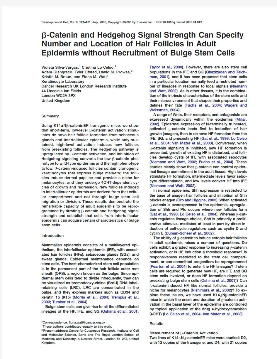
Figure1.Titration ofβ-Catenin Signaling In
Vitro and In Vivo
(A–D)Cultured keratinocytes from wild-type
(wt),D2,and D4mice were examined by West-
ern blotting(A–C)or transient transfection with
a TOPFLASH luciferase reporter(D).
(A)Comparison of relative levels of?Nβ-
cateninER by immunoblotting with anti-ER
or,as a loading control,anti-actin antibody.
(B and C)Comparison of relative levels of
endogenousβ-catenin and?Nβ-cateninER
in total Triton X-100soluble lysates(B)or the
supernatant from hypotonic cell lysates(C).
Upper band detected with anti-β-catenin is
?Nβ-cateninER as it was also detected with
anti-ER.
(D)Cells were untreated(U)or treated with
increasing4OHT concentrations,from2×
10–10(?10)to2×10–6(?6)M,for24hr.Rela-
tive light units(RLU)are shown.Bars repre-
sent means of at least three replicates in up
to four experiments±SEM.Asterisks indi-
cate significant increase relative to untr.
(E–K)Sections of wild-type(E)and D4trans-
genic(F–K)tail skin stained with anti-ER.
Skin was either untreated(E–G)or treated for
24hr with the4OHT doses shown.(G and H)
SG;(I–K)IFE.Brackets in(F)indicate
patches of higher(**)or lower(*)transgene
expression.Scale bars equal100?m in(G)
and(J),50?m in(E)and(F).
(L)One unit of tail epidermis is outlined in
red,corresponding to three HF with associ-
ated SG and IFE.
(M and N)%units with ectopic CDP expres-
sion in D2(M)and D4(N)epidermis treated
with low(L),medium(M),or high(H)4OHT
dose.
(O)Average number of CDP-positive patches
per unit.Mean±SEM of three experiments
shown in(M)and(N).d,days.
(Lo Celso et al.,2004).In previous work,we used a sin-To examineβ-catenin-mediated transcriptional acti-gle dose of4OHT,1mg per mouse(Lo Celso et al.,
vation,we transfected wild-type,D2,and D4keratino-2004).For the present experiments,we used three con-cytes with a luciferase construct containing an en-centrations:0.5(low dose),1.5(medium dose),or3mg
hancer with multiple Lef1/Tcf binding sites(TOPFLASH) (high dose)per mouse,applying4OHT every second day.or,as a negative control,one with mutated Lef1/Tcf To determine the effects of transgene copy number
binding sites(FOPFLASH).4OHT treatment of cells ex-and4OHT concentration onβ-catenin signaling,we pressing?Nβ-cateninER transfected with FOPFLASH generated spontaneously immortalized lines of kerati-
did not result in induction of luciferase(Lo Celso et al., nocytes.D4keratinocytes expressed approximately2004,and data not shown).TOPFLASH activation oc-
curred within8hr of addition of4OHT and was greater 2-fold more?Nβ-cateninER protein than D2keratino-
cytes(Figure1A).?Nβ-cateninER ran as a band of ap-at24hr(data not shown).
proximately110kDa,slightly higher than endogenous
We compared TOPFLASH activation in wild-type,D2,β-catenin.In lysates of total Triton soluble proteins,and D4keratinocytes24hr after adding a range of con-?Nβ-cateninER was considerably less abundant than
centrations of4OHT(Figure1D).There was no tran-endogenousβ-catenin(Figure1B).In extracts depleted scriptional activation at any concentration of4OHT in
wild-type cells.In D2cells,there was activation only at of the pool ofβ-catenin bound to E-cadherin(Zhu and
Watt,1999),?Nβ-cateninER and endogenousβ-catenin the highest4OHT concentration.In D4cells,there was were similar in abundance(Figure1C).Thus,in trans-
significant activation at concentrations of2×10?8M genic epidermis the amount ofβ-catenin available for and higher.The maximum activation achieved in D2 signaling is approximately2-fold that in wild-type epi-
and D4cells was the same,even though D4cells ex-dermis.pressed more?Nβ-cateninER protein.
Titration ofβ-Catenin and Shh Signaling
123
We next examined whether there was regional varia-pearance of ectopic CDP expression,the proportion of tion in transgene expression in vivo.While staining with
units with ectopic CDP,and the number of patches of an antibody to the mutant ER confirmed that all basal ectopic CDP per unit(Figures1M and1O).Ectopic CDP cells of the IFE,ORS,and periphery of the SG ex-
expression was first observed at day6with medium pressed the transgene(Lo Celso et al.,2004),there was and high4OHT concentrations and at day9with the heterogeneity in expression levels,with higher levels
low dose(Figures2A and2C).CDP was most readily being detected in the sebaceous gland than the IFE or induced in the SG(Figure2C),while the HF was most ORS(Figures1E and1F).IFE expression was hetero-
refractory(Figure1M).Medium-dose4OHT induced a geneous,with patches of cells at the junction between higher number of CDP-positive patches than low-dose the ORS and the IFE expressing higher levels than cells
4OHT,and at all doses,the number of positive patches in other regions(Figure1F).The same expression pat-per unit increased over time(Figure1O;compare Fig-tern was observed in both transgenic lines and also in
ures2D and2F).At the high4OHT dose,the number of K14MycER mice(data not shown;Arnold and Watt,CDP-positive patches was lower than at the medium 2001).
dose,but high-dose patches were larger(data not We next examined sections of tail skin from mice that shown).
had been treated for24hr with three doses of4OHT.
β-catenin activation in K14?Nβ-cateninER mice in-In untreated epidermis,ER immunoreactivity was most duces the underlying mesenchyme to form a dermal abundant at cell-cell borders(Figure1G).After treat-
papilla,surrounded by a cup-like sheath of keratino-ment with3mg4OHT,cell border staining was reduced cytes(Figure2D,arrow;Lo Celso et al.,2004;Van Mater (Figure1H)and there was punctate staining in the nu-
et al.,2003).In D2mice,formation of dermal papillae cleus and cytoplasm(Figure1I;Lo Celso et al.,2004).(red in Figure2A)was4OHT dose dependent and oc-In epidermis treated with0.5mg4OHT,cell-cell border
curred after induction of ectopic CDP.
staining was predominant(Figure1J),while with1.5D4epidermis responded more rapidly than D2to mg,staining was primarily nuclear and cytoplasmic
4OHT(Lo Celso et al.,2004),and by1day ectopic CDP (Figure1K).The same dose-dependent redistribution of expression was observed(Figures1N,2G,and2H).In ?Nβ-cateninER was observed in IFE,SG,and HF in
D4mice,the IFE was most sensitive and the HF most both lines(data not shown).There was no evidence that refractory(Figures1N and2G).Although CDP expres-activation ofβ-catenin by4OHT triggered epidermal
sion was observed along most of the D4ORS,distinct apoptosis,necrosis,or inflammation(data not shown).projections of CDP-positive epithelium and dermal pa-We conclude that different concentrations of4OHT
pillae were hardly ever observed(Figure2A;compare result in different levels ofβ-catenin activation and that Figures2J and2E).The correlation between the num-?Nβ-cateninER is activated to approximately the same
ber of CDP-positive patches and4OHT dose and length extent in cells of the SG,IFE,and HF ORS.Transgene of treatment was less pronounced in D4than D2mice; copy number also affects activation,because D4cells
patch size tended to be greater and individual patches were more responsive than D2cells to low concentra-merged with one another(Figure1O;compare Figures tions of4OHT.
2D and2J).
We conclude that different regions of the epidermis
differed in their responsiveness toβ-catenin activation. Control of Hair Follicle Number and Location
SGs were most responsive and HFs most refractory. by Transgene Copy Number and4OHT Dose
Regional variation in transgene expression did not ap-To evaluate ectopic hair follicle differentiation,we used
pear to determine where ectopic HF formed in the IFE an antibody to CCAAT displacement protein(CDP)that
(data not shown;see Figure1F).However,it might ex-is expressed primarily in the base of the follicle,known
plain why D2SG were most sensitive to HF induction as the bulb(Braun et al.,2003).Induction of CDP,to-
(Figure1M).HF morphogenesis progressed further in gether with Lef1and keratin17,is an early indicator of
D2than in D4mice(Figure2A).
ectopic HF formation(Lo Celso et al.,2004).
We performed whole-mount labeling of4OHT-treated
mouse tail skin(Braun et al.,2003)to facilitate quantita-Hedgehog Signaling Is Necessary for?-Catenin-
tion(Figures1L–1O).Figure2shows the morphology of
Induced Formation of New Hair Follicles
the ectopic follicles,both schematically(Figure2A)and To investigate howβ-catenin induced new hair follicles, in representative CDP-labeled whole mounts(Figures
we isolated RNA from whole dorsal skin of triplicate 2B–2J).6-week-old female D2mice,comparing untreated skin In wild-type and untreated K14?Nβ-cateninER tail
with skin treated either for1day with1mg4OHT or skin,the hair follicles have prominent sebaceous harvested after seven daily https://www.doczj.com/doc/006539934.html,ing Affymet-glands and are clustered in groups of three.We defined
rix chip technology,we analyzed>18,000transcripts, one HF triplet and adjacent interfollicular epidermis as representing>14,000genes.Genespring software was one epidermal unit(Figure1L).The percentage of units
used to normalize the raw data and identify genes with in which ectopic expression of CDP occurred in the IFE a t test p value of less than0.05and a change in relative (Figure2A;arrowheads in Figure2J),SG(arrows in Fig-
expression levels of at least3-fold in4OHT-treated ure2C),and HF ORS(arrow in Figure2E)is shown for transgenics.150probe sets were identified as>3-fold each4OHT concentration and each transgenic line in
upregulated and13probe sets>3-fold downregulated Figures1M and1N.The number of CDP-positive patches after7days of4OHT treatment.The full list of known
genes,as well as the normalization and filter parame-per positive unit is shown in Figure1O.
In D2mice,the4OHT dose affected the time of ap-ters,are available as Supplemental Data with this arti-
Developmental Cell
124
Figure2.Effects on D2and D4Transgenic
Epidermis of Activating?Nβ-CateninER with
Different Concentrations of4OHT for Dif-
ferent Lengths of Time
4OHT was applied at0.5mg(L),1.5mg(M),
and3mg(H)doses.
(A)Schematic representation of results,
showing outer root sheath(ORS)of a single
HF with associated SG and IFE.Green,CDP
expression;red,new dermal papillae(DP).
Arrows:time points analyzed.Letters corre-
spond to whole mounts in(B)–(J).
(B–J)Tail whole mounts immunolabeled for
CDP.Transgenic lines,4OHT doses,and time
of treatments(days,d)are indicated.Dashed
lines demarcate hair follicles and sebaceous
glands in(B),(C),(F),and(G).Arrows in(C)
indicate CDP expression in SG,in(D)a der-
mal papilla,and in(E)an outgrowth arising
from ORS.Arrowheads in(G),(H),and(J)in-
dicate new outgrowths arising from IFE.
Scale bars equal100?m.
cle online.The raw chip data files are available at http://whether Hedgehog signaling was required for ectopic https://www.doczj.com/doc/006539934.html,/geo/info/linking.html(accession
HF formation.
number GSE1579).K14?Nβ-catenin transgenics were treated with1mg Some of the upregulated genes corresponded to
4OHT±cyclopamine to block Hedgehog signaling(Fig-knownβ-catenin targets or markers of HF differentia-ures3A–3H;van den Brink et al.,2004).In untreated tion(see,for example,Gat et al.,1998;Lo Celso et al.,
and cyclopamine-treated epidermis,Gli1was mainly 2004;Van Mater et al.,2003),including cyclin D1,hair cytoplasmic(Figures3K and3M;Niemann et al.,2003), keratins,CDP,and Lef1.The increase in cyclin D1and
whereas after4OHT treatment alone,Gli1was primarily other cell-cycle regulators is consistent with the local nuclear(Figure3L).Cyclopamine blocked the induction
of anagen(Figures3A and3B)and reduced the appear-increase in proliferation that occurs in response to
β-catenin(Lo Celso et al.,2004).ance of ectopic HF(Figures3E and3F)in D2mice, Some of the genes that were most highly induced
thereby converting the D2phenotype to wild-type. were members of the Hedgehog signaling cascade:Cyclopamine did not completely normalize the D4phe-Shh was upregulated4.8-fold,Ptch26.6-fold,Gli18.4-
notype,but converted it to a D2phenotype(Figures3C fold,and Gli23.6-fold.In addition,N-Myc,a target of and3D):the new follicles became more pronounced Shh(Oliver et al.,2003),was upregulated8-fold at7
and dermal papilla formation was stimulated(Figures days(see Supplemental Data).Increased expression of3G and3H).
Shh and Ptc has previously been observed when
The morphological observations were confirmed by β-catenin is activated in the epidermis(Gat et al.,1998;staining whole mounts with a range of antibodies.Kera-Lo Celso et al.,2004).Since Shh drives proliferation
tin17expression induced byβ-catenin in the IFE and during normal hair follicle anagen(Oro and Higgins,SG of D2mice(Figure3I)was largely inhibited,and ker-
atin17expression was confined to the ORS(Figure3J), 2003)andβ-catenin activation results in local increases
in proliferation(Lo Celso et al.,2004),we investigated as in wild-type epidermis(data not shown;Lo Celso et
Titration ofβ-Catenin and Shh Signaling
125
Figure3.Hedgehog Signaling Is Required for
β-Catenin-Induced Hair Follicle Formation
D2(A,B,E,F,I,J,N,O,R,S)and D4(C,D,
G,H,K–M,P,Q,T,U)mice were untreated
(K),treated with4OHT alone(A,C,E,G,I,L,
N,P,R,T),or treated with4OHT and cyclo-
pamine(B,D,F,H,J,M,O,Q,S,U).
(A–D)H&E-stained sections of dorsal(A and
B)or tail(C and D)epidermis.
(E–U)Tail whole mounts labeled with anti-
bodies to K14(E–H),K17(I and J),Gli1(K–M),
CDP(N–Q),or Ki67(R–U).Arrows in(K)and
(L)show nuclear Gli1staining.Dashed lines
outline SG(J),ORS(N,O,R),or HF and SG
(Q,S–U).Scale bars equal100?m in(A)–(J)
and(N)–(U)and50?m in(K)–(M).
al.,2004).Ectopic expression of CDP in D2epidermis LRC in the bulge had declined significantly.This did not (Figure3N)was also inhibited by cyclopamine(Figure
correlate with increased apoptosis(data not shown)but 3O).In D4epidermis,the size of the ectopic CDP with stimulation of LRC to divide.Some LRC became patches was reduced(Figures3P and3Q).As ex-
Ki67positive,and lightly BrdU-labeled cells appeared in pected,cyclopamine treatment led to a reduction in the bulge(Figure4G;see also Braun et al.,2003). epidermal proliferation,as evaluated by Ki67staining
K15expression in the bulge of untreated HF(Morris (Figures3R–3U).et al.,2004)corresponded to the zone of LRC(Figures
4H and4I).K15expression in the bulge was preserved Lineage Reprogramming Occurs Independently
in both transgenic lines,even at high4OHT concentra-of Bulge Stem Cells tions and late time points(Figures4J and4K and data We used two markers to examine the effect ofβ-catenin
not shown)when LRC had been completely lost(Figure activation on the hair follicle stem cell compartment:4K).We conclude thatβ-catenin-induced HF formation
was initiated without involvement of bulge LRC and retention of BrdU label and expression of K15.In wild-
type mice and untreated transgenic mice,LRC were that even when LRC divided and lost their label,the concentrated in the bulge,and there were scattered
original bulge remained,as detected by K15expression. LRC in the sebaceous glands and IFE(Figure4A;Braun As further evidence thatβ-catenin induction of HF et al.,2003).There was no evidence of LRC depletion
could occur independently of bulge stem cells,we used in the bulge of D2HF treated with4OHT(medium dose)the K14promoter for lineage tracing in order to provide for up to14days(Figure4B)or D4mice treated for up
a positive marker of IFE cell progeny.We generated tri-to9days(Figures4D and4E).Thus,at times when ec-ple transgenic mice by crossing D2K14?Nβ-cateninER topic hair follicle formation was well advanced in IFE
mice with K14CreER mice(Vasioukhin et al.,1999; and SG(Figure2A),bulge LRC had not been lost and Hong et al.,2004)and Rosa26Cre-Reporter mice in there was no evidence that they had migrated from
which a stop codon flanked by LoxP sites has been their original location.inserted upstream of theβ-galactosidase gene(Sori-
ano,1999).The mice were treated for21days with1.5 By28days of treatment of D2mice(Figure4C)and15
days of treatment of D4mice(Figure4F),the number of mg4OHT,and then the skin was harvested and exam-
Developmental Cell
126
Figure4.?Nβ-CateninER-Induced Hair Folli-
cles Are Not Derived from Existing Bulge
Cells
(A–K)Transgenic line(D2or D4)and days(d)
of4OHT treatment(medium dose)are shown.
(A–F)Green,BrdU;red,keratin14.In(A),lo-
cations of sebaceous glands(SG),hair folli-
cle outer root sheath(ORS),bulge(BG),and
bulb(BL)are indicated;arrows show scat-
tered LRC in SG and IFE.
(G)Double labeling for Ki67(green)and
LRC(red).
(H–K)K15expression.
(H)Wild-type epidermis stained with sec-
ondary antibody alone,showing nonspecific
SG labeling.
(I–K)Double labeling for K15(red)and LRC
(green).No SG remain in(J)and(K).
(L–P)Lineage tracing in K14CreER×R26R×
K14?Nβ-cateninER D2transgenics treated
with1.5mg4OHT for2weeks.Blue,XGal;
brown,CDP.
(L and M)Back skin.
(N–P)Whole mounts of tail epidermis.Posi-
tion of HF ORS is indicated in(H)and(N).
Ectopic HF in IFE,identified by CDP expres-
sion,were either completely XGal positive
(white asterisks),completely negative(red
asterisks),or mixed(black asterisks).Box in
(N)shows region of IFE with ectopic CDP ex-
pression;similar regions of IFE from other
whole mounts are shown at higher magnifi-
cation in(O)and(P).Scale bars equal100
?m in(A)–(H)and(J)–(N)and50?m in(I).
ined for expression of CDP andβ-galactosidase in con-for3weeks,left the skin untreated for3weeks,and ventional sections(Figures4L and4M)and whole
then applied4OHT for a further1or2weeks(Figures mounts(Figures4N–4P).5A–5F).Withdrawal of4OHT caused preexisting folli-The efficiency of K14CreER-mediated recombination
cles to cease the growth phase(anagen)of the hair depended on the dose of4OHT(A.G.and F.M.W.,un-cycle and enter the resting phase(Figures5A and5B)
and caused ectopic follicles to regress(Figures5A and published observations).Under the4OHT treatment
conditions used,β-catenin was activated in all K14-5B).When4OHT was reapplied,the original follicles re-positive cells(Figures1F–1K),but there was patchy in-
entered anagen(Figure5D);ectopic follicles regrew and duction ofβ-galactosidase(Figures4M and4N).CDP-were wider than before(Figures5A,5C,and5D).Occa-positive,?Nβ-cateninER-positive epithelial outgrowths
sionally three distinct hair shafts were found associated in the IFE were either uniformlyβ-galactosidase posi-with a single follicle,excluding the possibility that the tive(white asterisks),uniformly negative(red asterisks),
regrowth phenotype was due simply to existing club or a mixture of positive and negative cells when at the hairs reentering anagen(Figure5E).
boundary between recombined and nonrecombined
On reapplication of4OHT,the number of sites of ec-IFE regions(black asterisks)(Figures4L,4M,4O,and topic CDP in the ORS returned to the same level as
after the first treatment(Figure5F),suggesting that the 4P).Theβ-galactosidase status of ectopic follicles
nearly always matched that of the surrounding IFE(Fig-original ectopic follicles regrew.The reason why on re-ures4N–4P).This strongly suggests that the majority of
growth the ectopic follicles were wider than before may new follicles were derived from immediately adjacent be that dermal papilla cells were induced over a wider IFE rather than from neighboring hair follicles,support-
region than following the first treatment(data not ing the conclusion that bulge LRC were not involved.shown).Although someβ-catenin-induced follicles do
form a mature hair shaft(Figure5E;Gat et al.,1998;Lo
Celso et al.,2004),the regrowth phenotype we ob-?-Catenin-Induced Follicles Can Undergo Cycles
of Growth and Regression and Provide Niches served could not be interpreted as a true hair growth for Neural Crest-Derived Cells
cycle(Millar,2002),since it was entirely4OHT depen-To evaluate whether?Nβ-cateninER-induced follicles dent,and the rudimentary outgrowths,while positive could undergo cycles of growth and regression,we in-
for CDP,Lef1,and K17,usually lacked the inner root duced new follicles in D2epidermis with1.5mg4OHT sheath marker trichohyalin(Lo Celso et al.,2004).
Titration ofβ-Catenin and Shh Signaling
127
Figure 5.β-Catenin-Induced Hair Follicles
Can Undergo Cycles of Growth and Regres-
sion and Provide a Niche for Melanocytes
and Dermal Papilla Cells
(A–E)H&E-stained sections of D2transgenic
dorsal epidermis treated with4OHT for3
weeks,then harvested immediately(A),left
untreated for3weeks(+3w)and harvested
(B),or treated with4OHT for a further1(+1w)
(C)or2(+2w)(D and E)weeks.CDP staining
in brown(E).Arrows in(A)–(D)show ectopic
follicles.Arrows in(E)show hair shaft forma-
tion;Cb,club hair;OHS,old hair shaft;NHS,
new hair shaft.
(F)Number of ectopic CDP-positive out-
growths per existing HF in experiment il-
lustrated in(A)–(E).Error bars correspond to
standard deviation.
(G–O)Whole mounts of wild-type(wt)and D2
transgenic(tg)tail epidermis,untreated(H)
or treated with a low(G)or medium(I–O)
dose of4OHT for number of days shown.
Epidermis was labeled with L-Dopa(H–K)or
with antibodies to the proteins shown.In(H),
albino wild-type mouse is negative control
for L-Dopa staining.Brackets in(H)–(J)indi-
cate SG.
(G)Cuff of keratinocytes at base of ectopic
follicle into which dermal papilla cells insert.
(I)Melanocytes in follicle bulb(arrow)and
IFE(arrowhead).
(J and K)Melanocytes in HF induced from
SG(white arrowhead)and ORS(arrow)are
shown.Insert in(J)shows high-magnifica-
tion view of ectopic HF arising from ORS,
with melanocyte indicated by arrow.
(L)c-Kit-positive cells in infundibulum(ar-
row)and bulge(arrowhead)are shown;HF
and SG are indicated by dotted line.
(M)Dotted lines demarcate new HF.
(N and O)Arrows indicate nestin-positive
dermal papilla cells;arrowheads show scat-
tered nestin-positive cells in ORS(N)and IFE
(O).Upper inset in(O)shows dendritic nes-
tin-positive cells in ORS and lower inset shows
clustered nestin-positive cells in dermal pa-
pilla of an ectopic HF.Dotted lines demar-
cate HF and SG.
Scale bars equal100?m in(A)–(E)and(H)–
(M)and50?m in(G).
Normal hair follicles provide a niche for neural crest epidermis,c-Kit-positive cells appeared in IFE immedi-derivatives such as melanocytes(Nishimura et al.,
ately adjacent to the HF after9days of4OHT treatment 2002).We visualized the tyrosinase activity of differenti-(data not shown),and by21days they were found in ated melanocytes using L-Dopa as substrate.Albino
ectopic HF induced in all regions of the epidermis(Fig-mouse epidermis provided a negative control(Figure ure5M,dotted line indicates position of ectopic fol-
licles).
5H).In wild-type tail epidermis,differentiated melano-
cytes were concentrated at the base of the hair follicles In whole mounts of4OHT-treated K14?Nβ-cateninER (Figure5I,arrow),as reported previously(Nishimura et
epidermis,a cuff of keratinocytes surrounding an in-al.,2002),and small numbers were also scattered in the dentation at the base of the new follicles indicated der-IFE(Figure5I,arrowhead).L-Dopa-positive melano-
mal papilla formation(Figures2D and5G).Dermal pa-cytes were present in the ectopic follicles induced by pilla cells in wild-type and4OHT-treated transgenic skin β-catenin in SG(Figures5J and5K,arrowheads),IFE,
expressed not only alkaline phosphatase(data not and HF(insert)(Figures5J and5K).shown;see Lo Celso et al.,2004)but also nestin,a Undifferentiated melanocytes(melanoblasts)were
marker of neural crest-derived stem cells(Figure5N, detected with anti-c-Kit antibody(Nishimura et al.,arrow;Figure5O,lower insert;Fernandes et al.,2004). 2002).In wild-type epidermis,there were c-Kit-positive
In addition,small numbers of nestin-positive cells were cells in the HF infundibulum(arrow)and the bulge(ar-scattered along the length of wild-type and induced fol-rowhead)(Figure5L).In4OHT-treated?Nβ-cateninER
licles(Figures5N and5O,arrowheads;upper inset in
Developmental Cell
128
Figure6.β-Catenin Activation Induces Formation of Keratinocytes that Have Characteristics of Bulge Stem Cells
(A–F)Whole mounts(A–C)and sections from tail(D)or back(E and F)epidermis of wt untreated(B and E)or D2transgenic mice treated with 1.5mg4OHT for number of days shown or for21days.Ectopic follicles are demarcated with dotted lines in(A)and(C).Brackets indicate position of bulge in untreated follicle(E)and more extensive CD34labeling followingβ-catenin activation(D and F).Scale bars equal100?m in(A)–(F).
(G–L)FACS selection and culture of keratinocytes from dorsal epidermis of adult wild-type(G,I,K,L)and D2transgenic(H,J–L)mice treated with1.5mg4OHT for3weeks.Cells isolated from epidermis were double labeled for CD34andα6integrin.Cells with low forward and side scatter(circled in[G]and[H])were either sorted directly onto culture dishes(All sorted,[K]and[L])or further fractionated intoα6single positive(green boxes in[I]and[J])or CD34/α6double positive populations(red boxes in[I]and[J]).In(I)and(J),left red boxes show cells with lower surfaceα6than in right red boxes.
(K and L)Colony-forming efficiency of FACS-sorted keratinocytes.
(K)Dishes stained with Rhodamine B.
(L)Representative of two separate experiments,in each of which cells from triplicate wild-type or transgenic mice were pooled.White bars, wild-type;black bars,transgenic cells.Standard deviations shown.
Figure5O);these were dendritic in morphology and al.,1995).The majority of undifferentiated cells(inside lacked the Merkel cell marker K18(data not shown).red circles in Figures6G and6H)expressedα6integrin
(Figures6I and6J and data not shown).In4OHT-treated
wild-type epidermis,approximately4%of undifferenti-Cells in?-Catenin-Induced Hair Follicles Express
ated cells expressed high levels of both CD34andα6 Markers of Bulge Stem Cells
integrin,while2%were CD34positive and had low To investigate whetherβ-catenin-induced HF contained
levels ofα6integrin(Figure6I).In D2epidermis,the cells with characteristics of bulge stem cells,we
proportion of cells with high CD34andα6integrin was stained whole mounts and histological sections of skin
similar(3.5%in the profile shown),but the CD34-posi-with antibodies to K15and CD34(Figures6A–6F and
tive/α6integrin-low population was increased to about data not shown).In wild-type follicles,expression of
15%(Figure6J).
both markers was confined to the permanent portion of
Three cell fractions were selected for in vitro clonal the follicle(Figures4I,6B,and6E;Morris et al.,2004;
analysis(Figure6K):the total low forward and side scat-Trempus et al.,2003).In tail and back epidermis from
ter population(“All sorted”),α6-positive/CD34-negative D2and D4mice treated with4OHT for28days,expres-
cells(green gates,Figures6I and6J),andα6/CD34 sion of both markers was observed in all sites of new
double-positive cells,irrespective of whether integrin HF formation(Figures6A,6C,6D,and6F and data not
expression was high or low(red gates,Figures6I and shown),often extending along the length of the new folli-
6J).After14days of culture without4OHT,there was cles.CD34staining in ectopic follicles was slightly weaker
no significant difference between the colony-forming than in the preexisting bulge(Figures6E and6F).
efficiency of each fraction in wild-type dorsal epidermis We performed FACS of disaggregated keratinocytes
(Figures6K and6L).In transgenic cultures,the colony-from back(Figures6G–6L)or tail(data not shown)skin,
forming efficiency of the total and theα6-positive/ gating out the differentiated cells on the basis of high
forward and side scatter(Figures6G and6H;Jones et CD34-negative population was slightly increased,while
Titration ofβ-Catenin and Shh Signaling
129
the double-positive population had twice the colony-proliferation of LRC(Braun et al.,2003),and we have
not observed migration of LRC upwards toward the SG forming efficiency of wild-type(Figure6L).In all three
and IFE in any of these models(cf.Oshima et al.,2001; fractions,the size of individual colonies was larger in
Taylor et al.,2000).Even when LRC eventually divided transgenic compared to wild-type keratinocytes(Figure
in response to prolongedβ-catenin activation,thereby 6K).We conclude thatβ-catenin-induced HF contain
losing the BrdU label,the location of the original bulge cells with three characteristics of bulge stem cells:
was still detectable by expression of K15and CD34 clonal growth in culture and expression of K15and
(Morris et al.,2004;Trempus et al.,2003).
CD34.
Even though new follicle formation preceded prolifer-
ation of LRC,we could not formally exclude the possi-Discussion
bility that very small numbers of LRC,perhaps even a
single cell,were required to found new follicles in the By expressing different levels of?Nβ-cateninER in epi-
IFE.Lineage tracing experiments,however,showed dermal cells and applying different concentrations of
that this was unlikely.Whenever a new CDP-positive 4OHT,we can control the level ofβ-catenin transcrip-
epithelial outgrowth formed in the IFE,itsβ-galactosi-tional activation.The concept that cells exhibit a
dase expression matched the surrounding IFE.Further-graded response to a gradient ofβ-catenin regulatory
more,when the new follicle formed at the boundary be-activity was previously established in studies of ES cell
tweenβ-galactosidase-positive and-negative regions, lines with different APC mutations(Kielman et al.,2002).
that follicle would be partβ-galactosidase positive and However,the?Nβ-cateninER transgene has the advan-
partβ-galactosidase negative,indicating that it was not tage that transcriptional activity can be titrated within
clonal in origin.It has long been proposed that mouse individual cells,both in vivo and in culture.
IFE is organized into“epidermal proliferative units”:at As Figure2illustrates,the response of adult epider-
the base of each lies a single stem cell surrounded by mis toβ-catenin activation was more complex than a
committed progenitors(transit amplifying cells)(Potten simple switching on of de novo HF formation.With
and Morris,1988).If this is indeed the case,the implica-increasingβ-catenin activation,the timing and location
tion of the nonclonal origin of IFE hair follicles is that of ectopic HF formation were altered.In both trans-
IFE-committed progenitors can be redirected to adopt genic lines,the ORS,including the bulge,of existing HF
a HF fate.Alternatively,IFE stem cells in mouse,as in was most refractory to induction in tail epidermis.This
human(Jones et al.,1995),may be clustered.
is consistent with evidence that bulge cells are quies-
Remarkably,not only was new follicle formation initi-cent cells in an environment that makes them refractory
ated without any detectable involvement of existing to a range of signaling pathways(Tumbar et al.,2004).
bulge cells,but new follicles contained cells that ex-Factors that are upregulated in the bulge include the
pressed K15and CD34and were clonogenic in culture. Wnt antagonist Dickkopf-3(Morris et al.,2004;Tumbar
Thus,cells with bulge stem cell characteristics can be et al.,2004)and Tcf3,which can repress Wnt signaling
formed from nonbulge epidermal cells.Ectopic follicles (Merrill et al.,2001).
could undergo4OHT-dependent cycles of growth and During embryonic development,there is a complex
regression.In response to reapplication of4OHT,there interplay between Hedgehog and Wnt signaling(Taipale
was no further increase in the number of HF,but the and Beachy,2001),and inappropriate activation of
ectopic follicles enlarged.This implies that there is either pathway is associated with a range of epidermal
some mechanism to limit the number of new follicles: cancers(Oro and Higgins,2003;Gat et al.,1998;Lo
it might involve the same type of intercellular signaling Celso et al.,2004;Niemann et al.,2003).Shh was events that control the spacing of new follicles during upregulated in response toβ-catenin activation in embryonic development(Millar,2002).
K14?Nβ-cateninER epidermis,and inhibition of Shh The generation of new HF from IFE leads us to pro-with cyclopamine reducedβ-catenin-induced HF for-pose that given the appropriate signal,any prolifera-mation in D2transgenic mice.tion-competent cell can form a new HF expressing both In the absence of cyclopamine,4OHT-induced HF ORS and bulge markers.β-catenin activation in cul-morphogenesis progressed further in D2than in D4tured human epidermal cells increases the number of mice,resulting in induction of dermal papillae and epi-stem cells(Zhu and Watt,1999);whether this occurs thelial outgrowths from the ORS.In D2epidermis,lin-through expansion of the preexisting stem cell pool,as eage reprogramming was accompanied by only local we originally proposed,or through generation of new increases in proliferation,whereas in D4epidermis,pro-stem cells from committed progenitors remains to be liferation was more widespread and the follicles be-explored(Pearton et al.,2004).The increased size of came grossly thickened.The ability of cyclopamine to the colonies formed by K14?Nβ-cateninER keratino-convert the D4to the D2phenotype suggests that the cytes is interesting:since the cells were cultured in the D4phenotype results from Hedgehog-induced prolifer-absence of4OHT,it must reflect an intrinsic increase in ation being too extensive to allow proper morphogene-proliferative potential.A role of Wnt signaling in increasing sis.Hedgehog antagonists may be useful in the treat-stem cell number has now been reported in a wide ment of epidermal tumors in which either Wnt or range of tissues,including the blood,mammary gland, Hedgehog signaling is activated(Niemann et al.,2003;and nervous system(Liu et al.,2004;Megason and Taipale and Beachy,2001).McMahon,2002;Reya et al.,2003).
Ectopic HF formation preceded any obvious change In addition to expressing bulge markers,β-catenin-in proliferation or migration of bulge LRC.The effects induced follicles were populated by melanocytes and of c-Myc and?NLef1on lineage selection also precede
formed dermal papillae.The ability of ectopic follicles
Developmental Cell
130
nolabeled as described previously(Braun et al.,2003;Niemann et to provide a home for melanocytes is of interest be-
al.,2003).To visualize melanocytes,epidermal sheets were incu-cause melanocyte stem cells are dependent on interac-
bated with0.2%L-Dopa(Acros Organics,Netherlands)for3–16hr tions with the HF bulge(Nishimura et al.,2002).It is
at37°C in the dark.Images were obtained using a Zeiss510confo-also intriguing that both melanocytes and dermal pa-cal microscope as described by Braun et al.(2003).
pilla cells are neural crest derived(Fernandes et al.,To quantify CDP expression in whole mounts,30–50units of epi-
2004)and Wnt dependent.Wnt1signaling is involved in
dermis(Figure1L)per tail were scored in three separate mice for
each experimental condition.The percentage of units with ectopic the expansion and differentiation of melanocytes,and
CDP expression and the number of ectopic CDP-positive patches ablation ofβ-catenin in neural crest stem cells leads to
per unit±SEM were determined.Within each unit IFE,SG,and HF lack of melanocytes(Le Douarin and Dupin,2003).
were scored separately.
Epidermal stem cells rarely feature in accounts of
adult stem cell populations that exhibit plasticity(Nie-Western Blotting and Luciferase Assay
mann and Watt,2002;Wagers and Weissman,2004).Spontaneously immortalized keratinocyte lines were generated
This is probably because when epidermal cells are sep-from adult mouse skin and cultured with a feeder layer,essentially
as described previously(Romero et al.,1999),except that skin tryp-arated from their neighbors and deprived of extracellu-
sinization was carried out at4°C and cells were disaggregated lar matrix attachment,they withdraw irreversibly from
without stirring.Cell lysis and Western blotting conditions are de-the cell cycle and initiate terminal differentiation(Watt,
scribed in Supplemental Data.
2002).Nevertheless,the finding that epidermalβ-cat-Keratinocytes were transiently transfected with Promega lucifer-
enin activation induces dermal papillae opens up an ase reporter constructs pRL(Renilla luciferase control)and TOP-
unexpected way in which the epidermis could influence FLASH or FOPFLASH(firefly luciferase)using Genejuice(Novagen).
Luciferase activities were measured as described previously(Lo the homing,localization,and properties of a range of
Celso et al.,2004).
different cell types.Adult dermal cells have multilineage
potential,including formation of neural precursors(Fer-
FACS and Clonogenicity Assay
nandes et al.,2004)and cells of the erythroid and
Keratinocytes were isolated and cultured from adult dorsal skin as myeloid lineages(Lako et al.,2002).Not only can epi-
described by Romero et al.(1999),with the minor modifications dermalβ-catenin signaling control selection of the epi-described above.Three further changes were made to increase dermal lineages,it may also stimulate the differentiation colony-forming efficiency:keratinocytes were grown on collagen-
coated dishes(BD Biocoat),the feeder layer was confluent,and of other cell types by re-creating some of the inductive
the basal FAD culture medium was calcium free.1000–5000kerati-interactions that occur during normal epidermal devel-
nocytes were plated per35mm dish.After14days,the cultures opment.
were fixed with4%PFA and stained with1%Rhodamine B.Colo-
nies were defined as clusters of five or more keratinocytes. Experimental Procedures For two-color FACS,cell suspensions were incubated with FITC
or biotin-conjugated primary antibodies for30min on ice,washed Generation and Experimental Treatment of Mice twice for10min at4°C,then incubated with streptavidin PE for30 The D2and D4lines of K14?Nβ-cateninER transgenic mice were min on ice.Cell viability was assessed by7AAD(BD Bioscience) generated as described(Lo Celso et al.,2004).For lineage marking staining.Cells were sorted with a FACSVantage sorter according to studies,the D2line was crossed with mice that were homozygous CD34andα6integrin expression(Morris et al.,2004)after gating for the Rosa26Cre reporter(R26R)(Soriano,1999)and K14CreER out dead cells and cells with high forward and side scatter. (Vasioukhin et al.,1999;Hong et al.,2004).At the start of each
experiment,the mice were6to8weeks old,and therefore in the
resting phase of the hair cycle.Wild-type,nontransgenic litter-
Supplemental Data
mates were used as controls.
Supplemental Data include two tables and Supplemental Experi-The?Nβ-cateninER transgene was activated by topical applica-
mental Procedures and can be found with this article online at tion of4-hydroxytamoxifen(4OHT;Sigma)dissolved in acetone.Tail
https://www.doczj.com/doc/006539934.html,/cgi/content/full/9/1/121/DC1/. skin was treated by applying4OHT with a paintbrush(0.5,1.5,or3
mg per mouse every second day).For the microarray experiment,
1mg4OHT was applied with a micropipette to a clipped area of
Acknowledgments
dorsal skin either once or every day for7days.In some experi-
ments,mice received daily topical applications of cyclopamine(50
We are grateful to everyone who provided us with advice or rea-?M in ethanol)and4OHT every second day.LRC were generated
gents,in particular Dr.Ron McKay.We received superb technical as described previously(Braun et al.,2003).
support from the CR-UK Biological Resources,Histopathology,
FACS,and Experimental Pathology Units and the Affymetrix Facil-Antibodies ity.This work was supported by CR-UK,an EU FP5Network,and The antibodies used have been described previously(Braun et al.,by the Association for International Cancer Research.A.G.is the 2003;Niemann et al.,2003;Lo Celso et al.,2004),except for the recipient of a Marshall Sherfield Fellowship.
following:anti-CD34(RAM34;BD Pharmingen),c-Kit(01902-D;
Pharmingen),and nestin(nestin130;gift from R.McKay)(Marvin et
Received:August17,2004
al.,1998).
Revised:February11,2005
Accepted:April26,2005
Histochemistry,Immunolabeling of Sections,
Published:July5,2005
and Whole Mounts
Conventional frozen or fixed,paraffin-embedded sections were
References
prepared and immunolabeled as described previously(Lo Celso et
al.,2004).LacZ staining was performed with X-Gal as described
Arnold,I.,and Watt,F.M.(2001).c-Myc activation in transgenic previously(Soriano,1999);when sections were double labeled,the
mouse epidermis results in mobilization of stem cells and differenti-antibody incubation was performed after the X-Gal incubation and
ation of their progeny.Curr.Biol.11,558–568.
the antibody was visualized with DAKO Visualization Systems(Lo
Celso et al.,2004).Braun,K.M.,Niemann,C.,Jensen,U.B.,Sundberg,J.P.,Silva-Var-Whole mounts of tail epidermal sheets were prepared and immu-
gas,V.,and Watt,F.M.(2003).Manipulation of stem cell proliferation
Titration ofβ-Catenin and Shh Signaling
131
and lineage commitment:visualisation of label-retaining cells in response:a critical role for N-myc in proliferation of neuronal pre-
https://www.doczj.com/doc/006539934.html,A100,7331–7336. wholemounts of mouse epidermis.Development130,5241–5255.
Oro,A.E.,and Higgins,K.(2003).Hair cycle regulation of Hedgehog Duman-Scheel,M.,Weng,L.,Xin,S.,and Du,W.(2002).Hedgehog
signal reception.Dev.Biol.255,238–248.
regulates cell growth and proliferation by inducing Cyclin D and
Cyclin E.Nature417,299–304.Oshima,H.,Rochat,A.,Kedzia,C.,Kobayashi,K.,and Barrandon,
Y.(2001).Morphogenesis and renewal of hair follicles from adult Fernandes,K.J.,McKenzie,I.A.,Mill,P.,Smith,K.M.,Akhavan,M.,
multipotent stem cells.Cell104,233–245.
Barnabe-Heider,F.,Biernaskie,J.,Junek, A.,Kobayashi,N.R.,
Toma,J.G.,et al.(2004).A dermal niche for multipotent adult skin-Pearton,D.J.,Ferraris,C.,and Dhouailly,D.(2004).Transdifferentia-derived precursor cells.Nat.Cell Biol.6,1082–1093.tion of corneal epithelium:evidence for a linkage between the seg-
regation of epidermal stem cells and the induction of hair follicles Fuchs,E.,Tumbar,T.,and Guasch,G.(2004).Socializing with the
during embryogenesis.Int.J.Dev.Biol.48,197–201.
neighbors:stem cells and their niche.Cell116,769–778.
Potten,C.S.,and Morris,R.J.(1988).Epithelial stem cells in vivo.J. Gat,U.,DasGupta,R.,Degenstein,L.,and Fuchs,E.(1998).De
Cell Sci.Suppl.10,45–62.
novo hair follicle morphogenesis and hair tumors in mice express-
ing a truncated beta-catenin in skin.Cell95,605–614.Reya,T.,Duncan,A.W.,Ailles,L.,Domen,J.,Scherer,D.C.,Willert,
K.,Hintz,L.,Nusse,R.,and Weissman,I.L.(2003).A role for Wnt Ghazizadeh,S.,and Taichman,L.B.(2001).Multiple classes of stem
signalling in self-renewal of haematopoietic stem cells.Nature423, cells in cutaneous epithelium:a lineage analysis of adult mouse
409–414.
skin.EMBO J.20,1215–1222.
Romero,M.R.,Carroll,J.M.,and Watt,F.M.(1999).Analysis of cul-Hong,K.U.,Reynolds,S.D.,Watkins,S.,Fuchs,E.,and Stripp,B.R.
tured keratinocytes from a transgenic mouse model of psoriasis: (2004).Basal cells are a multipotent progenitor capable of renewing
effects of suprabasal integrin expression on keratinocyte adhesion, the bronchial epithelium.Am.J.Pathol.164,577–588.
proliferation and terminal differentiation.Exp.Dermatol.8,53–67. Jones,P.H.,Harper,S.,and Watt,F.M.(1995).Stem cell patterning
Soriano,P.(1999).Generalized lacZ expression with the ROSA26 and fate in human epidermis.Cell80,83–93.
Cre reporter strain.Nat.Genet.21,70–71.
Kielman,M.F.,Rindapaa,M.,Gaspar,C.,van Poppel,N.,Breukel,
Taipale,J.,and Beachy,P.A.(2001).The Hedgehog and Wnt signal-C.,van Leeuwen,S.,Taketo,M.M.,Roberts,S.,Smits,R.,and
ling pathways in cancer.Nature411,349–354.
Fodde,R.(2002).Apc modulates embryonic stem-cell differentia-
Taylor,G.,Lehrer,M.S.,Jensen,P.J.,Sun,T.T.,and Lavker,R.M. tion by controlling the dosage of beta-catenin signaling.Nat.
(2000).Involvement of follicular stem cells in forming not only the Genet.32,594–605.
follicle but also the epidermis.Cell102,451–461.
Lako,M.,Armstrong,L.,Cairns,P.M.,Harris,S.,Hole,N.,and Ja-
Trempus,C.S.,Morris,R.J.,Bortner,C.D.,Cotsarelis,G.,Faircloth, hoda,C.A.(2002).Hair follicle dermal cells repopulate the mouse
R.S.,Reece,J.M.,and Tennant,R.W.(2003).Enrichment for living haematopoietic system.J.Cell Sci.115,3967–3974.
murine keratinocytes from the hair follicle bulge with the cell sur-Le Douarin,N.M.,and Dupin,E.(2003).Multipotentiality of the neu-
face marker CD34.J.Invest.Dermatol.120,501–511.
ral crest.Curr.Opin.Genet.Dev.13,529–536.
Tumbar,T.,Guasch,G.,Greco,V.,Blanpain,C.,Lowry,W.E.,Rendl, Liu, B.Y.,McDermott,S.P.,Khwaja,S.S.,and Alexander, C.M.
M.,and Fuchs,E.(2004).Defining the epithelial stem cell niche in (2004).The transforming activity of Wnt effectors correlates with
skin.Science303,359–363.
their ability to induce the accumulation of mammary progenitor
van den Brink,G.R.,Bleuming,S.A.,Hardwick,J.C.,Schepman, https://www.doczj.com/doc/006539934.html,A101,4158–4163.
B.L.,Offerhaus,G.J.,Keller,J.J.,Nielsen,
C.,Gaffield,W.,van De-Lo Celso,C.,Prowse,
D.M.,and Watt,F.M.(2004).Transient activa-
venter,S.J.,Roberts,D.J.,and Peppelenbosch,M.P.(2004).Indian tion of beta-catenin signalling in adult mouse epidermis is sufficient
Hedgehog is an antagonist of Wnt signaling in colonic epithelial to induce new hair follicles but continuous activation is required to
cell differentiation.Nat.Genet.36,277–282.
maintain hair follicle tumours.Development131,1787–1799.
Van Mater,D.,Kolligs,F.T.,Dlugosz,A.A.,and Fearon,E.R.(2003). Marvin,M.J.,Dahlstrand,J.,Lendahl,U.,and McKay,R.D.(1998).
Transient activation of beta-catenin signaling in cutaneous kerati-A rod end deletion in the intermediate filament protein nestin alters
nocytes is sufficient to trigger the active growth phase of the hair its subcellular localization in neuroepithelial cells of transgenic
cycle in mice.Genes Dev.17,1219–1224.
mice.J.Cell Sci.111,1951–1961.
Vasioukhin,V.,Degenstein,L.,Wise,B.,and Fuchs,E.(1999).The Megason,S.G.,and McMahon,A.P.(2002).A mitogen gradient of magical touch:genome targeting in epidermal stem cells induced dorsal midline Wnts organizes growth in the CNS.Development by tamoxifen application to mouse https://www.doczj.com/doc/006539934.html,A 129,2087–2098.96,8551–8556.
Merrill,B.J.,Gat,U.,DasGupta,R.,and Fuchs,E.(2001).Tcf3and Wagers,A.J.,and Weissman,I.L.(2004).Plasticity of adult stem Lef1regulate lineage differentiation of multipotent stem cells in cells.Cell116,639–648.
skin.Genes Dev15,1688–1705.
Watt,F.M.(2002).Role of integrins in regulating epidermal adhe-Millar,S.E.(2002).Molecular mechanisms regulating hair follicle sion,growth and differentiation.EMBO J.21,3919–3926. development.J.Invest.Dermatol.118,216–225.
Zhu,A.J.,and Watt,F.M.(1999).β-catenin signalling modulates Morris,R.J.,Liu,Y.,Marles,L.,Yang,Z.,Trempus,C.,Li,S.,Lin,proliferative potential of human epidermal keratinocytes indepen-J.S.,Sawicki,J.A.,and Cotsarelis,G.(2004).Capturing and profiling dently of intercellular adhesion.Development126,2285–2298. adult hair follicle stem cells.Nat.Biotechnol.22,411–417.
Niemann,C.,and Watt,F.M.(2002).Designer skin:lineage commit-
ment in postnatal epidermis.Trends Cell Biol.12,185–192.
Niemann,C.,Unden,A.B.,Lyle,S.,Zouboulis,C.C.,Toftgard,R.,
and Watt,F.M.(2003).Indian hedgehog andβ-catenin signaling:
role in the sebaceous lineage of normal and neoplastic mammalian
https://www.doczj.com/doc/006539934.html,A100Suppl.1,11873–11880.
Nishimura,E.K.,Jordan,S.A.,Oshima,H.,Yoshida,H.,Osawa,M.,
Moriyama,M.,Jackson,I.J.,Barrandon,Y.,Miyachi,Y.,and Nishi-
kawa,S.(2002).Dominant role of the niche in melanocyte stem-
cell fate determination.Nature416,854–860.
Oliver,T.G.,Grasfeder,L.L.,Carroll, A.L.,Kaiser, C.,Gillingham,
C.L.,Lin,S.M.,Wickramasinghe,R.,Scott,M.P.,and Wechsler-
Reya,R.J.(2003).Transcriptional profiling of the Sonic hedgehog
解剖科学进展 Progress of Anatomical Sciences 2014 Sep, 20(5): 436~439 β-catenin 基因真核表达载体的构建及蛋白的表达和定位 1211*1 张成宏,沈 涛,佟宇鑫,李 妍,李 丰(中国医科大学 1. 基础医学院细胞生物学教研室 细胞生物学卫生部重点实验室,2. 附属盛京医院骨一科,辽宁 沈阳 110004) Construction of eukaryotic plasmid of human β-catenin gene and the expression and localization of its fusion protein 1211*1 ZHANG Cheng-hong , SHEN Tao , TONG Yu-xin , LI Yan , LI Feng (1. Department of Cell Biology, the Key Laboratory of Cell Biology of Ministry of Education and Public Health of China, College of Basic Medical Sciences, and 2. Department of Orthopedic Surgery of Shengjing Hospital, China Medical University, Shenyang 110004, China) 【Abstract】 Objective To construct the expression plasmid of beta-catenin (or β-catenin) gene and identify the expression and localization of its fusion protein. Methods Total mRNA was extracted from NIH3T3 cells, cDNA was formed by reverse transcription. The β-catenin coding sequence was amplified by polymerase chain reaction (PCR) and subcloned into pEGFP-C1 vector. After the target region was identified by enzyme digestion and sequencing, the plasmid was transfected into NIH3T3 cells. The expression of the recombinant plasmid in NIH3T3 cells was detected by Western blot. The localization of pEGFP-β-catenin in NIH3T3 cells was observed with laser scanning confocal microscopy. Results The length of the fragment identified by restriction enzyme digestion was 2346bp. The expression of pEGFP-β-catenin fusion protein with a molecular weight of 115kDa was detected by Western blot. The pEGFP-β-catenin fusion protein was mostly localized in the membrane and cytoplasm of NIH3T3 cells. Conclusion The recombinant plasmid of β-catenin gene was successfully cloned into eukaryotic expressing vector, and the pEGFP-β-catenin fusion protein was mostly localized in the membrane and cytoplasm of NIH3T3 cells. [Key words]β-catenin; plasmid construction; NIH3T3 cell; gene expression and localization 【摘要】 目的 构建β-catenin真核表达载体并证实融合蛋白在细胞内的表达及定位。方法 提取工具细胞NIH3T3的mRNA,反转录为cDNA。PCR扩增β-catenin基因cDNA全长,并将其亚克隆至pEGFP-C1表达载体中。进一步将构建的重组质粒进行酶切和测序鉴定,并转染到工具细胞NIH3T3细胞中,提取细胞蛋白进行Western blot检测。最后利用激光扫描共聚焦显微镜观察pEGFP-β-catenin在NIH3T3 细胞内的定位。结果 β-catenin基因cDNA全长克隆到了真核表达载体pEGFP-C1中,酶切鉴定片段为2346 bp,并测序成功。Western blot检测到了GFP-β-catenin融合蛋白表达,分子量约为115kDa。pEGFP-β-catenin在工具细胞NIH3T3细胞中主要定位于细胞膜和细胞质。结论 成功构建了β-catenin基因cDNA全长的真核表达载体,pEGFP-β-catenin蛋白在NIH3T3细胞中主要定位于细胞膜和细胞质。 【关键词】 β-catenin基因;质粒构建;NIH3T3细胞;基因表达与定位 【中图分类号】 Q257 【文献标志码】 A 【文章编号】 1006-2947(2014)05-0436-04 【收稿日期】2014-01-05 【基金项目】国家自然科学基金资助项目(81372337)和辽宁省 自然科学基金项目(2013021090) * 通讯作者 (To whom correspondence should be addressed) 连接蛋白(catenins)是一类与上皮性钙粘素(E-cadherin)细胞内区连接的胞浆蛋白包括α-[12]catenin、 β-catenin、 γ- catenin和p120-catenin等。其中β- catenin是经典的 Wnt信号转导通路的关键蛋[3]白,在胚胎发育和肿瘤发生中起着重要的作用。 ,,β-catenin基因位于染色体3p21由 16 个外显子组成,其中cDNA全长2346bp。研究表明β-catenin 是一种多功能蛋白,即是E-cadherin复合体的重要组成部分,调节细胞间的粘附;又是经典Wnt 信号传导通路的关键调控点,介导Wnt信号通路从膜经质进[4,5]核的传递,在胚胎发育及肿瘤发生中起关键作用。 本研究通过构建β-catenin基因cDNA全长的真核表达载体,并证实了其在细胞内的表达与定位, ,
Wnt/β-catenin信号通路与发育和疾病研究进展Wnt信号通路是参与发育过程的关键信号网络,能够参与组织特化和细胞迁 移等的发育过程。Wnt信号通路在成体动物组织内稳态的维持过程中同样发挥着重要的作用,异常的Wnt信号常与多种癌症的发生密切相关。本文概述了近两年来Wnt信号通路的激活机制、与其他功能蛋白和通路间的交互影响及其在发育和疾病方面的最新进展。 1Wnt/β-catenin信号通路概述 β-catenin是一个多功能蛋白分子。细胞未激活时,细胞质中的β-catenin与细胞粘附分子相互结合,共同定位在细胞连接处,起维持细胞连接的作用,其余大部分自由β-catenin被降解。当有特殊刺激诱导时,β-catenin降解受到抑制,并转移到细胞核中与转录因子TCF和LEF家族相互作用调节基因表达。由Wnt 配体触发依赖于β-catenin的信号通路被称为canonical Wnt/β-catenin信号通路。然而另一些Wnt配体能够通过不依赖β-catenin的机制引发Wnt信号通路的激活。哺乳动物中,Wnt家族包括19个成员,其配体家族有10个,共受体有LRP5和6、Ryk和Ror2。不同Wnt与配体的组合能够触发细胞特异性复合体的形成以及特异信号通路的激活并产生不同的效应[1]。研究表明其它受体和配体同样能够影响Wnt或β-catenin信号通路,例如R-spondins信号通路能够通过孤儿G蛋白偶联的受体促进Wnt/β-catenin信号通路的活性[2];此外,粘附分子NCAM诱发的信号亦能促进β-catenin的转录活性[3]。Luckert等[4]通过两种蛋白组学的方法研究一条通路中的多个蛋白,发现了一种特定肝癌细胞系是如何响应canonicalWnt配体Wnt3a,稳定β-catenin,并同时响应noncanonical Wnt配体Wnt5a。然而在某些特定情况下,两种配体对蛋白量或是蛋白磷酸化的状态起到的作用是截然相反的。 2Wnt/β-catenin信号通路与发育和疾病 2.1Wnt/β-catenin信号通路在神经发育中的作用一些Wnt配体触发的信号通路涉及钙离子信号。钙离子信号的不对称变化能促使轴突转向。Akiyama等[5]揭示了神经生长因子如何通过三磷酸肌醇(IP3)信号通路调节神经轴突转向。Hutchins研究小组描述了一条能介导轴突生长并指引胼胝体跨越中线发育的信号通路。该通路由Wnt5a触发,通过Ryk介导IP3的产生,增加细胞内的钙离子,激活钙-钙粘素依赖的蛋白激酶CaMKII。另有研究表明,Wnt5a能通过Ryk 和Frizzled介导皮质轴突转向[6]。此外,Wnt信号通路在神经退行性疾病亦能发挥作用。Wexler等[7]研究了canonical Wnt信号通路配体Wnt1对神经祖细胞的影响,并进行了基因组范围分析,发现多种神经退行性疾病相关的基因发生改变。另有研究表明canonical Wnt信号通路与精神分裂症相关蛋白间存在联系[8-11]。 2.2Wnt/β-catenin信号通路与干细胞在被称为“Canonical”的Wnt信号通路中,Wnt与Frizzled和LRP共受体结合,从而抑制β-catenin降解复合体活性,使β-catenin聚集并转移到细胞核中调节基因表达。该通路在发育过程中发挥着重要
糖尿病患者的皮肤感染可见有疖、痈、蜂窝织炎、糖尿病性大疱病、毛囊 导致Wnt 信号通路的重要变化。Wnt 信号途径未激活时,β-catenin 与GSK3,Axin, 大肠腺瘤样息肉基因(APC)组成复合物,GSK3β可以将β-catenin 磷酸化,磷酸化的β -catenin 通过β-TRCP 连接泛素,进而由蛋白酶降解,维持胞内β-catenin 的稳定。此时 核内的LEF/TCF,与Groucho 和CtBP 抑制蛋白结合,抑制基因转录。当经典的Wnt 信号途径被激活后,分泌到胞外的Wnt 与跨膜受体Lrp5/Lrp6 以及Fzd 结合形成复合物,然后Lrp5/Lrp6 的胞内段被某个蛋白激酶(目前尚不清楚)磷酸化,Dishevelled 被磷酸化 而激活,进而抑制GSK3β活性,多蛋白复合体随之解离,未被降解的β-catenin 在胞 质内大量聚集,当胞内β-catenin 达到一定的水平时,形成的游离的β-catenin 进入胞核内,取代抑制蛋白与转录因子LEF1/TCF 结合,调控下游基因的转录 [11-12] 。 Wnt/β-catenin 途径是调控细胞生长增殖的关键途径,在胚胎发育中起着重要作用 [13] 。新近的研究表明,Wnt/β-catenin 信号可通过激活下游靶基因(如cyclinD1 和c-myc) 促进多种类型干细胞及前体细胞自我更新的能力,包括胚胎干细胞 [14] 、肠干细胞 [15] 、 表皮干细胞 [16] 、造血干细胞 [17] 等。在CNS 发育过程中,Wnt/β-catenin 通路同样具有 重要作用。Wnt1 的缺失引发中脑、小脑及脊髓的严重缺陷 [18-20] ,Wnt3a 的缺失则引起 海马的整体丧失 [21] 。上述现象可归结为Wnt 的缺失扰乱了脑室区干细胞或前体细胞的 增殖。脑室区的神经上皮前体细胞持续表达稳态的β-catenin 使前体细胞储蓄量增加, 从而扩大了大脑皮质区。J. Viti 等将逆转录病毒携带的Wnt7a 转染小鼠前脑后,促进 神经前体细胞增殖 [22] 。N. Israsena 等从小鼠皮质分离NSCs,使之过表达β-catenin, 在bFGF 存在下,继发神经球的形成显著增加。由此进一步提示Wnts 促进NSCs 增殖 的功能 [23] 。 纵上所述,Wnt/β-catenin 在神经发育过程中神经干细胞增殖分化过程发挥了重要 的作用。众所周知,在胚胎发育过程中,由于完整脉管系统尚未形成,无法提供血供, 因此神经系统的发育是处于低氧(1~5%O 2
1狐狸、刺猬和青蛙 中国民间故事 阅读导航…… 这是一个有趣的民间故事。 狐狸、刺猬和青蛙得到了一块黄油,坏心眼的狐狸想独吞这块黄油,于是想了很多点子。可没想到,它的鬼点子统统失败了。最后,黄油落到了聪明的青蛙手里。 这个故事告诉我们:正义如果胜过邪恶,就得比邪恶更有力量;善良的人要对付坏心眼而狡猾的人,就必须比他更有智慧。 狐狸、刺猬和青蛙一起走路,突然看到了一块黄油。 青蛙说:“一分三,大家各吃一份。”刺猬赞成,可是狐狸想独吞这块黄油,不同意。他想了一个歪点子,说:“这里有一个坑,比赛一下,看谁先跳进坑里去,又最先从坑里跳出来,谁就得到这块黄油。” 刺猬听了,就绕着坑来回转,想跳进去。 狐狸也朝坑边走来。 只有青蛙慢慢在后爬,跟着狐狸走。
狐狸走到坑边时,青蛙把他的尾巴咬住了。青蛙暗想:狐狸从坑里跳出来时,一定会向上翘尾巴。果然是这样。青蛙借着狐狸的尾巴先跳到了坑里,并且还比狐狸出来得快。 狐狸失算了,但总想捞到这一块黄油。 狐狸又想了一个比赛办法:“我们三个谁活的年岁最大,这黄油就是谁的。” 刺猬说:“我活了一百年。” 狐狸说:“我活了一千年。” 青蛙难过得哭了。 “为什么哭?”刺猬、狐狸一齐问。 青蛙说:“我大儿子假如在世的话,有狐狸你这样大;我第二个儿子假如活着的话,有刺猬你这样大。” 这样,狐狸、刺猬只能跟青蛙的孩子比,更别说青蛙自己了。 狐狸、刺猬都被说住了。狐狸又失算了。 狐狸一转身,又想出了一个比赛办法:谁喝酒醉得快,谁就拿这块黄油。 刺猬说:“我喝一杯酒就会醉。”
狐狸说:“我一闻到酒就醉了。” 这时,青蛙摇晃着身子,好像喝醉了酒。 刺猬、狐狸问:“你怎么了,为什么摇摇晃晃的?” “我一听别人说酒就会醉。”青蛙大声地说。 刺猬、狐狸都愣住了。 狐狸又是算了。 最后,黄油落到了青蛙的手里。 积累金沙滩…… 狐狸失算了,但总想捞到这一块黄油。 就是因为有了正义感,人才成为人,而不成为狼。 ——弗兰西斯·培根正义的怒火一旦燃烧起来,最骄傲的阴谋者也逃不了他的斧头的威严。——莎士比亚 开心训练场…… 1、看拼音写词语 (dú tūn) (àn xiǎng) (guǒ rán) (shī suàn)
以赛亚伯林与“狐狸和刺猬”说 以赛亚?伯林与“狐狸和刺猬”说 2012 年09 月16 日16:43 字号:T|T 0 人参与0 条评论打印转发 犹太观念史家以赛亚?伯林(IsaiahBerlin , 1909-1997 年) 近些年来在中国思想界与读书圈中,可谓名声卓着。伯林的重要着作,除《马克思传》之外,已基本引进国内。作为 个俄裔思想家,伯林的诸多论述,其论题都与俄国相关。尤 有代表性的文集,是《俄国思想家》。该书最为经典性的篇 章《刺猬与狐狸》,就是通过对托尔斯泰与陀思妥耶夫斯基 观念差异的论述,区分了两种类型的知识分子及其对待世界 的态度。 伯林讲,思想家分刺猬与狐狸两种:刺猬之道,以贯之 ( 元主义);狐狸狡诈,却性喜多方(多元主义) 。这就是伯林 着名的刺猬与狐狸论。 这不是说伯林没有偏爱。他有偏爱。伯林爱的是狐狸。所以,伯林常讲的,不是“刺猬型”的卢梭、黑格尔、谢林、马克思,
而是冷门的“狐狸”们,如维科、赫尔德、赫尔岑等等。在他看来,这些在当代世界早已被遗弃的思想家,才是多元自由主义、消极自由的最好实践者,是对抗按照一元主义方案设计的极权社会的最好良药。 伯林擅谈,关于两种自由的讨论,基本上隐藏在两个话题之中,其一为犹太人的身份与国家问题;另一个就是俄国知识分子的内在冲突与国家问题。终其一生,念兹在兹。我想,这两者,都和伯林的身份有一定关系。 俄国是伯林的故国,怎么理解故国兴亡?如何看待“老大哥” 的兴衰?这是观念史家伯林最关切的,也是我们关切的。 伯林1909 年出生于当日俄国的里加,后移居彼得堡,1920 年举家移居英国。对于彼得堡这座城市,伯林再熟悉不过了。 这座城市,在世纪的历史上四度更名,第一个叫圣?彼得堡,此名译自德语( SanktPeterburg ),为彼得大帝钦定; 1914 年一战爆发,俄罗斯与德国鏖战东线,为了避嫌,彼得堡遂改成俄国名,成了彼得格勒;1924 年,列宁逝世,彼得格勒又更名为列宁格勒,1991 年,苏联崩塌,列宁格勒又改为彼得堡。 20 世纪俄罗斯的历史,犹如伯林故国旧首彼得堡的名字一般,城头变幻大王旗,三十年河东,三十年河西。恰好,高寿的伯林,全看见了。怎么理解这个复杂的20 世纪,需要从历史深处去寻找。观念史家的办法,往往是从思想根源上来探
第21讲基因突变和基因重组 考点1基因突变 一、可遗传变异和不可遗传变异 在光学显微镜下可见的可遗传变异为染色体变异, 的变异为基因突变、基因重组,只在减数分裂过程发生的变异为基因重组,真、原核生物和病毒共有的变异类型为基因突变。 二、基因突变 1.基因突变的实例:镰刀型细胞贫血症
(1)图示中a 、b 、c 过程分别代表DNA 复制、转录和翻译。突变发生在a(填字母)过程中。 (2)患者贫血的直接原因是血红蛋白异常,根本原因是发生了基 因突变,碱基对由=====A T 突变成=====T A 。 2.基因突变的概念 DNA 分子中发生碱基对的替换、增添和缺失,而引起的基因结构的改变。 3.发生时间 主要发生于有丝分裂间期或减Ⅰ分裂前的间期。 4.诱发基因突变的因素(连线) 类型 举例 引发突变原因 ①物理因素 a .亚硝酸、碱基类似物 Ⅰ.影响宿主细胞DNA ②化学因素 b .某些病毒的遗传物质 Ⅱ.损伤细胞内DNA ③生物因素 c .紫外线、X 射线 Ⅲ.改变核酸碱基 答案: 5.基因突变的特点 (1)普遍性:一切生物都可以发生。 (2)随机性:生物个体发育的任何时期和部位。 (3)低频性:自然状态下,突变频率很低。 (4)不定向性:一个基因可以向不同的方向发生突变。
(5)多害少利性:大多数基因突变对生物体是有害的,但有些基因突变,可使生物获得新性状,适应改变的环境。 6.基因突变的结果 产生一个以上的等位基因。 7.意义 (1)新基因产生的途径; (2)生物变异的根本来源; (3)提供生物进化的原始材料。 判断正误(正确的打“√”,错误的打“×”) 1.观察细胞有丝分裂中期染色体形态可判断基因突变发生的位置。(×) 2.有丝分裂前期不会发生基因突变。(×) 提示:基因突变不只发生在分裂间期。引起基因突变的因素分为外部因素和内部因素,外部因素对DNA的损伤不仅发生在间期,而是在各个时期都有;另外,外部因素还可直接损伤DNA分子或改变碱基序列,并不是通过DNA的复制来改变碱基对,所以基因突变不只发生在间期。 3.基因突变不一定会引起生物性状的改变。(√) 4.基因突变不一定都产生等位基因。(√) 提示:病毒和原核细胞的基因组结构简单,基因数目少,而且一般是单个存在的,不存在等位基因。因此,真核生物基因突变可产生它的等位基因,而原核生物和病毒基因突变产生的是一个新基因。 5.基因突变不一定都能遗传给后代。(√) 提示:基因突变如果发生在有丝分裂过程中,一般不遗传,但有些植物可能通过无性生殖传递给后代。如果发生在减数分裂过程中,可以通过配子传递给后代。 6.由基因B1突变的等位基因B2可能是由于碱基对替换或碱基
[6]章浩军,范文东,余裕昌.伤寒论之燥病研究[J].福建中医学院学报,2010,20(1):38-40. [7]张小平.张锡纯经方应用与发挥实用[J].中医药杂志,2011,27(6):412-413. [8]黄东生,张公奇曹颖甫治而不验医案三则浅析[J].陕西中医学院学报,2011,34(2):19-20. [9]李艳滨,康红浅谈伤寒方治“胃不和”之“卧不安”[J].中国民间疗法,2011,34(2):19-20. [10]黄圆媛.伤寒论石膏类汤方的配伍应用[J].中国民族民间医药,2010,(20):13,15. [11]许艳红.糖尿病视网膜病变的中医药治疗近况[J].光明中医, 收稿日期:2013-12-05 基金项目:国家自然科学基金(81202954);江苏省高校优势学科建设工程资助项目(1006);江苏省六大高峰人才项目(2012-WS- 037);江苏省中医药管理局领军人才项目(LJ200908) 作者简介:冯程程(1988-),女,江苏如东人,硕士研究生,研究方向:肿瘤的中西医结合治疗和科研工作。 通讯作者:王瑞平(1961-),女,江苏启东人,教授,博士研究生导师,主任医师,博士,研究方向:肿瘤的中西医结合治疗。E-mail: wrp61@163.com. 2011,26(6):1230. [12]赵惠,周春祥.张仲景论治皮肤瘙痒病浅析[J].河南中医,2011,31(12):1342-1343. [13]袁长津.从瘟疫学说浅谈“非典”的中医辨治(二)[J].湖南中医药导报,2003,9(7):7-9. [14]李红卫.喘息型慢性支气管炎急性发作期105例辨治体会[J].光明中医,2009,24(2):266. [15]南振军,陈亚文,陈荣.痰定喘汤治疗喘息型慢性支气管炎87例[J].现代中医药,2003,2:19-20. [16]程刘海,张茂平,王明杰.论麻杏石甘汤证无表证[J].河南中医,2008,28(12):3-4 [17]王改敏.伤寒杂病论麻杏类方研究概况[J].四川中医,2007,25(9):37-40. [18]刘朝阳.麻黄杏仁甘草石膏汤加味治疗流行性感冒38例[J].河南中医,2009,29(5):441-442. [19]亢志兰.略谈石膏之药理作用及其临床应用[J].光明中医,2006,21(5):16-17. [20]郭文娟,烟建华.药物组合结合病机理论探索证内涵的新思路[J].北京中医药大学学报,2009,32(6):374-375. [21]聂惠民.论《伤寒杂病论》的组方特色与经方创研优势[J].中医杂志,2006,47(11):806-808. DOI?10.13192/j.issn.1000-1719.2014.06.096 恶性肿瘤β-catenin表达及中药对其干预的研究进展 冯程程1,邹玺2,周锦勇2,吴坚2,王瑞平2 (1.南京中医药大学第一临床医学院,江苏南京210046;2.江苏省中医院,江苏南京210029) 摘要:β-连环素(β-catenin)是一种多功能蛋白质,广泛存在于各种类型的细胞中,它的异常表达在恶性肿瘤的增殖、分化、凋亡和侵袭、转移等相关行为中发挥了重要的作用。研究发现,一些中药复方及相关中药单体的抗肿瘤作用与其干预β-catenin异常表达相关。这些研究可以成为指导研究天然药物靶向治疗肿瘤中的一部分,为开发新型抗肿瘤药物打下坚实的基础。 关键词:恶性肿瘤;β-连环素;中药;研究进展 中图分类号:R273文献标志码:A文章编号:1000-1719(2014)06-1306-03 Expression ofβ-catenin in Malignant Tumor and Interventional Effects of Chinese Medicine in Its Treatment FENG Chengcheng1,ZOU Xi2,ZHOU Jinyong2,WU Jian2,WANGRuiping2 (1.Nanjing University of Chinese Medicine,Nanjing210046,Jiangsu,China; 2.Jiangsu Province Hospital of Traditional Chinese Medicine,Nanjing210029,Jiangsu,China) Abstract:β-catenin is a kind of multifunctional protein,which widely exists in various types of cells.Its abnormal expression plays an important role in malignant tumor proliferation,differentiation,apoptosis,invasion,metastasis and other related behav-iours.Researches found that some Chinese medicine compound and the TCM monomer's anti-cancer effects are related to their in-tervention of theβ-catenin abnormal expression.These researches can be part of our guidance in the studies of natural medicine's targeted treatment of cancer,which lays a solid foundation for the development of new anti-tumor drugs.Key words:cancer;β-catenin;traditional Chinese medicine;research progress β-连环素(β-catenin)是最近研究发现的一种癌基因,它的异常表达在恶性肿瘤的增殖、分化、凋亡和侵袭、转移等生物学行为中发挥了重要的作用。诸多研究发现,一些中药可以通过干预β-catenin的表达起到抗肿瘤的作用,现将近年来相关研究作一综述。 1β-catenin的结构特点及生物学特性 β-catenin最早作为黏附分子被发现[1],后续研究发现β-catenin是一种多功能蛋白,主要位于胞膜上,
β-catenin与恶性肿瘤关系的研究进展 姜 涛1,刘庆华2,陈峻青1 【指示性摘要】β-连环素(β-catenin,β-cat)作为胞浆内的一种重要多功能蛋白,具有介导细胞粘附和在W nt信号传导途径中起重要作用的双重功能。目前对恶性肿瘤的研究表明,β-catenin基因突变及蛋白的过表达与肿瘤的发生、发展和预后密切相关,阻断β-catenin信号通路的异常激活有望成为治疗恶性肿瘤的新手断。 【关键词】β-catenin;W nt;恶性肿瘤 【中图分类号】R730.23 【文献标识码】A 【文章编号】1672-4992-(2007)06-0888-03 β-catenin是环连蛋白家族中的一员,也是一种多功能蛋白质,它能与20多种蛋白质结合,是新原癌基因的产物,最早是在对细胞粘附分子E-cadherin的研究过程中发现的。它是一种具有介导细胞粘附及信号传导双重活性的多功能蛋白。近年来对恶性肿瘤的发病、侵袭转移机制进行了大量的研究,发现β-catenin与恶性肿瘤有密切的关系。 1 β-ca ten i n的研究现状 1.1 β-ca ten i n的结构特点及生物学特性 β-catenin为环连蛋白家族的成员之一,分子量约为90kd。β-catenin的肽链约约有800个氨基酸组成,主要分三个结构域:氨基端可使得β-catenin翻译后保持稳定[1],羧基端负责激活靶基因的转录,另一个重要的区域称ar m区域因结构紧密可防止蛋白水解,是β-catenin与其配体相互作用的基础,进一步发挥多种功能。β-catenin主要有两个功能,它是一重要的细胞粘附分子和细胞骨架成分,同时又是W nt信号通路中的一个重要组成部分[3],β-catenin在发育过程中指导机体的正常发育;在肿瘤细胞中,促进细胞增殖,抑制细胞凋亡。 1.2 β-ca ten i n的调节 β-catenin在细胞内的水平受两组蛋白质的功能竞争来调节:一组为破坏复合体,由GSK-3β、Axin及APC组成。它能够使β-catenin磷酸化,并进一步被泛酸化,最后被泛酸依赖性的蛋白水解酶水解。而另一组为拮抗破坏复合体的蛋白类,由CKIε、DSH及G BP/Frat组成,当W nt信号刺激,CKIε使D sh磷酸化,释放G BP/Frat,并结合与Axin联系的GSK-3β,从而减少β-catenin的磷酸化,其降解受抑制。另外,近年来研究发现许多蛋白、细胞因子和细胞分化诱导剂均是β-catenin的调控因素。转化生长因子(TGF-β)所介导的T AK-1/NLK相关途径是β-catenin的负性调控因 【收稿日期】 2006-09-13 【修回日期】 2006-10-19 【作者单位】 1中国人民解放军93277部队,辽宁 沈阳 110000 2中国医科大学附属第一医院肿瘤外科,辽宁 沈阳 110001 【作者简介】 姜涛(1979-),男,黑龙江安达市人,在读硕士,主要从事腹部恶性肿瘤的基础与临床研究。 【通讯作者】 刘庆华(1951-),男,辽宁沈阳人,教授,主任医师,硕士生导师,主要从事胃肠癌转移的分子机制及临床应 用研究。素;维甲酸、1、25-二羟维生素D 3 也可抑制β-catenin/ LEF/TCF复合体的活性;I CAT通过阻止β-catenin/T细胞因子4复合物的形成而对W nt信号传导途径进行负调控来发挥抑癌作用。 2 β-ca ten i n与恶性肿瘤发生的关系 β-catenin在胞浆内的积聚加速了其与下游效应子Tcf/ Lef的作用,β-catenin参与信号传导与肿瘤发生的机制可能为:①β-catenin突变后与APC结合下降导致胞浆内游离的β-catenin水平升高。②β-catenin突变使GSK-3β磷酸化位点受阻,β-catenin降解障碍。③β-catenin/Tcf/Lef复合体被激活转录使靶基因上调促进肿瘤发生。 2.1 β-ca ten i n蛋白异常表达 β-catenin在细胞异常表达可见于人类多种恶性肿瘤。细胞内所有游离的β-catenin以及与E-cadherin、APC蛋白等结合形成复合体的β-catenin共同形成一个“胞内β-catenin库”,可以互换达到一种平衡。一旦这种平衡被打破,则可能造成恶性肿瘤的发生[6]。Dong Kyun Wo等[7]研究胃癌时发现β-catenin蛋白的核内堆积与其基因的突变有相关性,故推测β-catenin异常的亚细胞定位及积聚与细胞恶性转化、增殖密切相关。近年在结直肠癌、胰腺癌、甲状腺癌、肝癌、食管癌、膀胱癌、黑色素瘤等恶性肿瘤也得到类似结论。 2.2 β-ca ten i n编码基因CTNNB1突变 编码β-catenin的基因称CT NNB1[8],定位于染色体3p21,全长23.2kb,共有16个外显子。其中第3个外显子某些密码子(Ser33、Ser37、Ser45、Thr41)所编码蛋白质区域构成了β-catenin蛋白的NH2末端正是GSK-3β的磷酸化位点。其缺失或突变会导致β-catenin不与GSK-3β结合成复合物,当GSK-3β磷酸化时可不被降解。这样胞浆内游离的β-catenin积累,与Tcf-4结合形成复合物,进行核转位,启动靶基因转录。在一些肿瘤组织中常可检测到这些位点的缺失或突变,Morin对4株结肠癌细胞株CT NNB1的序列分析中发现,T CT116和S W48结肠癌细胞株均存在CT NNB1突变,TCT116细胞株中存在Ser45密码子的缺失,而S W48细胞株则存在33密码子的点突变,Ser33→Tyr。Van Nhieu[21]等对35例原发性肝癌的研究表明,有34%(12/35)的肝癌组织中有CT NNB1外显子3的缺失或错义突变,在肝癌组织中的膜性β-catenin和胞内β-catenin的表达比邻近正常肝组织明显增高,且核内β-catenin的异常高表达占43%,与CT2 NNB1的突变存在显著相关性。因此,认为CT NNB1突变所 ? 8 8 8 ?MODERN ONC OLOGY,Jun12007,VO I115,NO16
【部编语文】二年级阅读理解专项训练及答案 一、二年级语文下册阅读理解训练 1.阅读下文,回答问题 海滨小城(节选) ①海边是一片沙滩,沙滩上遍地是各种颜色、各种花纹的贝壳。这里的孩子见得多了,都不去理睬这些贝壳,贝壳只好寂寞地躺在那里。远处响起了汽笛声,那是出海捕鱼的船队回来了。船上满载着银光闪闪的鱼,还有青色的虾和蟹,金黄色的海螺。船队一靠岸,海滩上就喧闹起来。 ②小城里每一个庭院都栽了很多树。有桉树、椰子树、橄榄树、凤凰树,还有别的许多亚热带树木。初夏,桉树叶子散发出来的香味,飘得满街满院都是。凤凰树开了花,开得那么热闹,小城好像笼罩在一片片红云中。 ③小城的公园更美。这里栽着许许多多榕树。一棵棵榕树就像一顶顶撑开的绿绒大伞,树叶密不透风,可以遮太阳,挡风雨。树下摆着石凳,每逢休息的日子,石凳上总是坐满了人。 ④小城的街道也美。除了沥青的大路,都是用细沙铺成的,踩上去咯吱咯吱地响,好像踩在沙滩上一样。人们把街道打扫得十分干净,甚至连一片落叶都没有。 ⑤这座海滨小城真是又美丽又整洁。 (1)选文的中心句:________。 (2)围绕中心,短文重点描写了海滨小城的________、________、________、________。(3)选文是按照________的结构来描写海滨小城的,选文第②自然段是按________的结构来具体描写沙滩的。 A.总分 B.分总 C.总分总 【答案】(1)这座海滨小城真是又美丽又整洁 (2)沙滩;树;公园;街道 (3)B;A 【解析】【分析】(1)考查找中心句的方法。中心句是一段文章中处于中心地位的句子。它在全文中起主导作用,是这段话中最重要的句子。“找中心句”是概括段意的一种形式,有些文章的段落就是围绕中心句写的。我们在阅读时找准了中心句,也就把握了这段话的主要内容,概括了段意。这段话的中心句在句尾:这座海滨小城真是又美丽又整洁。 (2)考查对课文内容的理解能力。解答时要带着问题细读课文整体感知文章内容,就能找到答案。围绕中心,短文重点描写了海滨小城的沙滩、树、公园、街道。 (3)整体感知课文内容可知,选文是按照分总的结构来描写海滨小城的,选文第②自然段是按总分的结构来具体描写沙滩的。 故答案为:(1)这座海滨小城真是又美丽又整洁。(2)沙滩、树、公园、街道。(3)B A 【点评】(1)考查学生掌握找中心句的方法并运用。(2)此题考查筛选相关信息的能力。(3)本题考查分析作品结构的能力。 2.阅读下文,回答问题。
β-catenin/TCF复合物的检测 一、参照Zea-Longa线拴法复制大脑中动脉缺血模型(MCAO)。 二、评分及针刺 按Zausinger六分法对其神经功能进行评分,造模后评分去除评分为4分和5分的动物,2分的分为针刺组和非针刺组,加上假手术组和正常组,每组2只做预实验,针刺组取人中穴120次/分,针刺1分钟,每12小时针刺一次,共3天。 三、提取细胞浆和细胞核蛋白 抽提蛋白的所有步骤都需在冰上或4℃进行(采用武汉博士德生物的亚细胞结构胞浆和胞核蛋白抽提试剂盒Cytoplasmic and Nuclear Protein Extraction Kit ) 1.提前准备好CER和NER放在冰上,并加入PMSF 至最终浓度为1mM,配制成组织匀浆液,把组织剪切成尽可能细小的碎片,在PBS 里面匀浆制成细胞悬液,500Xg 离心2~3 分钟收集细胞,弃上清,估计细胞沉淀体积,快速地将沉淀的细胞重悬于5-10倍体积的Cytoplasmic Extraction Reagent(CER)中,剧烈振荡混匀15 秒,使细胞完全悬浮并分散开。(如果细胞沉淀没有完全悬浮并分散开,可以适当延长振荡混匀时间),放置在冰上孵育10-30 分钟。 2.把组织称重后,把组织尽可能切成非常细小的碎片,按每50 毫克组织加入500 微升蛋白抽提液Cytoplasmic Extraction Reagent(CER)的比例加入裂解液,并在玻璃匀浆器内充分匀浆(超声要慎用,可能破坏弱的相互作用),匀浆需在冰浴或4℃进行。匀浆后把匀浆液转移到塑
料离心管内,冰浴放置15分钟。 3.高速剧烈振荡混匀5 秒,4℃12000~16000xg 离心10 分钟。 4.立即吸取上清至一预冷的塑料管中,即为抽提得到的细胞浆蛋白。可进行后续的Western、免沉淀和免疫共沉淀等操作,也可以-70℃冻存。(千万不要触及沉淀,可以在沉淀上方保留极小体积的上清,以免触及沉淀。) 5.完全吸尽残余的上清,加入2倍体积的Nuclear Extraction Reagent(NER)另一说为50微升,上清尽量去净,避免细胞浆蛋白污染。 6.高速剧烈振荡混匀15秒,使沉淀完全悬浮并分散开。然后放回冰浴中孵育30min,每隔5分钟再剧烈振荡混匀10~20 秒,4℃12000~16000g 离心10 分钟。 7.立即吸取取上清至一预冷的塑料管中,即为抽提得到的细胞核蛋白。可进行后续的操作,也可以-70℃冻存。 四、(此步可不要)Protein G Agarose或Protein A-G Agarose预纯化裂解液 1.预处理细胞核蛋白,每1ml裂解物加入50ul山羊IgG(通常情况下0.1ug-3ug较常用),冰浴1小时。 2. 加100 μl Preotein A/G agarose beads,4°C缓慢摇动10-30分钟 3. 14,000 x g 4°C离心10分钟 4. 取上清,弃沉淀 为提高蛋白回收率,可将Preotein A/G agarose beads(上述沉淀)用裂解液洗涤1-2次,所得上清和前面的合在一起
GasderminA3基因突变对小鼠毛囊β-catenin表达影响的初步研究 雷明星1,杨恬2,连小华2,杨力1(1重庆大学生物工程学院,国家“111计划”基地,重庆400044,2第三军医大学基础医学部细胞生物学教研室,重庆400038) 摘要目的:探讨GasderminA3(GSDMa3)基因突变前后β-catenin 在毛囊中表达的变化。方法:采用石蜡切片免疫组化S-P法,RT-PCR,western 等方法检测GSDMa3基因突变鼠和C57BL/6(B6)鼠随着毛囊的生长期,退化期,静止期等,检测出生后11d,16d,18d,24d等各个时相点小鼠毛囊β-catenin 的表达。结果:结果表明GSDMa3基因突变鼠不管在总体上,还是在各个时期,β-catenin在毛囊中的表达明显高于C57BL/6鼠的表达;在生长期,β-catenin表达于内外根鞘和毛母质细胞中,在GSDMa3基因突变鼠的表达强于B6鼠;在退化期,β-catenin主要表达于外根鞘和毛母质细胞中,在突变鼠的表达更强于B6鼠;在静止期,β-catenin主要表达在突变鼠的外根鞘细胞中,而在B6鼠的毛囊中表达很少。结论:GSDMa3基因可能通过调节β-catenin的表达,影响毛囊的生长和发育。 关键词:GSDMa3;基因突变;毛囊;β-catenin 中图法分类号: R322.995;R329.28;R392. 11文献标识码:A Preliminary study on impact of GasderminA3 gene mutation on β-catenin expression in mouse hair follicle LEI Ming-xing1,YANG Tian2,YANG Li1,LIAN Xiao-hua1(1College of Bioengineering, Chongqing University, Chongqing 400044; 1Department of Cell Biology, College of Medicine, Third Military Medical University, Chongqing 400038, China) Abstract:Objective To study the expression changes of β-catenin in the hair follicle before and after GasderminA3 gene mutation. Method Using paraffin section and immunohistochemical S-P, RT-PCR, western to detect the expression of β-catenin in the hair follicle in GSDMa3 mutant and C57BL/6(B6)mouth with anagen、catagen and telogen respectively on postnatal 11d、16d、18d and 24d. Results The results showed thatβ-catenin expression in the GSDMa3 gene mutations was significantly higher than C57BL/6 mouth not only in general, but also at different phase points. On anagen, β-catenin expresses in the inner and outer root sheath and hair matrix cells, with stronger expression in GSDMa3 gene mutant mouth than in B6 mouth; On catagen, β-catenin mainly expresses in the outer root sheath and hair matrix cells, with more stronger expression in GSDMa3 gene mutant mouth than in B6 mouth; in telogen,β-catenin expresses in the outer root sheath cells in the mutant mouth while little in the hair follicle of B6 mouth.Conclusion GSDMa3 gene affects the hair follicle growth and development may be by regulating the expression of β-catenin. Key words: GSDMa3;gene mutation;hair follicle;β-catenin