Ferritin elisa kit
- 格式:pdf
- 大小:129.61 KB
- 文档页数:14
猪附红细胞体病实验室elisa法试剂盒检测血清抗体操作规程以下是猪附红细胞体病(PRRS)实验室ELISA法试剂盒检测血清抗体的操作规程:1. 准备工作:- 所有实验操作必须在洁净的实验室条件下进行。
- 所有试剂和设备要保持干净和无污染。
- 所有操作者必须戴合适的个人防护装备,如实验手套和口罩。
2. 样品准备:- 需要测试的血清抗体必须首先经过离心,以去除任何凝块或杂质。
- 将离心后的血清抗体转移到新的离心管中,并标记好每个样品的标识符。
3. 负对照和阳性对照的准备:- 准备适量的负对照和阳性对照血清。
负对照血清应来自未感染PRRS病毒的猪,阳性对照血清应来自已感染PRRS病毒的猪。
- 将负对照和阳性对照血清转移到新的离心管中,并标记好每个样品的标识符。
4. ELISA板的准备:- 取出所需数量的试剂盒ELISA板,并将其标记以便区分每个样品。
- 将所需数量的ELISA板样品孔加装样品:每个孔加入100μl样品(血清、负对照和阳性对照),按照标识符进行分配。
- 在每个样品孔中加入相同数量的稀释缓冲液,使总体积达到200μl。
5. 孵化过程:- 用塑料封膜密封ELISA板,并在37℃孵化器中孵育1小时。
- 孵化结束后,小心地移除塑料封膜。
6. 洗涤过程:- 将ELISA板排空,但不要使吸附于底部的红细胞沉淀。
- 向每个孔中加入300μl洗涤缓冲液,轻轻搅拌板以混合液体。
- 将液体完全排出,并重复此洗涤过程3次。
7. 底物加入:- 向每个孔中加入100μl表面过氧化物酶底物溶液。
- 在室温下保护孔,避免阳光直接照射。
- 在孔中形成蓝色反应后,开始计时。
8. 反应停止:- 在20分钟的反应时间后,向每个孔中加入100μl停止液。
- 用洗涤缓冲液冲洗每个孔1-2次,以确保停止液完全混合。
9. 光密度读取:- 使用ELISA板读取器读取每个孔的吸光度值(OD)。
- 将各样品的OD值与阳性对照的OD值进行比较,以确定血清抗体的存在与否。
铁蛋白(FER)测定试剂盒(胶乳免疫比浊法)适用范围:用于体外定量测定人体血清中的铁蛋白的含量。
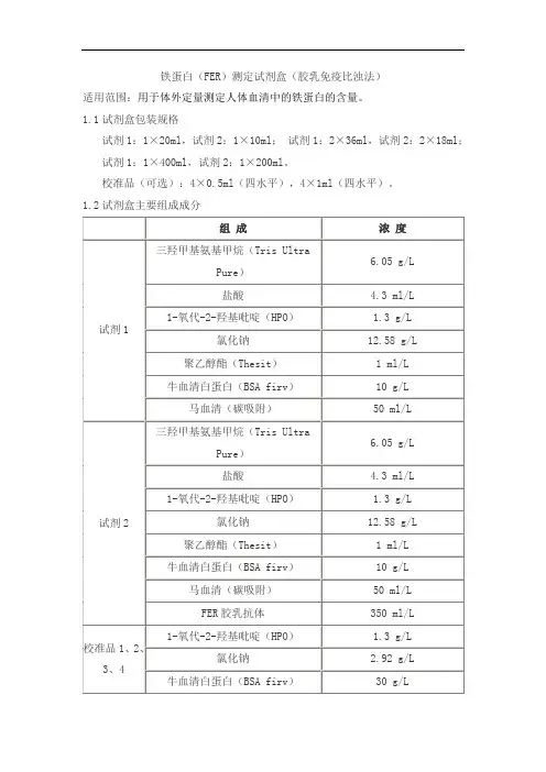
1.1试剂盒包装规格试剂1:1×20ml,试剂2:1×10ml;试剂1:2×36ml,试剂2:2×18ml;试剂1:1×400ml,试剂2:1×200ml。
校准品(可选):4×0.5ml(四水平),4×1ml(四水平)。
1.2试剂盒主要组成成分2.1 外观液体双试剂:试剂1无色澄清液体;试剂2 乳白色悬浊液。
校准品:浅黄至棕红色液体。
2.2 净含量液体试剂的净含量不得低于标示体积。
2.3 试剂空白吸光度在37℃、660nm波长、1cm光径条件下,试剂空白吸光度应不大于2.0。
2.4 分析灵敏度测定浓度为400ng/ml样本时,吸光度变化绝对值(|ΔA|)应不小于0.03。
2.5 线性范围在(6,450)ng/ml范围内,线性相关系数r不小于0.996,在(50,450)ng/ml 区间内线性相对偏差不大于±15%,(6,50]ng/ml区间内线性绝对偏差不大于±7.5ng/ml。
2.6 重复性重复测试两份高低浓度的样本,所得结果的变异系数(CV%)应不大于8%。
2.7 批间差不同批号试剂测试同一份样本,测定结果的批间相对极差应不大于10%。
2.8 准确度相对偏差:相对偏差应不超过±10%。
2.9 校准品溯源性依据GB/T 21415-2008《体外诊断医疗器械生物样品中量的测量校准品和控制物质赋值的计量学溯源性》的要求,校准品溯源至NIBSC生产的有证参考物质(WHO 94/572)。
2.10 稳定性效期稳定性:试剂盒在2℃~8℃下有效期为12个月。
取失效期的试剂盒进行检测试验结果满足2.3、2.4、2.5、2.6、2.8的要求。
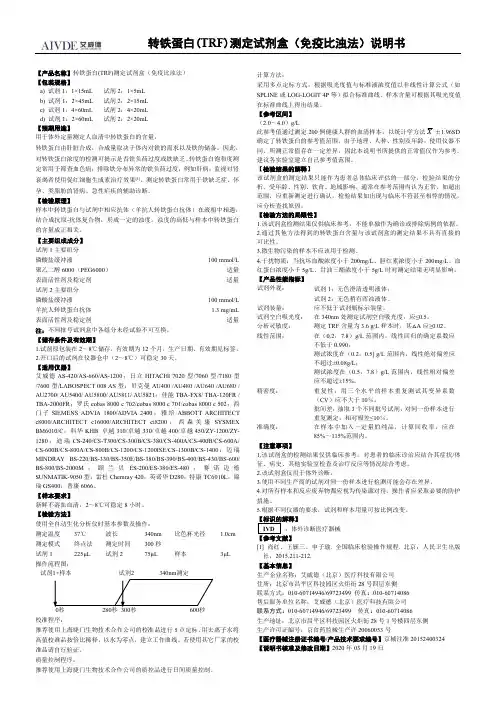
转铁蛋白(TRF)测定试剂盒(免疫比浊法)说明书【产品名称】转铁蛋白(TRF)测定试剂盒(免疫比浊法)【包装规格】a)试剂1:1×15mL试剂2:1×5mLb)试剂1:2×45mL试剂2:2×15mLc)试剂1:4×60mL试剂2:4×20mLd)试剂1:2×60mL试剂2:2×20mL【预期用途】用于体外定量测定人血清中转铁蛋白的含量。
转铁蛋白由肝脏合成,合成量取决于体内对铁的需求以及铁的储备。
因此,对转铁蛋白浓度的检测可提示是否铁负荷过度或铁缺乏。
转铁蛋白饱和度测定常用于筛查血色病;排除铁分布异常的铁负荷过度,例如肝病;监视对肾衰竭者使用促红细胞生成素治疗效果[1]。
测定转铁蛋白常用于铁缺乏症、怀孕、类脂肪的肾病、急性疟疾的辅助诊断。
【检验原理】样本中转铁蛋白与试剂中相应抗体(羊抗人转铁蛋白抗体)在液相中相遇,结合成抗原-抗体复合物,形成一定的浊度。
浊度的高低与样本中转铁蛋白的含量成正相关。
【主要组成成分】试剂1主要组分磷酸盐缓冲液100mmol/L 聚乙二醇6000(PEG6000)适量表面活性剂及稳定剂适量试剂2主要组分磷酸盐缓冲液100mmol/L 羊抗人转铁蛋白抗体 1.3mg/mL 表面活性剂及稳定剂适量注:不同批号试剂盒中各组分未经试验不可互换。
【储存条件及有效期】1.试剂原包装在2~8℃储存,有效期为12个月,生产日期、有效期见标签。
2.开口后的试剂在仪器仓中(2~8℃)可稳定30天。
【适用仪器】艾威德AS-420/AS-660/AS-1200;日立HITACHI7020型/7060型/7180型/7600型/LABOSPECT008AS型;贝克曼AU400/AU480/AU640/AU680/ AU2700/AU5400/AU5800/AU5811/AU5821;佳能TBA-FX8/TBA-120FR/ TBA-2000FR;罗氏cobas8000c702/cobas8000c701/cobas8000c502;西门子SIEMENS ADVIA1800/ADVIA2400;雅培ABBOTT ARCHITECT c8000/ARCHITECT c16000/ARCHITECT ci8200;西森美康SYSMEX BM6010/C;科华KHB卓越310/卓越330/卓越400/卓越450/ZY-1200/ZY-1280;迪瑞CS-240/CS-T300/CS-300B/CS-380/CS-400A/CS-400B/CS-600A/ CS-600B/CS-800A/CS-800B/CS-1200/CS-1200ISE/CS-1300B/CS-1400;迈瑞MINDRAY BS-220/BS-330/BS-350E/BS-380/BS-390/BS-400/BS-430/BS-600/ BS-800/BS-2000M;颐兰贝ES-200/ES-380/ES-480;赛诺迈德SUNMATIK-9050型;雷杜Chemray420;英诺华D280;特康TC6010L;锦瑞GS400;普康6066。

铁蛋白用途:用免疫学方法定量测定人血清或血浆中铁蛋白含量。
电化学发光免疫测定试剂,适用于罗氏Elecsys1010、2010和E170免疫测定分析仪。
概述:铁蛋白的分子量较大(440KD),由含24个亚单位的蛋白质外壳(脂铁蛋白)以及含约2500个Fe3+离子的铁核心两部分组成(在肝脏和脾脏中的铁蛋白)。
铁蛋白有形成聚合体的倾向,一旦在储存器官的细胞中过量存在时,易浓缩成半结晶状的血铁黄素并出现在溶酶体中。
用等电聚焦技术可以将铁蛋白分成20余种异质体。
它们之间的区别主要是含酸性的H亚单位与弱硷性的C亚单位的不同。
硷性异质体(主要存在于肝、脾和骨髓中)起长期储存铁的作用。
酸性异质体主要存在于心血管、胎盘和肿瘤组织中。
它们含铁量较低,其功能可能是铁转运的中间体。
铁蛋白的检测适用于了解体内铁代谢的状况。
在治疗初期检测铁蛋白可反映当时体内铁的储量,可以早期发现网织内皮系统中铁储存的不足。
在临床上,20ng/ml的阈值可以有效地判断准潜伏期铁不足并提示铁储存的耗竭。
正常情况下储存铁可用于血红蛋白的合成。
低于12ng/ml的铁蛋白阈值时,判断为潜伏期铁不足。
以上二种判断值,不需要进一步的实验室参考资料,甚至在血像提供的形态学指标仍然正常的情况下,仍是如此。
同时如伴有小细胞低色素性贫血,即可提示存在铁不足。
如果铁蛋白水平较高,又排除了供铁不正常的可能性,即反映体内铁过量的状况。
400ng/ml为判断阈值。
铁蛋白升高还可见于下列肿瘤:急性白血病、何杰金氏病、肺癌、结肠癌、肝癌和前列腺癌。
检测铁蛋白对肝脏转移性肿瘤有诊断价值,76%的肝转移病人铁蛋白含量高于400ng/ml。
升高的原因可能是由于细胞坏死、红细胞生成被阻断或肿瘤组织中合成增多。
两种单克隆抗体M-4.184和M-3.170在测定中用于形成夹心复合物。
原理:采用双抗体夹心法原理,整个过程18分钟完成。
・ 第1步:15µl 标本、生物素化的抗铁蛋白单克隆抗体和钌(Ru)标记的抗铁蛋白单克隆抗体混匀,形成夹心复合物。
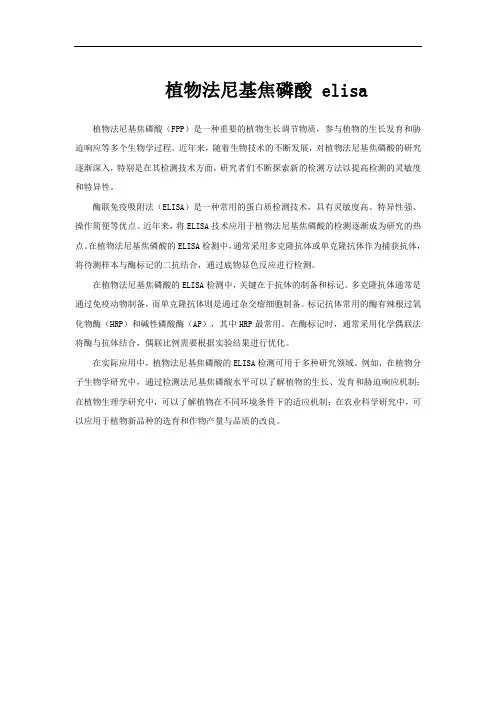
植物法尼基焦磷酸 elisa
植物法尼基焦磷酸(FPP)是一种重要的植物生长调节物质,参与植物的生长发育和胁迫响应等多个生物学过程。
近年来,随着生物技术的不断发展,对植物法尼基焦磷酸的研究逐渐深入,特别是在其检测技术方面,研究者们不断探索新的检测方法以提高检测的灵敏度和特异性。
酶联免疫吸附法(ELISA)是一种常用的蛋白质检测技术,具有灵敏度高、特异性强、操作简便等优点。
近年来,将ELISA技术应用于植物法尼基焦磷酸的检测逐渐成为研究的热点。
在植物法尼基焦磷酸的ELISA检测中,通常采用多克隆抗体或单克隆抗体作为捕获抗体,将待测样本与酶标记的二抗结合,通过底物显色反应进行检测。
在植物法尼基焦磷酸的ELISA检测中,关键在于抗体的制备和标记。
多克隆抗体通常是通过免疫动物制备,而单克隆抗体则是通过杂交瘤细胞制备。
标记抗体常用的酶有辣根过氧化物酶(HRP)和碱性磷酸酶(AP),其中HRP最常用。
在酶标记时,通常采用化学偶联法将酶与抗体结合,偶联比例需要根据实验结果进行优化。
在实际应用中,植物法尼基焦磷酸的ELISA检测可用于多种研究领域。
例如,在植物分子生物学研究中,通过检测法尼基焦磷酸水平可以了解植物的生长、发育和胁迫响应机制;在植物生理学研究中,可以了解植物在不同环境条件下的适应机制;在农业科学研究中,可以应用于植物新品种的选育和作物产量与品质的改良。
蓝耳病毒(PRRSV)ELISA检测试剂盒操作说明一、介绍:VDPro® PRRSV-VR或LV抗体检测试剂盒是用ELISA来定量检测猪蓝耳病毒(美洲株或欧洲株)抗体。
试剂盒是用重组PRRSV(美洲株或欧洲株)的N蛋白包被,用来检测血清中N 蛋白抗体。
通过监测猪群中PRRSV抗体滴度,这种血清学定量检测方法能够提供有效地信息,用来预防和控制该疾病。
二、试验原理:VDPro® PRRSV-VR,PRRSV-LV抗体检测试剂盒是通过间接ELISA检测血清中PRRSV抗体,用重组N蛋白包被,酶标抗体为猪抗IgG。
实验操作步骤是样品稀释,加样,孵育,洗涤;再加入酶标抗体,如果血清中含有N蛋白抗体,则抗体会与酶标板上包被的重组N蛋白结合,酶标抗体也会与血清中N蛋白抗体结合,加入底物后,颜色将会变深。
如果血清中没有N蛋白抗体,颜色变化会很小,或不发生变化。
三、试剂盒内容:1、PRRSV 重组N蛋白包被板 5块2、10倍洗液 200毫升3、血清稀释液 200毫升4、酶结合抗体 50毫升5、阳性血清对照 2毫升6、阴性血清对照 2毫升7、显色底物 60毫升8、终止液 30毫升四、采样标准①尽可能采集新鲜血清样品。
②采集全血,分离出血清后,将血清存放在-20度冰箱,以备使用。
③不要使用溶血或污染严重的样品④大中型猪场监测时,每群(后备母猪、经产母猪、断奶仔猪等)采样不少于35头份;五、注意事项:1、使用之前请仔细阅读使用说和操作明;2、试剂盒储存条件:4-8℃;六、实验前准备:1、洗液10倍稀释:即20ml洗液,加入180ml去离子水混匀,备用。
2、TMB底物:使用前恢复到室温,因为低温可能导致不显色。
七、样品稀释:1、准备好1ml样品稀释板或微量试管;2、样品按100倍稀释,分步稀释:即先加入90微升样品稀释液,再加入10微升的样品血清,混合均匀;再取90微升样品稀释液,加入10微升经过第一步稀释的血清,混合均匀即可。
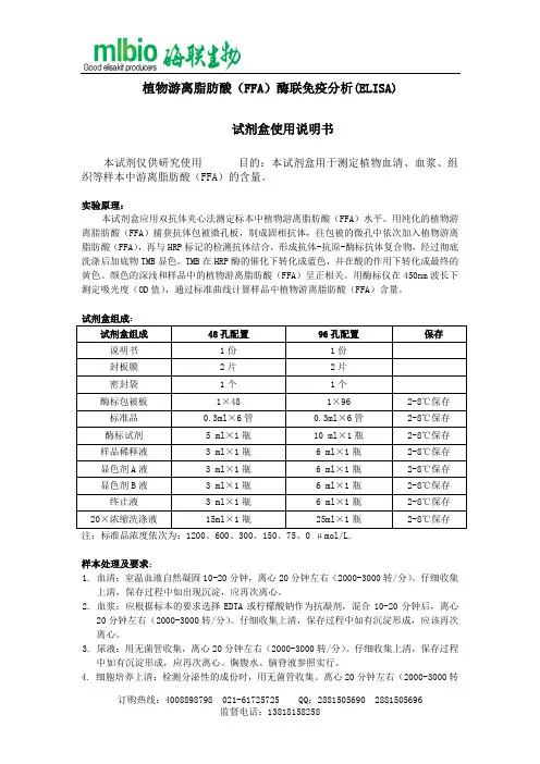
植物游离脂肪酸(FFA)酶联免疫分析(ELISA)试剂盒使用说明书本试剂仅供研究使用目的:本试剂盒用于测定植物血清、血浆、组织等样本中游离脂肪酸(FFA)的含量。
实验原理:本试剂盒应用双抗体夹心法测定标本中植物游离脂肪酸(FFA)水平。
用纯化的植物游离脂肪酸(FFA)捕获抗体包被微孔板,制成固相抗体,往包被的微孔中依次加入植物游离脂肪酸(FFA),再与HRP标记的检测抗体结合,形成抗体-抗原-酶标抗体复合物,经过彻底洗涤后加底物TMB显色。
TMB在HRP酶的催化下转化成蓝色,并在酸的作用下转化成最终的黄色。
颜色的深浅和样品中的植物游离脂肪酸(FFA)呈正相关。
用酶标仪在450nm波长下测定吸光度(OD值),通过标准曲线计算样品中植物游离脂肪酸(FFA)含量。
试剂盒组成:样本处理及要求:1. 血清:室温血液自然凝固10-20分钟,离心20分钟左右(2000-3000转/分)。
仔细收集上清,保存过程中如出现沉淀,应再次离心。
2. 血浆:应根据标本的要求选择EDTA或柠檬酸钠作为抗凝剂,混合10-20分钟后,离心20分钟左右(2000-3000转/分)。
仔细收集上清,保存过程中如有沉淀形成,应该再次离心。
3. 尿液:用无菌管收集,离心20分钟左右(2000-3000转/分)。
仔细收集上清,保存过程中如有沉淀形成,应再次离心。
胸腹水、脑脊液参照实行。
4. 细胞培养上清:检测分泌性的成份时,用无菌管收集。
离心20分钟左右(2000-3000转/分)。
仔细收集上清。
检测细胞内的成份时,用PBS(PH7.2-7.4)稀释细胞悬液,细胞浓度达到100万/ml左右。
通过反复冻融,以使细胞破坏并放出细胞内成份。
离心20分钟左右(2000-3000转/分)。
仔细收集上清。
保存过程中如有沉淀形成,应再次离心。
5. 组织标本:切割标本后,称取重量。
加入一定量的PBS,PH7.4。
用液氮迅速冷冻保存备用。
标本融化后仍然保持2-8℃的温度。
转铁蛋白检测试剂盒(免疫比浊法)说明书转铁蛋白检测试剂盒(免疫比浊法)说明书【产品名称】通用名称:转铁蛋白检测试剂盒(免疫比浊法)英文名称:TRF Kit【包装规格】R1:R2 ,1×30ml;1×10ml1×60ml;1×20ml 2×60ml;2×20ml【预期用途】转铁蛋白检测试剂盒临床上用于定量测定人体血清中转铁蛋白的含量。
转铁蛋白(TRF)连接上铁离子之后可以防止铁中毒以及通过肾的流失。
其水平的升高常见于铁缺乏症、怀孕、雌性激素的控制以及类脂肪的肾病。
其水平的降低常见于睾丸激素的控制、感染、急性的炎症、某些类型的肾炎、血色素缺失、急性的疟疾以及营养不良。
【检验原理】人体中的转铁蛋白与试剂中抗人转铁蛋白抗体在缓冲液中快速形成抗原抗体复合物,使反应液出现浊度。
当反应液中保持抗体过剩时,形成的复合物随抗原量增加而增加,反应液的浊度亦随之增加,在340nm以终点法检测吸光度变化,与校准品对照,即可计算出未知蛋白的含量。
【主要组成成分】组成主要成分R1 NaH2PO4缓冲液R2 抗人转铁蛋白抗体注意不同批号的试剂盒的组分不能混用。
校准品:用户自行购买利德曼公司的多项高值免疫标准液,标准值见说明书;质控品:用户自行购买利德曼公司多项免疫质控血清,质控值见说明书;【储存条件及有效期】1.包装试剂均应在2?,8?避光储存,可稳定至标签所示失效日期;2( 试剂有效期为12个月;3( 开瓶有效期:10天(开瓶后在2?,8?保存);【适用仪器】包装规格适用机型1×30ml;1×10ml 日立7060、1×60ml;1×20ml 日立7170、东芝-40 2×60ml;2×20ml 日立7020、奥林巴斯AU640、贝克曼CX4 【样本要求】1、标本为离心或分离除去血液凝块的新鲜血清。
2、血清样本在2~8?储存不超过一周。
马传染性贫血病毒CELISA抗体检测试剂盒技术参数[作用与用途]本试剂盒采用竞争ELlSA技术,用于检测马血清中马传染性贫血病毒抗体,检测结果可用于马传染性贫血感染的辅助诊断。
【主要成分】______________________________________________________________________________采集待检马的血液,先将全血置37°C条件放置30分钟,后置2~8℃下放置1小时,待血清析出后收集血清。
若血清析出不佳,可将析出液以5000r∕min,离心5分钟,收集上清。
【操作方法】1 .使用前所有试剂应恢复至室温(15~25°C)o试剂应轻轻地旋转或震荡予以混合。
2 .将10倍浓缩洗涤液用双蒸水稀释10倍后备用。
3 .将待检血清样品、阴性对照血清和阳性对照血清与酶标抗原按照1:1 (V/V)混合均匀,推荐以下步骤: 吸取待检血清样品、阴性对照血清和阳性对照血清各60μl,按顺序分别加入60μl的酶标抗原,充分混匀,置37C 孵育30分钟。
4 .取出包被板,将100μl孵育后的样品加入对应的孔中,在37C孵育30分钟。
5 .弃去孔内液体,每孔加入洗涤液250μl,孵育3分钟,弃去洗涤液,洗涤4次,每次拍干。
6 .将底物溶液A和底物溶液B等体积混合均匀,作为底物溶液,现用现配。
向各反应孔中加入底物溶液100μl∕JL,置室温(15-25βC)避光显色5分钟。
7 .加入终止液,50μl∕孔,轻微震荡混合均匀后,在450nm的波长下读数(应在加入终止液后5分钟内完成读数),并记录结果。
8 .计算公式物件I虔_ ______ 阴性对照血清平均OD45θnm 值一待检血清样品OD45Onm 值____押例率二阴性对照血清平均OD450nm值一阳性对照血清平均OD45θnm值【结果判定】1 .试验结果有效的条件是:阴性对照血清的平均OD450nm值>1.0:阳性对照血清的平均OD450nm值<0.20。
铁蛋白(Ferritin)测定试剂盒(电化学发光免疫分析法)适用范围:本试剂盒用于体外定量测定人体血清样本中铁蛋白(Ferritin)的含量。
1.1产品型号/规格:50人份/盒、100人份/盒。
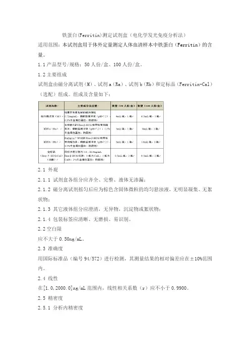
1.2主要组成试剂盒由磁分离试剂(M)、试剂a(Ra)、试剂b(Rb)和定标品(Ferritin-Cal)(选配)组成。
组成及含量如下:2.1 外观2.1.1 试剂盒各组分应齐全、完整、液体无渗漏;2.1.2 磁分离试剂摇匀后应为棕色含固体微粒的均匀悬浊液,无明显凝集、无絮状物;2.1.3 其它液体组分应澄清,无异物,沉淀物或絮状物;2.1.4 包装标签应清晰、无磨损、易识别。
2.2空白限应不大于0.50ng/mL。
2.3 准确度用国际标准品(编号94/572)进行检测,其测量结果的相对偏差应在±10%范围内。
2.4 线性在[1.0,2000.0]ng/mL范围内,线性相关系数(r)应不小于0.9900。
2.5 精密度2.5.1 分析内精密度在试剂盒的线性范围内,浓度为(40.0±10ng/mL)和(400.0±80.0ng/mL)的样品检测结果的变异系数(CV)应不大于8%。
2.5.2 批间精密度在试剂盒的线性范围内,用3个批号试剂盒分别检测浓度为(40.0±10ng/mL)和(400.0±80.0ng/mL)的样品,检测结果的变异系数(CV)应不大于15%。
2.6 效期末稳定性本产品效期为15个月,试剂盒在2~8℃下保存至有效期末进行检测,检测结果应符合2.1、2.2、2.3、2.4、2.5.1的要求。
2.7 溯源性依据GB/T21415-2008《体外诊断医疗器械生物样品中量的测量校准品和控制物质赋值的计量学溯源性》的要求,定标品溯源至国际标准品(编号94/572)。
铜蓝蛋白ELISA试剂盒说明书铜蓝蛋白ELISA试剂盒说明书供应商:上海樊克生物有限公司规格:96T/48T保存条件:2-8℃低温保存保质期:6个月,所有试剂盒均提供最新批次。
用途:科研实验等领域科学研究,不得用于临床诊断。
上海樊克经营Elisa试剂盒、国产/进口elisakit、血清,培养基,实验耗材仪器,抗体,细胞,酶等生物化学试剂,种属齐全,价格实惠,质量有保证,提供实验代测服务. 欢迎电咨询!铜蓝蛋白ELISA试剂盒说明书Note on the use of enzyme standard instrument:1 sample name file settings due to the part of the test sample is not necessarily continuous, the name of the sample is particularly important. Also should do the sample file name and absorbance value documents remain consistent, so if and then transferred out absorbance file, sample file name can automatically at the same time, V; if large numbers of samples with different micropore plate number difference (in the form of file suffix).2. Sample program file setting on a laboratory with a test may use different manufacturers kit, but different KIT provisions set control number, threshold criteria and standards may be inconsistent enzyme mark instrument the sample program is also inconsistent. In order to facilitate the use of the template can be used in the document with the suffix to be distinguished.试剂盒组成:铜蓝蛋白ELISA试剂盒说明书Operational matters needing attention:The reagent should label storage instructions, return to room temperature before use. After thinning of the standard products should be discarded, can not be saved.No - in the experiment of the plate should be immediately put back the bag, sealed to prevent spoilage.- no other reagents should be packaged or covered. Do not mix different batches of reagents. Before use.] using disposable suction head to avoid cross contamination and draw the termination of the liquid and the substrate of a and B liquid, avoid using a portion of the metal sample adding device.Plastic container configuration - use clean washing liquid. All samples of ingredients and mix well before use in the kit.The A substrate should be volatile, avoid long time to open the lid. Substrate B is sensitive to light and avoid prolonged exposure to light. Avoid hand contact, toxic. After completion of the experiment should be immediately read od.The addition of reagents should be consistent in order to ensure that all reaction plate hole incubation time.The temperature in accordance with the amount of liquid and the sequence of instructions indicated time, sterile operation铜蓝蛋白ELISA试剂盒说明书试剂盒组成:封板膜:2片(48)/2片(96)说明书:1份密封袋:1个标准品:???2700ng/L 0.5ml×1瓶0.5ml×1瓶??2-8℃保存酶标包被板:??1×48 ????1×96 ????????2-8℃保存样品稀释液:?3ml×1瓶? 6?ml×1瓶????2-8℃保存显色剂A液:?3ml×1瓶6?ml×1瓶????2-8℃保存显色剂B液:?3ml×1瓶6?ml×1瓶????2-8℃保存终止液:?????3ml×1瓶6ml×1瓶????2-8℃保存浓缩洗涤液:(20ml×20倍)×1瓶(20ml×30倍)×1瓶???2-8℃保存铜蓝蛋白ELISA试剂盒说明书试剂盒性能1. 灵敏度:最小的浓度小于1号标准品。
呋喃妥因代谢物(AHD)ELISA检测试剂盒说明书一、概要用来检测呋喃妥因代谢物的最常用方法是LC-UV,LC-MS和LC-MS/MS。
酶联免疫方法结合了色谱技术,采用AHD衍生物的特异性抗体,具有很高的精确度和灵敏度,较低的操作技术要求和短暂的检测时间,检测样本量大等特点在检测中很好的表现出来。
二、试验原理本试剂盒采用间接竞争ELISA方法,在酶标板微孔条上预包被偶联抗原,样本中的残留物的AHD经衍生化后和微孔条上预包被的偶联抗原竞争抗呋喃妥因代谢物的衍生物抗体,加入酶标二抗后,用TMB底物显色,样本吸光值与其所含残留物AHD代谢物的含量成负相关,与标准曲线比较即可得出样本中呋喃妥因代谢物的残留量。
三、适用范围可定性、定量检测动物组织(鸡、猪、牛、鱼、虾等)、牛奶和蜂蜜中呋喃妥因代谢物的残留量。
四、交叉反应率呋喃妥因代谢物…………………………………………100%呋喃唑酮代谢物…………………………………小于0.1%呋喃它酮代谢物…………………………………小于0.1%硝基糠腙(呋喃西林)代谢物…………………小于0.1%呋喃唑酮………………………………………………小于1%呋喃它酮………………………………………………小于1%呋喃妥因………………………………………………13%呋喃西林…………………………………………小于1%五、使用单位需自备的设备及试剂设备:┅┅微孔板酶标仪450nm/630nm┅┅旋转蒸发仪/氮气吹干装置┅┅均质器┅┅振荡器┅┅涡旋仪┅┅离心机┅┅天平:感量0.01g┅┅刻度移液管:10ml┅┅洗耳球┅┅容量瓶:100ml、1L┅┅玻璃试管:10ml┅┅聚苯乙烯离心管:50ml┅┅微量移液器:单道 20ml~200ml、100ml~1000ml 多道 250ml试剂:┅┅乙酸乙酯(分析纯)┅┅正己烷(或正庚烷)(分析纯)┅┅三水合磷酸氢二钾(分析纯)┅┅甲醇(分析纯)┅┅二水合亚硝基铁氰化钠(Na2Fe(CN)5·NO·2H2O)(分析纯)(供奶样用)┅┅七水合硫酸锌(ZnSO4·7 H2O) (分析纯)(供奶样用)┅┅浓盐酸┅┅氢氧化钠(分析纯)┅┅去离子水六、提供的材料与试剂1、96孔酶标板×1块(包被有偶联抗原)2、标准液×6瓶:(1ml/瓶)0ppb,0.1ppb,0.3ppb,0.9ppb,2.7ppb,8.1ppb3、高浓度标准品:(1ml/瓶) 100ppb4、酶标二抗12ml …………………… 红色帽5、抗体工作液7ml …………………… 绿色帽6、底物液 A 液7ml …………………… 白色帽7、底物液 B 液7ml …………………… 红色帽8、终止液7ml …………………… 黄色帽9、20×浓缩洗涤液40ml……………………… 透明帽10、2×浓缩复溶液 50ml……………………… 蓝色帽11、2-硝基苯甲醛15.1mg…………………… 黑色帽七、溶液的配制配液1: 衍生化试剂向装有2-硝基苯甲醛的试剂瓶中加甲醇溶解定容至10ml(浓度为10mM)。
铁蛋白(Fer)测定试剂盒(量子点荧光免疫层析法)
适用范围:用于体外定量测定人血清、血浆、全血中铁蛋白(Fer)的含量。
1.1 包装规格
10人份/盒,20人份/盒,25人份/盒,30人份/盒,40人份/盒,50人份/盒。
1.2 主要组成成分
2.1 物理性状
2.1.1外观
试剂盒应组分齐全,内外包装均应完整,标签清晰,液体试剂无渗漏。
2.1.2 膜条宽度
产品的膜条宽度应≥2.5 mm。
2.1.3 液体移行速度
液体移行速度应不低于10 mm/min。
2.1.4 净含量
试剂盒中稀释液净含量的相对偏差应不超过±15%。
2.2 准确度
用Fer国家标准品(150540)作为样本进行测定,其测定结果的相对偏差应不超过±15%。
2.3 检出限
检出限应不高于5.0 ng/mL。
2.4 线性
在线性区间[5,1000] ng/mL内,相关系数r应不低于0.9900。
2.5 重复性
分别用浓度为(40±10)ng/mL和(400±80)ng/mL的样本各重复测定10次,其变异系数(CV)应不大于15%。
2.6 批间差
用三个批号试剂盒分别测定浓度为(40±10)ng/mL和(400±80)ng/mL两个区间样本,所得结果的批间变异系数(CV)应不大于15%。
2.7 效期稳定性
4℃~30℃保存,有效期为24个月。
取到期后2~3个月的试剂盒进行测定,测定结果应符合2.2、2.3、2.4、2.5项的要求。
(本试剂盒仅供体外研究使用,不用于临床诊断!)产品货号:E-EL-R3018产品规格:96T/48T/24T大鼠铁蛋白(FE)酶联免疫吸附测定试剂盒使用说明书Rat FE(Ferritin) ELISA Kit使用前请仔细阅读说明书。
如果有任何问题,请通过以下方式联系我们:销售部电话************,************技术部电话************QQ客服800110755具体保质期请见试剂盒外包装标签。
请在保质期内使用试剂盒。
联系时请提供产品批号(见试剂盒标签),以便我们更高效地为您服务。
用途该试剂盒用于体外定量检测大鼠血清、血浆或其它相关生物液体中FE浓度。
灵敏度、检测范围、特异性和重复性●灵敏度:9.38ng/mL。
●检测范围:15.63-1000ng/mL。
●特异性:可检测样本中的大鼠FE,且与其它类似物无明显交叉反应。
●重复性:板内,板间变异系数均<10%。
检测原理本试剂盒采用双抗体夹心ELISA法。
用抗大鼠FE抗体包被于酶标板上,实验时样品(或标准品)中的大鼠FE会与包被抗体结合,游离的成分被洗去。
后依次加入生物素化的抗大鼠FE抗体和辣根过氧化物酶标记的亲和素,抗大鼠FE抗体与结合在包被抗体上的大鼠FE结合,生物素与亲和素特异性结合而形成免疫复合物,游离的成分被洗去。
加入显色底物(TMB),TMB在辣根过氧化物酶的催化下呈现蓝色,加终止液后变成黄色。
用酶标仪在450nm波长处测OD值,FE浓度与OD450值之间呈正比,通过绘制标准曲线计算出样品中FE的浓度。
试剂盒组成及保存未拆封的试剂盒可在2-8℃保存一周;如果一周以后才使用试剂盒,请拆开试剂盒并按照下表中的条件分别保存各组分。
试剂体积以实际发货版说明书为准。
相关试剂在分装时会比标签上标明的体积稍多一些,请在使用时量取而非直接倒出。
试验所需自备物品1.酶标仪(450nm波长滤光片)2.高精度移液器,EP管及一次性吸头:0.5-10μL, 2-20μL, 20-200μL, 200-1000μL3.37℃恒温箱,双蒸水或去离子水4.吸水纸5.加样槽注意事项1.试验中请穿着实验服并戴乳胶手套做好防护工作。
铁蛋白(FER )测定试剂盒(化学发光法)2. 性能指标2.1 试剂盒性能指标2.1.1 外观试剂盒包装应完整,各组分应齐全,品名、批号和有效期应清晰。
各瓶试剂外观应完整,标签应清晰,无破损,无渗漏。
M 试剂为黄褐色磁珠悬浮液,沉淀属于正常现象;R 试剂为澄清透亮液体,没有沉淀和悬浮物。
2.1.2净含量试剂盒各规格净含量应符合表1的要求。
表1 净含量要求2.1.3 准确度 用国家标准品(150540)作为样本进行检测,其测量结果的相对偏差不超过 ±10%。
2.1.4 检出限检出限应不高于 3ng/mL 。
2.1.5 线性线性范围 10ng/mL ~2000ng/mL ,相关系数 r 应不低于 0.9900。
2.1.6重复性批内变异系数(CV)应≤10%。
2.1.7批间差批间变异系数(CV)应≤15%。
2.2校准品性能指标2.2.1外观校准品外包装应完整,标签标示应清晰,为澄清透亮液体。
2.2.2装量校准品装量应不少于标示量。
2.2.3校准品赋值准确性用经校准品校准的化学发光免疫分析仪检测国家标准品(150540),结果的偏倚在±10%内。
2.2.4均匀性a)瓶内均匀性:CV≤10%;b)瓶间均匀性:CV≤10%。
2.3质控品性能指标2.3.1外观质控品外包装应完整,标签标示应清晰,为澄清透亮液体。
2.3.2装量质控品装量应不少于标示量。
2.3.3预期结果用经校准品校准的化学发光免疫分析仪检测质控物,结果应在靶值范围内。
2.3.4均匀性a)瓶内均匀性:CV≤10%;b)瓶间均匀性:CV≤10%。
铁蛋白(Ferr)测定试剂盒(化学发光免疫分析法)适用范围:本产品用于体外定量测定人血清中的铁蛋白的含量。
1.1 产品规格试剂盒规格为48人份/盒、96人份/盒。
1.2 主要组成成分表1 铁蛋白(Ferr)测定试剂盒(化学发光免疫分析法)主要组成成分a) 酶结合物以含牛血清白蛋白的缓冲液配制的联接HRP的Ferr单克隆抗体,其中含ProClin300做为防腐剂。
b) 校准品校准品主要以小牛血清为稀释液,其中含ProClin300做为防腐剂,校准品A~F 中含Ferr的目标浓度分别约为含0µg/L、30µg/L、110µg/L、210µg/L、280µg/L、370 µg/L。
校准品具体浓度详见标签及试剂盒参数IC卡。
c) 发光液发光液A主要成份为鲁米诺,发光液B主要成份为过氧化脲,两者均以pH8.6 的Tris-HCl缓冲液配制。
d) 包被微孔板包被有Ferr单克隆抗体的白色聚苯乙烯微孔板,用铝箔袋真空包装。
e) 质控品(备选)以正常人血清为基质制备的冻干品,其中含ProClin300做为防腐剂,靶值浓度范围QCⅠ(30.00 µg/L~50.00 µg/L)QCⅡ(320.00 µg/L~480.00 µg/L)。
质控品具体浓度详见质控品参数表。
不同批号试剂盒中的相同组分不能互换。
2.1 外观a)液体组分应澄清,无沉淀或絮状物,实际装量应不小于标示装量;b)冻干组分呈白色或淡黄色疏松体,加水后应在3分钟内完全溶解;c)所有组分均无包装破损,标示清楚。
2.2 准确度使用试剂盒校准品校准后测定国家标准品(编号:150540),国家标准品的实测浓度与标示浓度的偏差在±10%之间。
2.3线性在[2,1000 ]µg/L范围内,相关系数不低于0.9900。
2.4 精密度2.4.1重复性:CV≤10%(手工操作),CV≤8%(全自动)。
Elisa Kits
1 Elisa Kit原理
Elisa(酶联免疫):是免疫酶技术的一种,是将原抗体反应的特异性与酶反应的敏感性相
结合而建立的一种新技术。
Elisa的技术原理是:将酶分子与抗体(或抗原)结合,形成稳定的酶标抗体(或抗原)结合物,当酶标抗体(或抗原)与固相载体上的抗原(或抗体)
结合时,即可在底物溶液参与下,产生肉眼可见的颜色反应,颜色的深浅与抗原或抗体的
量成比例关系,使用Elisa检测仪,即酶标仪,测定其吸收值可作出定量分析。
此技术具有特异、敏感、结果判断客观、简便和安全等优点。
用来测定生物样品中蛋白质或其他(潜在)抗原物质的浓度。
针对待测物的特异性抗体被吸附在一个不溶性的载体表明,如聚氯乙烯板。
添加已知量的物品。
这样待测物品被
抗体结合。
清洗载体,针对这一待测物质的第二个位点添加二抗,这一份子携带了一个酶,它会使下一个试剂出现颜色变化。
颜色变化的强度能被光度计测定,并同待测物质的标准
溶液进行对比。
ELSA广泛应用于临床医学和兽医学的诊断及科研当中。
2 Elisa Kit配置
(1)酶标板
(2)标准溶液
(3)底物
(4)单抗和二抗
(5)PBS-磷酸缓冲液作为稀释液
(6)终止溶液。
RayBio®Human Ferritin ELISA KitUser ManualRayBio® Human FerritinELISA Kit Protocol(Cat#: ELH-Ferritin-001)RayBiotech, Inc.We Provide You With ExcellentProtein Array System and Service Tel:(Toll Free)1-888-494-8555 or 770-729-2992; Fax:770-206-2393; Web: Email: info@RayBiotech, Inc.RayBio® Human FerritinELISA Kit ProtocolTABLE OF CONTENTSI.Introduction (2)II.Reagents (2)III.Storage (3)IV.Additional Materials Required (3)V.Reagent Preparation (4)VI.Assay Procedure (6)VII.Assay Procedure Summary (7)VIII.Calculation of ResultsA.Typical Data (7)B.Sensitivity (8)C.Recovery (8)D.Linearity (8)E.Reproducibility (9)IX.Specificity (9)X.Troubleshooting Guide (10)I. INTRODUCTIONThe RayBio® Human Ferritin ELISA (Enzyme-Linked Immunosorbent Assay) kit is an in vitro enzyme-linked immunosorbent assay for the quantitative measurement of human Ferritin in serum, plasma, cell culture supernatants and urine. This assay employs an antibody specific for human Ferritin coated on a 96-well plate. Standards and samples are pipetted into the wells and Ferritin present in a sample is bound to the wells by the immobilized antibody. The wells are washed and biotinylated anti-human Ferritin antibody is added. After washing away unbound biotinylated antibody, HRP-conjugated streptavidin is pipetted to the wells. The wells are again washed, a TMB substrate solution is added to the wells and color develops in proportion to the amount of Ferritin bound. The Stop Solution changes the color from blue to yellow, and the intensity of the color is measured at 450 nm.II. REAGENTS1.Ferritin Microplate (Item A): 96 wells (12 strips x 8 wells) coated withanti-human Ferritin.2.Wash Buffer Concentrate (20x) (Item B): 25 ml of 20x concentratedsolution.3.Standards (Item C): 2 vials of recombinant human Ferritin.4.Assay Diluent A (Item D): 30 ml diluent buffer, 0.09% sodium azide aspreservative. For Standard/Sample (serum/plasma) diluent.5.Assay Diluent B (Item E): 15 ml of 5x concentrated buffer. ForStandard/Sample (cell culture medium/urine) diluent.6.Detection Antibody Ferritin (Item F): 2 vial of biotinylated anti-humanFerritin (each vial is enough to assay half microplate).7.HRP-Streptavidin Concentrate (Item G): 8 μl 20,000x concentratedHRP-conjugated streptavidin.8.TMB One-Step Substrate Reagent (Item H): 12 ml of 3,3’,5,5’-tetramethylbenzidine (TMB) in buffer solution.9.Stop Solution (Item I): 8 ml of 2 M sulfuric acid.III. STORAGEMay be stored for up to 6 months at 2o to 8o C from the date of shipment. Standard (recombinant protein) should be stored at -20 o C or -80o C (recommended at –80 o C) after reconstitution. Opened Microplate Wells or reagents may be stored for up to 1 month at 2o to 8o C. Return unused wells to the pouch containing desiccant pack, reseal along entire edge.Note: the kit can be used within one year if the whole kit is stored at -20 o C. Avoid repeated freeze-thaw cycles.IV. ADDITIONAL MATERIALS REQUIRED1Microplate reader capable of measuring absorbance at 450 nm.2Precision pipettes to deliver 2 μl to 1 ml volumes.3Adjustable 1-25 ml pipettes for reagent preparation.4100 ml and 1 liter graduated cylinders.5Absorbent paper.6Distilled or deionized water.7Log-log graph paper or computer and software for ELISA data analysis.8Tubes to prepare standard or sample dilutions.V. REAGENT PREPARATION1.Bring all reagents and samples to room temperature (18 - 25°C) beforeuse.2.Sample dilution: Assay Diluent A (Item D) is used for dilution ofserum/plasma samples. Recommend 2 - 20 fold dilution forserum/plasma samples; 1x Assay Diluent B (Item E) can be used fordilution of cell culture supernates/urine.3.Assay Diluent B should be diluted 5-fold with deionized or distilledwater before use.4. Preparation of standard: Briefly spin the vial of Item C and then add 400 μl Assay Diluent A (for serum/plasma samples) or 1x Assay Diluent B (for cell culture supernates/urine) into Item C vial to prepare a 200 ng/ml standard. Dissolve the powder thoroughly by a gentle mix. Pipette 270 μl Assay Diluent A or 1x Assay Diluent B into each tube. Use the 200 ng/ml standard solution to produce a dilution series (shown below). Mix each tube thoroughly before the next transfer. Gently vortex to mix. Assay Diluent A or 1x Assay Diluent B serves as the zero standard (0 ng/ml).5. If the Wash Concentrate (20x) (Item B) contains visible crystals, warm to room temperature and mix gently until dissolved. Dilute 20 ml of Wash Buffer Concentrate into deionized or distilled water to yield 400 ml of 1x Wash Buffer.6. Briefly spin the Detection Antibody vial (Item F) before use. Add 100 μl of 1x Assay Diluent B into the vial to prepare a detection antibody concentrate. Pipette up and down to mix gently (the concentrate can be stored at 4°C for 5 days). The detection antibody concentrate should be diluted 80-fold with 1x Assay Diluent B and used in step 4 of Part VI Assay Procedure.180 μl 200 80 32 12 5.12 2.04 0.81 0 ng/ml ng/ml ng/ml ng/ml ng/ml ng/ml ng/ml ng/mlStandard, ItemC + 400 μl180μl 180 μl 180 μl 180 μl 180 μl7.Briefly spin the HRP-Streptavidin concentrate vial (Item G) and pipetteup and down to mix gently before use. HRP-Streptavidin concentrateshould be diluted 20,000-fold with 1x Assay Diluent B.For example: Briefly spin the vial (Item G) and pipette up and down to mix gently . A dd 2 μl of HRP-Streptavidin concentrate into a tube with 198 μl 1x Assay Diluent B to prepare a 100-fold diluted HRP-Streptavidin solution (don’t store the diluted solution for next day use). Mix through and then pipette 50 μl of prepared 100-fold dilutedsolution into a tube with 10 ml 1x Assay Diluent B to prepare a final10,000 fold diluted HRP-Streptavidin solution.VI. ASSAY PROCEDURE:1.Bring all reagents and samples to room temperature (18 - 25°C) beforeuse. It is recommended that all standards and samples be run at least induplicate.2.Add 100 μl of each standard (see Reagent Preparation step 2)andsample into appropriate wells. Cover well and incubate for 2.5 hours atroom temperature or over night at 4°C with gentle shaking.3.Discard the solution and wash 4 times with 1x Wash Solution. Wash byfilling each well with Wash Buffer (300 μl) using a multi-channel Pipette or autowasher. Complete removal of liquid at each step isessential to good performance. After the last wash, remove anyremaining Wash Buffer by aspirating or decanting. Invert the plate and blot it against clean paper towels.4.Add 100 μl of 1x prepared biotinylated antibody (Reagent Preparationstep 6) to each well. Incubate for 1 hour at room temperature withgentle shaking.5.Discard the solution. Repeat the wash as in step 3.6.Add 100 μl of prepared Streptavidin solution (see Reagent Preparationstep 7) to each well. Incubate for 45 minutes at room temperature with gentle shaking.7.Discard the solution. Repeat the wash as in step 3.8.Add 100 μl of TMB One-Step Substrate Reagent (Item H) to each well.Incubate for 30 minutes at room temperature in the dark with gentleshaking.9.Add 50 μl of Stop Solution (Item I) to each well. Read at 450 nmimmediately.VII. ASSAY PROCEDURE SUMMARY1. Prepare all reagents, samples and standards as instructed.2. Add 100 μl standard or sample to each well.Incubate 2.5 hours at room temperature or over night at 4o C.3. Add 100 μl prepared biotin antibody to each well.Incubate 1 hour at room temperature.4. Add 100 μIncubate 45 minutes at room temperature.5. Add 100 μl TMB One-Step Substrate Reagent to each well.Incubate 30 minutes at room temperature.6. Add 50 μl Stop Solution to each well.Read at 450 nm immediately.VIII. CALCULATION OF RESULTSCalculate the mean absorbance for each set of duplicate standards, controls and samples, and subtract the average zero standard optical density. Plot the standard curve on log-log graph paper or using Sigma plot software, with standard concentration on the x-axis and absorbance on the y-axis. Draw the best-fit straight line through the standard points.A. TYPICAL DATAThese standard curves are for demonstration only. A standard curve must be run with each assay.B. SENSITIVITYThe minimum detectable dose of Ferritin is typically less than 0.6 ng/ml. C. RECOVERYRecovery was determined by spiking various levels human Ferritin into human serum, plasma and cell culture media. Mean recoveries are as follows:Sample Type Average % Recovery Range (%)Serum 97.32 86-105Plasma 103.1 93-112Cell culture media 96.46 85-93D. LINEARITYSample Type Serum Plasma Cell Culture Media 1:2 Average % of Expected 97.78 102.8 105.4Range (%) 86-106 91-110 96-1131:4 Average % of Expected 101.1 75.54 86.67Range (%) 91-110 67-81 75-94E. REPRODUCIBILITYIntra-Assay: CV<10%Inter-Assay: CV<12%IX.SPECIFICITYCross Reactivity: This ELISA kit shows no cross-reactivity with the following cytokines tested: human Angiogenin, BDNF, BLC, CNTF, ENA-78, FGF-4, IL-1α, IL-1β, IL-2, IL-3, IL-4, IL-5, IL-6, IL-7, IL-8, IL-9, IL-11, IL-12 p70, IL-12 p40, IL-13, IL-15, IL-309, IP-10, FGF-4, FGF-6, FGF-7, G-CSF, GDNF, GM-CSF, IFN-γ, IGFBP-2, IGF-BP-3, IGF-BP-4, Leptin (OB), MCP-1, MCP-2, MCP-3, MDC, MIF, MIG, MIP-1α, MIP-1 β, MIP-1δ, PARC, PDGF, RANTES, SCF,SDF-1alpha, TARC, TGF-β, TIMP-1, TIMP-2, TNF-α, TNF-β, TPO, VEGF.X. TROUBLESHOOTING GUIDEProblem Cause Solution1. Poor standardcurve1. Inaccurate pipetting 1. Check pipettes2. Improper standard dilution 2. Ensure a brief spin of Item Cand dissolve the powder thoroughly by a gentle mix.2. Low signal 1.Too brief incubationtimes 1. Ensure sufficient incubation time; assay procedure step 2 may change to over night2. Inadequate reagent volumes or improper dilution 2. Check pipettes and ensure correctpreparation3. Large CV 1. Inaccurate pipetting 1. Check pipettes4. High background 1. Plate is insufficientlywashed 1. Review the manual for proper wash. If using a platewasher, check that all ports areunobstructed.2. Contaminated wash buffer 2. Make fresh wash buffer5. Low sensitivity 1. Improper storage of theELISA kit 1. Store your standard at<-20o C afterreconstitution,others at 4 o C. Keep substrate solution protected from light2. Stop solution 2. Stop solution shouldbe added to eachwell before measureRayBio® ELISA kits:Over 200 ELISA kits, custom ELISA kit choose from over 500 list visit for details.This product is for research use only.©2004 RayBiotech, Inc.。