CFSE使用方法
- 格式:pdf
- 大小:209.74 KB
- 文档页数:6


细胞悬液置于离心管底部。
离心管横放,将PBS滴加在靠近管口的管壁上。
再将CFSE加在PBS滴上。
维持离心管横向水平,拧上管盖,迅速垂直倒置离心管于涡旋器上涡旋。
(可参考JOVE网上的CFSE标记视频)
步骤:
1. 常规方法分离PBMC,细胞计数后300g离心10分钟。
弃上清,加入1%FBS/PBS液体重悬,洗涤一次,再用合适体积数目的1%FBS/PBS重悬,取出2×10^6l的细胞悬液,离心弃上清。
2. 用0.1%FBS/PBS稀释CFSE储存液(5000×,5mM)至1umol/L。
3. 用0.5ml上述CFSE稀释液重悬细胞,轻柔混匀。
4. 37 ℃放置10min。
5. 加入1ml冰冷的RPMI 1640 完全培养液,4℃,5分钟以中止染色。
6. 300g室温离心5分钟。
弃上清,加入冰冷的含10%FBS 的完全1640培养液洗2次。
最后用0.4ml 培养基重悬。
7. 将上述细胞悬液加入96孔培养板,每孔105个细胞,(阳性对照用PHA刺激(1ug/ml))
8. 37 ℃, 5%CO2 避光培养3天。
9. 吸出培养液,24小时内上机检测。
(7AAD排除死细胞)
17. 在FACScalibur流式细胞仪上检测,用Cell Quest软件获取数据,然后用软件分析。

cfse染料波长-回复CFSE染料波长是指一种荧光染料的激发和发射波长。
CFSE (Carboxyfluorescein succinimidyl ester)是一种广泛应用于细胞生物学研究中的荧光染料。
它可以通过与细胞内的活性蛋白结合,以及通过其荧光信号的激发和发射来可视化和跟踪细胞的生物活动。
在本文中,我们将一步一步地探讨CFSE染料波长的相关知识。
首先,我们将了解CFSE染料的激发波长。
CFSE染料的激发波长通常为492nm。
这意味着当CFSE染料暴露在492nm的激发光下时,它会吸收能量并转化为发射荧光信号。
接下来,我们将详细了解CFSE染料的发射波长。
CFSE染料的发射波长通常为517nm。
这意味着在CFSE染料吸收激发光并转化为发射光时,它会产生具有517nm波长的荧光信号。
然而,需要注意的是,CFSE染料的激发和发射波长可以受到实验条件的影响而发生变化。
例如,在某些特定的实验设定中,可能需要使用不同的激发波长以达到最佳的荧光信号强度。
在这种情况下,研究人员可以选择适当的激发光源,并调整实验条件以获得最佳结果。
除了CFSE染料的激发和发射波长之外,还有一些其他与此相关的因素值得进一步探讨。
例如,荧光染料的量子产率和荧光强度可能会受到其化学结构和环境条件的影响。
此外,荧光染料与细胞内蛋白质或其他生物分子的结合方式及其影响也是研究人员需要考虑的因素之一。
在CFSE染料的应用中,其荧光信号的强度和稳定性对细胞生物学研究非常重要。
因此,研究人员通常会在实验之前对荧光染料进行仔细的优化和验证。
他们会通过使用不同的实验条件,包括激发光源和荧光检测系统等,来确定最佳的CFSE染料波长和设置。
总结起来,CFSE染料波长的理解对于细胞生物学研究的进展至关重要。
通过了解其激发和发射波长,并在实验条件中进行适当的优化,研究人员可以使用CFSE染料来可视化和跟踪细胞的生物活动,从而为细胞生物学研究的发展做出贡献。
殷佳琪资土01班配位化学螯合剂在选矿中的应用摘要近年来,螯合捕收剂的发展取得了飞速进步,一些研究及实践的资料证明,螯合捕收剂的浮选性能与它们的螯合特性密切相关。
本文从配位原子、捕收性能、捕收机理、药剂种类等方面,总结了近年来的研究发现,并以苯甲羟肟酸为例,展示了螯合类捕收剂在工业中的运用。
关键词:螯合捕收剂捕收机理药剂种类工业1螯合剂的简介1.1 应用方向在浮选药剂发展过程中,第一代混合捕收油早已过时,第二代离子型水溶性捕收力强的浮选药剂(如黄药、黑药等)已经历了70余年,越来越无法满足目前世界范围内日渐贫、细、杂矿石的浮选分离要求。
近年来,人们都把注意力转向第三代非离子型高选择性浮选剂上,特别是螯合捕收剂更以其卓越的选择性深受人们关注[1]。
因为金属螯合物比普通的离子型和共价型金属盐更稳定,长期以来将螯合剂当作选择性更好的捕收剂,并且,它们似乎可以代替常规的捕收剂,从已知的分析化学中的分离金属方法也可以看出这一点[2,3]。
螯合剂在选矿中的运用主要包括浮选和选择性絮凝。
这两种工艺的主要表面化学原理十分相似。
它们分选的选择性基本上都是取决于药剂在矿物一水界面上的选择性吸附。
这种选择性实际上是药剂的官能团在矿物表面吸附点的亲和力的函数。
由于螯合型官能团对某些金属离子具有较高的专一性,因而可以认为它是选矿药剂的理想组成部分。
1.2 螯合剂和螯合作用螯合型药剂至少必须有两个原子同时由金属配位。
这些原子通常是O、N、S和P。
“配位”物质提供的这些给予体原子称为“配位体”。
如果单个配位体分子或离子不止有一个原子与金属离子配位,便使共自身围绕中心原子弯成螯状,形成复杂的环状结构,称为“螯合物”(来源于希腊语中的“蟹钳”)根据配位体在带正电的金属离子周围配位区域内的配位位置数目是二、三、四、五、六,可将它们相应地称作二元环.、三元环、四元环、五元环和六元环。
下面给出的例子为二乙基二硫代氨基甲酸脂螯合剂的S一S型配位体与镍形成的二员环(l:2)[4]。

心脏内皮细胞的分离概述说明以及解释1. 引言1.1 概述心脏内皮细胞是构成心脏血管系统的重要组成部分,具有维持血管结构稳定、调节血压和参与免疫反应等多种功能。
因此,研究心脏内皮细胞的分离方法对于心血管疾病的诊断、治疗以及药物筛选等方面具有重要意义。
本文旨在概述并解释心脏内皮细胞的分离方法,包括定义和功能介绍、分离方法的概述以及它们之间的优劣比较。
此外,我们还将详细讨论心脏内皮细胞分离过程中的步骤和操作技巧,如材料准备、操作环境要求、细胞分离酶选择与使用方法以及培养基配制与使用注意事项等。
在实验结果和数据解读部分,我们将介绍心脏内皮细胞分离后效率和纯度评估指标,并展示实验结果并进行详尽解读。
此外,我们还会将结果与预期进行比较,并探讨其与现有研究进展之间的联系。
最后,在结论与未来展望部分,我们将总结心脏内皮细胞分离方法的优缺点,并探讨其在心脏内皮细胞研究中的潜在应用前景。
同时,我们还会提出该领域研究进一步发展的方向和探索的方向。
通过本文的阐述,希望能够为研究人员提供有关心脏内皮细胞分离方法的全面指导,促进对心脏内皮细胞功能和相关疾病机制的深入理解,并为相关治疗方法的开发和改进提供新思路。
2. 心脏内皮细胞的分离:心脏内皮细胞是一种位于血管内壁上的细胞,其主要功能包括调节血管张力、维持心脏功能和参与免疫反应等。
在很多研究中都需要对心脏内皮细胞进行分离以便进一步的实验操作和研究。
本部分将概述心脏内皮细胞的定义和功能,并对常用的心脏内皮细胞分离方法进行简要介绍,并对这些方法进行优劣比较。
2.1 心脏内皮细胞的定义和功能:心脏内皮细胞是一种存在于血管壁以及相关组织中的特化上皮细胞,其具有重要的生理功能。
首先,它们构成了血管内壁,起到隔离并调控外部环境与体液之间物质交换的作用。
其次,心脏内皮细胞可以通过释放各种生物活性物质来参与调节血管张力,因此在血压控制和循环系统平衡方面发挥着关键作用。
此外,它们还可以通过分泌细胞外基质和参与免疫反应等方式对心脏功能进行支持和调节。

cfse染料波长-回复CFSE(carboxyfluorescein succinimidyl ester)染料是一种常用的细胞示踪染料,也被称为洋红荧光素。
它在生物医学和细胞研究中被广泛使用,因其明亮的荧光信号和良好的稳定性而备受青睐。
在这篇文章中,我们将一步一步地回答关于CFSE染料波长的问题,帮助读者更好地了解和使用这种重要的生物染料。
首先,我们需要了解CFSE染料的基本特性。
CFSE属于荧光素家族,其化学结构中包含苯并吡喃环并带有甲苯基的共轭二烯。
由于其结构的独特性,CFSE具有强大的荧光特性,可以有效地被激发和发射荧光信号。
CFSE染料的激发波长为494纳米,而发射波长为521纳米。
这意味着,当CFSE染料受到494纳米的激发光照射时,它会发出521纳米的荧光信号。
这一波长范围的选择是基于CFSE染料的吸收光谱和发射光谱的特性,以及所使用的荧光显微镜的光源和检测器的选择。
在细胞实验中,CFSE染料被广泛用作细胞示踪剂,可以用于追踪和定量细胞增殖、移行和分化等过程。
其原理是将CFSE染料与目标细胞一起孵育,然后通过细胞的代谢活性将CFSE染料转化为CFSE缀合物。
这些缀合物可在细胞的质膜内累积并由CFSE释放荧光信号。
通过测量细胞中的荧光强度,我们可以了解细胞在不同时间点的变化,用于评估细胞增殖率、迁移速度或分化程度。
除了波长的选择外,使用CFSE染料还需要考虑许多其他因素。
首先,选择适当的染料浓度非常重要。
通常,细胞染色需要在一定浓度范围内进行,以确保荧光信号的强度和稳定性。
对于CFSE染料,推荐使用的浓度范围为0.5到5微克/毫升。
其次,染色时间的选择也很重要。
在染色的初期阶段,CFSE染料被细胞摄取并在细胞内进行代谢转化。
随着时间的推移,CFSE染料开始积累并释放荧光信号。
染色时间的选择应基于特定细胞类型和实验目的,通常在30分钟至24小时之间。
最后,CFSE染料的稳定性也需要考虑。
CFSE活细胞标记的特点
CFSE:羧基荧光素二醋酸盐琥珀酰亚胺酯(5-or6-(N-Succinimidyloxycarbonyl)-3',6'-O,O'-diacetylfluorescein)。
优点:
1、活体标记:可以追踪细胞在体内的分裂增殖过程。
2、时间长:标记的荧光可以维持长达数周之久。
3、安全:不需使用放射性同位素。
4、相关性高:与H3掺入法结果一致,可代替后者。
5、特定跟踪:可以与细胞亚群特定抗体结合使用。
6、简单:操作简单,可靠。
7、稳定:进入细胞后荧光不受内环境影响。
8、结果客观:对细胞生理活动没有任何影响。
9、经济实惠:费用经济,效率高。
用途:
1、细胞增殖检测(尤其是淋巴细胞的增殖检测)。
2、体内示踪。
3、细胞荧光标记。
4、细胞毒性检测。
其他:
1CO2培养箱,移液器等。
2、激发波长:500nm
3、发射波长:520nm。
准备:1.20ml 预冷5%FBS(HI)-PBS2.50ml 预冷MACS buffer3.70um滤网4.2.5ml RBC lysis5.LS column6.50ml 37度预热PBS7.50ml 37度预热complete culture medium(RPMI 1640 +10% HeatInactivated FBS+10 mM HEPES+1 mM penicilline streptomycin+ 50 mM 2-mercaptoethanol)(一)CD3抗体包被取96孔板每孔加入100ul 10ug/ml anti-CD3抗体(PBS)密封4度过夜。
(二)淋巴细胞获取1.小鼠的脾脏结摘取于10ml 冷5%FBS-PBS中。
2.于70um滤网研磨滤过,500g 4度5min离心后弃上清。
3.2.5ml RBC lysis裂红5 min,10ml 冷5% PBS终止。
4.500g 4度5min 离心后弃上清,10ml MACS buffer重悬,计数。
留取105个细胞进行FACS。
(三)磁珠分选naïve CD8a+ T细胞1.Centrifuge cell suspension at 300×g for 10 minutes. Aspirate supernatant completely. 2.Resuspend cell pellet in 400 μL of MACS buffer per 108 total cells. 3.Add 100 μL of Naive CD8a+ T Cell Biotin-Antibody Cocktail per 108 total cells. 4.Mix well and incubate for 5 minutes in the refr igerator (2−8 °C). 5.Add 200 μL of MACS buffer per 108 total cells. 6.Add 200 μL of Anti-Biotin MicroBeads per 108total cells. Add 100 μL of CD44 MicroBeads per 108 total cells.7.Mix well and incubate for an additional 10 minutes in the refrigerator (2−8 °C).8.Wash cells by adding 10ml of MACS buffer per 108 cells and centrifuge at 300×g for 10 minutes. Aspirate supernatant completely.9.Resuspend up to 108cells in 500 μL of MACS buffer.10.Place LS column in the magnetic field of a suitable MACS Separator.11.Prepare column by rinsing with 3 mL of MACS buffer. 12.Apply cell suspension onto the column. Collect flow-throughcontaining unlabeled cells, representing the enriched naive CD8a+ T cell fraction. 13.Wash column with 3 mL of MACS buffer. Collect unlabeled cells thatpass through, representing the enriched naive CD8a+ T cells, andcombine with the flow-through from step 3. 14.Determine cell number. 留取105个细胞进行FACS。
脐带间充质干细胞生物学有效性及临床研究进展摘要:目的:研究脐带间充质干细胞(hMSCs)生物学有效性。
方法:本实验以PHA刺激淋巴细胞作为反应体系,观察hMSCs与淋巴细胞共培养时增殖的变化,培养体系分为阴性对照组、阳性对照组、实验组。
结果:在共培养接触体系中不同数量hMSCs对淋巴细胞增殖具有抑制作用,且hMSCs浓度越高,抑制作用越强,具有剂量依赖性。
结论:间充质干细胞在不同类型的炎症环境中,可由静息状态转变成激活状态,通过细胞间相互作用和分泌可溶性免疫调控因子,调节多种免疫细胞的增殖、分化、活化以及炎症因子的分泌功能。
MSC的免疫调控功能是其临床应用中重要的生物学有效性质量属性,对这一质量属性的有效评价,是指导MSC产品研发及临床应用中最为相关的质量控制内容之一。
近年来人们对MSC免疫调控功能的认识及其与临床应用的相关性,在此基础上提出了针对治疗性MSC产品免疫调控功能的评价策略,为在我国建立和完善MSC生物学有效性评价体系奠定基础。
关键词:脐带间充质干细胞;物学有效性;免疫调控功能前言:间充质干细胞是具有高度自我更新和多向分化潜能的多能干细胞,在体外不同的诱导条件下可分化为骨细胞、软骨细胞、脂肪细胞、肌腱、肌肉细胞以及神经细胞等,其免疫原性低,几乎影响免疫系统的所有细胞,具有免疫调节作用[1]。
但其免疫调节作用机制不明确,我们通过研究hMSCs对异体淋巴细胞增殖的影响,进一步探讨hMSCs的免疫调节作用的可能机制,为hMSCs应用于临床提供一定基础。
1材料与方法1.1 材料脐带间充质干细胞(hMSCs)和淋巴细胞(PBMC)均由生产提供。
间充质干细胞无血清培养基;牛血清(FBS);10XPBS;灭菌注射用水;PHA(植物血凝素);CFSE;无水DMSO;荧光抗体;48孔板;流式细胞仪。
1.2 方法1.2.1 hMSCs表型鉴定调整细胞浓度,使用荧光抗体进行标记,不加抗体作为空白对照;室温避光孵育,洗涤后检测。
碳硫高速分析仪操作规程前言碳硫高速分析仪是一种用于快速准确测量固体材料中碳和硫含量的仪器。
本操作规程的目的是为了确保使用者能够正确操作仪器,保证测试结果的准确性和可靠性。
设备准备在进行任何操作之前,首先要确保设备已经正常运转并处于可使用状态。
具体操作步骤如下:1.打开仪器电源,并等待电源指示灯亮起。
2.确认设备的供气管道连通良好,并且空气压力稳定在0.5-0.7Mpa之间。
3.启动仪器并进入测试界面。
如果发现异常,请联系维修人员处理。
样品准备在进行样品测试之前,需要对样品进行一定的准备工作。
具体操作步骤如下:1.样品应当是完全干燥的,并且经过了粉碎和混合。
2.取出一定量的样品,称重之后放入进样池中。
3.在进样池中注入盖板里的氧化剂。
4.关闭进样池盖,点击“测试”按钮,开始测试。
测试操作在进行测试操作时,需要按照正确的步骤进行操作。
具体步骤如下:1.在测试完成之前,不要将进样池盖打开。
2.点击“测试”按钮,等待测试进程完成。
3.测试完成后,点击“结果”按钮,查看测试结果。
4.点击“清洗”按钮,清洗进样池,结束测试。
后续操作测试完成后,需要对设备进行一定的后续操作,以确保设备的安全和可靠性。
具体步骤如下:1.关闭气源。
2.关闭仪器电源。
3.清洗进样池和周围区域,将仪器恢复到初始状态。
注意事项在进行操作过程中,需要注意以下事项:1.在测试过程中,不要打开进样池盖。
2.如果发现样品异常,如有异味或者发生爆炸现象,请立即停止测试并联系维修人员处理。
3.不要将进样池放置在阳光照射下的地方。
结论本操作规程详细介绍了使用碳硫高速分析仪过程中的设备准备、样品准备、测试操作、后续操作等内容,并且对注意事项进行了说明。
只有正确使用和操作才能保证仪器测试数据的准确性和可靠性,确保调制的最终合成的制剂、零部件符合质量要求,生产正常进行。
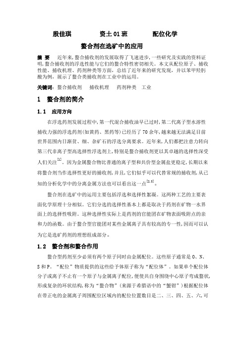
CellTrace ™ Cell Proliferation KitsCatalog nos. C34554, C34557, C34564IntroductionThe CellTrace ™ Cell Proliferation Kits provide versatile and well-retained cell tracing reagents in a convenient and easy-to-use form. Each kit contains a CellTrace ™ reagent (CellTrace ™ CSFE, CellTrace ™ Violet, or CellTrace ™ Far Red) in single-use vials to permit small scale experiments without preparing excess quantities of stock solution. TheCellTrace ™ reagents easily diffuse into cells and bind covalently to intracellular amines, resulting in stable, well-retained fluorescent staining that can be fixed with aldehyde fixatives. Excess unconjugated reagent passively diffuses to the extracellular medium, where it can be quenched with complete media and washed away.Spectral Characteristics CellTrace ™ CSFE (Cat. no. C34554)The approximate excitation and emission peaks of this product after hydrolysis are 492 nm and 517 nm, respectively. Cells labeled with CellTrace ™ CSFE reagent can be visualized by fluorescence microscopy using standard fluorescein filter sets or analyzed by flow cytometry in an instrument equipped with a 488-nm excitation source.CellTrace ™ Violet (Cat. no. C34557)The approximate excitation and emission peaks of this product after hydrolysis are 405 nm and 450 nm, respectively. Cells labeled with CellTrace ™ Violet reagent can be visualized by fluorescence microscopy using standard DAPI filter sets or analyzed by flow cytometry in an instrument equipped with a 405-nm excitation source.CellTrace ™ Far Red (Cat. no. C34564)The approximate excitation and emission peaks of this product after hydrolysis are 630 nm and 661 nm, respectively. Cells labeled with CellTrace ™ Far Red reagent can be visualized by fluorescence microscopy using standard Cy ®5 filter sets or analyzed by flow cytometry in an instrument equipped with a 633/635-nm excitation source.Table 1Contents and storageBefore StartingMaterials Required but NotProvided• Cells of interest as a single-cell suspension• Phosphate-buffered saline (PBS) or similar protein-free buffer • Culture media containing protein such as FBS or BSA • Flow cytometerCautionNo data are available addressing the mutagenicity or toxicity of CellTrace ™ reagents (Component A). Handle the DMSO dye solution with caution because DMSO is known to facilitate the entry of organic molecules into tissues. Always wear suitable protective clothing, gloves, and eye/face protection when handling this reagent. Dispose of the reagents in compliance with all pertaining local regulations.Storage and HandlingUpon receipt, store the kit components desiccated at ≤–20°C until required for use. When stored properly DMSO and dry CellTrace ™ reagents are stable for at least one year. Allow the product to warm to room temperature before opening vials. Stocksolutions of CellTrace ™ reagents should be used only on the day of preparation. Avoid repeated freezing and thawing of kit contents.Figure 1 Generational tracing using CellTrace ™ Reagents. (A) Cell proliferation was followed for 8 generations using the CellTrace ™ Violet reagent. Human peripheral blood mononuclear cells were harvested and stained with CellTrace ™ Violet reagent prior to stimulation with mouse anti-human CD3 (Cat. no. MHCD0300) and Interleukin-2 (Cat. no. PHC0027) for 7 days. The discrete peaks in this histogram represent successive generations of live, CD4 positive cells. Analysis was completed using an Attune ® Acoustic Focusing Cytometer with 405-nm excitation and a 450/40 bandpass emission filter . The unstimulated parent generation is indicated in red. (B) and (C) Cell Proliferation was followed for 7 generations using the CellTrace ™ CFSE and CellTrace ™ Far Red reagents, respectively. Human T lymphocytes were harvested and stained with CellTrace ™ CFSE or CellTrace ™ Far Red reagent prior to stimulation with anti-human CD3 (Cat. no. MHCD0300) for 5 days. The discrete peaks in these histograms represent successive generations of live, SYTOX ® Green (Cat. no. S34860) negative cells. The unstimulated parent generation is indicated in blue (CellTrace ™ CFSE) or purple (CellTrace ™ Far Red). Analysis was completed using an Attune ® Acoustic Focusing Cytometer with 488 nm excitation and a 530/30 nm bandpass emission filter for CellTrace ™ CFSE. Analysis was completed for CellTrace ™ Far Red using an Attune ® Acoustic Focusing Cytometer with 638 nm excitation and a 660/20 nm bandpass emission filter.CellTrace ™ CFSE Fluorescence P e r c e n t o f M ax103104105106801004020060CellTrace ™ Violet FluorescenceC e l l C o u n t10310410510620001000102400003000CellTrace ™ Far Red FluorescenceP e r c e n t o f M a x103104105106801004020060ABCExperimental ProtocolsGeneral Guidelines• The following methods have been optimized for monitoring cell proliferation of human T and B lymphocytes.• In other cell types and applications, we recommend titration of the CellTrace ™reagents to determine the optimal staining concentration. CellTrace ™ stock solutions may be diluted in DMSO for this purpose.• Recommended concentration for CellTrace ™ staining is 1–10 μM. A dye concentration of 5–10 μM is recommended for tracking five or more generations, while 1–2 μM may be sufficient for tracking less than five generations.• Start with a single cell suspension in PBS for uniform cell labeling.• Uniform cell labeling can also be improved by gently agitating cells during staining.• A low flow rate should be used for analysis on hydrodynamic focusing cytometers to ensure separation of distinct generational peaks. All collection rates will provide equivalent results when analyzing on an Attune ® Acoustic Focusing Cytometer.• To ensure appropriate instrument setup, include a sample of unstimulated control cells in proliferation experiments using CellTrace ™ reagents.Table 2 Preparation of CellTrace ™ stock solutionsLabeling Cells in SuspensionThe following protocol has been optimized for cell concentrations of up to 106 cells/mL. Dye concentration may need to be increased for samples with >106 cells/mL.1.1 Prepare CellTrace ™ stock solution immediately prior to use by adding the appropriatevolume of DMSO (Component B) to one vial of CellTrace ™ reagent (Component A) and mixing well (see Table 2, above). 1.2 Add 1 µL of CellTrace ™ stock solution in DMSO (prepared in Step 1.1) to each mL of cellsuspension in PBS for a final working solution (see Table 2, above, for concentration). 1.3 Incubate the cells for 20 minutes at room temperature or 37°C, protected from light.1.4 Add five times the original staining volume of culture medium (containing at least1% protein) to the cells and incubate for 5 minutes. This step removes any free dye remaining in the solution.1.5 Pellet the cells by centrifugation and resuspend them in fresh pre-warmed completeculture medium.1.6 Incubate the cells for at least 10 minutes before analysis to allow the CellTrace ™ reagentto undergo acetate hydrolysis. 1.7 Proceed with cell stimulation, incubation, or analysis.Alternate Method for LabelingCells in Suspension The following protocol has been optimized for cell concentrations up to 106 cells/mL.Dye concentration may need to be increased for samples with >106 cells/mL.2.1 Prepare CellTrace™ stock solution immediately prior to use by adding the appropriatevolume of DMSO (Component B) to one vial of CellTrace™ reagent (Component A) andmixing well (see Table 2, page 3).2.2 Pellet the cells by centrifugation and remove the supernatant.2.3 Dilute the CellTrace™ DMSO stock solution in pre-warmed (37°C) phosphate-bufferedsaline (PBS) or other protein-free buffer to the desired working concentration (1–10 µM).2.4 Gently resuspend the cells in the PBS dye solution (prepared in Step 2.1).2.5 Incubate the cells for 20 minutes at room temperature or 37°C, protected from light.2.6 Add five times the original staining volume of culture medium (containing at least1% protein) to the cells and incubate for 5 minutes. This step removes any free dyeremaining in the solution.2.7 Pellet the cells by centrifugation and resuspend them in fresh, pre-warmed completeculture medium.Incubate the cells for at least 10 minutes before analysis to allow the CellTrace™ reagent2.8to undergo acetate hydrolysis.Proceed with cell stimulation, incubation, or analysis.2.9Alternate Method for LabelingAdherent Cells3.1 Prepare CellTrace™ stock solution immediately prior to use by adding the appropriatevolume of DMSO (Component B) to one vial of CellTrace™ reagent (Component A) andmixing well (see Table 2, page 3).3.2 Grow the cells to the desired density on coverslips or flasks filled with the appropriateculture medium.3.3 Dilute the CellTrace™ DMSO stock solution in pre-warmed (37°C) phosphate-bufferedsaline (PBS) or other protein-free buffer to the desired working concentration (1–10 µM).This is the loading solution.3.4 Remove the culture medium from the cells and replace it with the loading solution(prepared in Step 3.3).3.5 Incubate the cells for 20 minutes at 37°C.3.6 Remove the loading solution, wash the cells twice with culture medium containing atleast 1% protein, and replace with fresh, pre-warmed complete culture medium.Incubate the cells for at least 10 minutes before analysis to allow the CellTrace™ reagent3.7to undergo acetate hydrolysis.Proceed with cell stimulation, incubation, or analysis.3.8Optional: Fixation andPermeabilizationLabel the cells with CellTrace™ reagent according to one of the protocols listed above.4.14.2 Before fixation, wash and resuspend the cells in PBS or other protein-free buffer.4.3 Fix the cells for 15–20 minutes at room temperature using an aldehyde-based fixativesuch as paraformaldehyde, protected from light.4.4 Wash the cells with PBS.4.5 If desired, permeabilize the cells using any appropriate protocol. CellTrace™ reagentscovalently bind to cells and will not wash out after permeabilization.4.6 Following permeabilization, wash the cells with PBS.4.7 Resuspend the cells in PBS prior to analysis.Combining CellTrace™Reagents with otherFluorescent Markers5.1 Label the cells with CellTrace™ reagent according to one of the protocols listed above.5.2 Resuspend the cells in a buffer appropriate for the subsequent staining applications (seeProduct List, below).5.3 Apply stains for immunophenotyping, DNA content, apoptosis, or other markers asrecommended for each stain.References1. J Cell Biol 101, 610 (1985);2. J Cell Biol 103, 2649 (1986);3. J Immunol Methods 171, 131 (1994);4. J Exp Med 184, 277 (1996);5. J Immunol Methods 133, 87 (1990);6. Transplant Proc 24, 2820 (1992);7. Current Protocols in Cytometry, J. P. Robinson, Ed., (1998) pp 9.11.1-9.11.9.;8. Cytometry Part A, 79A: 95–101 (2011); 9. Current Protocols in Immunology, R. Coico, Ed., (2008) pp 7.10.1-7.10.24.10 June 2014Purchaser NotificationThese high-quality reagents and materials must be used by, or directl y under the super v ision of, a tech n ically qualified individual experienced in handling potentially hazardous chemicals. Read the Safety Data Sheet provided for each product; other regulatory considerations may apply.Obtaining SupportFor the latest services and support information for all locations, go to .At the website, you can:• Access worldwide telephone and fax numbers to contact Technical Support and Sales facilities • Search thr ough frequently asked questions (FAQs)• Submit a question directly to Technical Support (techsupport@ )• Search for user documents, SDSs, vector maps and sequences, application notes, formulations, handbooks, certificates of analysis, citations, and other product support documents• Obtain information about customer training • Download software updates and patchesSDSSafety Data Sheets (SDSs) are available at /sds .Certificate of AnalysisThe Certificate of Analysis provides detailed quality control and product qualification information for each product. Certificates of Analysis are available on our website. Go to /support and search for the Certificate of Analysis by product lot number, which is printed on the product packaging (tube, pouch, or box).Limited Product WarrantyLife Technologies Corporation and/or its affiliate(s) warrant their products as set forth in the Life Technologies’ General Terms and Conditions of Sale found on Life Technologies’ website at /termsandconditions . If you have any questions, please contact Life Technologies at /support .DisclaimerLIFE TECHNOLOGIES CORPORATION AND/OR ITS AFFILIATE(S) DISCLAIM ALL WARRANTIES WITH RESPECT TO THIS DOCUMENT, EXPRESSED OR IMPLIED, INCLUDING BUT NOT LIMITED TO THOSE OF MERCHANTABILITY, FITNESS FOR A PARTICULAR PURPOSE, OR NON-INFRINGEMENT. TO THE EXTENT ALLOWED BY LAW, IN NO EVENT SHALL LIFE TECHNOLOGIES AND/OR ITS AFFILIATE(S) BE LIABLE, WHETHER IN CONTRACT, TORT, WARRANTY, OR UNDER ANY STATUTE OR ON ANY OTHER BASIS FOR SPECIAL, INCIDENTAL, INDIRECT, PUNITIVE, MULTIPLE OR CONSEQUENTIAL DA MAGES IN CONNECTION WITH OR ARISING FROM THIS DOCUMENT, INCLUDING BUT NOT LIMITED TO THE USE THEREOF.Important Licensing InformationThese products may be covered by one or more Limited Use Label Licenses. By use of these products, you accept the terms and conditions of all applicable Limited Use Label Licenses.All trademarks are the property of Thermo Fisher Scientific and its subsidiaries, unless otherwise specified. Cy is a registered trademark of GE Healthcare UK Limited. ©2014 Thermo Fisher Scientific Inc. All rights reserved.Product List Current prices may be obtained from our website or from our Customer Service Department.Cat. no. Product Name Unit SizeC34554 CellTrace ™CSFE Cell Proliferation Kit *for flow cytometry*...................................................... 1 kitC34557 CellTrace ™Violet Cell Proliferation Kit *for flow cytometry*......................................................1 kit C34564 CellTrace ™Far Red Cell Proliferation Kit *for flow cytometry*.................................................... 1 kit Related ProductsMHCD0300 Purified Mouse anti-human CD3 ............................................................................ 500 μL PHC0026 Recombinant Human Interleukin-2.......................................................................... 10 μgS10274 SYTOX ® AADvanced ™Dead Cell Stain Kit *500 tests* ........................................................ 5 × 0.5 mL S10349 SYTOX ® AADvanced ™ Dead Cell Stain Kit *100 tests* ........................................................... 0.2 mLS34859 SYTOX ® Red Dead Cell Stain *for flow cytometry* *1000 tests*...................................................1 mL S34860 SYTOX ®Green Dead Cell Stain *for flow cytometry* *1000 tests* ................................................. 1 mL111.31D DynaBeads ®CD3/CD28 T Cell Expander...................................................................... 2 mL08-0022SA OpTmizer ™T-Cell Expansion SFM........................................................................... 500 mL 10439-016 Fetal Bovine Serum, ES Cell-Qualified ....................................................................... 100 mL 14190-136 Dulbecco’s Phosphate-Buffered Saline (D-PBS) (1X), liquid...................................................... 1000 mL。