《临床化学》常用分析方法(全)
- 格式:docx
- 大小:66.01 KB
- 文档页数:4

临床化学常用分析方法临床化学是研究人体组织、体液中各种化学物质的定性和定量分析的科学。
临床化学的常用分析方法主要包括分光光度法、比色法、化学发光法、电化学分析法、质谱法等。
下面将对其中几种常用的分析方法进行介绍。
1.分光光度法:分光光度法是利用物质吸收或发射电磁波的特性进行定性和定量分析的方法。
在临床化学中常用的分光光度法有紫外-可见吸收光度法和原子吸收光度法。
紫外-可见吸收光度法用于分析有机化合物或含有色团的无机化合物。
通过测量被分析物吸收或透射的光的强度来确定物质的浓度。
该方法灵敏度高,快速,准确度较高。
原子吸收光度法适用于分析微量金属元素。
通过电子能级的跃迁来测定分析物中金属元素的含量。
该方法可以对多种金属元素进行分析。
2.比色法:比色法是利用化学反应使物质的颜色发生变化,然后根据产生的颜色进行定性和定量分析的方法。
在临床化学中常用的比色法有显色反应法和络合反应法。
显色反应法是通过观察显色反应的颜色变化来进行分析的方法,常用于血液中葡萄糖、脂类、尿液中氨基酸、蛋白质等的定性和定量分析。
络合反应法是通过分析物与试剂形成稳定络合物而产生颜色来进行分析的方法。
常用于镁、铁、钙等金属离子的分析。
3.化学发光法:化学发光法是利用发光化学反应来进行定性和定量分析的方法。
在临床化学中,化学发光法常用于检测荧光标记的抗体与抗原结合的程度,从而进行免疫测定。
常用的化学发光法有酶化学发光法、电化学发光法和化学发光免疫分析法。
4.电化学分析法:电化学分析法是通过测量电极在电解质溶液中电流、电势和电荷等物理量的变化,来进行定性和定量分析的方法。
在临床化学中常用的电化学分析法有电位滴定法和安培法。
电位滴定法是根据滴定曲线上的电位变化来测定溶液中其中一种物质的含量的方法。
常用于血液中氢离子浓度(pH值)的测定。
安培法是利用电极的电流变化与被测物质的浓度成正比的原理进行定量分析的方法,常用于血液中酶的活性测定。
5.质谱法:质谱法是根据质量谱仪测定被测物质分子的相对分子质量和相对丰度,来进行定性和定量分析的方法。
Kaplan: Clinical Chemistry, 5th EditionClinical References - Methods of AnalysisAlbumin in UrineGraham Jones iName: Albumin in urineClinical Significance: A marker of renal damage in diabetes and other conditions. Also amarker of risk for cardiovascular disease.Common name: MicroalbuminuriaRefer to Chapter 30, Renal Function, in the 5th edition of Clinical Chemistry: Theory, Analysis, Correlation.Students’ Quick Hyperlink Review•Measurand•Principles of analysis and current usage•Reference and preferred methods•Specimen•Interferences•Urine albumin reference intervals•Interpretation•Urine albumin performance goals•References•Urine albumin methods tableMeasurandHuman albumin is a single polypeptide chain of 585 amino acids with 17 internal disulphide bonds but without carbohydrate side chains. Albumin in the circulation has microheterogeneity due to structural flexibility, ligand binding, and other factors. The transition of albumin from the serum to the urine has the potential to markedly increase the structural variability by fragmentation, internal cleavage, oxidation, or selective tubular resorption, and multiple forms of albumin have been identified in urine [1,2]. This variability in albumin has the potential to lead to standardization difficulties with assays and is known to produce different results for individual patients using different assays [3,4]. The development of assays for albumin fragments detectable by HPLC but not by standard immunoassays, the so-called immunochemical non-reactive albumin, has raised the possibility of improved sensitivity for detection of early renal damage [2]. There is a need to identify a measurand which is the most clinically relevant andi Albumin in UrineNew methodFifth edition: Graham Jonesanalytically suitable to provide standardized assays for urine albumin [5]. The structure of albumin in urine and its possible effect on various assays has been the subject of an extensive review [6].Principles of Analysis and Current UsageAssays for albumin in urine can be divided into three main categories: routine laboratory assays, point-of-care assays, and other reference or developmental assays. Owing to the issues mentioned above with regard to variability in the structure of urine albumin, there are some systematic and patient-specific differences between results from different assays, depending on the measurement technology and the antibody specificity.The vast majority of routine laboratories use immunoassays to quantitate albumin in urine. These may be structured as nephelometric or turbidimetric homogenous immunoassays or heterogenous competitive or noncompetitive immunoassays and may use monoclonal or polyclonal antibodies. These assays are generally purchased from diagnostic companies and are available for use on high-volume chemistry or immunoassay analyzers, as well as in manual assay formats such as enzyme-linked immunosorbent assay (ELISA) or radioimmunoassay. These commercial assays generally provide sufficient sensitivity to measure urine albumin down to concentrations found in healthy individuals, often with limits of detection below 5 mg/L, and are able to separate patients with normal albumin excretion from those with increased excretion. In addition, these assays have precision characteristics able to meet biological variation criteria with claimed coefficient of variation (CV) values for total precision of < 5%. For determination of the albumin-creatinine ratio, there is also the need to measure urine creatinine concentrations. Because the presence of noncreatinine chromogens in urine is much less than in plasma, routine creatinine assays are generally acceptable for this purpose. If a timed sample is received for calculation of the albumin excretion rate, the urine volume must be measured using appropriate scales or a volumetric flask. Given the high prevalence of diabetes, most laboratories need a method with high capacity to meet the clinical needs.There are a number of point-of-care methods available for urine albumin measurement using different assay formats and technologies. These may be semiquantitative or quantitative, and some also measure creatinine to allow calculation of the albumin-creatinine ratio. Point-of-care testing is of particular use in the clinical setting where a rapid return of laboratory results is difficult [7], although in any setting, the provision of a result for use within the same medical consultation as the sample collection can be beneficial. Since positive results can be referred to a laboratory for further analysis, a point-of-care test used as a screening test should have sufficient sensitivity to avoid missing positive results. The use of a high-quality quantitative point-of-care analyzer may avoid the need for referral of positive samples. Below are examples of point-of-care devices for measurement of urine albumin.The Siemens DCA Vantage analyzer (previously known as the DCA 2000) uses a monoclonal antibody agglutination technique for albumin, with a simultaneous chemical creatinine assay. These tests are performed on a single 40 µL sample; results are available in 7 minutes, using a disposable cartridge and a portable analyzer. The system achieves good precision, with CV < 5% for albumin and < 3% for creatinine, with quantitation of albumin down to 5 mg/L in a laboratory evaluation [8] with similar performance when used in remote locations [7].The Haemocue instrument is small, portable analyzer which uses disposable cuvettes preloaded with reagents. The system uses immunoturbidimetry and produces a result in 90 seconds from 18 µL of sample. The reporting range is 5 to 150 mg/L; however, precision was poorer than seen in laboratory methods at all concentrations tested, with within-run CVs of between 10% and 13% for patient samples with albumin concentrations above 20 µg/L [9,10], although overall a good correlation with routine laboratory methods has been achieved [10].The Siemens Clinitek system provides semiquantitative results for both albumin concentration and the albumin/creatinine ratio, using a regent strip with dye-binding techniques. The strips may be read in a small reader device, and results higher than 20 mg/L are reported as positive. Precision for the system cannot be easily determined, because the results are reported in large increments. A number of interferences are listed in the product information, including hematuria, soaps, dyes, and some drugs, as well as high levels of urine protein. An evaluation of patient samples showed that approximately 12% of patients with laboratory urine albumin results below 20 mg/L were falsely reported as elevated and 11% of samples with low-positive lab results (20 to 55 μg/L) were reported as negative [11]. A higher false-positive rate has been described in children [12], and it has been recommended that low-positive results be confirmed with laboratory testing [10].The Roche Micral reagent strips are dipped into a urine sample and the resultant immunologically-mediated color formation visually compared with a semiquantitative chart. The method has demonstrated acceptable between-user correlation [13].The lowest positive result is a color intensity associated with a nominal value of 20 mg/L. This decision point has been shown to have a high false-positive rate in two studies, indicating the need for laboratory follow-up if available [14,15].Measurement of urine albumin by HPLC detects different fragments of albumin, compared to immunoassay as described above. This leads to higher results, especially in the low range, with consequent higher detection rates for microalbuminuria when standard decision points are used [16,17]. More recent work has indicated possible co-elution of other proteins with albumin leading to overestimation of albumin by this method [18]. The differences between HPLC and immunoassay highlights the need for agreed reference methods and materials.Reference and Preferred MethodsThere are currently no reference methods or reference materials for urine albumin listed on the Joint Committee for Traceability in Laboratory Medicine (JCTLM) database [19]. In the absence of specific reference materials for urine albumin, most manufacturers reference their assays to human serum albumin—for example, using CRM470.The preferred methods for routine use are immunoassays for urine albumin with additional measurement of urine creatinine to allow calculation of the albumin/creatinine ratio. For many routine laboratories, this type of technology has the advantages of high throughput and performance on a routine chemistry or immunoassay analyzer. The required key performance characteristics are described further below.SpecimenThe amount of albumin in urine can be expressed in a number of different formats which require different samples [6]. These reporting formats include the following:Albumin Excretion Rate (AER)Albumin excretion rate is commonly expressed as mg per 24 hours or micrograms per minute. The former requires a 24-hour sample, whereas the latter may use a 24-hour sample or other timed period, such as an overnight collection. Both samples require close attention to start and finish times, as well as avoiding over- and under-collection from other causes. AER is considered to be the gold standard but is not generally recommended for routine use because of these collection difficulties.Urine Albumin/Creatinine Ratio (ACR)Expressing the albumin concentration in a spot sample as a ratio to urine creatinine is a method to reduce the effect of patient hydration on the albumin concentration. ACR is measured in a spot sample, preferably a first morning sample; a random sample is acceptable, but daily activity may lead to false-positive results. The units are mg/g creatinine or mg/mmol creatinine. The use of creatinine to correct for hydration also adds an influence of muscle mass to the result with larger people, who produce more creatinine, giving lower results for the same albumin excretion. This is seen with the different decision points for males and females recommended by some bodies. The ACR is recommended by the American Diabetes Association (ADA) for urine albumin testing [20]. These recommendations also indicate that results from at least 2 out of 3 samples over a 6-month period are used to confirm significant changes in albumin excretion status. Urine Albumin Concentration (UAC)The UAC is reported as mg/L or the equivalent µg/mL and is measured in a spot sample—like the ACR, preferably a first morning sample, but a random sample is acceptable. Some studies have shown minimal difference between the sensitivity of ACR and UAC for increased AER, so some authors recommend the use of UAC for general purposes, because this removes the requirement for creatinine measurement [21].No preservatives are usually required for urine albumin collections, and manufacturers recommendations should be consulted if a preservative is required. The sample may be stored at room temperature for up to 7 days and 1 month at 4°C to 8°C, according to World Health Organization (WHO) guidelines [22]. It is possible that bacterial contamination may affect stability at room temperature, so earlier cooling may be preferred. Storage at −20°C causes breakdown to fragments which are measured in some assays but not others, but this is not seen at −80°C, and long-term storage is possible at this temperature [3,23].InterferencesBiological causes for increased urine albumin excretion other than kidney damage include fever, exercise, heart failure, marked hyperglycemia, and hypertension [20]. Additionally, collection shortly after ejaculation may elevate results owing to the albumin content of semen. If these causes are identified, repeat testing at an appropriate time may be indicated to further evaluate positive results.Homogenous immunoassays such as turbidimetry are at risk of producing falsely low results due to a prozone effect [24]. Routine procedures to identify this problem may include (1) measurement of all samples neat and in dilution to confirm linear dilution, (2) measurement neat and with additional albumin added to confirm complete recovery, or (3) testing for high total protein with a dipstick to identify samples where excess albumin is likely [25].Attention should also be given to the possibility of carry-over effects when serum and urine are run on the same analyzer, given the > 1000-fold difference in albumin concentrations in the two sample types.Reference IntervalsDecision points for interpretation of urine albumin are not based on population reference intervals but rather on outcome-based consensus decision points. Different professional bodies have made slightly different recommendations, and the table below is based on the data from the American Diabetes Association nephrology guidelines [26]. Laboratories are encouraged to adopt their national guidelines where these are available. Of note, the decision point of 30 mg/g creatinine is also recommended as an indication of renal damage in the nondiabetic population [27].TABLE 1: Decision Points for Interpretation of Urine Albumin.*Spot Samples Timed SamplesAlbumin/Creatinine Ratio Albumin Excretion Rate AlbuminConcentration**mg/mol mg/24 hours µg/minmg/L mg/g orµg/mgNormal <30 <30 <2.5 (male)<30 <20<3.5 (female)30 -299 20-199 Microalbuminuria 30-299 2.5-29 (male)3.5-29(female)Macroalbuminuria ≥300 ≥30 ≥300 ≥200*Based on ADA Nephropathy guidelines [26].**Albumin concentration decision point from KDOQI [27].InterpretationUrine albumin is primarily measured as a marker of the risk of development of renal damage in diabetic patients. It is now becoming established as a marker for renal damage in nondiabetic patients, owing to vascular disease associated with hypertension, elevated lipids, and other standard risk factors. An elevated urine albumin is also an established marker of cardiovascular risk in the diabetic and nondiabetic populations [28], and this risk may extend down to results within the currently accepted “normal” range [29]. The benefit of urine albumin in the microalbumin range compared to measurement of total protein is the increased sensitivity provided by albumin. Once a result is in the macroalbumin range, the significance is the same as frank proteinuria.The response to the finding of an elevated urine albumin should be increased attention to risk factors, with the aim of reducing the risk of further damage to the kidneys or other organs. Performance GoalsLike many other analytes in urine, the concentration of albumin may vary considerably from day to day. A CV for within-subject biological variation of 36% is listed on the Biological Variation database on the Westgard website [30], although a recent review has shown marked variation in the estimates for this parameter, with the central tertile for all studies showing a range of 28% to 47% [6]. Thus assays with values for total analytical CVs below 7% will meet optimal precision requirements of less than a quarter of the within-subject variation for most estimates of this parameter. Given that urine albumin/creatinine ratios continue to provide information down to 10 mg/g (1.1 mg/mmol), the ability to measure albumin down below 5 mg/L is an advantage when measuring dilute samples. These criteria can be met by most laboratory-based immunoassays, including immunoturbidimetric assays that may be run on routine chemistry analyzers. By contrast, most point-of-care analyzers, particularly the semiquantitative methods, are unable to provide good analytical performance near upper limit of normal, although they are clearly able to identify higher levels of albumin within the “microalbuminuria” range.References1 Candiano G, Musante L, Bruschi M, Petretto A, Santucci L, Del Boccio P et al. Repetitivefragmentation products of albumin and alpha-1-antitrypsin in glomerular disease associated with nephrotic syndrome. J Am Soc Nephrol 2006:17:3139-3148.2 Osicka TM, Comper WD. Characterization of immunochemically nonreactive urinaryalbumin. Clin Chem 2004;50:2286-2291.3 Svridov D, Drake SK, Hortin GL. Reactivity of urinary albumin (microalbumin) assayswith fragmented or modified albumin. Clin Chem 2008;54:61-68.4 Comper WD, Jerums G, Osika TM. Differences in urinary albumin detected by fourimmunoassays and high-performance liquid chromatography. Clin Biochem 2004;37:105-111.5 Becker GJ. Which albumin should we measure? Kidney Int 2004;66(suppl 92):S16-17.6 Miller WG, Bruns DE, Hortin GL et al. Current issues in measurement and reporting ofurinary albumin. Clin Chem 2009;55:24-38.7 Shephard MDS, Gill JP. An innovative Australian point-of-care model for urinealbumin/creatinine ratio testing that supports diabetes management in indigenous medical services and has international application. Ann Clin Biochem 2005;42:208-215.8 Parsons MP, Newman DJ, Newall RG, Price CP. Validation of a point-of-care assay for theurinary albumin/creatinine ratio. Clin Chem 1999;45:414-417.9 Von Schenck H. Validation of albumin determined in urine with the HemoCue point-of-care analyzer. Scand J Clin Lab Invest 2003;63:119-126.10 Sarafidis PA, Riehle J, Bogojevic Z, Basta E, Chugh A, Bakris GL. A comparativeevaluation of various methods for microalbuminuria screening. Am J Nephrol2008;28:324-329.11 Pugia MJ, Lott JA, Luke KE, Shihabi ZK, Wians FH, Phillips L. Comparison of instrumentread dipsticks for albumin and creatinine in urine with visual results and quantitativemethods. J Clin Lab Analysis 1998;12:280-284.12 Meinhardt U, Ammann RA, Fluck C, Diem P, Mullis PE. Microalbuminuria in diabetesmellitus. Efficacy of a new screening method in comparison with timed overnight urinecollection. J Diabetes Complications 2003;17:254-257.13 Mogensen CE, Viberti GC, Peheim E et al. Multicenter evaluation of the Micral Test- IItest strip, an immunological rapid test for the detection of microalbuminuria. Diabetes Care1997;20:1642-1646.14 Parikh CR, Fischer MJ, Estacio R, Schrier RW. Rapid microalbuminuria screening in type2 diabetes mellitus: simplified approach with Micral test strips and specific gravity.Nephrol Dial Transplant 2004;19:1881-1885.15 Incerti J, Zelmaovitz T, Camargo JL, Gross JL, de Azevedo MJ. Evaluation of tests formicroalbuminuria screening in patients with diabetes. Nephrol Dial Transplant2005:20:2402-2407.16 Brinkman JW, Bakker SJ, Gansevoort RT, Hillege HL, Kema IP, Gans RO et al. Whichmethod for quantifying urinary albumin excretion gives what outcome? A comparison ofimmunonephelometry with HPLC. Kidney Int 2004;66:S69-S7517 Polkinhorne KR, Su Q, Chadban SJ, Shaw JE, Zimmet PZ, Atkins RC. Populationprevalence of albuminuria in the Australian Diabetes, Obesity, and Lifestyle (AusDiab)Study: immunonephelometry compared with high-performance liquid chromatography.Am J Kidney Dis 2006:47:604-613.18 Denis Sviridov D, Meilinger B, Drake SK, Hoehn GT, Hortin GL. Coelution of otherproteins with albumin during size-exclusion HPLC: implications for analysis of urinaryalbumin. Clin Chem 2006;52:389-397.19 Joint Committee for Traceability in Laboratory Medicine website. Available at</en/committees/jc/jctlm/> Accessed 05.28.2008.20 American Diabetes Association. Standards of medical care in diabetes. Diabetes Care2008;31(suppl 1):S12-S5421 Gansevoort RT, Verhave JC, Hillege HL, Burgerhof JGM, Bakker SJL, De Zeeuw D, DeJong PE. The validity of screening based on spot morning urine samples to detect subjectswith microalbuminuria in the general population. Kidney Int 2005;67:S28-S35.22 World Health Organization. Use of anticoagulants in diagnostic laboratory investigations.Available at<http://whqlibdoc.who.int/hq/2000/WHO_DIL_00.4.pdf>23 Parekh RS, Kao WH, Meoni LA, Ipp E, Kimmel PL, La Page J et al. Family Investigationof Nephropathy and Diabetes Research Group.Reliability of urinary albumin, total protein,and creatinine assays after prolonged storage: the family investigation of nephropathy anddiabetes. Clin J Am Soc Nephrol 2007;2:1156-1162.24 Jury DR, Mikkelsen DJ, Dunn PJ. Prozone effect and the immunoturbidimetricmeasurement of albumin in urine. Clin Chem 1990;36:1518-1519.25 Bakker AJ, Bierma-Ram A, Keidel H, Syperda H, Zijlstra A. (Micro)albuminuria: antigenexcess detection in the Roche Modular analyser. Clin Chem 2005;51:1070-1071.26 Molitch ME, DeFronzo RA, Franz MJ, Keane WF, Mogensen CE, Parving HH, SteffesMW. American Diabetes Association.Nephropathy in diabetes. Diabetes Care2004;27(Suppl 1):S79-83.27 Levey AS, Eckardt KU, Tsukamoto Y, Levin A, Coresh J, Rossert J et al. Definition andclassification of chronic kidney disease: a position statement from Kidney Disease:Improving Global Outcomes (KDIGO). Kidney Int 2005;67:2089-2100.28 Sarnak MJ, Levey AS, Schoolwerth AC, Coresh J, Culleton B, Hamm LL et al. Kidneydisease as a risk factor for development of cardiovascular disease: American HeartAssociation scientific statement. Circulation. 2003;108:2154-2169.29 Xu J, Knowler WC, Devereux RB, Yeh J, Umans JG, Begum M et al. Albuminuria withinthe “normal” range and risk of cardiovascular disease and death in American Indians: the Strong Heart Study. Am J Kid Dis 2007;49:208-216.30 Ricos C. Biological variation database. Available at <> Accessed05.02.2008.Clinical References - Methods of Analysis 5-9 Table 1: Albumin in Urine Methods Summary TableMethod 1: ImmunoturbidimetrySensitivity (mg/L): Typically < 10Principle: Anti-albumin antibodies react with albumin in the sample to scatter light,reducing transmitted light. Absorbance change can be measured at a range ofwavelengths in the visible range. A homogenous immunoassay.Usage: Widely used on routine chemistry analyzers and in Haemocue point-of-caredeviceComments: Prozone effect must be consideredMethod 2: ImmunonephelometrySensitivity (mg/L): Typically < 10Principle: Anti-albumin antibodies react with albumin in the sample to scatter light.Light is detected at an angle to the incident light.Usage: In common useComments: Requires specific analyzer with nephelometric capacityMethod 3: RadioimmunoassaySensitivity (mg/L): <5Principle: Competitive immunoassayUsage: Uncommon in routine use.Comments: The first measurement system for urine albumin in the microalbuminuricrangeMethod 4: HPLC (AusAM technologies)Sensitivity (mg/L): <5Principle: Zorbax preparative GF-250 HPLC columnUsage: Uncommon in routine useComments: Detects both immuno and non-immunoreactive intact albumin with possible detection of other proteinsMethod 5: Semiquantitative dipstick – dye binding (Clinitek)Sensitivity (mg/L): Approximately 20Principle: Binding to high-affinity sulfonephthalein dye with color change. Quantitation by comparison with color chart or Clinitek Reader using reflectometry. Colour chart is10, 30, 80, 150 mg/L. Creatinine can also be measured on the same system.Usage: Point-of-care deviceComments: Semiquantitative only. Visible hemoglobin or myoglobin in sample canaffect result. Colored drugs or dyes may mask true response. Low-positive results require confirmation.Method 6: Semiquantitative immunological dipstick (Micral)Sensitivity (mg/L): Approximately 20Principle: Detection of albumin with albumin-enzyme complex with substrate to formcolored pad. Comparison with color chart at 0, 10, 20, 50, 100 mg/L.Usage: Common in point-of-care settingComments: Useful screening test. No equipment required.Time of expose to urine iscritical. Low-positive results require confirmation.。

化学分析方法化学分析方法是化学领域中非常重要的一部分,它主要是通过实验手段对物质进行定性、定量的分析,从而揭示物质的组成、结构和性质。
化学分析方法广泛应用于工业生产、环境监测、食品安全、药品研发等领域,对于推动科学研究和社会发展起着重要作用。
一、定性分析方法。
定性分析是确定物质中某种化学成分的种类和性质的方法。
常见的定性分析方法包括化学反应法、光谱分析法和色谱分析法等。
其中,化学反应法是通过观察物质与特定试剂发生的化学反应来判断物质中某种成分的有无,比如酸碱中和反应、沉淀生成反应等。
光谱分析法则是利用物质对辐射的吸收、发射或散射来确定其成分,包括紫外可见光谱、红外光谱、质谱等。
而色谱分析法则是利用物质在固定相和流动相间的分配行为来进行分离和分析,如气相色谱、液相色谱等。
二、定量分析方法。
定量分析是确定物质中某种化学成分的含量的方法。
常见的定量分析方法包括重量法、容量法和仪器分析法等。
重量法是通过测定物质的质量来确定其中某种成分的含量,包括直接称量法、滴定法等。
容量法则是通过测定滴定液的用量来确定物质中某种成分的含量,包括酸碱滴定法、络合滴定法等。
仪器分析法是利用各种化学仪器进行分析,如原子吸收光谱、电化学分析、色谱-质谱联用等,能够实现高灵敏度、高精确度的定量分析。
三、综合分析方法。
综合分析方法是将定性分析和定量分析相结合,通过多种手段对物质进行全面分析。
常见的综合分析方法包括光谱分析-色谱分析联用、质谱-核磁共振联用、色谱-质谱-质谱联用等。
这些方法能够充分发挥各种分析技术的优势,实现对复杂物质的全面分析。
四、实验操作与安全。
在进行化学分析实验时,需要严格遵守实验操作规程,正确使用实验仪器和试剂,做好实验记录和数据处理。
同时,要注意实验室安全,做好防护措施,避免事故发生。
总结。
化学分析方法是化学领域中不可或缺的重要手段,它在科学研究和生产实践中发挥着重要作用。
通过定性分析和定量分析,可以全面了解物质的组成和性质,为科学研究和工程技术提供可靠的数据支持。

化学分析方法
化学分析方法是化学领域中非常重要的一部分,它主要用于确定物质的成分和性质,为化学实验和生产提供了重要的依据。
化学分析方法主要包括定性分析和定量分析两大类,其中定性分析是指确定物质的成分和性质,而定量分析则是确定物质中某种成分的含量。
在化学分析方法中,常用的定性分析方法包括颜色反应法、沉淀法、气体检测法等。
其中,颜色反应法是通过观察物质在特定条件下产生的颜色变化来确定其成分和性质,这种方法简单易行,广泛应用于实验室中。
沉淀法则是通过加入适当的沉淀剂,使待测物质发生沉淀反应,从而确定其成分和性质。
气体检测法则是利用气体在特定条件下的化学反应来确定物质的成分和性质,通常用于气体成分的分析。
而在定量分析方法中,常用的包括重量分析法、容量分析法、电位分析法等。
重量分析法是通过测定物质的质量来确定其中某种成分的含量,这种方法精度较高,常用于固体和液体物质的分析。
容量分析法则是通过滴定的方法来确定物质中某种成分的含量,这种方法操作简便,适用于溶液中成分的分析。
电位分析法是通过测
定物质在电位变化时的反应来确定其中某种成分的含量,这种方法对于一些特殊的物质分析具有重要意义。
除了上述常用的化学分析方法外,还有许多新的分析方法不断涌现,如光谱分析法、质谱分析法、色谱分析法等,这些新方法在分析灵敏度、分辨率和速度上都有了很大的提高,为化学分析提供了更多的选择。
总的来说,化学分析方法在化学实验和生产中起着至关重要的作用,它不仅可以帮助我们确定物质的成分和性质,还可以为化学研究和工业生产提供有力的支持。
随着科学技术的不断发展,化学分析方法也在不断创新和完善,相信在未来的发展中,它将发挥更加重要的作用。
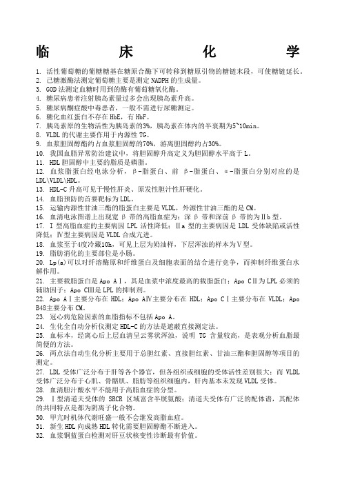
临床化学1. 活性葡萄糖的葡糖糖基在糖原合酶下可转移到糖原引物的糖链末段,可使糖链延长。
2. 己糖激酶法测定葡萄糖主要是测定NADPH的生成量。
3. GOD法测定血糖时用到的酶有葡萄糖氧化酶。
4. 糖尿病患者注射胰岛素量过多会出现胰岛素升高。
5. 糖尿病酮症酸中毒患者,一般不需进行尿糖测定。
6. 糖化血红蛋白不存在HbE,有HbF。
7. 胰岛素原的生物活性为胰岛素的3%,胰岛素在体内的半衰期为5~10min。
8. VLDL的代谢主要作用于内源性TG。
9. 血浆胆固醇酯约占血浆胆固醇的70%,游离胆固醇约占30%。
10. 我国血脂异常防治建议中,将胆固醇升高定义为胆固醇水平高于L。
11. HDL胆固醇中主要的脂质是磷脂。
12. 血浆脂蛋白经电泳分析,β-脂蛋白、前β-脂蛋白、α-脂蛋白分别对应的是LDL\VLDL\HDL。
13. HDL-C升高可见于慢性肝炎、原发性胆汁性肝硬化。
14. 血脂预防的首要靶标为LDL。
15. 运输内源性甘油三酯的脂蛋白主要是VLDL,外源性甘油三酯的是CM。
16. 血清电泳图谱上出现宽β带的高脂血症为;深β带和深前β带的为Ⅱb型。
17. I型高脂血症的主要病因LPL活性降低;Ⅱa型的主要病因是LDL受体缺陷或活性降低;Ⅳ型主要病因是VLDL合成亢进。
18. 血浆至于4度冷藏10h,可见上层为奶油样,下层浑浊的样本为Ⅴ型。
19. 脂肪消化的主要部位是小肠。
20. Lp(a)可以对纤溶酶原和纤维蛋白及细胞表面的结合进行竞争,而抑制纤维蛋白水解作用。
21. 主要载脂蛋白是Apo AⅠ,其是血浆中浓度最高的载脂蛋白;Apo CⅡ为LPL必须的辅助因子;Apo CⅢ是LPL的抑制剂。
22. Apo AⅠ主要分布在HDL;Apo AⅣ主要分布在HDL;Apo CⅠ主要分布在VLDL;Apo B48主要分布CM。
23. 冠心病危险因素的血脂指标不包括Apo A。
24. 生化全自动分析仪测定HDL-C的方法是遮蔽直接测定法。

临床医学检验技术(士):临床化学常用分析技术考点巩固真题一1、单选下列何种情况干扰血清总蛋白的双缩脲法()A.血清呈乳糜状B.高血糖C.高血钾D.高血钠E.高血钙正确答案:A参考解析:乳糜血影响样品空白吸光度的测定(江南博哥),故对一般定量测定方法有影响。
2、单选血清钠的检测()A.原子吸收分光光度法B.电化学分析技术C.散射比浊法D.荧光分析法E.电泳技术正确答案:B参考解析:锌的检测常采用原子吸收分光光度法;DNA的检测常采用荧光分析法;蛋白质的分离与鉴定常采用电泳技术;血清钠的检测常采用电化学分析技术;微量蛋白的检测常采用散射比浊法。
3、单选蛋白质的定量分析中,下列何种方法灵敏度最高()A.酶联免疫法B.比色法C.散射免疫比浊法D.透射免疫比浊法E.化学发光法正确答案:E参考解析:化学发光法可以测到pg甚至fg水平,灵敏度最高。
4、单选下列试验中血标本受溶血影响最小的是()A.K+测定B.LD测定C.血清铁测定D.Na+-测定E.AST测定正确答案:D参考解析:本题正确答案为D。
5、单选符合朗伯比尔定律的有色溶液稀释时,其最大吸收峰的波长位置改变是()A.不移动,但峰高值降低B.不移动,但峰高值增大C.向长波长方向移动,且峰高值增大D.向短波长方向移动,且峰高值增大E.向短波长方向移动,且峰高值降低正确答案:A参考解析:溶液中溶质不变则最大吸收峰波长不变,峰高值与溶液浓度成正比。
6、单选琼脂糖凝胶电泳用pH值为8.6的巴比妥缓冲液可以把血清蛋白质分成五条区带,由正极向负极数起,它们的顺序是()A.白蛋白、α1-球蛋白、α2-球蛋白、β-球蛋白、γ-球蛋白B.白蛋白、α1-球蛋白、α2-球蛋白、γ-球蛋白、β-球蛋白C.白蛋白、β-球蛋白、α1-球蛋白、α2-球蛋白、γ-球蛋白D.白蛋白、β-球蛋白、α1-球蛋白、γ-球蛋白、α2-球蛋白E.α1-球蛋白、α2-球蛋白、β-球蛋白、γ-球蛋白、白蛋白正确答案:A参考解析:琼脂糖凝胶电泳用pH值为8.6的巴比妥缓冲液可以把血清蛋白质分成五条区带,由正极向负极数起它们的顺序是白蛋白、α1-球蛋白、α2-球蛋白、β-球蛋白、γ-球蛋白。
临床化学检验基本操作一、引言临床化学检验是医学实验室中常用的检验方法之一,通过对血液、尿液、体液等样本进行化学成分的分析,可以帮助医生确定患者的健康状况、诊断疾病以及指导治疗方案。
本文将介绍临床化学检验的基本操作流程。
二、样本采集与处理1. 血液样本采集:采用无菌注射器抽取静脉血,要求患者空腹采集,避免进食、饮水和药物的影响。
采集后,将血液样本转移到试管或离心管中,使用离心机离心10分钟,分离血清或血浆。
2. 尿液样本采集:要求患者清洁干净的容器收集中段尿液。
尿液样本应该及时送到实验室进行分析,避免样本变质。
3. 体液样本采集:根据不同的体液类型,采取相应的采集方法。
例如,胸腹水可以通过穿刺吸引的方式采集。
三、常规化学检验1. 血液化学检验:包括血糖、血脂、电解质、肝功能、肾功能等项目。
根据检测项目不同,可选择不同的分析方法,如酶法、光度法、电极法等。
2. 尿液化学检验:主要检测尿常规、尿蛋白、尿糖、尿酮体等项目。
常用的检验方法有比色法、电极法、免疫法等。
3. 体液化学检验:包括胸腹水、脑脊液、关节液等样本的化学成分分析。
常用的检验方法有电解质测定、蛋白质定量、细胞计数等。
四、仪器操作1. 光度计操作:根据不同检测项目的要求,选择适当的波长,并将试管中的样本放入光度计中进行测量。
2. 电极法操作:将电极插入样本中,根据仪器的指示进行测量。
根据不同项目的要求,可能需要进行校准和调整。
3. 离心机操作:根据样本的要求,选择合适的离心速度和时间进行离心。
离心后,将上清液或上清液转移到另一个容器中进行进一步的分析。
五、质量控制1. 内部质量控制:通过每天运行内部质控样本,检验仪器的准确性和稳定性。
根据质控样本的结果,评估仪器的性能,及时调整仪器参数。
2. 外部质量控制:参加外部质量评价计划,定期将样本送到指定实验室进行检验,与其他实验室的结果进行比对,评估实验室的准确性和可靠性。
六、数据分析与结果解释1. 数据分析:根据检验结果,进行数据的统计和分析。
临床化学检测的高通量方法在临床医学领域,化学检测是一种常见而重要的手段,可以通过检测人体内的化学物质来评估健康状况、诊断疾病、监测治疗效果等。
随着科技的不断发展,高通量方法逐渐应用于临床化学检测中,以提高检测速度、准确性和效率。
本文将介绍临床化学检测的高通量方法及其应用。
一、什么是高通量方法高通量方法是指在相同时间内处理大量样本的技术。
在临床化学检测中,高通量方法可以通过同时处理多个样本、自动化操作和并行检测来提高检测效率。
与传统的逐个检测方法相比,高通量方法具有显著的优势,不仅能够节约时间和成本,还可以提高数据的准确性和稳定性。
二、高通量方法在临床化学检测中的应用1. 微生物检测:高通量方法可以应用于微生物检测中,通过快速扩增和检测微生物的核酸序列,可以快速准确地诊断感染病原体,并且对药物耐药性的检测也可以大大提高效率。
2. 药物代谢动力学研究:在药物研发过程中,了解药物的代谢动力学十分重要。
高通量方法可以通过检测药物在体内代谢的速度和产物,为药物设计和剂量调整提供支持。
3. 癌症早期诊断:高通量方法可以在癌症早期诊断中发挥重要作用。
通过检测体内的肿瘤标志物和遗传变异,可以提前发现癌症的存在和发展趋势,有利于及早实施治疗。
4. 遗传性疾病筛查:高通量方法在遗传性疾病筛查中具有广泛应用。
通过检测DNA、RNA和蛋白质的变异,可以迅速准确地筛查出携带遗传疾病的个体,为基因治疗和预防措施提供依据。
5. 新型生物标志物鉴定:高通量方法可以广泛应用于新型生物标志物的鉴定。
通过大规模的样本检测和数据分析,可以发现与疾病相关的潜在标志物,为疾病诊断和治疗提供新的思路和方法。
三、高通量方法的优势和挑战高通量方法的应用在临床化学检测中具有诸多优势。
首先,高通量方法可以同时处理大量样本,提高检测效率和吞吐量。
其次,高通量方法可以自动化操作,减少人为误差,保证数据的准确性和稳定性。
此外,高通量方法可以实现多指标并行检测,为综合分析和结果解读提供全面的信息。
临床医学检验技术(师):临床化学常用分析技术考试答案二1、单选在区带电泳中,能产生电荷效应和分子筛效应的支持介质有()A.淀粉、醋酸纤维素薄膜、纤维素B.纤维素C.硅胶D.淀粉、琼脂糖、聚丙烯酰胺凝胶(江南博哥)E.硅胶、纤维素、醋酸纤维素薄膜正确答案:D参考解析:以淀粉胶、琼脂或琼脂糖凝胶、聚丙烯酰胺凝胶等作为支持介质的区带电泳法称为凝胶电泳。
2、单选测定光束通过溶液混悬颗粒后的光吸收或光散射程度的定量方法是()A.荧光定量分析法B.散射光谱分析法C.发射光谱分析法D.吸收光谱分析法E.原子吸收光度法正确答案:B参考解析:透射和散射光谱分析法主要测定光线通过溶液混悬颗粒后的光吸收或光散射程度,常用法为比浊法,又可称为透射比浊法和散射比浊法。
3、单选超速离心时离心机的转速为()A.4000r/minB.8000r/minC.10000r/minD.20000r/minE.30000r/min正确答案:E4、单选带电化合物的分子大小和形状以及带净电荷的多少等性质可影响迁移率,但带净电荷多少是取决于()A.电泳时的电场强度B.电泳时缓冲液的离子强度C.电泳时缓冲液的pHD.电泳时通电时间的长短E.以上都不是正确答案:C5、单选用于纯化酶和受体蛋白的最好方法()A.盐析法B.电泳法C.亲和层析法D.有机溶剂沉淀法E.吸附分离法正确答案:C参考解析:亲和层析法是利用待分离物质和它的特异性配体间具有特异的亲和力,从而达到分离目的的一类特殊层析技术。
具有专一亲和力的生物分子对主要有:抗原与抗体、DNA与互补DNA或RNA、酶与它的底物或竞争性抑制剂、激素与它们的受体、维生素和它的特异性结合蛋白、糖蛋白与它相应的植物凝集素等。
亲和层析纯化过程简单、高分辨率。
对分离含量极少又不稳定的活性物质尤为有效。
6、单选下列何种方法可用于获得纯蛋白质().A.透析B.亲和层析C.免疫固定电泳D.离子交换层析E.琼脂糖凝胶电泳正确答案:B参考解析:亲和层析是利用待分离物质和它的特异性配体间具有特异的亲和力,从而达到分离的目的,亲和层析可用于纯化生物大分子,稀释液的浓缩、不稳定蛋白质的贮藏、分离核酸等。
医学检验主管检验师资格考试复习资料生物化学(11)临床化学常用分析技术一、光谱分析(分光光度技术)利用各种化学物质所具有的发射、吸收或散射光谱谱系的特征,来确定其性质、结构或含量的技术,称为光谱分析技术。
特点:灵敏、快速、简便。
是生物化学分析中最常用的分析技术。
分类(一)可见及紫外分光光度法分光光度法的理论基础是朗伯-比尔定律。
mber-Beer定律:A=k·b·cA为吸光度k—吸光系数b—光径,单位:cmc—溶液浓度,单位:g/L2.摩尔吸光系数:在公式“A=k·b·c”中,当c=1mol/L,b=1cm时,则常数k可用ε表示。
3.比吸光系数:在公式“A=k·b·c”中,当c为百分浓度(w/v),b为cm时,则常数k可用E%表示,称为比吸光系数或百分吸光系数。
(二)原子吸收分光光度法原子吸收分光光度法是基于元素所产生的原子蒸气中待测元素的基态原子,对所发射的特征谱线的吸收作用进行定量分析的一种技术。
即在一定条件下,原子的吸光度同原子蒸气中待测元素基态原子的浓度成正比。
常用的定量方法有:标准曲线法、标准加入法、内标法。
1.标准曲线法:将一系列浓度不同的标准溶液按照一定操作过程分别进行测定,以吸光度为纵坐标,浓度为横坐标绘制标准曲线。
在相同条件下处理待测物质并测定其吸光度,即可从标准曲线上找出对应的浓度。
由于影响因素较多,每次实验都要重新制作标准曲线。
2.标准加入法:把待测样本分成体积相同的若干份,分别加入不同量的标准品,然后测定各溶液的吸光度,以吸光度为纵坐标,标准品加入量为横坐标,绘制标准曲线,用直线外推法使工作曲线延长交横轴,找出组分的对应浓度。
本法的优点是能够更好地消除样品基质效应的影响,较为常用。
3.内标法:在系列标准品和未知样品中加入一定量样本中不存在的元素(内标元素),分别进行测定。
以标准品与内标元素的比值为纵坐标,标准品浓度为横坐标绘制标准曲线,再根据未知样品与内标元素的比值依曲线计算出未知样品的浓度。
《临床化学》常用分析方法
一、电泳分析
在直流电场中,带电粒子向带符号相反的电极移动的现象称为电泳。
二、光谱分析(分光光度技术)
利用各种化学物质所具有的发射、吸收或散射光谱谱系的特征,来确定其性质、结构或含量的技术,称为光谱分析技术。
具有灵敏、快速、简便等特点,是生物化学分析中最常用的分析技术。
发射光谱分析:荧光分析法和火焰光度法
吸收光谱分析:可见及紫外光分光光度法、原子吸收分光光度法散射光谱分析:比浊法
三、层析技术
层析法是利用不同物质理化性质的差异而建立起来的技术。
所有的层析系统都由固定相和流动相组成。
四、离心技术
离心技术分为制备离心技术和分析离心技术。
制备离心技术主要用于物质的分离、纯化,而分析离心技术主要用来分析样品的组成。
五、电化学分析技术
利用物质的电化学性质,测定化学电池的电位、电流或电量的变化进行分析的方法称为电化学分析法。
包括电位法、电导法、电容量分析法。
离子选择电极法:离子选择电极(ISE)能对某特定离子产生响应,在一定范围内,其电位与溶液中特定离子活度的对数呈线性关系,因此可用离子选择电极来定量分析某些离子的活度或浓度。
代谢物浓度检测技术和血清酶催化活性浓度
一、酶反应动力学原理
酶反应动力学主要研究酶催化反应的过程与速率,以及各种影响酶催化速率的因素,定量时的观察对象是总单位时间内底物的减少或产物增加的量。
当底物[S]>>Km时,公式可近似为V=Vmax;从理论上说只有测定的是酶Vmax,反应速度才和酶量成正比。
二、酶活性的定义及单位
在实验规定的条件下(温度、最适pH、最适底物浓度时),在1分钟内催化1μmol底物发生反应所需的酶量作为1个酶活力国际单位(U)。
习惯用U/L来表示体液中酶活性浓度。
三、测定酶活性浓度的两大类方法
(一)固定时间法(取样法)
(二)连续监测法:酶耦联法(指示酶、辅助酶)
(三)影响酶活性测定的因素
1.底物浓度的影响:底物的种类和浓度。
多种底物中,Km最小的底物往往是此酶的生理底物。
2.反应体系的最适pH、缓冲液的种类和浓度。
3.温度的控制:37℃。
四、代谢物浓度酶法测定
(一)代谢物酶促终点法测定
1.一步法:最简单的底物法测定。
2.酶促耦联法:工具酶(指示酶、辅助酶)
在临床生化测定中,最常用的耦联指示系统有两个:一个是脱氢酶系统,另一个为氧化酶系统。
脱氢酶系统:测定NAD+或NADP+在340nm处的吸光度增高或下降来计算被测物的浓度。
但会受到内源性脱氢酶及其底物如乳酸脱氢酶和乳酸/丙酮酸的干扰。
(二)动力学法测定:实际操作中,测定两个固定时间的吸光度差值,只要此期间待测物消耗<5%,就可以采用标准浓度对照法计算样本浓度,所以动力学法有时又称为固定时间法。
与终点法相比,动态法测定中待测物无须完全转化,故工具酶的用量较少。
产物的堆积和样品色原对动态法影响较小,而对终点测定法影响较大。
如碰到乳糜或溶血,在做终点法测定时,有时需设样本空白。