Synthesis and Characterization of PEG Dimethacrylates and__Their Hydrogels
- 格式:pdf
- 大小:267.58 KB
- 文档页数:8

第52卷第11期 辽 宁 化 工 Vol.52,No.11 2023年11月 Liaoning Chemical Industry November,2023基金项目:广东轻工职业技术学院2021年度大学生科研项目(项目编号:XSKYL202121);广东轻工职业技术学院第二十一届“挑战杯”大学生课外学术科技作品竞赛立项项目;广东轻工职业技术学院2022年度创新创业精致育人项目(项目编号:JZYR202218);广东轻工职 业技术学院2022年度创新创业教育教学改革项目(项目编号:CYJG202210)。
收稿日期: 2022-10-14助剂对聚醋酸乙烯酯乳胶涂料黏度和光泽度的影响范欣蕾,刘颖诗,梁家宪,侯欣桦,佘家康,谢梓良,蔡楚冰,李永莲*,罗媛媛(广东轻工职业技术学院 生态环境技术学院,广东 广州 510300)摘 要:为了研究几种常见助剂对聚醋酸乙烯酯乳胶涂料黏度和光泽度的作用及影响,探索了丙烯酸钠盐、羟乙基纤维素、十二酯醇、OP -10、丙二醇等助剂在不同使用量时聚醋酸乙烯酯乳胶涂料的黏度和光泽度。
结果表明:随着分散剂丙烯酸钠盐用量的增大,乳胶涂料的黏度也随之增大,然后减小,分散剂丙烯酸钠盐的合适用量为6.0 g;增稠剂羟乙基纤维素用量增大时,乳胶涂料的黏度也增大;成膜助剂十二醇酯用量增加时,乳胶涂料的黏度先降低再增加,十二醇酯合适用量为2.0 g;乳化剂OP -10的用量增多时,乳胶涂料的黏度总体有下降趋势,OP -10合适添加量为 0.3 g。
聚醋酸乙烯酯乳胶涂料的光泽度都在2.2%~2.3%之间,各助剂的增减对其影响不大。
关 键 词:聚醋酸乙烯酯乳胶涂料;丙烯酸钠盐;羟乙基纤维素;OP -10;十二酯醇;黏度;光泽度 中图分类号:TQ633 文献标识码: A 文章编号: 1004-0935(2023)11-1581-04聚醋酸乙烯酯(PVAc)乳胶涂料,是一种重要的乳液胶黏剂,其优点很多,例如原料来源丰富且价格低廉、操作工艺简单、初期黏接强度高等。


基因载体peg化壳聚糖的制备及其表征文章标题:《深度探究基因载体peg化壳聚糖的制备及其表征》一、引言在生物医学领域中,基因载体peg化壳聚糖作为一种新型的载体材料,具有良好的生物相容性和可降解性,被广泛应用于基因转染和基因治疗领域。
本文将深度探究基因载体peg化壳聚糖的制备方法及其表征技术,旨在帮助读者全面了解该载体材料的特性和应用。
二、基因载体peg化壳聚糖的制备1. 壳聚糖的选择与预处理在制备基因载体peg化壳聚糖时,首先需要选择合适的壳聚糖原料,并进行预处理,如脱乙酰化处理、碱水解处理等,以提高其水溶性和生物相容性。
2. PEG化修饰将经过预处理的壳聚糖与聚乙二醇(PEG)进行共价结合或物理混合,形成peg化壳聚糖,通过调节PEG的分子量和壳聚糖与PEG的摩尔比,可以控制基因载体的粒径和稳定性。
3. 表面修饰利用化学交联或其他表面修饰技术,改善peg化壳聚糖的荷电性和靶向性,提高其在细胞内的基因递送效率。
三、基因载体peg化壳聚糖的表征1. 粒径和分布利用动态光散射技术(DLS)和透射电子显微镜(TEM)对peg化壳聚糖基因载体的粒径和分布进行表征,以评估其在基因递送过程中的稳定性和渗透性。
2. 荷电性通过电泳方法测定peg化壳聚糖基因载体的电荷密度,并利用zeta电位仪测定其在不同pH值下的表面电位,以评估其在体内外环境中的稳定性和荷电性。
3. 体外释放动力学借助离心管超滤、高效液相色谱等技术,对peg化壳聚糖基因载体在不同条件下的基因释放动力学进行表征,以评估其在基因递送过程中的缓释效果和递送效率。
四、个人观点与展望基因载体peg化壳聚糖作为一种多功能的生物医用材料,具有广阔的应用前景。
在未来的研究中,可以深入探究其在肿瘤治疗、基因编辑和干细胞治疗等领域的应用,开发更加高效和安全的基因递送系统,为临床治疗提供更多选择。
五、总结本文从基因载体peg化壳聚糖的制备和表征两个方面进行了全面的探讨,介绍了其制备方法、表征技术及应用前景。
上转换发光材料在太阳能电池中的应用鲁明11S009078概要:本文通过对上转换发光材料在燃料敏化太阳能电池中的应用的学习,主要介绍二氧化钛上转换发光粉制备,并对其在染料敏化电池上的应用进行研究。
一背景目前染料敏化太阳能电池的最高光电转换效率可达11%以上[1,2]。
料敏化太阳能电池主要以N3, N-719 染料作为敏化剂[1~5], 与TiO2组成光阳极, 对太阳光进行吸收并转换为电能. 染料分子可被视为“电子泵”, 这个泵的驱动力就是太阳光, 常用的N3, N-719 染料对太阳光的吸收范围主要在290~700 nm 之间[3~5], 但对占太阳光全部能量高达43%的红外光却利用甚微, 所以利用光转换材料将红外光转换为染料可以充分吸收利用的可见光对于提高电池的光电转换效率具有重要的意义. 由于稀土离子的特殊性质, 即存在未充满的4f 壳层, 具有丰富的能级和4f 电子的跃迁特性, 使稀土发光成为研究的热点, 并在荧光、监视器、X 射线成像、光通信等领域得到了广泛的应用[6~10]。
本文通过水热法制备Er3+和Yb3+共掺的TiO2:(Er3+, Yb3+)上转换发光粉, 将其组装在染料敏化太阳能电池中, 使原来利用率极低的红外光转化为染料可以充分吸收的可见光, 从而提高电池的光电转化效率。
二实验原料与仪器二氧化钛(P25), 钛酸四正丁酯、异丙醇、聚乙二醇(分子量20000)、碘、碘化锂、4-叔丁基吡啶(TBP)、OP 乳化剂(Triton X-100)、氧化铒、氧化镱、氢氧化锂, 均为分析纯, 购于中国医药集团上海化学试剂公司; 敏化染料N-719; 可控温磁力搅拌器(C-MAG HS4, 德国IKA); 马弗炉(上海实验电炉厂); 短弧氙灯/汞灯稳流电源(CHF-XM-500W, 北京畅拓科技有限公司)、电化学分析仪/工作站CHI660C(上海辰华仪器有限公司); 导电玻璃基片(FTO,北京建筑材料研究院); X 射线粉末衍射仪(D8-advance); 紫外-可见分光光度计(UV-2550); FSP920 荧光光谱仪。

聚乙二醇二羧酸水解聚乙二醇二羧酸(Polyethylene Glycol Diacid, PEG-DA)是一种重要的化学物质,在医药、化妆品和工业领域都有广泛的应用。
聚乙二醇二羧酸的水解性质使其在药物传递系统、聚合物材料和润滑剂等领域发挥了重要的作用。
本文将从深度和广度的角度来评估聚乙二醇二羧酸的水解特性,并探讨其在不同领域的应用。
一、什么是聚乙二醇二羧酸?聚乙二醇二羧酸是一种聚合物,由聚乙二醇和二羧酸组成。
聚乙二醇是一种具有良好溶解性和润滑性的化学物质,而二羧酸是一种含有两个羧基(-COOH)的有机酸。
将这两种化合物结合在一起形成聚乙二醇二羧酸,可以带来有趣的水解性质。
聚乙二醇二羧酸的水解性质使其在药物传递系统中发挥了重要的作用。
聚乙二醇二羧酸可以被人体酶解为多个单体,并在体内逐渐释放出活性物质。
这种控制释放的特性使得聚乙二醇二羧酸成为制造缓释药物的理想载体。
二、聚乙二醇二羧酸的应用1. 聚乙二醇二羧酸在医药领域的应用聚乙二醇二羧酸在医药领域有多种应用。
它可以被用作药物传递系统的载体,控制药物的释放速度。
在一些肿瘤治疗方面,聚乙二醇二羧酸可以帮助将药物定向释放到肿瘤组织中,减少对正常组织的损伤。
聚乙二醇二羧酸还可以用于制备人工关节和缓解关节炎等疾病的治疗。
2. 聚乙二醇二羧酸在化妆品领域的应用聚乙二醇二羧酸在化妆品领域也有广泛的应用。
由于其良好的润滑性和保湿性,聚乙二醇二羧酸可以用于制造护肤品、洗发水和沐浴露等产品。
它还可以作为防晒剂和染发剂的成分,为消费者提供更好的化妆效果和舒适感。
3. 聚乙二醇二羧酸在工业领域的应用除了医药和化妆品领域,聚乙二醇二羧酸在工业领域也有各种应用。
它可以用作聚合物材料的添加剂,提高聚合物材料的性能和可塑性。
聚乙二醇二羧酸还可以用于制造润滑剂、染料和塑料等产品。
三、个人观点和理解我个人认为聚乙二醇二羧酸的水解特性为其在医药、化妆品和工业领域的应用提供了诸多可能性。

阳离子水性封闭型聚异氰酸酯交联剂的制备与性能孙祥;韦军【摘要】以异佛尔酮二异氰酸酯(IPDI)和三乙醇胺(TEOA)制备支化型异氰酸酯基(-NCO)封端的预聚体,分别以N-甲基二乙醇胺(MDEA)和聚乙二醇(PEG)为亲水扩链剂,最后用二甲基吡唑(DMP)封闭剩余的-NCO基团得到阳离子水性封闭型聚异氰酸酯交联剂,并用傅里叶变换红外光谱(FT-IR)表征了交联剂的结构.主要研究了预聚反应时异氰酸酯指数(R1值)以及亲水扩链剂的种类对交联剂性能的影响.研究表明:亲水扩链剂的种类对交联剂的解封温度有显著的影响,当以MDEA为亲水扩链剂、R1值为1.9时,交联剂的性能达到最佳.【期刊名称】《涂料工业》【年(卷),期】2016(046)005【总页数】5页(P60-64)【关键词】阳离子;水性;封闭聚异氰酸酯;交联剂【作者】孙祥;韦军【作者单位】常州大学材料科学与工程学院,江苏常州213164;盐城工学院材料工程学院,江苏盐城224051【正文语种】中文【中图分类】TQ630.4+3随着人们环保意识的加强,水性涂料越来越受到关注,水性封闭型聚异氰酸酯交联剂作为水性涂料固化剂以其安全稳定,环境友好等特点[1-2]被广泛应用于汽车、家具等的涂层、丝绸以及粘合剂等领域[3-6]。
水性封闭型聚异氰酸酯交联剂主要是在非亲水预聚体中接入部分亲水性基团使整个分子达到亲水的效果,而且封闭型聚异氰酸酯可以将活性异氰酸酯基团(—NCO)保护起来,避免其与空气中的水反应,高温释活后的—NCO可重新与活泼氢发生反应,从而提高产品的物理性能[7-9]。
根据亲水基团的离子性质,水性封闭型聚异氰酸酯交联剂可分为阳离子型、阴离子型以及非离子型,其中,阳离子型聚异氰酸酯交联剂由于具有良好的润湿性、抗菌防霉等特点,在化纤整理与复合[10]、木器涂料[11]和阴极电泳漆[12]等领域有着广阔的应用前景,但目前针对阳离子型聚异氰酸酯交联剂的研究报道相对较少。
PET薄膜用水性聚氨酯胶黏剂的制备宋有信;鲍俊杰;陶灿;许戈文;黄毅萍;程芹【摘要】以聚对苯二甲酸-3-甲基-1,5-戊二醇酯二醇(TPA-1000)、聚乙二醇(PEG-2000)为软段、异佛尔酮二异氰酸酯(IPDI)、一缩二乙二醇(DEG)、2,2-二羟甲基丙酸(DMPA)为硬段,合成了一系列水性聚氨酯胶黏剂,用于PET薄膜的粘接.用傅里叶变换红外光谱(FTIR)表征了聚氨酯的结构,同时对聚氨酯胶膜进行了拉伸、耐水性、DSC和T型剥离等测试.结果表明,随着聚乙二醇含量的减少,大大增加了胶膜的力学性能和耐水性能,胶膜的热稳定性能有所提高,粘接强度先增加后减小,质量比TPA-1000:PEG-2000=1:1时,T型剥离强度达到最大值4.55 N/25 mm.%A series of waterborne polyurethane adhesive for PET film were synthesized using poly -to-ben-zene-two-methyl anhydride-3-methyl-1,5-pentanediol ester glycol(TPA-1000)and polyethylene glycol (PEG-2000)as soft segments,isophorone diisocyanate(IPDI),diethylene glycol(DEG)and 2,2-dimethylol propionic acid(DMPA)as hard segments.They were using for PET films bonding.The structure of polyurethane films were characterized by Fourier transform infrared spectroscopy(FTIR).At the same time, the polyurethane films was subjected to tensile test,water resistancetest,differential scanning calorimetry (DSC)and T type stripping test.The results show that with the decrease of the content of polyethylene glycol,the mechanical properties and water resistance of the films increase greatly;The thermal stability of the films increase and the bond strength increase first and then decrease.The results show that when the mass ratioTPA-1000:PEG-2000=1:1,the bonding strength reach a maximum of 4.55 N /25 mm.【期刊名称】《应用化工》【年(卷),期】2018(047)004【总页数】4页(P737-740)【关键词】聚对苯二甲酸-3-甲基-1,5-戊二醇酯二醇;聚对苯二甲酸乙二醇;聚乙二醇;水性聚氨酯;粘接强度【作者】宋有信;鲍俊杰;陶灿;许戈文;黄毅萍;程芹【作者单位】安徽大学化学化工学院安徽省绿色高分子材料重点实验室水基高分子材料安徽省工程技术研究中心,安徽合肥 230601;安徽大学化学化工学院安徽省绿色高分子材料重点实验室水基高分子材料安徽省工程技术研究中心,安徽合肥230601;安徽大学化学化工学院安徽省绿色高分子材料重点实验室水基高分子材料安徽省工程技术研究中心,安徽合肥 230601;安徽大学化学化工学院安徽省绿色高分子材料重点实验室水基高分子材料安徽省工程技术研究中心,安徽合肥230601;安徽大学化学化工学院安徽省绿色高分子材料重点实验室水基高分子材料安徽省工程技术研究中心,安徽合肥 230601;安徽大学化学化工学院安徽省绿色高分子材料重点实验室水基高分子材料安徽省工程技术研究中心,安徽合肥230601【正文语种】中文【中图分类】TQ311作为多用途的环保材料,水性聚氨酯(WPU)由于其优异的性能,广泛应用于织物整理剂、胶黏剂、涂料等领域[1-3]。
两亲性纳米胶束载药系统的研究进展摘要本文综述了由两亲性共聚物制备纳米胶束用于载药系统的研究进展,并进一步介绍这些载药系统的优点及应用。
关键词两亲性共聚物纳米胶束前言两亲性共聚物是同时含有亲油性与亲水性高分子链段的大分子物质只有独特的溶液性质,聚集特性,表面活性,生物相容性,溶液选择性等。
两亲性高分子在选择性溶剂中发生微相分离,可以形成具有疏溶剂核与溶剂化壳的自组装结构——聚合物纳米胶束[1]是研究得较多的一种非常重要的药物载主要用于对疏水难溶药物的增溶作用。
在肿瘤的治疗上目前采用的主要是化疗,即利用化学药物杀、抑制肿瘤细胞的生长繁殖和促进肿瘤细胞的分化,但是化疗治疗肿瘤在杀伤肿瘤细胞的同时,也将正常细胞和免疫(抵抗)细胞一同杀灭,化疗依然无法根治肿瘤且药物利用度不高。
肿瘤耐药的机制错综复杂经典的产生耐药的原因是抗肿瘤药物在进入肿瘤组织后无法到达靶细胞内的分子靶点或者无法达到有效的胞内浓度。
而与传统剂型相比,纳米载药体系的优点是粒径10—100nm,能在血液中长时间循环并保持稳定;在靶位表现更好的生物膜穿透性能;可保护核苷酸,防止被核酸酶降解。
具有缓释、控释与靶向给药的特点,提高了生物利用度;降低了毒副作用;增加了药物稳定性;丰富了药物的剂型选择,减少了用药量等在纳米铁微粒表面包覆一层聚合物后,可以固定蛋白质或酶,以控制生物反应。
很多纳米颗粒在体内的吸收和分布具有一定的规律。
如肿瘤血管对纳米颗粒有较高的通透性,因此可用纳米载体携带药物靶向作用于肿瘤组织。
另外,还可以利用纳米载体的一些特异的物理性质向靶位点转运药物。
通过连接特异性抗体和配体介导载体由细胞内吞途径被摄取或通过干扰技术从基因水平减少外排蛋白表达纳米载体能够克服外排蛋白而使更高浓度的药物在胞内蓄积。
另外随着新型刺激响应性材料的出现药物在肿瘤细胞内的释放时间和释放位置可通过采用不同种类和比例的聚合物进行调节也开发出了可同时包载多种药物的纳米载体使药物同时达到肿瘤部位可控制药物释放的纳米载体已成为现实。
journal of materials chemistry b 模板-回复Journal of Materials Chemistry B 模板是一种用于撰写材料科学领域论文的标准格式。
这篇文章将按照指定的主题,逐步回答问题,并在文章的结构中使用Journal of Materials Chemistry B 模板。
Introduction (简介):在这一部分,我们将简要介绍文章的主题,并解释为什么这个主题在材料科学领域中有重要意义。
Materials and Methods (材料与方法):在这一部分,我们将描述所用到的材料和实验方法。
我们将解释材料的来源、实验的具体步骤以及所采用的测量和分析技术。
Results and Discussion (结果与讨论):在这一部分,我们将展示并讨论实验结果。
我们将逐步回答一些问题,例如我们的实验结果是否符合预期,我们的实验结果与先前的研究结果有何不同之处,和我们的实验结果的潜在应用等。
Conclusion (结论):在这一部分,我们将总结文章的主要发现,并指出未来可能的研究方向。
References (参考文献):在这一部分,我们将列出我们在文章中引用的相关文献。
根据上述Journal of Materials Chemistry B 模板的指导,我们将逐步回答问题。
Introduction (简介):本文将着眼于探讨一种新型材料的合成与应用。
这种材料被广泛认为在光电设备方面具有巨大的潜力。
通过研究这种材料,我们可以更好地了解其性质和可能的应用,并为光电器件的发展做出贡献。
Materials and Methods (材料与方法):我们选择的材料是一种新型有机小分子材料。
该材料在市场上容易获得,我们将通过一系列的实验来改变其化学结构,以改善其光电性能。
我们首先确认了材料的纯度,并使用X射线衍射(XRD)和扫描电子显微镜(SEM)对其形貌和晶体结构进行了表征。
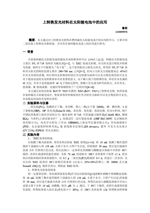
Synthesis and Characterization of PEG Dimethacrylates andTheir HydrogelsSheng Lin-Gibson,*,†Sidi Bencherif,†James A.Cooper,†Stephanie J.Wetzel,†Joseph M.Antonucci,†Brandon M.Vogel,†Ferenc Horkay,‡and Newell R.Washburn†Polymers Division,National Institute of Science and Technology,Gaithersburg,Maryland20899-8543,and Section on Tissue Biophysics and Biomimetics,Laboratory of Integrative and Medical Biophysics,NICHD, National Institutes of Health,Bethesda,Maryland20892Received February26,2004Facile synthesis and detailed characterization of photopolymerizable and biocompatible poly(ethylene glycol) dimethacrylates(PEGDM)and poly(ethylene glycol)urethane-dimethacrylates(PEGUDM)are described. Poly(ethylene glycol)s of various molecular masses(M n)1000to8000g/mol)were reacted with methacrylic anhydride or with2-isocyanatoethyl methacrylate to form PEGDMs and PEGUDMs,respectively.PEGDMs were also prepared by a microwave-assisted route to achieve fast reaction conversions under solvent free bined analyses of1H NMR and MALDI-TOF MS confirmed the formation of prepolymers of high purity and narrow mass distribution(PD<1.02).Aqueous solutions of the PEGDMs and PEGUDMs (10%and20%by mass fraction)were photopolymerized to yield hydrogels.Bovine chondrocytes,seeded in the hydrogels,were used to assess the biocompatibility.Preliminary rheology and uniaxial compression measurements showed varied mechanical response,and biocompatibility studies showed that cells are completely viable in both types of hydrogels after two weeks.IntroductionHydrogels produced by photopolymerization have been investigated extensively as biomaterials in applications such as scaffolds for tissue engineering,drug delivery carriers,in the prevention of thrombosis,post-operative adhesion forma-tion,and as coatings for biosensors.1The photopolymeriza-tion process allows the hydrogel to be generated in vitro or in vivo from a low viscosity solution of monomer,oligomer, or low molecular mass polymer(macromer)by a free radical pathway in a minimally invasive manner.The chemical cross-linking results in hydrogels that contain a high water content yet possess mechanical properties similar to those of soft tissues.Another advantage of hydrogels is their high permeability to oxygen,nutrients,and other water-soluble metabolites,making them particularly attractive as scaffolds in tissue engineering applications.Although hydrogels have been studied as potential materi-als for bone,tendon,and nerve regeneration,it is cartilage tissue engineering that has shown the most promise.Chon-drocytes encapsulated in hydrogels retain their native form, and over time can generate native cartilage tissue.The use of photopolymerized hydrogels as opposed to natural physical gels such as alginate also allows for the material properties to be more easily adjusted.For example,a typical approach to control the hydrogel mechanical properties is to tailor the network cross-link density.This can be achieved by adjusting the molecular mass of the macromer or by varying the mass percent of macromer in the solutions.The cross-link density in fully cross-linked networks is directly proportional to the gel modulus and inversely proportional to the swelling.These are important considerations for tissue engineering in which the former affects transport properties and the latter deter-mines the materials functional practicality and influences cell behavior.In drug delivery applications,the pore or mesh size can be adjusted to control the drug release rate by varying the content,density,and length of the cross-linking groups.Several types of photopolymerizable hydrogels have been investigated for use as biomaterials.These include poly-(ethylene glycol)(PEG)acrylate derivatives,PEG meth-acrylate derivatives,poly(propylene fumarate-co-ethylene glycol)2and oligo(poly(ethylene glycol)fumarate)3that contains cross-linkable sites in the polymer backbone,poly-(vinyl alcohol)derivatives,4modified polysaccharides such as those from hyaluronic acid,and dextran methacrylates. We are particularly interested in PEG dimethacrylates (PEGDM)and similar PEGDM derivatives as model systems because PEG alone is bio-inert but can be easily modified to become bioactive.5Cross-linking by dimethacrylates have been shown to be biocompatible with the unreacted dimeth-acrylates having relatively low cytotoxicity.6,7In addition, PEGDMs and their copolymers and derivatives have been successfully used by several groups both in vitro and in vivo as scaffold materials.8There is a general consensus that the material properties and external stimulation strongly affect the cell response. The importance of PEGDM hydrogel cross-link density (controlled by PEGDM mass fraction in solution)on me-*To whom correspondence should be addressed.E-mail:slgibson@.†National Institute of Science and Technology.‡National Institutes of Health.1280Biomacromolecules2004,5,1280-128710.1021/bm0498777CCC:$27.50©2004American Chemical SocietyPublished on Web04/21/2004chanical properties and on the chondrocytes’ability toproduce cartilaginous tissues has been demonstrated byBryant and Anseth.9In addition,PEGDM co-photopolymer-ized with a degradable macromer,acrylate endcapped poly-(lactic acid)-b-poly(ethylene glycol)-b-poly(lactic acid)in thepresence of chondrocytes shows that biodegradable moietiesin hydrogels have significant effects in tissue generation.Despite the large number of studies currently available,thereis still a lack of a clear understanding of the correlationbetween material properties and cell response.Furthermore,after years of research,the physical properties of hydrogelsare still difficult to predict by theories due to nonidealitiesof the gel formation.These nonidealities include conversiondependent reactivity,cyclization and multiple cross-linking,and defects and nonhomogeneous cross-linking(also knownas spatial gel inhomogeneity).Well-defined model materialsare necessary for the preparation of hydrogels with highreproducibility and easily adjustable properties.We have prepared a series of controlled molecular mass(MM)PEGDMs and poly(ethylene glycol)urethane dimeth-acrylates(PEGUDM)of high purity and low polydispersity.PEGDMs were prepared both in solution and under solventfree conditions via a microwave-assisted route.The syntheticapproaches described herein are particularly straightforward.The dimethacrylate products were characterized by protonnuclear magnetic resonance(1H NMR)and matrix-assistedlaser desorption ionization time-of-flight mass spectrometry(MALDI-TOF MS).PEGDMs and PEGUDMs of differentmolecular masses were photocrosslinked to form hydrogels,and preliminary cell viability studies were conducted.Thematerial structure-property relationships and detailed cellresponse studies will be described in a later paper.Experimental Section15Materials.PEG(MM≈1000(1k)to8000g/mol(8k)),methacrylic anhydride(MA),2-isocyanatoethyl methacrylate(IEM),ethyl ether,and triethylamine(TEA)were purchasedfrom Sigma-Aldrich and used as received.Dichloromethanewas purchased from Sigma-Aldrich and dried over activatedmolecular sieves(4Å)prior to use.Photoinitiator Irgacure2959(I2959)was obtained from Ciba Specialty Chemicalsand used as received.Primary bovine chondrocytes werecultured in growth medium composed of Dulbecco’s modi-fied Eagle medium,10%fetal bovine serum,1%minimumessential medium(GIBCO,Invitrogen Corp),50µg/mL L-ascorbic acid2-phosphate(Sigma),and1%antibiotics (penicillin/streptomycin)(Mediatech,Inc.).Cell viability wasmeasured using Live/Dead Viability/Cytotoxicity Kit(L-3224)purchased from Molecular Probes Inc.Synthesis of PEGDM and PEGUDM.PEGDM andPEGUDM were prepared from the reaction of various PEGsand MA or IEM,respectively.An example of the synthesisof a5k PEGDM is as follows.PEG(5g,≈0.001mol),2.2equiv of MA(0.34g,0.0022mol),and TEA(0.2mL)werereacted in≈15mL of dichloromethane over freshly activatedmolecular sieves(≈3g)for4d at room temperature.Thesolution was filtered over alumina and precipitated into ethylether.The product was filtered and then dried in a vacuumoven overnight at room temperature.Microwave-Assisted Synthesis of PEGDM.PEG(0.2g) and a large excess of MA(up to5-fold excess)were mixed in a capped scintillation vial and placed in a commercial domestic microwave(GE,1100W)for various reaction times ranging from2to10min.Once the vial was cooled to room temperature,approximately2mL of ethyl ether was added and the vial was gently shaken to allow the PEGDM to precipitate.For the1k PEGDM,the vial was placed in a freezer to facilitate the precipitation process.Product was collected by filtration and dried in a vacuum oven. Characterization PEGDM and PEGUDM.High-resolu-tion,270MHz proton NMR spectra were taken on a6.35T JEOL GX270spectrometer manufactured by JEOL,Ltd. (Akishima,Japan).Deuterated chloroform was used as a solvent,and the polymer concentrations were varied between 2.5%and3.0%by mass fraction.All spectra were run at room temperature,15Hz sample spinning,45°tip angle for the observation pulse,and a10s recycle delay,for64scans. The standard relative uncertainty for molecular mass calcu-lated via1H NMR arises from the choice of baseline and is estimated to be8%.The MALDI matrix,dihydrobenzoic acid(DHB),and the PEGDM and PEGUDM were dissolved in1mL of THF. Sodium was used as the cationizing reagent in a1:1by volume ratio of THF solution(0.5mg/mL solution in THF) and PEGDM or PEGUDM/DHB solution.All MALDI samples were deposited on the target by electrospray.The MALDI-TOF MS was performed on a Bruker(Billerica, MA)REFLEX II in reflectron mode using delayed extraction and low-mass(i.e.,matrix-ion)blanking as previously described.10Each spectrum shown is the sum of75discrete laser shots and is shown without smoothing or background subtraction.Estimated expanded uncertainty reported for MM moments arises from the choice of baseline and laser power (5%).The estimated standard uncertainty in overall signal intensity from repeatability studies is15%.Preparation of Hydrogels.Photopolymerized hydrogels were prepared according to a previously described proce-dure.9PEGDM or PEGUDM(10%or20%by mass fraction) and aqueous I2959solution(0.05%by mass fraction)were mixed in distilled deionized water or growth medium when chondrocyte is encapsulated in the hydrogel.Cylindrical samples of3mm in height and6mm in diameter were cured with a long wavelength UV source(365nm,300µW/cm2) for10min to obtain hydrogels.All hydrogels maintain their structural integrity for the entire time under static culture. Bovine chondrocytes were seeded into hydrogels at a cell density of17000cell/mL to100000cell/mL gel.Cell viability within the cell-hydrogel scaffolds under static cultures was measured at14d.Characterization of Hydrogels.FTIR was used to measure the bulk reaction kinetics.The methacrylic vinyl contents of PEGDM were analyzed by Fourier transform infrared spectroscopy.The infrared samples were films, approximately0.3mm thick,prepared by solvent casting a film from a solution of PEGDM and photoinitiator in dichloromethane.Spectra were recorded at2cm-1resolution on a Magna System550FTIR(Nicolet Instrument Tech-PEG Dimethacrylates and Their Hydrogels Biomacromolecules,Vol.5,No.4,20041281nologies,Madison,WI)equipped with a DTGS detector.The co-addition of 64scans gave adequate signal-to-noise.In situ rheology measurements were used for assessing the reaction kinetics and shear modulus.Rheological mea-surements were performed on a stress-controlled Rheometrics SR-5000rheometer in a parallel plate configuration (quartz plates,40mm diameter).The instrument was raised onto a platform,and a quartz Pen-Ray 5.5W mercury UV lamp was mounted under the bottom plate,allowing in-situ monitoring of the hydrogel formation.For these studies,a multi-wave UV lamp was used.Aqueous PEGDM or PEGUDM solutions containing 0.05%I2959photoinitiator by mass fraction were loaded into the rheometer.The sample was exposed to UV for a short time (1-2min)to allow the sample to cure without external perturbation.The late time cure was monitored by measuring the storage and loss modulus (G ′and G ′′,respectively)at 1rad/s and 1Pa as a function of reaction time.Duplicate experiments showed excellent reproducibity with relative standard uncertainty of 3%.The shear modulus was also determined using uniaxial compression measurements performed using a TA.XT2I HR Texture Analyzer (Stable Micro Systems,U.K.).This ap-paratus measures the deformation ((0.001mm)as a function of an applied force ((0.01N).Cylindrical hydrogels (height 3mm,diameter 6mm)were deformed (at constant volume)between two parallel glass plates.The shear modulus,G ,was calculated from the nominal stress,σ(force per unit undeformed cross-section),using the equation 11where Λis the macroscopic deformation ratio (Λ)L /L 0,L and L 0are the lengths of the deformed and undeformed specimen,respectively).Measurements were carried out in triplicate at deformation ratios 0.6<Λ<1.No volume change or barrel distortion was detected.Cell viability was determined as follows.The Calcein AM and Ethidium homodimer-1were added to chondrocyte media at a concentration of 2µM/mL.This live/dead assay was then added to the cell-seeded hydrogels and incubated at 37°C in 5%CO 2for 5min.Live and dead cells were imaged using an inverted microscope Eclipse TE 300with a TE-FM Epi-Fluorescence attachment (Nikon,Inc.).Digital pictures were taken using a Nikon Coolpix 990digital camera.Results and DiscussionsSynthesis and Characterization of PEGDM and PE-GUDM.PEGDMs and PEGUDMs of high purity and lowpolydispersity are prepared as model materials for the formation of photocrosslinkable hydrogels.The PEG hy-droxyl endgroups react with methacrylic anhydride to form PEGDM or with 2-isocyanatoethyl methacrylate to form PEGUDM (Figure 1).Urethane linkages in the PEGUDM are incorporated as an approach to enhance the hydrogen bonding in hydrogels,thus providing an additional adjustable parameter controlling the material properties.For the solution synthesis of PEGDM,we choose to use the less reactive methacrylic anhydride as opposed to the more commonly used methacryloyl chloride.The byproduct formed in the reaction of PEG with methacrylic anhydride is methacrylic acid,rather than the triethylamine/HCl salt formed in the reaction with methacrylic chloride.Since methacrylic acid can be removed more easily than the salt byproduct,methacrylic anhydride was used in these reactions.This allows for a straightforward oligomer purification process,which consists of one filtration through alumna followed by a single precipitation with diethyl ether.Given sufficient reaction time,only a slight excess of methacrylate anhydride relative to PEG hydroxyl is necessary to achieve quantitative conversion.Proton NMR and MALDI-TOF MS together provide comprehensive information regarding the degree of meth-acrylate conversion and product purity.Figure 2shows a typical 1H NMR spectrum of PEGDM.PEG has one main chemical shift at δ≈3.64.Under the current measurement conditions,the ethylene glycol penultimate endgroups cannot be differentiated from those of internal ethylene glycol segment.The chemical shift of methylene protons on MA are δ≈5.80and 6.21.Upon reaction of PEG,these protons shift to δ≈5.57and 6.13(peaks b and c),respectively.Moreover,the protons adjacent to the methacrylategroupsFigure 1.Synthesis of PEGDM andPEGUDM.Figure 2.1H NMR of 3k PEGDM.σ)G (Λ-Λ-2)1282Biomacromolecules,Vol.5,No.4,2004Lin-Gibson et al.shift to δ≈4.30.The 1H NMR spectra for PEGDM shows the expected peaks,but the lack of additional peaks suggests that unreacted methacrylic anhydride,methacrylic acid byproduct,and triethylamine all have been quantitatively removed.MALDI-TOF MS is a powerful technique from which the molecular mass,molecular mass distribution,and endgroup functionalities can be determined.Since MALDI detects all species within a discrete molecular mass range,it can be used to determine the amount of PEGDM versus the amount other impurities,such as PEGs with only one hydroxyl reacted (PEG mono-methacrylate)and unreacted PEG in a mixture.A MALDI-TOF MS spectrum of a typical 1k-PEGDM (Figure 3a)clearly illustrates both the high degree of methacrylate conversion and narrow polydispersity.Upon a closer examination (Figure 3b),three sets of peaks are observed.The main series corresponds to Na +cationized PEGDM.A minor second series of peaks corresponds to K +cationized PEGDM,and the third minor series may be attributed to H +cationized species.H +and K +contamination may occur during sample preparation and is common for the MALDI analysis of PEG and PEG derivatives.12H +is due to the matrix acid,and K +is in the matrix salt.Although the molecular mass calculation for the third series of peaks agrees with H +cationized PEGDM,it also agrees with thosecalculated for Na +cationized PEG mono-methacrylate.One approach to qualitatively differentiate the origin of these peaks is by comparing the M n of these different series.In the scenario that the peaks represent H +cationized PEGDM,the M n of the minor series should only differ from the main series by 22u (MM difference between Na +and H +),where as the M n difference should be higher (MM difference of ≈56u between PEGDM and PEGM)if the third series correspond Na +cationized PEG mono-methacrylate.The MALDI-TOF MS spectra of PEGDMs prepared from different molecular mass PEGs are shown in Figure 4.Intrinsic to MALDI analysis,the relative signal intensities decrease and the breadth of the peak appears to increase as the molecular mass increases.Each molecular mass can be clearly distinguished with all oligomers displaying the expected molecular mass distribution.The degree of conver-sion is quantitatively assessed for each product.As mentioned previously,the combination of 1H NMR and MALDI-TOF MS is necessary to gather the full picture of the product purity and degree of methacrylate conversion.From 1H NMR analyses,the molecular mass of PEGDMs can be calculated by comparing the peak intensities of ethylene glycol protons adjacent to the methacrylate (peak e)to internal ethylene glycol protons (peak d)or by comparing the peak integrations of a methacrylate proton (end group proton)to an ethylene glycol proton.However,since the unreacted PEG hydroxyl groups cannot be distin-guished by 1H NMR due to overlapping with PEG protons,the molecular mass calculation must assume stoichiometric conversion.This is not necessarily true depending on the reaction conditions employed in the synthesis.MALDI provides complementary information as to the amount of dimethacrylate species as well as those of PEG mono-methacrylate and unreacted PEG.On the other hand,PEG and methacrylated PEG derivatives are fragile in the MALDI analysis and could fragment during the laser desorption.The fragmentation may lead to biasing and,therefore,affect the MM calculations.Proton NMR provides confirmation to the MALDI calculations.It is only when the two techniques agree that we can conclude that high reaction conversions have been achieved.The molecular mass results of all PEGDMs are listed in Table 1.For all PEGDMs,the number average molecular masses (M n )obtained by 1H NMRmatchFigure 3.MALDI-TOF MS of 1k PEGDM,(a)full spectra and (b)expansion showing one main set of peaks 44u apart due to Na +cationized PEGDM and two minor sets of peaks of PEGDM cationized by K +and H +,respectively.Figure 4.MALDI-TOF MS of a series of PEGDMs.PEG Dimethacrylates and Their Hydrogels Biomacromolecules,Vol.5,No.4,20041283closely to those calculated by plementary techniques thus conclusively demonstrate the high reaction conversion and low impurity in these dimethacrylates. We have also explored the use of microwaves to prepare PEGDMs.Microwave reactions have gained significant interest recently due to their ability to achieve fast reaction rates often without the need for an organic solvent.13,14In the conventional thermal reaction,energy is transferred to the material through convection,conduction,and radiation. Energy transfers thus rely on diffusion of heat from the surfaces of the material,which leads to nonuniform heating and may cause excess heating at the surface leading to side reactions.Microwave energy can be delivered directly to material through molecular interaction with the electromag-netic field and can increase the local reaction kinetics,which leads to significantly reduced reaction time.Microwave reactions are becoming more widely used in combinatorial chemistry and green(solvent free)chemistry.The use of microwave reaction for the synthesis of PEGDM is particu-larly straightforward.The reaction requires5min to reach completion under microwave irradiation as opposed to4d for solution reactions.In addition,microwave-assisted reac-tions do not require a solvent or catalyst,and the product can be precipitated simply by adding diethyl ether.For the microwave preparation of PEGDMs,the ratio of PEG to MA and optimized reaction times are important in achieving high reaction conversions.Whereas the solution reaction requires only a slight excess of MA relative to PEG, a near stoichiometric conversion by the microwave reaction is facilitated by a larger amount of MA relative to PEG.Table 2shows the effect of reaction time and reagent ratio on the reaction conversion.The effect of reaction time can be compared for the microwave reactions of1k PEGDM with the same reagent stoichiometric ratio of MA to PEG as the solution reaction,i.e.,2.2.The conversion increases with increased reaction time,but does not reach high conversion even after10min reaction time.Clearly the dominating effect in achieving high conversion is the MA to PEG ratio where a monotonic increase in reaction conversion is observed with an increased MA to PEG ratio.At a MA to PEG ratio of10, a near stoichiometric conversion can be achieved after5min. Since the microwave reaction is carried out neat,the lack of molecular mobility may require an excess of MA to be present locally.It is interesting to note that,although the reaction temperature becomes elevated during the microwave reaction,we do not detect cross-linking or any other side reaction in the product.Both the1H NMR and MALDI are nearly identical for PEGDMs prepared by the solvent approach or the microwave-assisted route.The effect of PEG molecular mass on conversion is also evaluated for the microwave-assisted reactions.Table2 shows the percent conversion calculated from the1H NMR results for1k and4k PEGDM reacted using various MA to PEG ratios for the same length of reaction time.Although PEGs are crystalline at room temperature,reaction mixtures are heated above the melting temperatures during microwave irradiation;thus,no significant differences between the reaction conversion for1k and4k PEG are observed. Solution preparation of PEGUDMs is done in a similar manner as the PEGDMs.The isocyanate group on IEM is reacted with PEG hydroxyl groups.Polyurethane and related polyurethane copolymers have been used in various biologi-cal applications,such as heart valve implants;therefore,the urethane linkages used in the current study are expected to have relatively low cytotoxicity.The urethane spacer pro-vides additional hydrogen bonding sites that may enhance the mechanical properties.Moreover,urethane linkages may be degradable under certain conditions.The PEGUDMs prepared from various PEG precursors are also characterized using a combination of1H NMR and MALDI-TOF MS. Figure5shows a typical1H NMR of3k PEGUDM.The expected peaks are observed for the PEGUDM products. The MALDI-TOF MS of PEGUDMs synthesized from various molecular mass PEGs are shown in Figure6.Similar to the PEGDM spectra,the intensities generally decrease and breadth of distribution increases with increased molecular mass.The intensity is slightly higher for the3k PEGUDM than for the2k PEGUDM since a higher reaction conversion is obtained for the3k polymer.The insert of Figure6shows the expanded spectra of the3k PEGUDM.Two series ofTable1.Molecular Mass Results(g/mol)of PEGDM Obtained Using1H NMR and MALDI-TOF MSPEG M n(NMR)M n(MALDI)M w(MALDI)PDI 1k104710641085 1.02 2k222221502178 1.01 3k342432363283 1.01 5k505746304681 1.01 8k833386808776 1.01Table2.Reaction Conversion of PEGDMs Synthesized by the Microwave Process Calculated by1H NMRPEG M n MA/PEG(mol:mol)reaction time(min)%conversion1k 2.22292.25402.21068458410596 4k 2.2537457910599Figure5.1H NMR of a3k PEGUDM.1284Biomacromolecules,Vol.5,No.4,2004Lin-Gibson et al.peaks are clearly distinguishable.The main series with higher peak intensities (S1)corresponds to Na +cationized PEGUDM.Endgroup analysis using the Polymerics software suggests that the minor series (S2)corresponds to Na +cationized PEG mono-urethane methacrylate.This is in agreement with the difference in M n calculated for the two series,which correlates well with the molecular mass difference of an endgroup.1H NMR confirms this peak assignment,and this is described in detail in the following paragraph.Table 3lists the reaction conversions for PEGUDM calculated by 1H NMR and MALDI-TOF MS.From 1H NMR,we can calculate an apparent molecular mass by comparing the methylene protons adjacent to the urethane (peak d)vs internal PEG protons (peak g).Since the molecular mass of PEG is known,we can calculate a theoretical M n of PEGUDM at 100%reaction conversion.It is thus possible to back calculate the reaction conversion by comparing the apparent M n to the theoretical M n .To calculate the reaction conversion from MALDI,the peak integrations of the two series (S1and S2)are first calculated.The total reaction conversion is then the sum of S1and half of S2.As shown in Table 3,the reaction conversions obtained from the very different techniques are statistically identical.It is noted that although the reaction conversions are relative high,the synthesis of PEGUDM does not reach a near stoichio-metric conversion as is the case for the PEGDM reactions.Preparation and Characterization of Hydrogels.The reaction kinetics of the cross-linking of bulk PEGDM and PEGUDM are monitored by FTIR.From the FTIR spectrum,a decrease in the C d C stretch and a shift in the C d O stretch are observed as the methacrylate groups react.As shown inFigure 7a,high vinyl group conversions can be achieved after 15min irradiation for the bulk reactions of PEGDM.Similar studies are carried out for PEGUDMs (Figure 7b).The bulk reaction kinetics appear to be similar for PEGDM andPEGUDM.Figure 6.MALDI-TOF MS of PEGUDMs prepared from different molecular mass PEGs.The insert shows an expanded spectrum of 3k PEGUDM.Table 3.Percent Reaction Conversion of PEGUDM Synthesis Calculated by 1H NMR and MALDI-TOF MSMALDI %conversion PEG NMR %conversionS1S2total 1k 958714932k 806436823k 898317925k 938713948k85663483Figure 7.FTIR of bulk photocure of (a)4k PEGDM and (b)5k PEGUDM monitored as a function of UV exposuretime.Figure 8.Storage modulus measured as a function of UV irradiation time for aqueous PEGDM (10%by mass fraction).PEG Dimethacrylates and Their Hydrogels Biomacromolecules,Vol.5,No.4,20041285Reaction kinetics and gel mechanical properties of hy-drogels are studied by in situ rheological studies.Hydrogels are prepared by loading low viscosity,photoinitiatior-containing,aqueous PEGDM or PEGUDM solutions between two parallel plates followed by irradiation to cross-link the dimethacrylates.Since the solutions are confined between parallel plates,a certain amount of stress is always built into the as-prepared hydrogels.When submersed in water or growth medium,these hydrogels can swell or contract depending on their thermodynamic state.In addition,PEG hydrogels produced by the free radical polymerization have high spatial inhomogeneity.Relationships between gel structures and properties will be the subject of a future paper.In the present study,only preliminary data determined by rheology on the gel modulus as-prepared in the confined stateis discussed.Two measurements are examined for each sample:a time-sweep measurement in which the storage modulus (G ′)and loss modulus (G ′′)are monitored as a function of irradiation time and a subsequent frequency sweep measurement at low strain amplitude.From the time sweep (Figure 8,only G ′is shown for clarity),the reaction kinetics can be qualitatively evaluated.During the course of the reaction,both G ′and G ′′increase,and G ′becomes greater than G ′′once gelation occurs.The storage modulus G ′increases as the reaction progresses for several orders of magnitude and eventually plateaus.The time at which the G ′reaches a plateau provides a rough estimate of the time it takes to complete the photocrosslinking reaction.It should be noted that the light source used in the rheological measurements was of a different intensity and wavelength;therefore,although the trends in the curing rate are correct,the kinetics determined here cannot be used directly to predict the rate of curing under long UV wavelength.The subsequent frequency sweep verifies the formation of a chemically cross-linked gel.It is also of interest to examine the slope of G ′as a function of frequency which provides qualitative information regarding the strength of the gel.All PEGDM and PEGUDM gels show relatively weak frequency depen-dence in the frequency range examined,confirming that the gels are in fact cross-linked and that gels as prepared are mechanically robust.The shear moduli of hydrogels were also measured using a uniaxial compression test and calculated using equations derived from the strain-energy function.Figure 9shows the shear modulus of PEGDM hydrogels prepared from different molecular mass oligomers (2k,4k,and 8k)and as a function of PEGDM mass fraction (varying from 10%to 30%).AsFigure 9.Shear modulus of hydrogels prepared using various PEGDM molecular masses and at various massfractions.Figure 10.Live stain (top)and dead stain (bottom)of PEGDM (left column)and PEGUDM (right column)hydrogels containing bovine chondrocytes.The cell density is 100000cell/mL.1286Biomacromolecules,Vol.5,No.4,2004Lin-Gibson et al.。