胸外科学英文影印版.上册(David C.Sabiston)思维导图
- 格式:xmin
- 大小:5.19 KB
- 文档页数:1


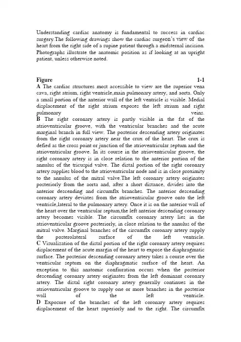
Understanding cardiac anatomy is fundamental to success in cardiac surgery.The following drawings show the cardiac surge on’s view of the heart from the right side of a supine patient through a midsternal incision. Photographs illustrate the anatomic position as if looking at an upright patient, unless otherwise noted.Figure 1-1 A The cardiac structures most accessible to view are the superior vena cava, right atrium, right ventricle,main pulmonary artery, and aorta. Only a small portion of the anterior wall of the left ventricle is visible. Medial displacement of the right atrium exposes the left atrium and right pulmonary veins.B The right coronary artery is partly visible in the fat of the atrioventricular groove, with the ventricular branches and the acute marginal branch in full view. The posterior descending artery originates from the right coronary artery near the crux of the heart. The crux is defied as the cross point or junction of the atrioventricular septum and the atrioventricular groove. In its course in the atrioventricular groove, the right coronary artery is in close relation to the anterior portion of the annulus of the tricuspid valve. The distal portion of the right coronary artery supplies blood to the atrioventricular node and is in close proximity to the annulus of the mitral valve.The left coronary artery originates posteriorly from the aorta and, after a short distance, divides into the anterior descending and circumflx branches. The anterior descending coronary artery deviates from the atrioventricular groove onto the left ventricle,lateral to the pulmonary artery. Once it is on the anterior wall of the heart over the ventricular septum,the left anterior descending coronary artery becomes visible. The circumflx coronary artery lies in the atrioventricular groove posteriorly, in close relation to the annulus of the mitral valve. Marginal branches of the circumflx coronary artery supply the posterolateral surface of the left ventricle.C Visualization of the distal portion of the right coronary artery requires displacement of the acute margin of the heart to expose the diaphragmatic surface. The posterior descending coronary artery takes a course over the ventricular septum on the diaphragmatic surface of the heart. An exception to this anatomic confiuration occurs when the posterior descending coronary artery originates from the left dominant coronary artery. The distal right coronary artery generally continues in the atrioventricular groove to supply one or more branches in the posterior wall of the left ventricle.D Exposure of the branches of the left coronary artery requires displacement of the heart superiorly and to the right. The circumflxbranch of the left coronary artery is in the atrioventricular groove, giving off marginal branches to supply blood to the posterolateral surfaces of the left ventricle. The obtuse marginal branch is usually near the base of the left atrial appendage. The obtuse marginal branch may not be visible on the surface of the heart when it lies within the myocardium. In this situation it can usually be identifid as a slightly yellowish discoloration of the otherwise red-brown myocardium. The anterior descending coronary artery takes a course along the ventricular septum anteriorly, providing one or more diagonal branches to supply blood to the anterolateral surfaces of the left ventricle. The size of the diagonal branches depends on the relative size of the circumflx marginal branches because the blood supply to the lateral wall of the ventricle is shared.Figure 1-2 Coronary veins of the right ventricle drain directly to the right atrium or through the besian veins to the right ventricle. Most of the venous return of the left ventricle is via the coronary sinus. Large cardiac veins located on the posterior surface of the heart collect through a common venous channel in the atrioventricular groove, which is anatomically related to the posterior portion of the annulus of the mitral valve. These veins drain into the right atrium at the coronary sinus. The orifie of the coronarysinusis located in close proximity to the septal portion of the annulus of the tricuspid valve. The sinoatrial node is located on the lateral surface of the right atrium at the junction with the superior vena cava at the origin of the crista terminalis. It occupies a considerable area, often greater than 1 cm in diameter, of the lateral wall of the right atrium.Three interatrial conduction pathways are thought to arise from the sinoatrial node. The anterior and medial interatrial conduction pathways course anterior and posterior to the orifie of the superior vena cava and through the atrial septum, anterior to the foramen ovale. The posterior interatrial conduction pathway follows the crista terminalis and crosses the atrial septum caudal to the foramen ovale on the su perior rim of the coronary sinus. The atrioventricular node is located in the flor of the right atrium at a point approximately one-third the distance along a line from the coronary sinus to the commissure of the anterior and septal leaflt of the tricuspid valve. The bundle of His generally follows this line to penetrate the annulus of the tricuspid valve and enter the ventricular septum just below the membranous portion. The triangle of Koch is bounded by the coronary sinus, the septal attachment of the tricuspid valve,and (laterally) a ridge of tissue referred to as the tendon of Todaro. The commissure of the septal and anterior leaflt of the tricuspid valve is usually well defied. The commissure of the septal and posterior leaflt of the tricuspid valve may be less obvious. This commissural cleft can generally be identifid by the relationships of the chordae tendineae of the papillary muscle attached to the leaflts. The commissure of the anterior and posterior leaflts may also be poorly defied, but this is not of great anatomic importance because these leaflts function as a unit to approximate the septal leaflt.C A magnifid view of the tricuspid valve shows the details of the septal leaflt, with the delicate chordaetendineae and papillary muscle attached. A portion of the posterior leaflt is seen, with the commissure between the posterior and septal leaflts. The coronary sinus is in the lower right corner of the image.D With the atrial septum removed, the anatomic relations of the right and left atrioventricular valves and the aortic valve are delineated at the firous body of the heart. Anatomically, the commissure of the septal and anterior leaflt of the tricuspid valve is closely related to the aortic annulus and the right atrium. Similarly, the noncoronary portions of the annulus of the aortic valve are closely related to the superior wall of the left atrium and the anterior leaf let and annulus of the mitral valve.E With the anterior wall of the right ventricle opened, the crista supraventricularis, with its septal and pari etal bands, separates the tricuspid valve annulus from the annulus of the pulmonary valve. Space between the crista supraventricularis and the pulmonary annulus formsthe infundibular chamber. The papillary muscle of the conus is attached to the ventricular septum caudal to the crista supraventricularis and marks the most distal portion of the bundle of His on the ventricular septum. The surface of the right ventricle is trabecular, making identifiation of small defects of the muscular portion of the ventricular septum extremely diffiult.F Right ventricle, normal. The thin wall of the rightventricle is compared with the thick wall of the leftventricle (behind and to the left of the image).Trabeculations are coarse in the right ventricle.The tricuspid valve septal leaflt is barely visible, attached by chordae to the papillary muscle of the conus on the ventricular septum. The pulmonary valve is separated from the tricuspid valve by the infundibular (conus) portion of the right ventricle. The pulmonary valve is attached directly to the right ventricular outflw tract, with myocardium com pletely surrounding it.G Right ventricle, normal. This specimen shows the three segments of the right ventricle. The smooth wall of the right ventricle directly below the tricuspid valve is the inflw segment. This gives way almost immediately to the coarsely trabeculated body of the right ventricle, constituting most of the pumping chamber. The free wall portion is reflcted to the left of the image, and the septal wall of the body of the right ventricle is on the right. The infundibular or conal segment of the right ventricle is smooth and is delineated by the ventricular-infundibular fold on the left and the trabecula septomarginalis on the right. These myocardial muscle bundles are minimally de fied in the normal right ventricle but become promi nent when the right ventricle is hypertrophied. These muscle bundles are also called the parietal and sep tal bands when they are suffiiently hypertrophied to obstruct the right ventricular outflw tract. In this state, a ridge forms on the infundibular septum, sep arating the body of the right ventricle from the conus portion, called the crista supraventricularis.Figure 1-2 (continued)Figure 1-3A The aorta (Ao) and pulmonary artery trunk (PA) are closely related anteriorly. The plane of the semilunar valves is different owing to the presence of the infun dibular or conal portion of the right ventricle. This extension of the right ventricular outflw tract carries the plane of the pulmonary valve higher than the aortic valve and directs the pulmonary trunk more posteriorly. The interleaflt triangle is the space between the firous attachments of the semilunar leaflts to the aorta or pulmonary artery. The firous connective tissue of the interleaflt triangle is looser and more flxible than other portions of the aortic root. The interleaflt triangle between the right andleft cusp of the aortic valve is closely related to the pulmonary trunk and the outflw tract of the rightventricle. The anterior interleaflt triangle of theaortic valve, located between the right (R) and posterior (P) or noncoronary cusps, extends to the membranous septum.B The anterior relationships of the aortic root are best appreciated and illustrated by removing the entire anterior wall of the right ventricle, leaving only the firous attachment structure of the pulmonary valve. The aortoventricular junction is on the sinus portionof the aorta, leaving the lowest portion of the aortic valve attached to the ventricle and the commissures above in the aorta. The right (R) coronary sinus ofthe aorta is related to the right ventricular outflw tract. The left anterior descending branch of the left coronary artery passes posterior to the pulmonary trunk, giving off the fist septal branch near the medial posterior commissure of the pulmonary valve.The fist septal branch courses toward the papillary muscle of the conus (medial papillary muscle).C The course of the left anterior descending coronary artery posterior to the pulmonary trunk is appreciated when viewed from above. The relationshipof the medial posterior commissure of the pulmonary valve to the origin of the fist septal branch is demonstrated.D Views of the superior aspect of the heart illustrate the central location of the aorta, with other valves dis played around it. IVC, inferior vena cava; LV, left ven tricle; RV, right ventricle; SVC, superior vena cava.E Posterior relations of the aortic valve demonstrate that the posterior interleaflt triangle between theleft (L) and posterior (P) noncoronary cusps is exactly in the middle of the anterior leaflt of the mitral valve. The shape of the mitral annulus is not round; rather, it conforms to the primarily round shape ofthe aortic outflw tract.F Views of the anterior leaflt of the mitral valve through the aortic root demonstrate the importanceof the conformity of the mitral valve to the left ventricular outflw tract.Figure 1-4A Aortic and mitral valves, anterior leaflt. The aortic valve is shown with typical thin, semilunar-shaped leaflts attached to the aorta by three commissures. The junction of the aorta and the left ventricular myocardium (aortoventricular junction) is partially bridged by the firous hinge point of the aorticvalve at the lowest point of the aortic sinus. Thereis no “annulus,” or circular firous ring, of the aortic valve; the valve is attached to the left ventricular outflw tract and aorta by the crown-shapedfirous hinge point. For convenience, surgeonsrefer to the “annulus” of the aortic valve to de scribe the diameter of the left ventricular outflw tract at the level of the aortoventricular junction after excision of the aortic valve. There is continuitybetween the aortic valve firous structure and the anterior leaflt of the mitral valve. The midpoint of the anterior leaflt of the mitral valve is directly below the commissure, between the left coronary leaflt and the noncoronary leaflt of the aortic valve. Chordae tendineae attaching the mitral valve to the papillary muscle are demonstrated. The left ventricular outflw tract leading to the aor tic valve and aorta is delineated by the anterior leaflt of the mitral valve and the wall of the left ventricle, shown here in cut section.B Aortic valve, close-up view. Note the thin, translu cent appearance of the aortic valve leaflts. The junction of the sinus aorta with the ascending aorta is called the sinotubular junction. This is an impor tant anatomic structure in aortic reconstructive sur gery. The ratio of the diameter of the sinotubular junction to the aortic “annulus” is normally 0.85 to 1.0. Although the aorta tends to enlarge somewhat with age, enlargement of the sinotubular junction due to aortic dilation resulting in a ratio greater than 1.0 is considered abnormal, regardless of the patient’s age.C Mitral valve. The anterior leaflt is to the left, the posterior leaflt is to the right, and the commissure is in between. Thin, translucent normal leaflts are at tached to the posterior papillary muscle by delicate but remarkably strong chordae tendineae. The distri bution of chordae from a single papillary muscle is to half the anterior leaflt and half the posterior leaf let. Note the chordae supporting the commissure.Figure 1-5A The heart is sectioned in the horizontal plane to demonstrate the left atrium, mitral valve, and left ventricular outflw tract during diastole. The anterior leaflt of the mitral valve moves toward the left ventricular outflw tract.B During systole, the anterior leaflt of the mitral valve apposes the posterior leaflt, closing the atrio ventricular orifie. At the same time, the left ventricu lar outflw tract is widely opened. Papillary muscles shorten to maintain the proper length as the ventricle moves inward during systole.C The unique shape of the mitral valve is demonstrated during systole and diastole. The mitral valve con forms to adjacent structures to allow maximal areafor flw at low pressure during diastole and to provide fim closure at high pressure during systole.Note that the anterior leaflt not only swings anteriorly into the subaortic left ventricle (left ventricular outflw tract) but also flxes to provide maximal opening.D Left ventricular inflw tract, normal. The left ventricular inflw tract is shown with the left atrium above, sepa rated from the left ventricle below by the mitral valve. The section is directly through the anterior papillary muscle so that some chordal attachments to the posterior leaflt of the mitral valve have been severed. The anterior leaflt is wider than the posterior leaflt. The anterior leaflt, however, constitutes one-third of the annular circumference, whereas the posterior leaflt is attached to two-thirds of the mitral valve annulus.Dual chordal attachment of the anterior leaflt to the anterior and posterior papillary muscles is present.The left ventricular wall is fiely trabeculated, com pared with the coarse trabeculation of the right ventricle (opening on the right side of the image). Notealso the thick left ventricular myocardial muscle mass compared with that of the thin right ventricle.E Left ventricular outflw tract, normal. The left ventric ular outflw tract is delineated by the anterior leaflt and the free wall of the left ventricle. This passage way conducts blood from the left ventricle to the aor tic valve and the aorta. The dual chordal attachmentof the anterior leaflt of the mitral valve to the two papillary muscles is appreciated.F This view of the pericardial reflctions over the great vessels with the heart excised is not one usually seen by surgeons. Because this is not actually a surgical perspective, these relationships are shown in the anatomic position for clarity. As the aorta leaves the pericardial sac, it is located anterior and to the rightof the bifurcation of the pulmonary artery. The superior and inferior venae cavae enter the pericardial sac on the right side. The pulmonary veins enter posteriorly on the right and left sides of the pericar dial sac. The left atrium is attached at its superior aspect by two folds of the pericardium, with a narrow space between them. Thus, the left atrium is a midline structure with mostly free pericardial space behind it. The channel above the pericardial reflc tion over the left atrium and below the aorta and pulmonary artery is referred to as the transverse sinus. The vertical passageway between the pulmo nary veins (through the left atrial pericardial reflc tion) is referred to as the oblique sinus. These sinuses have been used as pathways for coronary bypass grafts from the aorta to the posterior wallof the left ventricle.G The posterior wall of the excised heart is shown to appreciate the pericardial reflctions on the surfaceof the heart. The relatively narrow space between pericardial reflctions over the inferior vena cava is shown. The back wall of the left atrium is mostly free pericardial space, and the distance between the superior and inferior pericardial reflctions over the oblique sinus is demonstrated.Figure 1-5 (continued)。

七年级上册仁爱英语思维导All human beings desire happiness and seek to avoid suffering. No one wants problems or difficulties, but in life, we inevitably face various challenges. How we respond to these challenges determines our growth and success. The seventh-grade Renai English textbook provides valuable guidance on cultivating the right mindset and developing effective problem-solving skills.The book emphasizes the importance of a positive attitude. When faced with obstacles, it's easy to become discouraged and lose motivation. However, the textbook encourages students to embrace challenges as opportunities for personal growth. By maintaining a positive outlook, students can approach difficulties with resilience and determination, rather than succumbing to negative emotions. This mindset fosters a growth mindset, which is essential for continuous learning and improvement.Another key aspect highlighted in the textbook is the power of critical thinking. In today's rapidly changing world, the ability to analyze situations, identify patterns, and draw logical conclusions is invaluable. The textbook provides exercises and activities that stimulate students' analytical skills, encouraging them to questionassumptions, consider multiple perspectives, and think outside the box. By developing these skills, students become better equipped to tackle complex problems and make informed decisions.Effective communication is also a central theme in the textbook. Whether it's expressing ideas clearly, actively listening, or collaborating with others, strong communication skills are crucial for success in all areas of life. The textbook offers various scenarios and role-playing activities that help students practice their communication abilities, ensuring they can convey their thoughts and understand others' perspectives with clarity and empathy.Furthermore, the textbook emphasizes the importance of time management and organizational skills. In a world filled with distractions and competing demands, the ability to prioritize tasks, manage time effectively, and stay organized is essential. The textbook provides practical strategies and tools to help students develop these crucial skills, enabling them to achieve their goals and maintain a balanced life.Woven throughout the textbook is a strong emphasis on character development. The lessons and activities encourage students to cultivate virtues such as integrity, responsibility, and respect for diversity. By fostering these qualities, the textbook aims to shape well-rounded individuals who can contribute positively to societyand build meaningful relationships with others.In conclusion, the seventh-grade Renai English textbook offers invaluable guidance for students, equipping them with the mindset, skills, and character traits necessary for success in the modern world. By embracing its teachings, students can develop a positive attitude, critical thinking abilities, effective communication skills, time management strategies, and strong character. These qualities will not only benefit them academically but also prepare them for the challenges and opportunities that lie ahead in their personal and professional lives.。