肝内胆管细胞癌影像诊断及鉴别诊断 ppt课件
- 格式:ppt
- 大小:6.31 MB
- 文档页数:1


肝内胆管细胞癌影像诊断及鉴别诊断什么是肝内胆管细胞癌肝内胆管细胞癌(intrahepatic cholangiocarcinoma,ICC)是起源于肝内胆管内上皮细胞的恶性肿瘤,有着较高的侵袭性和复发率。
它是肝脏恶性肿瘤的一种,占据了大约10%-15%的比例。
影像学表现超声诊断超声是ICC最常用的影像学检查方法之一,可以帮助医生对其进行初步筛查,常见的表现为:1.肝内空泡状、囊性或实性占位,呈规则性或不规则形状。
2.以肝内外胆管为中心向周围生长,常表现为胆管扩张、轮廓不规则、甚至是胆管壁钙化。
3.肿块周边可见明显的血流,使得肝脏内外动脉、门静脉和肝静脉出现分支征象。
CT诊断CT是目前最常用的影像学检查方法之一,其具有高分辨率、短检查时间及易于操作的特点,既可作为初筛,也可用于进一步确认伴随病变、评估病变范围以及指导手术治疗。
常见的表现为:1.病灶轮廓不规则,可呈结节状、分叶状、不规则状等。
2.病灶密度不均匀,呈低密度、等密度或稍高密度等不同形态。
3.CT增强分为动脉期、门静脉期和平衡期,ICC的强化情况常表现为动脉期强化显著,门静脉期和平衡期强化较轻,且周边血管扩张程度明显。
MRI诊断MRI比CT有更好的分辨率,对于细胞癌的诊断更加明确及准确,同时具有不放射性、多个重现面、对软组织分辨率更好的优点。
常见的表现为:1.T1WI下呈现低或等信号,T2WI呈现不同程度的高信号。
2.DWI呈现高信号。
3.用gadoxetate钆盐增强的磁共振胆道成像(MR- cholangiography,MRC)可显示胆管扩张和胆管壁增厚及填充缺损等。
鉴别诊断ICC的鉴别诊断涉及到肝癌、肝血管瘤、转移瘤等。
以下是几种与之鉴别的临床特点:1.与肝癌的鉴别:肝癌常呈现中央坏死、斑点状或结节状强化,而ICC常表现为单个或多个不规则小结节和结节坏死等表现。
2.与肝血管瘤的鉴别:肝血管瘤表现为无集中居中强化的混合密度病灶,而ICC则表现为中心坏死部分增强的不规则肝内结节。
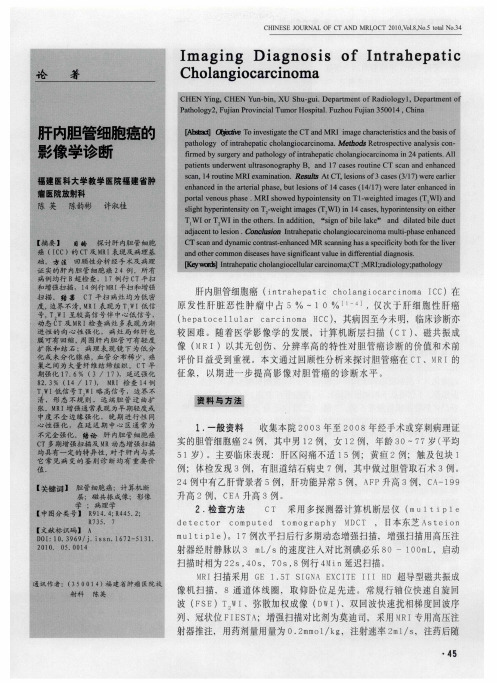
肝内胆管细胞癌核磁共振影像学诊断及鉴别诊断进展摘要:目的:探讨肝内胆管细胞癌患者的核磁共振成像表现。
方法:选取我院患者进行核磁共振影像检查肝内胆管细胞癌30例,共50个病灶。
观察核磁共振影像表现包括形态和边缘,密度、信号和强化特点,灶周和肝外改变,同时将病理改变与核磁共振影像表现相对照。
结果:核磁共振影像诊断肝内胆管细胞癌30例患者中,18例为结节型,12例为块状型。
经病理检验,30例患者镜检结果,14例患者可见典型的腺癌样结构,4例患者中为中低分化腺癌,4例患者为乳头状腺癌。
结论:肝内胆管癌的核磁共振影像主要表现为血供少,但有极少部分肿瘤表现为血供多。
肝内胆管细胞癌核磁共振影像表现具有特征性,并与其病理基础相关。
认识以上特征与病理改变,能提高诊断的准确性。
关键词;肝内胆管细胞癌;核磁共振影像学;鉴别诊断Antipathetic bile duct carcinoma progress in nuclear magnetic resonance (NMR) imaging diagnosis and differential diagnosisAbstract: Objective:On patients with antipathetic bile duct carcinoma of nuclear magnetic resonance (NMR) imaging manifestations.Methods:Select our hospital patients for nuclear magnetic resonance (NMR) imaging of 30 cases of antipathetic bile duct carcinoma, a total of 50 lesions.Observation of nuclear magnetic resonance (NMR) imaging performance including shape and edge, density, signal, and the characteristics of the reinforcement, stoveand extra hepatic change, at the same time the pathological changes and nuclear magnetic resonance (NMR) imaging performance.Results: Nuclear magnetic resonance (NMR) imaging in the diagnosis of antipathetic bile duct carcinoma of 30 cases, 18 cases of nodular type, 12 cases of block type.By pathological examination, microscopic examination of the 30 patients as a result, 14 patients with carcinoma of the typical sample structure, four patients in low differentiated carcinoma, 4 cases of papillary carcinoma.Conclusion:Antipathetic bile duct carcinoma, nuclear magnetic resonance (NMR) imaging is mainly characterized by less blood supply, but there are few tumor characterized by blood.Antipathetic bile duct carcinoma nuclear magnetic resonance (NMR) imaging demonstrated its characteristics, and is related to its pathological basis.Know more features and pathological change, can improve the accuracy of diagnosis.Keywords:Antipathetic bile duct carcinoma;Nuclear magnetic resonance imaging;The differential diagnosis肝内胆管细胞癌(ICC)是仅次于肝细胞癌的肝内第二常见的恶性肿瘤,发病率近年呈上升趋势,临床预后不佳。