消化道的X线诊断
- 格式:ppt
- 大小:14.01 MB
- 文档页数:100
![实践技能辅助检查之普通X线影像诊断(九)消化道穿孔[临床实践技能考试]](https://uimg.taocdn.com/3ea45e668e9951e79b8927e2.webp)
【星恒教育】临床实践技能考试:辅助检查
一、普通X 线影像诊断
(九)消化道穿孔
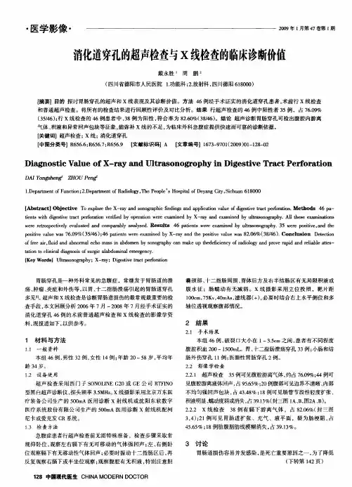
消化道穿孔常继发于胃肠道溃疡、创伤破裂、炎症及肿瘤,其中胃、十二指肠溃疡穿孔最为常见。
【X 线平片表现】
胃肠道穿孔表现有气腹、腹腔积液、腹脂线异常和麻痹性肠胀气,其中以游离气腹最重要且出现较早。
腹腔内游离气体可上浮到横膈与肝或胃之间,立位片表现为一侧或双侧膈下透亮的线条状或新月形气体影;气体可进入小网膜囊。
在中腹部腰椎右侧见气腔或气液腔;气体可进入腹膜后间隙,衬托出肾脏的外形轮廓。
图2-30 消化道穿孔(膈下游离气体)
消化道穿孔X 线平片考试技巧
模拟试题:
原网址:/sjjn/32192.html
2。
消化道穿孔x线诊断标准X-ray diagnosis is an essential tool in the assessment of gastrointestinal disorders, including the detection of gastrointestinal perforation. Gastrointestinal perforation refers to the formation of a hole or tear in the wall of the digestive tract, which allows the contents of the gastrointestinal system to leak into the abdominal cavity. This condition is considered a medical emergency and requires prompt diagnosis and treatment. X-ray imaging plays a crucial role in the diagnosis of gastrointestinal perforation, as it can provide valuable information about the location, extent, and possible complications associated with the perforation.When evaluating a patient for possible gastrointestinal perforation, the radiologist considers various factors to establish an accurate diagnosis. Firstly, the clinical history of the patient is essential. The radiologist needs to know the presenting symptoms, such as sudden and severe abdominal pain, fever, nausea, or vomiting, which are oftenindicative of a possible perforation. The patient's medical history, including any previous surgeries or gastrointestinal disorders, can also provide important clues.In addition to the clinical history, the radiologist carefully examines the X-ray images for signs of gastrointestinal perforation. One of the most common findings is the presence of free air within the abdominal cavity. Air can accumulate in the peritoneal cavity due to the leakage of gas from the perforated gastrointestinal tract. On X-ray, free air appears as radiolucent areas, often seen under the diaphragm or outlining the loops of bowel. This finding is highly suggestive ofgastrointestinal perforation.Other radiographic signs that may indicate gastrointestinal perforation include abnormal bowel gas patterns, such as dilated loops of bowel or the absence of bowel gas in the affected area. The presence of fluidlevels within the abdomen can also be observed, indicating the accumulation of fluid due to the leakage ofgastrointestinal contents. These findings, along with the clinical presentation, help the radiologist in making an accurate diagnosis of gastrointestinal perforation.However, it is important to note that X-ray imaging alone may not always be sufficient to establish adefinitive diagnosis of gastrointestinal perforation. In some cases, further imaging studies, such as computed tomography (CT) or ultrasound, may be required to confirm the diagnosis or provide additional information. CT scan can provide detailed images of the gastrointestinal tract, allowing for a more precise localization and characterization of the perforation. Ultrasound can be useful in certain cases, particularly in the evaluation of perforations in the upper gastrointestinal tract.In conclusion, X-ray imaging plays a crucial role in the diagnosis of gastrointestinal perforation. The radiologist carefully evaluates the clinical history and X-ray findings to establish an accurate diagnosis. The presence of free air, abnormal bowel gas patterns, and fluid levels within the abdomen are important radiographicsigns that may indicate gastrointestinal perforation. However, it is important to note that additional imaging studies, such as CT or ultrasound, may be necessary in some cases to confirm the diagnosis or provide further information. Prompt and accurate diagnosis of gastrointestinal perforation is essential for timely intervention and management of this potentially life-threatening condition.。

上消化道X线钡餐上消化道X线钡餐介绍:上消化道X线钡餐检查,是诊断上消化道疾病的主要手段之一,与纤维胃镜检查起到互补作用。
上消化道X线钡餐正常值:1.上腹部不适症状及上腹部肿块。
2.先天性胃肠道异常。
3.十二指肠手术后复查。
上消化道X线钡餐临床意义:异常结果:钡餐检查中见钡流方向异常,瘘管显示,肝胆系充盈显影。
需要检查的人群:胃及十二指肠和肝胆道因炎症、溃疡、肿瘤浸润等均可造成十二指肠胆囊或胆总管瘘及奥狄氏括约肌闭锁不全。
上消化道X线钡餐注意事项:不合宜人群:没有特殊说明,对钡餐出现反应的人群。
检查前禁忌:通常病人在检查前8-12h禁食,检查前4h禁饮水。
检查时要求:配合医生。
上消化道X线钡餐检查过程:X线钡餐检查,通常分食管、胃、十二指肠三段进行。
胃检查:口服不被胃粘膜吸收或吸附的显像剂后,经胃蠕动排入肠道,从胃内放射性下降可算出胃排空时间以了解胃的运动功能。
显像剂分99Tcm-SC或99Tcm-DT PA作成的固体或液体试验餐。
受检者空腹12 h,5 min内全部吃完固体或液体试验餐,并以1帧/5min的速度采集2 h,用ROI技术算出食物胃半排空时间。
食管检查:受检者禁食4 h以上,口含99Tcm-SC 37 MBq(1 mCi)的15 ml溶液,作一次快速吞咽,取立位以1帧/0.5s的速度采集60 s,以后每隔30 s作吞咽动作一次,共采集5 min。
用计算机ROI技术算出全食管通过时间及各段(上、中、下)通过时间和5 min内食管通过率。
十二指肠检查:受检者空腹4 h以上,检查方法同肝胆显像,待胆总管及十二指肠显影时,饮奶300 ml促胆汁排泄,以后每10 min采集1帧至60 min。
用ROI技术计算出十二指肠胃反流指数(DGRI)。

X线钡餐检查主要是查什么的在消化系统的检查工作通常情况下,一种比较常用的影像学检查方式就是X线钡餐检查,这种检查方式主要是确保或者在空腹的状态下食入钡餐,钡餐中含有胃酸不易溶解的硫酸钡物质,从而像摄入的食物一样成功的黏附在胃壁上,以此进一步有效检查患者的胃部是否出现不同程度的病变或者溃疡等相关方面的问题。
这种检查方式在当前临床实践中越来越被胃镜所代替,因为胃镜可以更加形象直观而且清楚,有效地看到病灶,进而采取患者的病理情况,进行精准的诊断。
然而该检查方式也有一定的可取之处,有自身的优势,因此在实际的检查过程中,对其也有广泛的应用。
需要注意的是,很多人并不清楚该检查方式具体检查什么,现在本文就具体说一下该检查方式,主要是检查什么的?一、钡餐是什么?钡餐主要指的是一种比较典型的白色斜方晶体,在针对消化道进行检查的过程中所应用的钡餐药用硫酸钡,也就是硫酸钡的悬浊液,不溶于水,不溶于酸,在水中的溶解度只有0.0024g/100g水。
用作白色颜料、纸和橡胶等的填充剂、X光透视肠胃时的药物等。
所以这种物质在水和脂质中是不会被溶解的,在胃肠道黏膜的吸附和检查孔中也不会被粘膜吸收,所以它对人体也没有任何的毒副作用。
钡餐造影检查的过程中也就是进行消化道钡剂造影,是指用硫酸钡作为造影剂,在X线的照射之下,针对患者的消化道是否出现溃疡或者病变等相关问题进行相对应的呈现和反应。
这种检查方式有着比较独特的作用和效能,与钡灌肠不同,钡餐造影的检查过程中可以有效通过口服的方式摄入相对应的造影剂,这样可以针对患者的整体的消化道,特别是确保上消化道的检查更为清晰明确,且经过有效的放射性检查,以此呈现出自身独特的优势。
其操作更为简单方便,可以减轻患者可能出现的不适感等等,同时检查结果也更精准,使口服医用硫酸钡制剂得到充分的应用,进一步显示胃肠道的造影检查结果。
二、X线钡餐检查主要是查什么的?X线钡餐检查的过程中,因为人体的相关器官组织在厚度密度等方面都有着十分显著的差异性,所以在具体检查过程中所呈现出的黑白的自然层次对比,有着十分显著的差异。