脑动脉系解剖
- 格式:ppt
- 大小:5.57 MB
- 文档页数:44
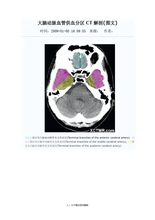
大脑动脉血管供血分区CT解剖(图文)时间:2009-01-08 16:09:55 来源:作者:浅蓝色部分为大脑前动脉终末支供血区(Terminal branches of the anterior cerebral artery),粉红色部分为大脑中动脉终末支供血区(Terminal branches of the middle cerebral artery),黄色部分为大脑后动脉终末支供血区(Terminal branches of the posterior cerebral artery)浅蓝色部分为大脑前动脉终末支供血区(Terminal branches of the anterior cerebral artery),粉红色部分为大脑中动脉终末支供血区(Terminal branches of the middle cerebral artery),黄色部分为大脑后动脉终末支供血区(Terminal branches of the posterior cerebral artery),绿色部分为脉络膜前动脉供血区(Anterior choroidal artery),浅蓝色部分为大脑前动脉终末支供血区(Terminal branches of the anterior cerebral artery),粉红色部分为大脑中动脉终末支供血区(Terminal branches of the middle cerebral artery),黄色部分为大脑后动脉终末支供血区(Terminal branches of the posterior cerebral artery),绿色部分为脉络膜前动脉供血区(Anterior choroidal artery),褐色部分为大脑前动脉深穿支供血区(Penetrating branches of the anterior cerebral artery),枣红色部分为大脑后动脉深穿支及后交通动脉供血区(Penetrating branches of the posterior cerebral artery and posterior communicating artery),浅蓝色部分为大脑前动脉终末支供血区(Terminal branches of the anterior cerebral artery),粉红色部分为大脑中动脉终末支供血区(Terminal branches of the middle cerebral artery),黄色部分为大脑后动脉终末支供血区(Terminal branches of the posterior cerebral artery),绿色部分为脉络膜前动脉供血区(Anterior choroidal artery),褐色部分为大脑前动脉深穿支供血区(Penetrating branches of the anterior cerebral artery),枣红色部分为大脑后动脉深穿支及后交通动脉供血区(Penetrating branches of the posterior cerebral artery and posterior communicating artery),亮红色部分为大脑中动脉深穿支供血(Penetrating branches of the middle cerebral artery)浅蓝色部分为大脑前动脉终末支供血区(Terminal branches of the anterior cerebral artery),粉红色部分为大脑中动脉终末支供血区(Terminal branches of the middle cerebral artery),黄色部分为大脑后动脉终末支供血区(Terminal branches of the posterior cerebral artery),绿色部分为脉络膜前动脉供血区(Anterior choroidal artery),枣红色部分为大脑后动脉深穿支及后交通动脉供血区(Penetrating branches of the posterior cerebral artery and posterior communicating artery),亮红色部分为大脑中动脉深穿支供血(Penetrating branches of the middle cerebral artery)浅蓝色部分为大脑前动脉终末支供血区(Terminal branches of the anterior cerebral artery),粉红色部分为大脑中动脉终末支供血区(Terminal branches of the middle cerebral artery),黄色部分为大脑后动脉终末支供血区(Terminal branches of the posterior cerebral artery),枣红色部分为大脑后动脉深穿支及后交通动脉供血区(Penetrating branches of the posterior cerebral artery and posterior communicating artery),亮红色部分为大脑中动脉深穿支供血(Penetrating branches of the middle cerebral artery)浅蓝色部分为大脑前动脉终末支供血区(Terminal branches of the anterior cerebral artery),粉红色部分为大脑中动脉终末支供血区(Terminal branches of the middle cerebral artery),黄色部分为大脑后动脉终末支供血区(Terminal branches of the posterior cerebral artery),浅蓝色部分为大脑前动脉终末支供血区(Terminal branches of the anterior cerebral artery),粉红色部分为大脑中动脉终末支供血区(Terminal branches of the middle cerebral artery),黄色部分为大脑后动脉终末支供血区(Terminal branches of the posterior cerebral artery),浅蓝色部分为大脑前动脉终末支供血区(Terminal branches of the anterior cerebral artery),粉红色部分为大脑中动脉终末支供血区(Terminal branches of the middle cerebral artery),黄色部分为大脑后动脉终末支供血区(Terminal branches of the posterior cerebral artery),浅蓝色部分为大脑前动脉终末支供血区(Terminal branches of the anterior cerebral artery),粉红色部分为大脑中动脉终末支供血区(Terminal branches of the middle cerebral artery),浅蓝色部分为大脑前动脉终末支供血区(Terminal branches of the anterior cerebral artery)(学习的目的是增长知识,提高能力,相信一分耕耘一分收获,努力就一定可以获得应有的回报)11 / 11下载文档可编辑。
大脑中动脉分段详解(简图+DSA+实物)大脑中动脉(MCA)是颈内动脉两个终支中较大的血管,缺血及梗塞最常累及此区。
大脑中动脉在解剖上一般分成4段或5段,分别为M1、M2、M3、M4、M5.M1段自颈内动脉分叉延伸至侧裂,包括分叉前及分叉后段。
M2段自膝部至侧裂顶及环状沟,包括6-8支主干动脉在侧裂内走行脑岛之上。
M3段自环状沟的顶部开始向外行走处,终止于侧裂表面。
M4段则为大脑中动脉的皮层支,其走出侧裂,弯曲分布在额叶、聚叶、顶叶岛盖上。
下图是___教授对大脑中动脉分段的一个前后位解剖图解,解说比较清楚,让人一目了然。
大脑中动脉的分段结构对于医学领域的从业者和研究人员来说至关重要。
M1段是单独的主干,分叉后段则可能是单干,双干、三干甚至更多。
M2段包括6-8支主干动脉在侧裂内走行脑岛之上。
M3段从环状沟的顶部开始向外行走处,终止于侧裂表面。
M4段则为大脑中动脉的皮层支,其走出侧裂,弯曲分布在额叶、聚叶、顶叶岛盖上。
DSA造影正位图和侧位图都能够更好地展示大脑中动脉的分支结构。
实物图也可以帮助医学工作者更好地了解大脑中动脉的结构。
大脑中动脉的分段结构的详细了解对于医学领域的从业者和研究人员来说至关重要。
大脑血管系统是人体重要的血液循环系统之一。
它由多个血管和神经组成,包括大脑前动脉(右侧和左侧)、额叶底内侧动脉、颈内动脉、动眼神经、大脑中动脉上干、大脑中动脉下干、前外侧中央动脉(外侧豆纹动脉)、脉络丛(膜)前动脉、大脑后动脉、后交通动脉、滑车神经、脑桥和三叉神经。
其中,大脑前动脉是大脑的主要血液供应来源之一。
它们分别位于大脑的左右两侧,负责向大脑的前部供血。
额叶底内侧动脉则位于大脑的内部,主要负责供应大脑的额叶底部区域。
颈内动脉是大脑血管系统的重要组成部分之一,负责向大脑供应血液。
动眼神经则是负责控制眼球运动的神经,也是大脑血管系统的一部分。
大脑中动脉上干和下干则分别位于大脑的中央区域,负责向大脑的中央区域供血。


脑动静脉系统详细解剖脑动静脉血管系统解剖一、脑血管的特点血管的分类动脉:大动脉:(弹性动脉)管壁含有大量的弹力膜(~)层主动脉肺动脉等中动脉:(肌动脉)管壁富含平滑肌细胞除主动脉和肺动脉外所有解剖学上有名称的动脉都属中动脉小动脉:也为肌性动脉中膜~层平滑肌管径在cm以下的动脉。
微动脉:无弹力内膜中膜只有~层平滑肌管径在cm以下的动脉。
血管的分类静脉:血管腔大管壁薄而柔软管壁内、中、外三层膜分界不明显外膜较中膜厚结缔组织内含有较多的平滑肌束管径在mm以上的静脉常有静脉瓣。
颈内动脉椎动脉一、脑的动脉基底动脉颈内动脉的分支眼动脉颈内动脉的海绵窦部颈内动脉的脑部Internalcarotidartery,Anteriorclinoidprocesssuperiorpart颈动脉管段c海绵窦段c交叉池段c前膝段c后膝段c颈内动脉颈外动脉系统甲状腺上动脉superiorthyroidartery舌动脉lingualartery面动脉facialartery颞浅动脉superficialtemporalartery上颌动脉maxillaryartery(脑膜中动脉middlemeningealartery)枕动脉occipitalartery耳后动脉posteriorauricularartery咽升动脉ascendantpharyngealartery颈外动脉系统脑动脉系人脑血供非常丰富左心室每分钟排血量为ml其中供应脑部的血液为ml占全身供血量的。
脑动脉系的特点脑动脉的主干及其主要分支均位于脑的腹侧面然后再回绕到脑的背侧面。
脑动脉可以分为皮质支和中央支(或回旋支与旁中央支)两类分支。
皮质支与中央支之间吻合甚少但皮质支与皮质支之间中央支与中央支之间却存在较多的吻合不过前者吻合丰富后者吻合相对较差。
脑动脉为肌型动脉管壁薄血管周围没有支持组织。
脑动脉内膜厚有发达的内弹力膜但中膜和外膜较薄仅含少量的弹力纤维没有外弹力膜因此脑动脉几乎没有搏动。