Epigenetics and Colorectal Cancer
- 格式:pdf
- 大小:855.19 KB
- 文档页数:28
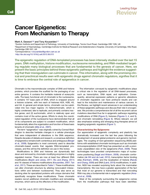
Leading EdgeReviewCancer Epigenetics:From Mechanism to TherapyMark A.Dawson1,2and Tony Kouzarides1,*1Gurdon Institute and Department of Pathology,University of Cambridge,Tennis Court Road,Cambridge CB21QN,UK2Department of Haematology,Cambridge Institute for Medical Research and Addenbrooke’s Hospital,University of Cambridge,Hills Road, Cambridge CB20XY,UK*Correspondence:t.kouzarides@/10.1016/j.cell.2012.06.013The epigenetic regulation of DNA-templated processes has been intensely studied over the last15 years.DNA methylation,histone modification,nucleosome remodeling,and RNA-mediated target-ing regulate many biological processes that are fundamental to the genesis of cancer.Here,we present the basic principles behind these epigenetic pathways and highlight the evidence suggest-ing that their misregulation can culminate in cancer.This information,along with the promising clin-ical and preclinical results seen with epigenetic drugs against chromatin regulators,signifies that it is time to embrace the central role of epigenetics in cancer.Chromatin is the macromolecular complex of DNA and histone proteins,which provides the scaffold for the packaging of our entire genome.It contains the heritable material of eukaryotic cells.The basic functional unit of chromatin is the nucleosome. It contains147base pairs of DNA,which is wrapped around a histone octamer,with two each of histones H2A,H2B,H3, and H4.In general and simple terms,chromatin can be subdi-vided into two major regions:(1)heterochromatin,which is highly condensed,late to replicate,and primarily contains inac-tive genes;and(2)euchromatin,which is relatively open and contains most of the active genes.Efforts to study the coordi-nated regulation of the nucleosome have demonstrated that all of its components are subject to covalent modification,which fundamentally alters the organization and function of these basic tenants of chromatin(Allis et al.,2007).The term‘‘epigenetics’’was originally coined by Conrad Wad-dington to describe heritable changes in a cellular phenotype that were independent of alterations in the DNA sequence. Despite decades of debate and research,a consensus definition of epigenetics remains both contentious and ambiguous(Berger et al.,2009).Epigenetics is most commonly used to describe chromatin-based events that regulate DNA-templated pro-cesses,and this will be the definition we use in this review. Modifications to DNA and histones are dynamically laid down and removed by chromatin-modifying enzymes in a highly regulated manner.There are now at least four different DNA modifications(Baylin and Jones,2011;Wu and Zhang,2011) and16classes of histone modifications(Kouzarides,2007;Tan et al.,2011).These are described in Table1.These modifications can alter chromatin structure by altering noncovalent interac-tions within and between nucleosomes.They also serve as docking sites for specialized proteins with unique domains that specifically recognize these modifications.These chromatin readers recruit additional chromatin modifiers and remodeling enzymes,which serve as the effectors of the modification.The information conveyed by epigenetic modifications plays a critical role in the regulation of all DNA-based processes, such as transcription,DNA repair,and replication.Conse-quently,abnormal expression patterns or genomic alterations in chromatin regulators can have profound results and can lead to the induction and maintenance of various cancers.In this Review,we highlight recent advances in our understanding of these epigenetic pathways and discuss their role in oncogen-esis.We provide a comprehensive list of all the recurrent cancer mutations described thus far in epigenetic pathways regulating modifications of DNA(Figure2),histones(Figures3,4,and5), and chromatin remodeling(Figure6).Where relevant,we will also emphasize existing and emerging drug therapies aimed at targeting epigenetic regulators(Figure1).Characterizing the EpigenomeOur appreciation of epigenetic complexity and plasticity has dramatically increased over the last few years following the development of several global proteomic and genomic technol-ogies.The coupling of next-generation sequencing(NGS)plat-forms with established chromatin techniques such as chromatin immunoprecipitation(ChIP-Seq)has presented us with a previ-ously unparalleled view of the epigenome(Park,2009).These technologies have provided comprehensive maps of nucleo-some positioning(Segal and Widom,2009),chromatin confor-mation(de Wit and de Laat,2012),transcription factor binding sites(Farnham,2009),and the localization of histone(Rando and Chang,2009)and DNA(Laird,2010)modifications.In addi-tion,NGS has revealed surprising facts about the mammalian transcriptome.We now have a greater appreciation of the fact that most of our genome is transcribed and that noncoding RNA may play a fundamental role in epigenetic regulation(Ama-ral et al.,2008).Most of the complexity surrounding the epigenome comes from the modification pathways that have been identified.12Cell150,July6,2012ª2012Elsevier Inc.Recent improvements in the sensitivity and accuracy of mass spectrometry (MS)instruments have driven many of these discoveries (Stunnenberg and Vermeulen,2011).Moreover,although MS is inherently not quantitative,recent advances in labeling methodologies,such as stable isotope labeling by amino acids in cell culture (SILAC),isobaric tags for relative and absolute quantification (iTRAQ),and isotope-coded affinity tag (ICAT),have allowed a greater ability to provide quantitative measurements (Stunnenberg and Vermeulen,2011).These quantitative methods have generated ‘‘protein recruit-ment maps’’for histone and DNA modifications,which contain proteins that recognize chromatin modifications (Bartke et al.,2010;Vermeulen et al.,2010).Many of these chromatin readers have more than one reading motif,so it is important to under-stand how they recognize several modifications either simulta-neously or sequentially.The concept of multivalent engagement by chromatin-binding modules has recently been explored by using either modified histone peptides (Vermeulen et al.,2010)or in-vitro-assembled and -modified nucleosomes (Bartkeet al.,2010;Ruthenburg et al.,2011).The latter approach in particular has uncovered some of the rules governing the recruit-ment of protein complexes to methylated DNA and modified histones in a nucleosomal context.The next step in our under-standing will require a high-resolution in vivo genomic approach to detail the dynamic events on any given nucleosome during the course of gene expression.Epigenetics and the Cancer ConnectionThe earliest indications of an epigenetic link to cancer were derived from gene expression and DNA methylation studies.These studies are too numerous to comprehensively detail in this review;however,the reader is referred to an excellent review detailing the history of cancer epigenetics (Feinberg and Tycko,2004).Although many of these initial studies were purely correl-ative,they did highlight a potential connection between epige-netic pathways and cancer.These early observations have been significantly strengthened by recent results from the Inter-national Cancer Genome Consortium (ICGC).Whole-genomeTable 1.Chromatin Modifications,Readers,and Their Function Chromatin Modification NomenclatureChromatin-Reader MotifAttributed Functionand Cit,citrulline.Reader domains:MBD,methyl-CpG-binding domain;PHD,plant homeodomain;MBT,malignant brain tumor domain;PWWP,proline-tryptophan-tryptophan-proline domain;BRCT,BRCA1C terminus domain;UIM,ubiquitin interaction motif;IUIM,inverted ubiquitin interaction motif;SIM,sumo interaction motif;and PBZ,poly ADP-ribose binding zinc finger.aThese are established binding modules for the posttranslational modification;however,binding to modified histones has not been firmly established.Cell 150,July 6,2012ª2012Elsevier Inc.13sequencing in a vast array of cancers has provided a catalog of recurrent somatic mutations in numerous epigenetic regulators (Forbes et al.,2011;Stratton et al.,2009).A central tenet in analyzing these cancer genomes is the identification of ‘‘driver’’mutations (causally implicated in the process of oncogenesis).A key feature of driver mutations is that they are recurrently found in a variety of cancers,and/or they are often present at a high prevalence in a specific tumor type.We will mostly concentrate our discussions on suspected or proven driver mutations in epigenetic regulators.For instance,malignancies such as follicular lymphoma contain recurrent mutations of the histone methyltransferase MLL2in close to 90%of cases (Morin et al.,2011).Similarly,UTX ,a histone demethylase,is mutated in up to 12histologi-cally distinct cancers (van Haaften et al.,2009).Compilation of the epigenetic regulators mutated in cancer highlights histone acetylation and methylation as the most widely affected epige-netic pathways (Figures 3and 4).These and other pathways that are affected to a lesser extent will be described in the following sections.Deep sequencing technologies aimed at mapping chromatin modifications have also begun to shed some light on the origins of epigenetic abnormalities in cancer.Cross-referencing of DNA methylation profiles in human cancers with ChIP-Seq data for histone modifications and the binding of chromatinregulators have raised intriguing correlations between cancer-associated DNA hypermethylation and genes marked with ‘‘bivalent’’histone modifications in multipotent cells (Easwaran et al.,2012;Ohm et al.,2007).These bivalent genes are marked by active (H3K4me3)and repressive (H3K27me3)histone modi-fications (Bernstein et al.,2006)and appear to identify transcrip-tionally poised genes that are integral to development and lineage commitment.Interestingly,many of these genes are targeted for DNA methylation in cancer.Equally intriguing are recent comparisons between malignant and normal tissues from the same individuals.These data demonstrate broad domains within the malignant cells that contain significant alter-ations in DNA methylation.These regions appear to correlate with late-replicating regions of the genome associated with the nuclear lamina (Berman et al.,2012).Although there remains little mechanistic insight into how and why these regions of the genome are vulnerable to epigenetic alterations in cancer,these studies highlight the means by which global sequencing plat-forms have started to uncover avenues for further investigation.Genetic lesions in chromatin modifiers and global alterations in the epigenetic landscape not only imply a causative role for these proteins in cancer but also provide potential targets for therapeutic intervention.A number of small-molecule inhibitors have already been developed against chromatin regulators (Figure 1).These are at various stages of development,andthreeFigure 1.Epigenetic Inhibitors as Cancer TherapiesThis schematic depicts the process for epigenetic drug development and the current status of various epigenetic therapies.Candidate small molecules are first tested in vitro in malignant cell lines for specificity and phenotypic response.These may,in the first instance,assess the inhibition of proliferation,induction of apoptosis,or cell-cycle arrest.These phenotypic assays are often coupled to genomic and proteomic methods to identify potential molecular mechanisms for the observed response.Inhibitors that demonstrate potential in vitro are then tested in vivo in animal models of cancer to ascertain whether they may provide therapeutic benefit in terms of survival.Animal studies also provide valuable information regarding the toxicity and pharmacokinetic properties of the drug.Based on these preclinical studies,candidate molecules may be taken forward into the clinical setting.When new drugs prove beneficial in well-conducted clinical trials,they are approved for routine clinical use by regulatory authorities such as the FDA.KAT,histone lysine acetyltransferase;KMT,histone lysine methyltransferase;RMT,histone arginine methyltransferase;and PARP,poly ADP ribose polymerase.14Cell 150,July 6,2012ª2012Elsevier Inc.of these(targeting DNMTs,HDACs,and JAK2)have already been granted approval by the US Food and Drug Administra-tion(FDA).This success may suggest that the interest in epige-netic pathways as targets for drug discovery had been high over the past decade.However,the reality is that thefield of drug discovery had been somewhat held back due to concerns over the pleiotropic effects of both the drugs and their targets. Indeed,some of the approved drugs(against HDACs)have little enzyme specificity,and their mechanism of action remains contentious(Minucci and Pelicci,2006).The belief and investment in epigenetic cancer therapies may now gain momentum and reach a new level of support following the recent preclinical success of inhibitors against BRD4,an acetyl-lysine chromatin-binding protein(Dawson et al.,2011; Delmore et al.,2011;Filippakopoulos et al.,2010;Mertz et al., 2011;Zuber et al.,2011).The molecular mechanisms governing these impressive preclinical results have also been largely uncovered and are discussed below.This process is pivotal for the successful progression of these inhibitors into the clinic. These results,along with the growing list of genetic lesions in epigenetic regulators,highlight the fact that we have now entered an era of epigenetic cancer therapies.Epigenetic Pathways Connected to CancerDNA MethylationThe methylation of the5-carbon on cytosine residues(5mC)in CpG dinucleotides was thefirst described covalent modifica-tion of DNA and is perhaps the most extensively characterized modification of chromatin.DNA methylation is primarily noted within centromeres,telomeres,inactive X-chromosomes,and repeat sequences(Baylin and Jones,2011;Robertson,2005). Although global hypomethylation is commonly observed in malignant cells,the best-studied epigenetic alterations in cancerare the methylation changes that occur within CpG islands, which are present in 70%of all mammalian promoters.CpG island methylation plays an important role in transcriptional regu-lation,and it is commonly altered during malignant transforma-tion(Baylin and Jones,2011;Robertson,2005).NGS platforms have now provided genome-wide maps of CpG methylation. These have confirmed that between5%–10%of normally unme-thylated CpG promoter islands become abnormally methylated in various cancer genomes.They also demonstrate that CpG hypermethylation of promoters not only affects the expression of protein coding genes but also the expression of various noncoding RNAs,some of which have a role in malignant trans-formation(Baylin and Jones,2011).Importantly,these genome-wide DNA methylome studies have also uncovered intriguing alterations in DNA methylation within gene bodies and at CpG‘‘shores,’’which are conserved sequences upstream and downstream of CpG islands.The functional relevance of these regional alterations in methylation are yet to be fully deciphered, but it is interesting to note that they have challenged the general dogma that DNA methylation invariably equates with transcriptional silencing.In fact,these studies have established that many actively transcribed genes have high levels of DNA methylation within the gene body,suggesting that the context and spatial distribution of DNA methylation is vital in transcrip-tional regulation(Baylin and Jones,2011).Three active DNA methyltransferases(DNMTs)have been identified in higher eukaryotes.DNMT1is a maintenance methyl-transferase that recognizes hemimethylated DNA generated during DNA replication and then methylates newly synthesized CpG dinucleotides,whose partners on the parental strand are already methylated(Li et al.,1992).Conversely,DNMT3a and DNMT3b,although also capable of methylating hemimethylated DNA,function primarily as de novo methyltransferases to estab-lish DNA methylation during embryogenesis(Okano et al.,1999). DNA methylation provides a platform for several methyl-binding proteins.These include MBD1,MBD2,MBD3,and MeCP2. These in turn function to recruit histone-modifying enzymes to coordinate the chromatin-templated processes(Klose and Bird,2006).Although mutations in DNA methyltransferases and MBD proteins have long been known to contribute to developmental abnormalities(Robertson,2005),we have only recently become aware of somatic mutations of these key genes in human malig-nancies(Figure2).Recent sequencing of cancer genomes has identified recurrent mutations in DNMT3A in up to25%of patients with acute myeloid leukemia(AML)(Ley et al.,2010). Importantly,these mutations are invariably heterozygous and are predicted to disrupt the catalytic activity of the enzyme. Moreover,their presence appears to impact prognosis(Patel et al.,2012).However,at present,the mechanisms bywhich Figure2.Cancer Mutations Affecting Epigenetic Regulators of DNA MethylationThe5-carbon of cytosine nucleotides are methylated(5mC)by a family of DNMTs.One of these,DNMT3A,is mutated in AML,myeloproliferative diseases(MPD),and myelodysplastic syndromes(MDS).In addition to its catalytic activity,DNMT3A has a chromatin-reader motif,the PWWP domain, which may aid in localizing this enzyme to chromatin.Somatically acquired mutations in cancer may also affect this domain.The TET family of DNA hydroxylases metabolizes5mC into several oxidative intermediates,including 5-hydroxymethylcytosine(5hmC),5-formylcytosine(5fC),and5-carbox-ylcytosine(5caC).These intermediates are likely involved in the process of active DNA demethylation.Two of the three TET family members are mutated in cancers,including AML,MPD,MDS,and CMML.Mutation types are as follows:M,missense;F,frameshift;N,nonsense;S,splice site mutation;and T,translocation.Cell150,July6,2012ª2012Elsevier Inc.15these mutations contribute to the development and/or mainte-nance of AML remains elusive.Understanding the cellular consequences of normal and aber-rant DNA methylation remains a key area of interest,especially because hypomethylating agents are one of the few epigenetic therapies that have gained FDA approval for routine clinical use(Figure1).Although hypomethylating agents such as azaci-tidine and decitabine have shown mixed results in various solid malignancies,they have found a therapeutic niche in the myelo-dysplastic syndromes(MDS).Until recently,this group of disor-ders was largely refractory to therapeutic intervention,and MDS was primarily managed with supportive care.However,several large studies have now shown that treatment with azacitidine, even in poor prognosis patients,improves their quality of life and extends survival time.Indeed,azacitidine is thefirst therapy to have demonstrated a survival benefit for patients with MDS (Fenaux et al.,2009).The molecular mechanisms governing the impressive responses seen in MDS are largely unknown. However,recent evidence would suggest that low doses of these agents hold the key to therapeutic benefit(Tsai et al., 2012).It is also emerging that the combinatorial use of DNMT and HDAC inhibitors may offer superior therapeutic outcomes (Gore,2011).DNA Hydroxy-Methylation and Its Oxidation Derivatives Historically,DNA methylation was generally considered to be a relatively stable chromatin modification.However,early studies assessing the global distribution of this modification during embryogenesis had clearly identified an active global loss of DNA methylation in the early zygote,especially in the male pronucleus.More recently,high-resolution genome-wide mapping of this modification in pluripotent and differentiated cells has also confirmed the dynamic nature of DNA methylation, evidently signifying the existence of an enzymatic activity within mammalian cells that either erases or alters this chromatin modification(Baylin and Jones,2011).In2009,two seminal manuscripts describing the presence of5-hydroxymethylcyto-sine(5hmC)offered thefirst insights into the metabolism of 5mC(Kriaucionis and Heintz,2009;Tahiliani et al.,2009).The ten-eleven translocation(TET1–3)family of proteins have now been demonstrated to be the mammalian DNA hydroxy-lases responsible for catalytically converting5mC to5hmC. Indeed,iterative oxidation of5hmC by the TET family results in further oxidation derivatives,including5-formylcytosine(5fC) and5-carboxylcytosine(5caC).Although the biological signifi-cance of the5mC oxidation derivatives is yet to be established, several lines of evidence highlight their importance in transcrip-tional regulation:(1)they are likely to be an essential intermediate in the process of both active and passive DNA demethylation,(2) they preclude or enhance the binding of several MBD proteins and,as such,will have local and global effects by altering the recruitment of chromatin regulators,and(3)genome-wide mapping of5hmC has identified a distinctive distribution of this modification at both active and repressed genes,including its presence within gene bodies and at the promoters of bivalently marked,transcriptionally poised genes(Wu and Zhang,2011). Notably,5hmC was also mapped to several intergenic cis-regu-latory elements that are either functional enhancers or insulator elements.Consistent with the notion that5hmC is likely to have a role in both transcriptional activation and silencing, the TET proteins have also been shown to have activating and repressive functions(Wu and Zhang,2011).Genome-wide mapping of TET1has demonstrated it to have a strong prefer-ence for CpG-rich DNA and,consistent with its catalytic function, it also been localized to regions enriched for5mC and5hmC. The TET family of proteins derive their name from the initial description of a recurrent chromosomal translocation, t(10;11)(q22;q23),which juxtaposes the MLL gene with TET1in a subset of patients with AML(Lorsbach et al.,2003).Notably, concurrent to the initial description of the catalytic activity for the TET family of DNA hydroxylases,several reports emerged describing recurrent mutations in TET2in numerous hematolog-ical malignancies(Cimmino et al.,2011;Delhommeau et al., 2009;Langemeijer et al.,2009)(Figure2).Interestingly,TET2-deficient mice develop a chronic myelomonocytic leukemia (CMML)phenotype,which is in keeping with the high prevalence of TET2mutations in patients with this disease(Moran-Crusio et al.,2011;Quivoron et al.,2011).The clinical implications of TET2mutations have largely been inconclusive;however,in some subsets of AML patients,TET2mutations appear to confer a poor prognosis(Patel et al.,2012).Early insights into the process of TET2-mediated oncogenesis have revealed that the patient-associated mutations are largely loss-of-function muta-tions that consequently result in decreased5hmC levels and a reciprocal increase in5mC levels within the malignant cells that harbor them.Moreover,mutations in TET2also appear to confer enhanced self-renewal properties to the malignant clones (Cimmino et al.,2011).Histone ModificationsIn1964,Vincent Allfrey prophetically surmised that histone modifications might have a functional influence on the regulation of transcription(Allfrey et al.,1964).Nearly half a century later, thefield is still grappling with the task of unraveling the mecha-nisms underlying his enlightened statement.In this time,we have learned that these modifications have a major influence, not just on transcription,but in all DNA-templated processes (Kouzarides,2007).The major cellular processes attributed to each of these modifications are summarized in Table1.The great diversity in histone modifications introduces a remarkable complexity that is slowly beginning to be ing transcription as an example,we have learned that multiple coexisting histone modifications are associated with activation,and some are associated with repression. However,these modification patterns are not static entities but a dynamically changing and complex landscape that evolves in a cell context-dependent fashion.Moreover,active and repres-sive modifications are not always mutually exclusive,as evi-denced by‘‘bivalent domains.’’The combinatorial influence that one or more histone modifications have on the deposition, interpretation,or erasure of other histone modifications has been broadly termed‘‘histone crosstalk,’’and recent evidence would suggest that crosstalk is widespread and is of great bio-logical significance(Lee et al.,2010).It should be noted that the cellular enzymes that modify histones may also have nonhistone targets and,as such,it has been difficult to divorce the cellular consequences of individual histone modifications from the broader targets of many of these16Cell150,July6,2012ª2012Elsevier Inc.enzymes.In addition to their catalytic function,many chromatin modifiers also possess‘‘reader’’domains allowing them to bind to specific regions of the genome and respond to information conveyed by upstream signaling cascades.This is important, as it provides two avenues for therapeutically targeting these epigenetic regulators.The residues that line the binding pocket of reader domains can dictate a particular preference for specific modification states,whereas residues outside the binding pocket contribute to determining the histone sequence specificity.This combination allows similar reader domains to dock at different modified residues or at the same amino acid displaying different modification states.For example,some methyl-lysine readers engage most efficiently with di/tri-methyl-ated lysine(Kme2/3),whereas others prefer mono-or unmethy-lated lysines.Alternatively,when the same lysines are now acet-ylated,they bind to proteins containing bromodomains(Taverna et al.,2007).The main modification binding pockets contained within chromatin-associated proteins is summarized in Table1. Many of the proteins that modify or bind these histone modifi-cations are misregulated in cancer,and in the ensuing sections, we will discuss the most extensively studied histone modifica-tions in relation to oncogenesis and novel therapeutics. Histone Acetylation.The Nε-acetylation of lysine residues is a major histone modification involved in transcription,chromatin structure,and DNA repair.Acetylation neutralizes lysine’s posi-tive charge and may consequently weaken the electrostatic interaction between histones and negatively charged DNA.For this reason,histone acetylation is often associated with a more ‘‘open’’chromatin conformation.Consistent with this,ChIP-Seq analyses have confirmed the distribution of histone acetyla-tion at promoters and enhancers and,in some cases,throughout the transcribed region of active genes(Heintzman et al.,2007; Wang et al.,2008).Importantly,lysine acetylation also serves as the nidus for the binding of various proteins with bromodo-mains and tandem plant homeodomain(PHD)fingers,which recognize this modification(Taverna et al.,2007).Acetylation is highly dynamic and is regulated by the competing activities of two enzymatic families,the histone lysine acetyltransferases(KATs)and the histone deacetylases (HDACs).There are two major classes of KATs:(1)type-B,which are predominantly cytoplasmic and modify free histones,and(2) type-A,which are primarily nuclear and can be broadly classifiedinto the GNAT,MYST,and CBP/p300families.KATs were thefirst enzymes shown to modify histones.The importance of thesefindings to cancer was immediately apparent,as one of these enzymes,CBP,was identified by its ability to bind the transforming portion of the viral oncoprotein E1A(Bannister and Kouzarides,1996).It is now clear that many,if not most,of the KATs have been implicated in neoplastic transformation,and a number of viral oncoproteins are known to associate with them.There are numerous examples of recur-rent chromosomal translocations(e.g.,MLL-CBP[Wang et al., 2005]and MOZ-TIF2[Huntly et al.,2004])or coding mutations (e.g.,p300/CBP[Iyer et al.,2004;Pasqualucci et al.,2011]) involving various KATs in a broad range of solid and hematolog-ical malignancies(Figure3).Furthermore,altered expression levels of several of the KATs have also been noted in a range of cancers(Avvakumov and Coˆte´,2007;Iyer et al.,2004).In some cases,such as the leukemia-associated fusion gene MOZ-TIF2,we know a great deal about the cellular conse-quences of this translocation involving a MYST family member. MOZ-TIF2is sufficient to recapitulate an aggressive leukemia in murine models;it can confer stem cell properties and reacti-vate a self-renewal program when introduced into committed hematopoietic progenitors,and much of this oncogenic potential is dependent on its inherent and recruited KAT activity as well as its ability to bind to nucleosomes(Deguchi et al.,2003;Huntly et al.,2004).Despite these insights,the great conundrum with regards to unraveling the molecular mechanisms by which histone acetyl-transferases contribute to malignant transformation has been dissecting the contribution of altered patterns in acetylation on histone and nonhistone proteins.Although it is clear that global histone acetylation patterns are perturbed in cancers(Fraga Figure 3.Cancer Mutations Affecting Epigenetic Regulators Involved in Histone AcetylationThese tables provide somatic cancer-associated mutations identified in histone acetyltransferases and proteins that contain bromodomains(which recognize and bind acetylated histones).Several histone acetyltransferases possess chromatin-reader motifs and,thus,mutations in the proteins may alter both their catalytic activities as well as the ability of these proteins to scaffold multiprotein complexes to chromatin.Interestingly,sequencing of cancer genomes to date has not identified any recurrent somatic mutations in histone deacetylase enzymes.Abbreviations for the cancers are as follows: AML,acute myeloid leukemia;ALL,acute lymphoid leukemia;B-NHL,B-cell non-Hodgkin’s lymphoma;DLBCL,diffuse large B-cell lymphoma;and TCC, transitional cell carcinoma of the urinary bladder.Mutation types are as follows:M,missense;F,frameshift;N,nonsense;S,splice site mutation;T, translocation;and D,deletion.Cell150,July6,2012ª2012Elsevier Inc.17。

不可切除胰腺癌的分子靶向药物治疗进展胡润,李俊蒽,姚沛,桂仁捷,段华新湖南师范大学附属第一医院,湖南省人民医院肿瘤科,长沙 410005通信作者:段华新,****************(ORCID: 0000-0001-9596-5013)摘要:胰腺癌作为消化系统最常见的恶性肿瘤之一,其发病率及死亡率正逐年上升,大多数胰腺癌患者因分期较晚而失去了手术机会。
尽管以吉西他滨、氟尿嘧啶为主的化疗方案在一定程度上延长了患者的生存期,但仍有部分患者因无法耐受化疗而失去治疗机会。
随着精准医疗时代的来临,分子靶向药物治疗展现出的优异疗效使其成为对抗肿瘤的重要治疗手段之一,但由于胰腺癌高度的异质性及复杂的免疫微环境,针对胰腺癌的分子靶向治疗并未取得显著效果,因此亟需探寻新的治疗靶点及药物攻克这一难题。
本综述基于胰腺癌常见分子靶点及肿瘤免疫相关靶点探究在不可切除胰腺癌中分子靶向药物治疗研究的最新进展,为胰腺癌患者提供新的治疗策略。
关键词:胰腺肿瘤;分子靶向治疗;免疫疗法基金项目:湖南省自然科学基金(2020JJ8084)Advances in molecular-targeted therapy for unresectable pancreatic cancerHU Run,LI Junen,YAO Pei,GUI Renjie,DUAN Huaxin.(Department of Oncology,The First Affiliated Hospital of Hunan Normal University, Hunan Provincial People’s Hospital, Changsha 410005, China)Corresponding author: DUAN Huaxin,****************(ORCID: 0000-0001-9596-5013)Abstract:Pancreatic cancer is one of the most prevalent malignant tumors of the digestive system, and its incidence and mortality rates are increasing year by year. Most patients with pancreatic cancer are unable to receive surgery due to the advanced stage. Although chemotherapy regimens based on gemcitabine and fluorouracil have prolonged the survival time of patients to some extent,some patients cannot tolerate chemotherapy and hence lose the opportunity for treatment. With the advent of the era of precision medicine, molecular-targeted therapy has exhibited an excellent therapeutic efficacy and has thus become one of the most important treatment techniques for tumors; however, due to the high heterogeneity of pancreatic cancer and its complicated tumor microenvironment, molecular-targeted therapy for pancreatic cancer has not achieved notable results. Therefore, it is imperative to seek new therapeutic targets and medications to overcome this issue. This article reviews the latest advances in the research on molecular-targeted therapy for unresectable pancreatic cancer based on common molecular targets and tumor immunity-related therapeutic targets, in order to provide new treatment strategies for patients with pancreatic cancer.Key words:Pancreatic Neoplasms; Molecular Targeted Therapy; ImmunotherapyResearch funding:Natural Science Foundation of Hunan Province of China (2020JJ8084)胰腺癌是一种起病隐匿、进展迅速、疗效及预后极差的恶性肿瘤,大多数患者确诊时已经属于晚期。

CpG岛甲基化现象与肿瘤的分子生物学特性及临床意义摘要: CIMP是基因启动子区CpG岛异常的甲基化现象。
CIMP的特点是大量抑癌基因同时发生甲基化,它与多种肿瘤的恶变密切相关。
目前,在结直肠癌、脑胶质瘤、乳腺癌、胃癌等多种肿瘤中均发现CIMP阳性亚型,且CIMP阳性肿瘤具有独特的病理表型,并与肿瘤预后密切相关。
本文主要表述近年研究中CpG岛甲基化在多种肿瘤中的分子生物学及病理学特征,以及CIMP阳性与不同肿瘤预后的关系。
关键词:CpG岛甲基化现象,结直肠癌,脑胶质瘤1.CpG岛的甲基化DNA甲基化状态的主要特征是胞嘧啶-磷酸鸟嘌呤(CpG)岛的甲基化[1]。
人类正常细胞接近70%的CpG岛二核苷酸发生甲基化,多集中在CpG岛密度低的区域,启动子区的CpG岛通常为非甲基化状态[2]。
CpG岛甲基化现象(CpG Island Methylator Phenotype,CIMP)是指启动子区CpG岛异常甲基化现象,抑癌基因的启动子区异常甲基化,导致抑癌基因转录沉默,与多种肿瘤形成密切相关[2,3]。
CIMP阳性分型被确定为一个独特的表观遗传学现象,在结直肠癌、神经胶质瘤、胃癌、乳腺癌、肝细胞癌、肾透明细胞癌等多种肿瘤中存在 [3,4,9,10,13,16,18,19]。
1.CIMP肿瘤的特征与临床意义1.CIMP与结直肠癌1999年Toyota等人在结直肠癌研究中发现CIMP现象,根据肿瘤特异性甲基化水平的高低,将结直肠癌分为两组,其中肿瘤特异性甲基化水平高的一组被定义为CIMP阳性[3]。
散发的微卫星不稳定(MSI-H)结肠癌中多数存在CIMP阳性[8],然而在Lynch综合症相关的MSI-H结直肠癌中CIMP阳性并不常见。
CIMP阳性与BRAF突变正相关,与核定位p27缺失的正相关,以及TP53突变、p21缺失、PTGS2过表达和p27胞浆错误定位的负相关性[5-8]。
散发MSI-H结肠癌中,CIMP阳性还与TGFBR2单核苷酸突变正相关[6-8]。

Cell:美国著名癌症中心发现联合两种表观遗传抑制剂逆转了肿瘤免疫逃避,强效治疗肺癌订阅号APExBIO研究意义近日,来自美国约翰霍普金斯大学悉尼金梅尔综合癌症中心(The Sidney Kimmel Comprehensive Cancer Center)的研究团队设计了一个新的顺序联合治疗方案——DNA甲基转移酶抑制剂(先)+HDAC抑制剂(后)——对非小细胞肺癌(NSCLC)具有强大的治疗效果。
该联合表观遗传治疗逆转了肿瘤免疫逃避,调节T细胞耗竭表型转向为记忆和效应T细胞表型,实现了通过阻止MYC驱动的细胞增殖和增强免疫信号传导介导的强大抗肿瘤免疫应答。
这些数据对于肺癌领域尤其是免疫相关的研究具有重大的意义,将表观遗传和免疫治疗相结合有望造福于更多的肺癌患者。
该论文发表于《Cell》。
图1 DNMTi+HDACi攻克肺癌的总机制肺癌(lung cancer)是一种常见的肺部恶性肿瘤,其发病率和死亡率增长最快,是威胁人类生命的最大恶性肿瘤之一。
免疫检查点(immune checkpoint)治疗的出现是一个巨大的进步,但只有极少数患者受益。
因此,寻求一个可以增强抗肿瘤免疫力并增加对免疫检查点治疗反应的高效组合是肺癌研究领域甚至其它肿瘤领域面临的主要挑战。
联合表观遗传治疗方案最常用的是用DNA甲基转移酶抑制剂(DNA methyltransferase inhibitors ,DNMTis)联合组蛋白脱乙酰酶抑制剂(histone deacetylase inhibitors,HDACis),前提是后者可以增强异常基因启动子DNA甲基化介导的异常沉默基因的重新表达。
然而,在HDACis的特定药理学特征、最佳给药策略以及与DNMTis最大协同作用的潜在机制这些方面,目前尚未引起重视。
锌螯合HDACis包含三大类:苯甲酰胺(benzamide),异羟肟酸(hydroxamic acid)和环四肽(cyclic tetrapeptides)。

名师精讲epigenetics表观遗传学的一二三四件小事!表观遗传学(epigenetics)是指在DNA序列不发生变化时,但基因表达却发生了可遗传的改变。
Epigenetics is the study of stable genetic modification that results in changes in gene expression and function without a corresponding alteration in DNA sequence.简单来说,epigenetics就是通过各种各样的信号刺激之后,我们身体内的各种基因发生了选择性的表达。
虽然这是一门新兴的学科,但是在Aleve生物中也是非常重要的一部分。
例如我们熟悉的细胞分化(differentiation),就是通过选择性的switch on/off certain gene. 甚至是对与很多疾病的诊断与治疗中,例如癌症,也可以通过研究各种基因的epigenetics,来去诊断具体的病理,究竟是哪一个不该表达的基因表达了还是那一个该表达的基因没有正确表达出来,这样就可以更加具有针对性去进行治疗,这也是未来精准化医疗personalized medicine的发展基础。
接下来我们就来看一下和epigenetics 相关的一些关键词吧!Introns and exons关键词:内含子;外显子;蛋白表达In most genes in humans and other multicellular organisms, the protein-coding sequence is split into segments (exons) that are separated by non-coding sequence (introns).Splicing关键词:剪切;去除内含子;选择性剪切导致蛋白结构改变When a gene is transcribed, the RNA polymerase traverses the entire sequence, exons and introns, to make the primary transcript. This is then processed, within the nucleus, by being physically cut at exon-intron boundaries; the exons are spliced together to make the mature mRNA, and the introns arediscarded. The machinery that does this, the spliceosome, is exceedingly complicated,Alternative splicing is often tissue-specific, and the different splice isoforms may have clearly different functions.Promoter关键词:启动子;允许RNA 聚合酶结合;基因可以表达In order to transcribe a gene, the RNA polymerase must attach to the DNA just upstream of the transcription start site. This region is called the promoter. Binding is determined by the DNA sequence, but also by sequence-specific binding of a whole set of other proteins that together constitute the transcription initiation complex.Enhancer关键词:增强子;加强RNA聚合酶结合;促进基因表达Enhancers are promoter-like sequences that are located some way away from the gene they regulate. They can be upstream or downstream of the gene, and in some cases up to a million base pairs away. Like promoters, they bind a variety of proteins, many of them tissue-specific, and the DNA loops round to bring them into contact with the promoterMethylation and acetylation关键词:DNA甲基化;乙酰化· Inactive genes can be wrapped tightly and become inaccessible – ensuring they are not read – (epigenetic silencing) ·Active genes are ‘looser’ and are more easily read and transcribedDNA methyltransferases关键词:DNA甲基化酶; 抑制转录They add methyl (-CH3) groups to DNA, specifically to the 5-position of cytosines that lie immediately upstream of guanines (so-called CpG dinucleotides, the p representing the phosphate joining adjacent nucleotides). 5-methyl cytosine base-pairs with guanine exactly the same as normal cytosine, but the methyl groups act as a signal to methyl DNA binding proteins, which in turn recruit other regulatory proteins.Histone modification关键词:组蛋白修饰;甲基化或乙酰化;调节基因表达Histone acetylation· An acetyl group (-COCH3) is added to one of the lysines in the histone structure.· Open up the structure and activates the chromatins.Histone methylation· A methyl group (-CH3) is added to a lysine. Depending on the position of lysine· Activation or inactivation以上就是关于调节基因表达的一些常见方法啦,不要忘记记笔记哦。
AbstractCancer is a disease that results from the successive accumu-lation of genetic and epigenetic alterations. Despite intense study, many unanswered questions about the nature of the contribution of epigenetic changes to carcinogenesis remain. In this review, we describe principles of epigenetics as they relate to our current understanding of carcinogenesis. There are a number of in vivo models of specifi c pathways of car-cinogenesis that are very useful for the characterization of epigenetic mechanisms that link environmental exposures or genetic susceptibility and cancer progression. Because epi-genetic alterations are thought to be reversible, they offer great promise for treatment of cancer. The use of animal models to evaluate the effects of decitabine and zebularine has elucidated the mechanisms of action and indicated the potential for these types of treatment. Ultimately, the great-est challenge lies in the integration of laboratory and epide-miologic data to best prevent and treat this deadly disease.Key Words: cancer; chromatin; epigenetics; histones; mechanisms; methylationIntroductionThe fi eld of cancer epigenetics has thrived on discover-ies from in vitro, in vivo, and human clinical and epi-demiologic studies. Results from these complimentary approaches have challenged the classic view of cancer, which has traditionally been hypothesized as a disease that results from the successive accumulation of genetic altera-tions in oncogenes and tumor-suppressor genes, which leads to uncontrolled cell growth. It is now appreciated that epi-genetic alterations contribute to carcinogenesis and a mechanis-tic link exists between environmental and dietary exposures and disease. Despite the rapidly developing breadth ofknowledge in this fi eld, many questions remain to under-stand the contribution of epigenetics to the carcinogenic pro-cess. Do environmental toxicants induce epigenetic changes to infl uence the initiation or promotion of cancer? Can epigenetic changes be initiators in the carcinogenic process, or are they a consequence of cellular transformation and genetic alterations? Most important, because epigenetic changes are largely thought to be reversible, can epigenetic therapy offer an avenue for cancer treatment? In this review, we will describe epigenetic changes in the context of carci-nogenesis and offer examples of models of cancer progres-sion and treatment that allow for the elucidation of the role epigenetics plays in cancer progression and treatment. First, we will introduce principles of epigenetic mecha-nisms in light of carcinogenesis. Then we will discuss how animal models contribute to our understanding of the contri-bution of epigenetics to understanding distinct pathways of carcinogenesis.DNA MethylationOne of the most extensively studied epigenetic mechanisms is the methylation of the fi fth carbon of a cytosine nucleotide to create 5-methylcytosine (5mC 1). The methyl group of 5mC lies in the major groove of the double helix and can interfere with transcription factor binding to prevent gene expression. Additionally, there is a class of methylated DNA-binding proteins, specifi cally MECP2 and the MBD family of proteins, which bind to methylated cytosines and repress gene transcription by blocking transcription factors. Cytosine pairs with guanine by means of a phosphate group, and this dinucleotide (CpG) has been a major focus of epi-genetic research because of its capacity to directly silence gene expression, particularly with respect to tumor-suppressor genes in carcinogenesis. CpG sites are unevenly distributed throughout the genome, concentrating in repetitive sequences such as tandem and interspersed repeats, distal gene regula-tory regions, and CpG islands (Bird 2002; Ehrlich 2009; Ehrlich et al. 1982). DNA methylation is highly dysregu-lated in cancer. Aberrant patterns of methylation arise, leading to hypomethylation of distal regulatory regions and repeti-tive elements along with hypermethylation of CpG islandsShami Virani, Justin A. Colacino, Jung H. Kim, and Laura S. RozekCancer Epigenetics: A Brief Review1Abbreviations that appear ≥3x throughout this article: 5mC,5-methylcytonsine; DNMT, DNA methyltransferase; H DAC, histone deacetylase.Shama Virani, BS, is a graduate student; Justin A. Colacino, MPH , is a graduate student; Jung H. Kim, PhD, is a postdoctoral fellow; and Laura S. Rozek, PhD, is an assistant professor, all at the Department of Environmental H ealth Sciences, University of Michigan School of Public H ealth, Ann Arbor.Address correspondence and reprint requests to Dr. Laura Rozek, University of Michigan School of Public Health, Department of Environmental Health Sciences, 6630 SPH Tower, 1415 Washington Heights, Ann Arbor, MI 48109 or email rozekl@.(Bird 2002; Ehrlich 2009). It has been known for some time that tumors from different sites display distinct CpG meth-ylation profiles (Esteller et al. 2001) and exhibit distinct pathways of carcinogenesis within tumor sites (Sartor et al. 2011; Shen et al. 2007). However, how CpG methylation re-lates to epidemiologic and clinical characteristics is not yet fully understood.Loss of DNA methylation was one of the fi rst epigenetic changes described in human cancer. The fi rst study of DNA methylation in human tumor tissue, using methylation-sensitive restriction enzyme digestion paired with Southern blotting, re-vealed that tumor tissues had a lower proportion of methylated cytosine than normal tissues (Feinberg and V ogelstein 1983). Shortly thereafter, whole genome enzymatic digests paired with high-performance liquid chromatography were used to show that overall 5mC content was inversely associated with tumor progression (Gama-Sosa et al. 1983). Since the publi-cation of these seminal studies, almost every type of cancer has been shown to have an overall defi ciency of 5mC com-pared with normal tissue, occurring specifi cally in intergenic repetitive regions, which increases genomic instability and promotes the progression of tumorigenesis.Repetitive ElementsRepetitive elements make up about half of the genome and are normally heavily methylated. In cancer, hypomethyl-ation of these genomic regions make up a large percentage of 5mC loss in cancers (Ehrlich 2009; Lander et al. 2001). Centromeric tandem repeats, adjacent-centromeric (juxta-centromeric) tandem repeats, and short- (Alu) and long-interspersed elements (LINE-1) are the most frequently studied repetitive elements in cancer that are found to be hypometh-ylated. Tandem repeats at and near the centromere play a role in keeping the DNA packaged into heterochromatin at the point of sister chromatid association, allowing for chromo-some stability. Hypomethylation of these regions can lead to chromatin decondensation and chromosome rearrangements through unstable translocations, leading to widespread ge-nomic instability (Eden et al. 2003). For example, in vitro experiments conducted to knock out DnmtI, a DNA methyl-transferase (DNMT1), in murine embryonic stem cells showed an increase in chromosomal translocations (Chen et al. 1998). Additionally, loss of heterochromatin can affect the copy number of genes involved in tumorigenesis (Eden et al. 2003; Ehrlich 2009; Kokalj-V okac et al. 1993). Hypometh-ylation of tandem repeats at or near centromeric regions contributes to tumorigenesis by unraveling the structure of the genome and amplifying genomic rearrangements (Kokalj-V okac et al. 1993). H owever, chromosomal abnormalities are not the only process that occurs in tumor cells, and this is signifi ed by other repetitive elements that are found to be hypomethylated in cancer.Alu and LINE-1 elements are retrotransposons—that is, genetic elements that have the ability to amplify themselves by means of RNA intermediates. These elements together make up about 30% of the genome (Chen et al. 1998). There are more than 500,000 LINE-1 elements in the ge-nome, although because of truncations, mutations, and dele-tions, only about 100 copies are functional. There are more than 1 million copies of Alu (Batzer and Deininger 2002). Both elements contain promoter sequences, which indicates their capacity for gene transcription if unregulated (Cordaux and Batzer 2009; Kazazian and Goodier 2002). In normal tissues, LINE-1 and Alu elements are silenced through DNA methylation; these elements are hypomethylated in cancer (Bird 2002; Thayer 1993). For example, it has been shown that hypomethylation of LINE-1 elements occurs in colorec-tal cancer early in tumorigenesis, disrupting normal patterns of gene expression (Suter et al. 2004). Hypomethylation of LINE-1 sequences has also been shown in urothelial and hepatocellular cancers (Jurgens et al. 1996; Takai et al. 2000). Alu elements, although less studied, have been shown to be hypomethylated with LINE-1 elements in prostate adenocarcinomas (Cho et al. 2007), pancreatic endocrine tumors, and carcinoid tumors (Choi et al. 2007). H ypo-methylation of LINE-1 and Alu elements was found to be strongly linked to genomic instability early in non-small-cell lung cancer, playing a potential role in the formation of lung neoplasias (Daskalos et al. 2009).Hypomethylation of these elements and their consequent activation has many implications for tumorigenesis. They can cause insertional mutagenesis and potentially disperse processed pseudogenes, which occur when spliced messen-ger RNA is reverse transcribed by L1 reverse transcriptase and reinserted into the genome. Transduction can occur when LINE-1 elements mobilize their 3’ and 5’ ends sepa-rately and carry them to new genomic locations. Rearrange-ments also take place when Alu and LINE-1 elements insert to potentially cause deletions or inversions in the genome (Kazazian 2004; Kazazian and Goodier 2002). This results in chromosomal abnormalities, aberrant gene expression, and overall genomic instability.Other targets of hypomethylation are the CpG sites found in promoter regions that are outside CpG islands. These are found in promoter regions of normally repressed genes and are methylated in normal cells (Bird 2002; Ehrlich 2002, 2009). In cancer cells, these regions are found to be hypo-methylated, affecting repression of normally silenced genes. For example, imprinted genes are normally monoallelically expressed; however, hypomethylation of CpG sites in pro-moter regions of these genes leads to their biallelic expres-sion and is linked to carcinogenesis (H olm et al. 2005). Hypomethylation of promoter regions leads to activation of otherwise silenced genes, promoting aberrant gene expres-sion, disruption of normal cellular processes, and overall ge-nomic instability.DNA MethyltransferasesAlthough cancer genomes are globally hypomethylated, some regions of the genome are found to be hypermethylated. Themechanism through which this occurs is DNMT overex-pression. DNA methylation is regulated by DNMTs that act as the methyl donors to the cytosine residue. Although fi ve members of the DNMT family have been discovered, only DNMT1, DNMT3a, and DNMT3b are known to contribute to the global pattern of cytosine methylation (Kulis and Esteller 2010; Okano et al. 1999). DNMT1 is classifi ed as a mainte-nance protein and appears to be involved in methylation of CpG sites in newly synthesized daughter DNA strands to match the methylation pattern of the parental strand. It also directly binds histone deacetylases to promote heterochroma-tin formation and silence gene activity (Bird 2002; Kulis and Esteller 2010; Li et al. 1992). DNMT3a and DNMT3b are classifi ed as de novo enzymes that are essential for establish-ment of mammalian development methylation patterns during embryogenesis and germ-cell development (Kulis and Esteller 2010).DNMT overexpression seems to be a common character-istic of tumors, although only DNMT1 and DNMT3a/b are implicated in tumorigenesis (Issa et al. 1993). It has been proposed that these enzymes cooperate to initiate and main-tain de novo methylation in cancer cells (Rhee et al. 2002). DNMT1 and DNMT3b have been shown to form a complex with oncogenic transcription factors to induce de novo meth-ylation of CpG islands in promoter regions (Di Croce et al. 2002). Patients with DNMT3a mutations had signifi cantly worse prognosis in acute myeloid leukemia (Ley et al. 2010). Therefore, DNMTs in cancer have a crucial role in the hy-permethylation that is found on CpG islands and its subse-quent downstream effects.The genomic regions that are targeted for hypermethyl-ation tend to be CpG islands. Contrary to individual CpG sites throughout the genome in intergenic regions, CpG is-lands are usually unmethylated in normal cells, regardless of gene expression (Jones and Laird 1999). However, there are very specifi c instances in which CpG islands are methylated in normal cells, such as in imprinted genes and X-chromosome inactivation.CpG Islands and Gene ExpressionCpG islands occupy approximately 60% of human gene pro-moters, most of which are constitutively expressed genes (Vu et al. 2000). A CpG island is generally defi ned as a 1000-kb stretch of DNA with GC content greater than 50%. The normal hypomethylated pattern of CpG islands is found to be consistent across various types of somatic tis-sues despite tissue-specific differences, illustrating that DNA methylation of these islands is not used as a regula-tory mechanism of gene expression (Cotton et al. 2011). Therefore, when a CpG island becomes aberrantly methyl-ated, it can have detrimental effects by stably silencing the associated gene (Cotton et al. 2011). The cancer cell ge-nome is characterized by hypermethylation of CpG islands in promoter regions (Edwards and Ferguson-Smith 2007; Jones and Laird 1999; Meehan et al. 1992; Riggs and Pfeifer 1992). In contrast with hypomethylation of intergenic CpG sites in cancer that lead to genomic instability, hyper-methylation of CpG islands promotes the progression of tumorigenesis by silencing tumor-suppressor genes. For example, PTEN, a protein that prevents rapid proliferation, is commonly hypermethylated in brain and thyroid cancers, whereas APC, a protein involved in cell-cycle regulation, cell–cell adhesion, and cell mobility, is inactivated by hyper-methylation in many lung, breast, and colorectal cancers (Fan and Zhang 2009; Hatziapostolou and Iliopoulos 2011; Illingworth and Bird 2009). Suppression of p16, a cell-cycle regulator, occurs in essentially all common human cancers (Ligget and Sidransky 1998). Inactivating these tumor sup-pressors directly promotes tumorigenesis due to lack of con-trol over cellular processes.In addition to tumor-suppressor genes, hypermethylation of other classes of genes such as DNA repair genes and tran-scription factors can indirectly lead to tumorigenesis through silencing of further downstream targets or accumulation of genetic errors. For example, GATA-4 and GATA-5 are tran-scription factors silenced in colorectal and gastric cancers (Alvarez-Nunez et al. 2006). Inactivation of DNA repair genes, such as O-6-methylguanine-DNMT, is commonly found in primary neoplasias (Esteller et al. 2000). Therefore, hypermethylation of CpG islands in cancers can affect mul-tiple pathways to promote carcinogenesis.Promoter hypermethylation is often an early event in tumorigenesis. Several mechanisms have been proposed for targeting CpG islands for hypermethylation. One explana-tion is that the location of these islands in genomic regions that have potentially undergone massive epigenetic repro-gramming leads to hypermethylation as a byproduct or for prevention of error (Bird 2002). Another explanation is that some gene promoters are targeted specifi cally by DNMTs complexed to oncogenic transcription factors (Okano et al. 1999). Finally, it has been proposed that hypermethylation is a result of histone marks created in a tumor-specifi c manner (Hatziapostolou and Iliopoulos 2011).Although it may appear that hypomethylation and hyper-methylation in cancer are opposing forces, the patterns usu-ally coexist within the same tumor, although they occur in different genomic regions. Further, the epigenetic abnormal-ities that occur because of hypo- and hypermethylation can interact in various ways to produce distinct subtypes of cancer. However, these patterns are stable but not irre-versible and remain fl exible as the cellular environment changes, contributing to the complexity of the cancer cell epigenome.The dysregulation of DNA methylation patterns ob-served in cancer does not occur independent of other epige-netic changes. Methylated DNA-binding proteins, which are attracted to methylated cytosine residues and contribute in gene silencing, have been shown to interact with a number of other partners involved in epigenetic regulation. In particu-lar, methylated DNA-binding proteins have been shown to interact with proteins that are involved in controlling the in-teraction between DNA and histones, the proteins involved in DNA packaging.Chromatin Remodeling in CancerThe estimated 1.8 linear meters of DNA in the human cell are organized into a 3-dimensional structure and compacted within the cell nucleus by means of associations with his-tones, the major DNA packaging proteins. These DNA–histone complexes are the primary components of chromatin, which makes up the bulk of the material in the nucleus. The basic chromatin unit is the nucleosome, which consists of a protein octamer containing pairs of each of the four core his-tone proteins (H2A, H2B, H3, H4). Nucleosome structures are highly conserved and repetitive throughout the genome, forming a “beads-on-a-string” structure. Nucleosomes are organized by histone protein H1, a linker protein found out-side the main histone octamer complex that binds to linker DNA at the entry and exit points of the nucleosome (Allan et al. 1980).There are two common higher levels of nucleosome or-ganization that are defi ned by the level of compaction of the nucleosome structures euchromatin and heterochromatin. Euchromatin is loosely packed and typically represents transcriptionally active genic regions due to the increased accessibility of the DNA in the nucleosome structure. Het-erochromatin is densely packed, with intense cytological nuclear staining due to the high density of nuclear proteins. Heterochromatin is further classifi ed into constitutive het-erochromatin, or permanently silenced chromatin, and fac-ultative heterochromatin, which is silenced chromatin that can become reactivated in response to appropriate genetic or environmental cues. Thus, throughout an organism’s lifetime, chromatin conformation is a fl uid, cell type–spe-cifi c process, and it is prone to restructuring in response to environmental or physiologic signals. Altered or abnormal chromatin conformation has also now been recognized as an epigenetic hallmark of many cancers.Chromatin conformation is controlled by chemical mod-ifi cations, mainly covalent modifi cations, of the N-terminus tails of the histone proteins that form the core of the nucleo-some. H istone modifi cations can affect the interaction be-tween histone proteins and DNA as well as between adjacent histone proteins. Histone modifi cation is a dynamic process, with enzymes catalyzing the addition of covalent modifi ca-tions (“writers”), their removal (“erasers”), and recognition of marks previously laid down (“readers”) (Wang et al. 2007). Dysregulation of each of these classes of enzymes has been associated with a variety of cancer types. Here, we will detail the functional consequences of aberrant control of these enzymes during the carcinogenic process for histone methylation and acetylation, the two best-characterized his-tone modifi cations.Histone MethylationHistone methylation has been widely shown to regulate tran-scription; methylation at specifi c histone tail residues is asso-ciated with both transcriptional activation and repression. Histone methylation occurs at both arginine and lysine resi-dues on the tails of histone proteins H3 and H4. A summary of enzymes that modify or read histone methylation marks that have been shown to be dysregulated in cancer is shown in Table 1. Lysine methylation is catalyzed by histone-lysine-N-methyltransferases, also known as K-methyltransferases, and involves the transfer of methyl groups from the cofactor S-adenosyl methionine. A key protein involved in control of stem cell maintenance and differentiation, EZH2 (enhancer of Zeste 2), is a K-methyltransferase that catalyzes the tri-methylation of H3K27 (Cao et al. 2002). EZH2 is a member of the polycomb repressive complex 2, a protein complex that involves both a K-methyltransferase and “reader” pro-teins that recognize H3K27me3. The H3K27me3 mark is normally involved in silencing genes related to development and stem cell differentiation, including the Hox gene cluster (Lewis 1978). In many cancers, however, EZH2 is overex-pressed both at the transcriptional and protein levels. EZH2 overexpression has been described as important in prostate cancer, where an increase in EZH2 protein staining in the cell nucleus was observed with a progression from benign to metastatic disease (Varambally et al. 2002). Further studies have identifi ed overexpression of EZH2 as a key feature in breast cancer, lymphomas, and glioblastomas, among other cancers (Kleer et al. 2003; Suvà et al. 2009; van Kemenade et al. 2001). In cancer cells, H3K27me3 has also been shown to repress gene expression independent of gene-promoter DNA methylation (Kondo et al. 2008), whereas in normal cells, EZH2 has been shown to control DNA methylation by interacting with DNMTs (Vire et al. 2006). Additionally, dysregulation of other members of the polycomb repressive complex, including proteins that interact with polycomb repressive complex 2 proteins following the transfer of the H3K27me3 mark by EZH2, have also been recently described. In contrast with the silencing histone modifi cation H3K27me3, histone methylation can also be a mark associated with tran-scriptional activation. The mixed lineage leukemia (MLL) is a K-methyltransferase that catalyzes the methylation of H3K4. MLL acts in opposition to polycomb repressive com-plex proteins, activating genes involved in development and differentiation (Milne et al. 2002). MLL genetic events, par-ticularly gene fusions and overamplifi cation, have been shown to be an important characteristic of leukemia. An experimental mouse model with an MLL–AF9 gene fusion introduced by homologous recombination led to the development of acute leukemia in all chimeric mice (Corral et al. 1996). A study of acute lymphoblastic leukemia patients with MLL transloca-tions found a unique gene expression profi le when compared with patients with conventional B-precursor acute lympho-blastic leukemia (Armstrong et al. 2002). Specifi cally, patients with MLL translocations were found to have multilineage gene expression, aberrantly overexpressing genes associated with early-stage hematopoiesis.H istone methylation marks are removed by a variety of enzymes, with marks at specific histone tail residues interacting with distinct histone lysine demethylases, or K-demethylases. JMJD2C is a K-demethylase that catalyzesthe removal of methylation marks from H3K9, a mark typi-cally associated with gene repression (Snowden et al. 2002). Amplifi cation of JMJD2C has been observed in a variety of cancers, including breast and esophageal cancer (Liu et al. 2009; Yang et al. 2000). Lysine specifi c demethylase 1, a K-demethylase that targets H3K9 and H3K4 methylation, has recently shown to be overexpressed in estrogen recep-tor–negative breast cancer (Lim et al. 2010), mesenchymal tumors (Schildhaus et al. 2011), and bladder cancers (Hayami et al. 2011). Although more research is necessary to fully understand the functional consequences of dysregulation of histone methylation, it is clear that K-demethylases and K-methyltransferases are important in the carcinogenic process and represent novel targets for therapy.Histone AcetylationUnlike histone methylation, which can be associated with transcriptional activation or repression based on the specifi c residue methylated, histone acetylation is strongly associ-ated with transcriptional activation. Histone acetylation oc-curs on lysine residues and is thought to enhance transcription by charge neutralization of the positively charged histones, decreasing their interaction with the negatively charged DNA phosphate backbone. Maintenance of histone acetylation marks and the dynamic state of chromatin conformation are controlled by histone acetyltransferases (HATs), also known as K-acetyltransferases, and histone deacetylases (HDACs1). HA Ts catalyze the addition of acetyl groups to histone lysinesHistone-modifying enzyme Targetmodifi cation Cellular function/related cancers ReferencesLysine methyltransferases (KMTs)MLL H3K4T ranscriptional activation; gene fusionsidentifi ed in leukemia Armstrong et al. 2002; Corral et al. 1996SETDB1H3K9T ranscriptional repression; amplifi ed in melanoma Ceol et al. 2011EZH2H3K27T ranscriptional repression; associatedwith tumor aggressivenessUpregulated in breast cancer, prostatecancer, lymphoma, glioblastoma Kleer et al. 2003; Suvà et al. 2009; van Kemenade et al. 2001; Varambally et al. 2002NSD1H3K36, H4K20T ranscriptional activation; gene fusionsin leukemia, multiple myeloma T aketani et al. 2009; Wang et al. 2007DOT1H3K79DNA damage repair; involved in leukemia Chang et al. 2010; Okada et al.2005; Tatum and Li 2011 Lysine demethylases (KDMs)LSD1H3K4, H3K9T ranscriptional repression; dysregulatedin breast cancer, upregulated in aggressiveprostate cancer Kahl et al. 2006; Lim et al. 2010; Wang et al. 2009JMJD2C H3K9T ranscriptional activation; rearrangedin lymphoma, amplifi ed in breast cancer andesophageal cancer Liu et al. 2009; Vinatzer et al. 2008; Y ang et al. 2000JMJD3H3K27T ranscriptional activation; upregulatedin aggressive prostate cancerXiang et al. 2007 Lysine methylation readersING4H3K4T umor suppressor; deleted in headand neck cancer and breast cancer,reduced expression in glioma Garkavtsev et al. 2004;Gunduz et al. 2005; Kim, Chin, et al. 2004; T apia et al. 2011BMI-1H3K27Oncogene; overexpressed in lymphoma,leukemia, colorectal and breast cancer Beà et al. 2001; Kim, Y oon, Kim, et al. 2004; Kim, Y oon, Jeong, et al. 2004; Lessard et al. 2003; Pietersen et al. 2008Table 1 Examples of histone methylation dysregulation in cancerusing acetyl coenzyme A as a cofactor and induce an open or permissive chromatin state, whereas HDACs remove acetyl groups and induce a closed or repressive state (Roth et al. 2001). The normal in vivo role of HATs and HDACs is often obfuscated in cancer, leading to an abnormal chromatin phenotype.There are three distinct families of H ATs: The Gcn5 family, the p300/CBP family, and the MYST family (Lee and Workman 2007). HATs from each of these families have been shown to play a role in carcinogenesis, from either inappropriate activation or repression of target gene activity. The Wnt signaling pathway, previously shown to be com-monly dysregulated in cancers, particularly those with a stem cell phenotype, has been shown to be augmented by the HAT Gcn5 in breast cancer (Chen et al. 2010). CBP (cyclic AMP response element-binding [CREB] protein) and p300, have been shown to be capable of acetylation of all four core histones as well as a number of other nonhistone proteins, including p53, Rb, E2F, and myb (Iyer et al. 2004). Loss of heterozygosity at either p300 or CBP has been detected in a large proportion of cancer cell lines examined, with 51% of cell lines experiencing loss at p300 and 35% experiencing loss at CBP (Tillinghast et al. 2003). These fi ndings suggest that both p300 and CBP are important tumor-suppressor genes that may be lost through loss of heterozygosity in a number of different cancers. MYST family HATs have been identifi ed as important in hematopoesis and, as such, also identifi ed as dysregulated in acute myeloid leukemia (Yang and Ullah 2007). In the M4/M5 subset of leukemia cases, a stable and recurrent translocation t(8;16)(p11;p13) causes a fusion between MOZ, a MYST family acetyltransferase, and CBP, leading to aberrant chromatin acetylation (Borrow et al. 1996). Similarly, MOZ is found fused to p300 following a t(8;22)(p11;q13) translocation observed in a subset of acute monocytic leukemia cases (Chaffanet et al. 2000).HDACs are enzymes that catalyze the removal of histone acetyl marks and are involved in transcriptional repression. HDACs, like HATs, also have nonhistone proteins as poten-tial substrates and are involved in the deacetylation of a number of proteins identifi ed as important in carcinogenesis, including p53, YY1, and STAT3 (Glozak et al. 2005). The 18 human proteins identifi ed with HDAC activity suggest that there is likely some redundancy in function between HDACs as well as the potential for different histone tail residues or other nonhistone proteins as targets.Studies of multistage models of carcinogenesis have identifi ed histone deactylation as an early step in the process (Fraga et al. 2005). Specifi cally, early loss of monoactylation of histone H4K16 was observed in a mouse model of multi-stage skin carcinogenesis. Additionally, a number of cancer cell lines, as well as primary lymphomas and colorectal ad-enomas, were also found to be hypoacetylated compared with normal cells, suggesting that histone deacetylation is a widespread event in cancer. HDACs are often overexpressed in many different tumor types, including breast (Krusche et al. 2005), prostate (Weichert, Röske, Niesporek, et al. 2008), and colorectal cancer (Weichert, Röske, Gekeler, et al. 2008). A study of the function of HDAC3, a class 1 HDAC, in cancer cells, found that long term knockdown by means of RNA interference led to inhibition of -catenin’s transloca-tion to the nucleus (Godman et al. 2008). In addition to dis-rupting Wnt signaling, H DAC3 inhibition also increased expression of the vitamin D receptor, rendering those cells more sensitive to the effects of vitamin D. The common pat-tern of H DAC deregulation in cancer cells has provided a novel target for chemotherapeutic intervention—the HDAC inhibitor. HDAC inhibitors, both natural and synthetic, have been widely used in the treatment of a number of diseases, including psychiatric diseases and cancer. There are two H DAC inhibitors currently approved by the US Food and Drug Administration for the treatment of cutaneous T-cell lymphoma—suberoylanilide hydroxaminc acid (vorinostat) and romidepsin. Additionally, there are a number of other HDAC inhibitors under investigation in early- and late-stage clinical trials, which may provide novel epigenetic therapies for cancer treatment.Animal Models of CarcinogenesisFindings from in vivo models of carcinogenesis can be used to predict how the most susceptible humans in the popula-tion may respond to genetic lesions or exposure to environ-mental carcinogens. Additionally, these studies can identify epigenetic biomarkers and provide insight into the specifi c mechanisms of tumor progression.There are a number of in vivo models of carcinogenesis that allow for the characterization of epigenetic mechanisms that link environmental exposures or genetic susceptibility and cancer progression. These models typically involve the induction of tissue-specific cancer through toxicant expo-sure or transgenic manipulation. A carefully designed ani-mal model can specifi cally characterize molecular pathways of carcinogenesis, providing evidence for a sequential series of epigenetic and genetic effects as a malignancy progresses from carcinoma in situ to metastatic disease. Often these models are particularly useful for elucidating the contribu-tion of epigenetic dysregulation of specifi c pathways in car-cinogenesis in a temporal fashion. Lung cancer is an example of a cancer where epidemiologic studies have identifi ed rel-evant exposures, but the early events in carcinogenesis are not well characterized (Betancourt et al. 2010; Jenkins et al. 2009). Exposure to 3-methylcholanthrene and diethylnitro-samine has been known for at least two decades to induce lung tumors in animal models (Henry et al. 1981; Schuller et al. 1988). These models have proven useful to understand the basic processes that underlie neoplastic lung adenocarci-noma initiation and progression. More recently, researchers have extended the use of these lung carcinogenesis models to understand the specifi c epigenetic mechanisms involved in lung cancer progression, including increases in promoter methylation of the cell-cycle regulator genes p27 and p57 (Liu et al. 2010). Epidemiologic studies have consistently identifi ed infl ammation as an important initiator and promoter。
PLOD3:一种潜在的直肠癌预后生物标志物①杨年钊戴大飞杨小龙姚军马佳慧赵军(皖南医学院第一附属医院胃肠外科,芜湖 241001)中图分类号R735.3文献标志码 A 文章编号1000-484X(2023)11-2335-08[摘要]目的:探讨PLOD3在直肠癌中的生物学功能及预后价值。
方法:GEPIA2、GENT2、TIMER、HPA、K-M Plot、cBioPortal、UALCAN、NetworkAnalys、GeneMANIA、Metascape、DAVID和SurvivalMeth用于分析PLOD3基因在直肠癌中的预后价值和生物学功能。
结果:与正常直肠组织相比,直肠癌中PLOD3基因的mRNA和蛋白呈显著高表达状态。
高表达PLOD3与直肠癌患者较短的总生存期(OS)显著相关。
PLOD3的表达水平与肿瘤免疫浸润细胞CD8+T呈负相关。
基因突变分析结果显示直肠癌患者中PLOD3基因突变率约为10%。
K-M生存分析发现在肿瘤突变负荷(TMB)高危组中,PLOD3的mRNA高表达与直肠癌患者较短的OS显著相关。
功能富集分析(KEGG、GO)显示直肠癌中PLOD3的生物学功能主要包括蛋白质羟基化、胶原蛋白生物合成和修饰酶、赖氨酸降解、细胞修饰氨基酸代谢过程、脂肪酸氧化。
直肠癌组织中PLOD3甲基化水平较对照组显著降低,高危组患者甲基化水平显著降低,高危组与较短的OS显著相关。
结论:PLOD3是直肠癌预后新的生物标志物,为直肠癌的诊断、寻找潜在治疗靶点、个体化治疗及预后评估提供参考。
[关键词]直肠癌;PLOD3(LH3);预后生物标志物;肿瘤免疫浸润;甲基化PLOD3: A potential novel prognostic biomarker for rectal cancerYANG Nianzhao, DAI Dafei, YANG Xiaolong, YAO Jun, MA Jiahui, ZHAO Jun. Department of General Surgery,the First Affiliated Hospital of Wannan Medical College, Wuhu 241001, China[Abstract]Objective:To investigate the prognostic and biological functions of PLOD3 in rectal cancer. Methods:GEPIA2,GENT2,TIMER,HPA,K-M Plot,cBioPortal,UALCAN,NetworkAnalys,GeneMANIA,Metascape,DAVID and SurvivalMeth were used to analyze the prognostic value and biological function of PLOD3 gene in rectal cancer. Results:Compared with normal rec‐tal tissue, the mRNA and protein expressions of PLOD3 in rectal cancer were significantly higher. High expression of PLOD3 was sig‐nificantly associated with poor overall survival (OS) in patients with rectal cancer. The expression level of PLOD3 was negatively corre‐lated with CD8+T cells. The results of gene mutation analysis showed significant PLOD3 gene changes (10%) in patients with rectal cancer, and K-M survival analysis found that in the high-risk group of tumor mutational burden (TMB), higher mRNA expression of PLOD3 was significantly associated with shorter OS in patients with rectal cancer. Through functional enrichment analysis (KEGG,GO), it was found that the biological function of PLOD3 in rectal cancer mainly include protein hydroxylation, collagen biosynthesis and modification enzymes, lysine degradation, cellular modification of amino acid metabolism, and fatty acid oxidation. The results of methylation analysis showed that the methylation level of PLOD3 in patients with rectal cancer was significantly lower than that in con‐trol group. The methylation level of patients in high-risk group decreased significantly, and the high-risk group was significantly related to the shorter OS. Conclusion:PLOD3 is a new biomarker for the prognosis of rectal cancer, which provide evidence and new sights for the diagnosis, potential therapeutic targets, individualized treatment and prognostic assessment of rectal cancer patients.[Key words]Rectal cancer;PLOD3(LH3);Prognostic biomarker;Tumor immune infiltration;Methylation结直肠癌是全球第三大常见癌症,约占所有实体瘤的10%,直肠肿瘤约占所有结直肠癌病例的40%,其中约50%表现为局部晚期疾病[1-2]。
·学术讲座·AcademicLecture·从肿瘤组织脱落后进入外周血的细胞称为循环肿瘤细胞(circulating tumor cells,CTCs),在1869年由Ashworth [1]首次提出。
CTCs 从肿瘤原发灶或转移灶逃逸后通过血管壁进入脉管系统[2],也可能通过蛋白水解或其他已存在的组织结构形成的通道移动[3]。
CTCs 在上皮-间质转化(epithelial-mes⁃enchymal transition,EMT)的过程中获得间质表型,变得更具有侵袭性[4-5]。
有研究证明肿瘤细胞必须经历间充质向上皮转化(mesenchymal-epithelial tran⁃sition,MET)后才能获得增殖能力,形成转移性肿瘤。
因此,有学者认为具有中间表型的肿瘤细胞可以最有效地在远处扩散和生长[6]。
CTCs 在外周血中数量极少,每105~107个血细胞中有1个CTC [7],因此捕获CTCs 成为一个挑战。
然而,CTCs 可以提供许多与疾病有关的信息,并且对临床诊疗有很高的参考价值。
现已经开发许多捕获方法,如目前唯一经美国FDA 批准用于检测的Cellsearch 系统,可以捕获外周血中的CTCs 并计数,用以研究CTCs 与肿瘤临床病理特性的相关性[8]。
肿瘤确诊往往依赖于组织活检,但这种有创操作无法反复实施[9]。
而CTCs 检测作为一种相对无创并可以重复进行的液体活检技术,在临床应用上有很好的潜在价值[10]。
目前富集CTCs 的技术大致可分为两类。
一类是根据CTCs 的表面抗原或其物理性质进行分选,而后通过识别表面的生物标记进行检测[11],即正性富集方式;另一类则是利用抗体与白细胞表面的CD45抗原结合后而被消耗,未被捕获的便是CTCs,即负性富集方式,这种方式可以避免细胞被损伤。
由于外周血中CTCs 的数量极少[7],因此理想的CTCs 捕获平台应该具有高捕获效率、高纯度和高产量的特性,并且一次可以处理较多的外周血量。
上皮性卵巢癌体系及胚系基因检测指南:ASCO推荐汇总本文作者:张欣欣责任编辑:张德普前沿据估计,2020年美国将有21,750例卵巢癌新确诊患者;尽管治疗取得了一些进展,但估计仍有13,940名妇女将死于该病。
卵巢癌在女性癌症死亡人数中排名第五,是女性生殖系统中死亡人数最多的一种癌症。
女性一生中患卵巢癌的风险约为1 / 78。
一生死于卵巢癌的几率大约是1/108。
卵巢癌的最大危险因素是有乳腺癌或卵巢癌的家族史,大约25%的卵巢癌是由遗传基因引起的。
在有家族史的卵巢癌患者中由BRCA1和BRCA2突变引起的占到40%,大约四分之一(6%的卵巢癌/输卵管癌/腹膜癌)是由BRCA1和BRCA2以外,包括许多与Fanconi贫血通路相关的基因或参与同源重组通路的基因引起的。
了解卵巢癌的潜在分子变化可以为患者提供更个性化的诊断、预测、预后和治疗策略,同时也可对其家庭成员采取具有临床意义医学措施。
许多医学协会建议对所有被诊断为卵巢癌的妇女进行基因检测,但实际上只有大约30%的妇女进行了基因检测。
此外,肿瘤诊疗参与者仍然缺乏足够的理解和/或资源以及如何将基因测试更好地融入他们实践的策略。
临床实践指南的目的是为临床医师(包括但不限于肿瘤学家,放射肿瘤学家,妇科肿瘤学家,和妇产科医师)和其他卫生保健从业者、护士和社会工作者、患者和护工等提供基于最佳支持证据来对基因测试的相关问题进行推荐。
在本文中,术语“胚系”指的是体内所有细胞的DNA序列,术语“体系”指的是肿瘤细胞的DNA发生的改变。
由于这是一个快速发展的主题,未来的方向和更新也将持续报告。
更多信息可在/gynecologic-cancer guidelines获得。
患者信息可在查询。
新的ASCO指南解决了临床实践中面临的真正挑战。
BRCA突变的高发率和针对这些突变的治疗手段的出现,使得有必要开发一种工具,指导肿瘤学诊疗参与者如何更好地将卵巢癌基因组检测纳入他们的实践。
cancer, and that these pathways involve both mutations and epigenetic alterations. For example, serrated polyps are associated with microsatellite instability and aberrant DNA methylation, whereas tubular adenomas more commonly arise via inactivation of the APC tumor suppressor gene and concurrent genetic alterations resulting from chromosomalinstability 3. At the present time, we appreciate that there are at least three, and likely more,polyp →cancer progression sequences that arise via different molecular events. These changes to the original model highlight that not only are there histologic differences incolorectal cancers but that there are important molecular differences amongst the cancers as well.We also now understand that there are thousands of molecular alterations found in theaverage colon cancer genome, and it is widely believed that only a subset of these alterations drive the cellular and clinical behavior of the cancer. These key molecular features of the tumors will ultimately be useful for understanding the environmental and genetic factors that lead to colorectal cancer, for predicting the natural history of the tumors, and fordetermining a specific cancer’s likelihood of responding to therapy. In fact, the molecular heterogeneity of colorectal cancer is believed to be one of the factors responsible for the significant variability in the treatment response among patients with the same stage of colorectal cancer 4. This is a consequence of the fact that, although there are defined molecular subtypes of colorectal cancer, the molecular subgroups cannot be accuratelydistinguished histologically or clinically at this time. The development of methods that can detect the significant epigenetic and genetic changes in cancers should permit theidentification of these molecular alterations, which can then be used as predictive markers.Efforts to characterize the molecular features of colorectal cancers have already led to the development of clinical assays that detect KRAS mutations, which is used to direct therapy with anti-EGFR monoclonal antibodies in patients with metastatic colorectal cancer 5.Epigenetic alterations, like gene mutations, contribute to the pathogenesis and molecular heterogeneity of these cancers. This is highlighted by the identification of a CpG island methylator phenotype (CIMP) subtype of colorectal cancer that has a distinct epigenome with a high frequency of methylated genes 6. Our understanding of epigenomic alterations in colorectal cancer, however, is less advanced than that of our understanding of genemutations, but substantial progress has recently been made in this area. In this review, we will provide an overview of the role of epigenetics in colorectal cancer, including the contribution of epigenetics to the molecular heterogeneity of colorectal cancer, and will discuss the clinical applications of these epigenetic alterations as biomarkers for early detection, diagnosis, prognostication and management of patients with colorectal cancer.The Molecular Pathogenesis of Colorectal CancerGene Mutations and Genomic InstabilityThe classic view of cancer is that it arises as a consequence of the accumulation ofmutations in key tumor-suppressor genes or oncogenes, which deregulate homeostatic functions and cause the transformation of normal cells into cancer cells. Sequencing of colon cancer genomes has revealed that there is only a small number of functionally important gene mutations out of the hundreds of mutations found in the average coloncancer genome. A recent analysis of approximately 13,000 genes revealed mutations in the coding sequence of approximately 67 genes in the average colon cancer genome, of which a subset of 12 genes were proposed to be the genes most likely to be involved with cancerformation in individual cancers 7. The current cancer genome sequencing efforts demonstrate that most cancer genomes carry hundreds of mutations, providing a major challenge to determine which of these mutations play a pathogenic role in the formation of the cancer (“driver mutations”) and which are merely a consequence of this process (“passenger NIH-PA Author Manuscript NIH-PA Author ManuscriptNIH-PA Author Manuscriptmutations”). Furthermore, colorectal cancers can be subgrouped based on the type of genomic instability that they display. They can be grouped into those with chromosomal instability (CIN), which is characterized by aneuploidy and chromosomal gains and losses,and those with microsatellite instability (MSI), which is identified by the presence offrequent insertion and deletion mutation in repetitive DNA sequences 8,9. Different patterns of mutant genes can be seen in the MSI vs. the CIN tumors suggesting that the underlying form of genomic instability in the cancer influences the susceptibility to and selection for specific mutations.The role of gene mutations in cancer formation can be appreciated in even the earliestlesions involved in the polyp →cancer progression sequence. The first identifiable histologic lesion implicated in colon cancer formation is the aberrant crypt focus. Dysplastic aberrant crypt foci can harbor mutations in APC , one of the most frequently mutated tumorsuppressor genes in colorectal cancer. Inactivation of APC leads to activation of theWingless/Wnt pathway, a common mechanism for initiating the polyp →cancer progression sequence 10. Subsequent to mutations in APC or other members of the Wnt signalingpathway, mutations in genes such as KRAS or TP53 occur and foster the clonal progression of the polyp cells to cancer 10,11. Progression can involve KRAS and TP53 mutations as well as mutations in genes that regulate important cell signaling pathways such as thetransforming growth factor –β (TGFB1) signaling pathway 12. Mutations in the type II TGF-β receptor (TGFBR2) occur in approximately 30% of colorectal cancers 13. The type of mutation that inactivates TGFBR2 depends upon the molecular subtype of the developing cancer cell 13,14. In addition, mutations and epigenetic alterations affecting other TGF-βsignaling pathway members, including SMAD2, SMAD4, RUNX3, and TSP1, have been identified in colon cancers 7,12,15–17. Ultimately, it is believed that although the underlying form of genomic or epigenomic instability determines the types of mutations that occur in colon cancer, the selective pressures that lead to the clonal evolution of the tumors are largely the same across all colorectal cancers. This concept can explain why certain genes are more frequently mutated in colon cancers than in other cancer types (e.g. breast cancer,prostate cancer) and vice versa.It has been proposed that gene mutations and epigenetic alterations contribute to coloncancer formation through the activation of oncogenic pathways and the inactivation of tumor suppressor genes that regulate hallmark behaviors of cancers 18. For example, KRAS is a proto-oncogene that is a downstream effector of the epidermal growth factor receptor. It signals through BRAF to activate the MAPK pathway. Mutations in KRAS or BRAF occur in approximately 55–60% of colorectal cancer, aberrantly activating the MAPK signaling pathway, inducing proliferation and suppressing apoptosis 19,20. The most common signaling pathways that carry mutant genes in colorectal cancer include not only the RAS-RAF-MAPK pathway, but also the PI3K pathway, WNT-APC-CTNNB1 pathway, and TGFB1-SMAD pathway 21. Insights into the biology of colorectal cancers have resulted from the identification of genes commonly mutated in these particular pathways in colon cancer and have led to paradigms that have informed the study of epigenetic alterations in cancer. These insights are also being currently used to develop new diagnostic and prognostic assays for colorectal cancer as well as therapies for colorectal cancer.Epigenetic Alterations and CancerOverview The study of gene mutations in colorectal cancer has established our current understanding of the role of molecular alterations in the development of colon cancer, and has led to the intense investigation of epigenetic alterations in cancer. Epigenetics broadly refers to heritable alterations in gene expression that are not mediated by changes in the DNA NIH-PA Author Manuscript NIH-PA Author Manuscript NIH-PA Author Manuscriptsequence. The epigenetic regulation of gene expression occurs in normal tissue and plays an important role in embryonic development, imprinting, and tissue differentiation 22. In 1982,aberrant epigenetic alterations were first discovered in colorectal cancer. Since that time,epigenetic research has revealed an epigenetic landscape consisting of a complex array of epigenetic regulatory mechanisms that control gene expression in both normal tissue and cancerous tissue 23,24 The epigenetic landscape is largely a reflection of factors thatdetermine the condensation state of the chromatin, which determines whether the DNA is accessible to proteins that control gene transcription. A relaxed or “open” chromatin state allows for gene transcription, whereas a condensed or “closed” chromatin state prevents gene transcription 22. The epigenetic mechanisms currently believed to play a role in cancer include: 1) DNA methylation of cytosine bases in CG rich sequences, called CpG Islands; 2)post-translational modifications of histones, which are proteins that form the nucleosomes,which regulate the packaging structure of the DNA (called chromatin); 3) microRNAs and noncoding RNAs; and 4) nucleosome positioning 22. In this review, we will focus on aberrant DNA methylation as it is the most extensively studied deregulated epigenetic mechanism in colorectal cancer. A number of excellent reviews on other classes ofepigenetic alterations, such as histone modifications, have recently been written, and the interested reader is directed to those reviews 25–28.DNA Methylation-OverviewDNA methylation refers to the enzymatic addition of a methyl group to the 5-position of cytosine by DNA methyltransferases (DNMT) to produce 5-methylcytosine, a normal base in DNA 29. Generally, the favored substrate for the DNMT’s is a CG dinucleotide sequence,which has been termed CpG. The majority of CpGs are methylated in normal mammalian cells with unmethylated CpGs being typically present only in regions of DNA called CpG Islands. CpG islands are regions in the genome where the percentage of the CpGdinucleotides is higher than would be expected based upon a random distribution ofnucleotides. Of interest, CpG’s are generally under-represented in the genome presumably due to the susceptibility of 5-methylcytosines to undergo transition mutations secondary to deamination 30. CpG islands are often defined as sequences greater than 200–500 bases in length with greater than 50% GC content and a ratio of observed to expected CpG ratio of greater than 0.631. CpG islands overlap the promoter region of 60–70% of genes and tend to be protected from methylation; however, they can become aberrantly methylated in cancer.Methylation of CpG islands within the promoter region is correlated with transcriptionalsilencing although it appears that decreased gene expression is only characteristic of a subset of methylated genes in colorectal cancer 32,33. (Figure 1) Methylation that occurs in CpG sites outside of promoter regions, termed gene body methylation, does not correlate with transcriptional inactivation. In fact, paradoxically, gene body methylation has beencorrelated with transcriptional activation 34.It is important to recognize that DNA methylation is a normal mechanism in the mammalian genome by which cells regulate gene expression, and that the majority of the CpG sites in the human genome, which are located outside of promoter regions, are heavily methylated.For example, DNA methylation is a major mechanism for X chromosome inactivation and genomic imprinting 35. As mentioned previously, the methylation state of a gene is regulated by a family of DNA methyltransferases (DNMTs), including, DNMT1, DNMT3a, andDNMT3b, which transfer methyl groups from s-adenosylmethionine to DNA in either a de novo fashion (DNMT3a and DNMT3b) or maintenance fashion (DNMT1)29,36,37. Other important roles for DNA methylation include silencing of repetitive centromeric sequences as well as transposons and other endoparasitic sequences; these later functions appear to be important for maintaining genomic stability 38–40. (Figure 1)NIH-PA Author Manuscript NIH-PA Author Manuscript NIH-PA Author ManuscriptGene methylation patterns are established during development by the de novo DNMTs and are critical for gene regulation. CpG methylation can lead to transcriptional inactivation via multiple mechanism, including directly inhibiting cis-binding elements, including the following transcription factors: AP-2, CREB, E2F, CBF and NF-KB 41–45. A prominent mechanism by which DNA methylation is thought to regulate gene expression is through cooperative interactions with enzymes that regulate the chromatin structure, which can induce a compacted chromatin environment that represses gene expression 33. Onemechanism involved in these cooperative interactions is based on a class of proteins called methyl-binding proteins. Methyl-binding proteins are a family of proteins that bind with high affinity to methylated DNA resulting in a cascade of events that can alter chromatin structure by recruiting proteins that can regulate histone acetylation, histone methylation,and chromatin remodeling, thereby condensing the chromatin and blocking access of transcription factors to the promoter region 33. These proteins include the methyl domain binding proteins: MeCP2, MBD1, MBD2, and MBD4; the zinc finger proteins Kaiso,ZBTB4, and ZBTB38; and the SET-and RING-finger associated proteins, UHFR1 andUHFR2 among others 28,46. The interaction between DNA methylation, histone modification and chromatin structure is complex, with abundant crosstalk. DNA methylation can impact chromatin structure, but the converse is also true. Because of the epigenetic crosstalkbetween DNA methylation and histone modification, aberrant DNA methylation can alter chromatin structure and gene expression, and dysregulation of histones and their modifying proteins may cause aberrant DNA methylation. Indeed polycomb group proteins (PcG) have been implicated in predisposing promoters to both normal methylation and cancer related methylation 46. PcG protein complexes, PRC1 and PRC2, regulate gene expression during embryonic development through their effects on histone modification and chromatincondensation. Polycomb mediated repression involves proteins commonly overexpressed in cancer, such as EZH2, and cooperates with aberrant DNA methylation to silence gene expression by regulating chromatin compaction 46. There is a close association betweenmethylated CpG islands and histones containing repressive post-translational modifications,such as H3K9me3 and H3K27me3, shown to be mediated by polycomb repressivecomplexes 47,48. This association has raised the possibility that abnormal activity of PRC2may be the mechanism for inducing aberrant DNA methylation in cancer 30.Recently, Feinberg and colleagues have extended the concept of CpG islands to “CpG island shores,” which are also abnormally methylated in cancer. CpG island shores refer to areas of less dense CpG dinucleotides within two kilobases upstream of a CpG island 49. Themethylation of CpG island shores is also associated with transcriptional inactivation and expression of splice variants. The methylation of these CpG island shores tends to be tissue specific, and has recently been shown to be altered in colorectal cancer 49,50. This driving role of aberrant methylation of CpG island shores in the development of cancer is still controversial and remains to be proven with additional studies, but if true, it couldpotentially explain certain phenomenon related to cancer epigenetics that are poorly understood at this time, such as cancer-related alterations in RNA splicing.DNA methylation and cancerAlthough the importance of gene mutations in the pathogenesis of cancer is widely accepted,the role of epigenetic alterations in cancer has been controversial until recently. The first epigenetic alteration in cancer was reported nearly three decades ago by Feinberg andVogelstein, who showed extensive global loss of 5’-methylcytosine content in colon cancers as compared to the normal colon 51. The global loss of DNA methylation predominantly affects CpG dinucleotides found in repetitive sequences of DNA, such as LINE-1 andsatellite repeats. This global hypomethylation can be found in the colon in an age-dependent fashion as well as in the early steps of the polyp →cancer progression sequence 52–54. Thus,NIH-PA Author Manuscript NIH-PA Author Manuscript NIH-PA Author Manuscriptit appears that the global hypomethylation of DNA is an early event in the colorectal cancer formation process. Studies of Dnmt1+/− mice demonstrate that the DNA hypomethylation may contribute to cancer formation by permitting or inducing genomic instability 39. It may also lead to loss of imprinting (LOI) which can result in the overexpression of imprinted genes like IGF2 and predispose to an individual to cancer 55,56.Following the discovery of DNA hypomethylation in cancer, the DNA methylation pattern of normally unmethylated CpG islands in gene promoters of many types of neoplasms was shown to be aberrantly hypermethylated, which is yet another distortion of normalmethylation patterns 52. Genome-wide studies of cancer epigenomes reveal 1–10% of CpG islands are aberrantly methylated, which suggests that thousands of gene promoters may be hypermethylated in the average cancer 57,58. The identification of hypermethylation of classic tumor suppressor genes, including CDKN2A, MLH1, CDH1, and VHL , provided indirect support for the functional role of aberrant DNA methylation in the cancer formation process 59–62. However, the significance of the global hypomethylation, and aberrant CpG island hypermethylation was not immediately evident leaving open the idea that the epigenetic alterations in cancers, including colorectal cancers are merely bystanderphenomenon in the cancer genome. The idea of epigenetic alterations being only passenger events in cancer has since been disproven over the last decade by innovative studies that have led to a richer understanding of the “driver” role of epigenetic alterations in thedevelopment of cancer 63–65. It is now accepted that both epigenetic alterations as well as gene mutations cooperate to drive the progression of normal cells to cancer cells. The aberrant hypermethylation of genes appears to be a common molecular mechanism for silencing tumor suppressor genes and can contribute to cancer formation through thetranscriptional repression of these genes. In fact, it appears that in the average colon cancer genome, many more genes are affected by aberrant methylation than by mutations, and that aberrant methylation is the only mechanism observed for the silencing of many genes incancer 46,66. Thus, while Knudson’s “two-hit hypothesis” initially referred to gene mutations,either germline or somatic 67, it is now understood that the “second hit” can be an epigenetic alteration. Both genetic and epigenetic events are comparable in the selective advantage that the event can confer to a cell’s ability to become a cancer cell. Interestingly, the specific genes that are commonly found to be methylated in colon cancer often differ from those commonly found to be methylated in other tumor types, suggesting that there is a selective process driving the occurrence of methylated genes in different tumor types.Mechanisms causing cancer related aberrant DNA methylationThe processes that mediate aberrant DNA hyper and hypo-methylation are currently under investigation. Although specific mechanisms have yet to be identified, it is now clear that DNA methylation is regulated through reciprocal interactions with histones, and that alterations in the post-translational state of histones are closely associated with cancerrelated alterations in DNA methylation 30. It is currently not clear if the aberrant methylation of genes in colon cancer results primarily from “selective targeting”, reflecting apredisposition of certain genes to become hypermethylated, “selective advantage”, reflecting the clonal outgrowth of cells that have methylated specific genes, or a combination of the two forces 30. With regards to mechanisms that could be involved with selective targeting,the two major competing models to explain the aberrant methylation of DNA observed in cancer are: 1) the enzymes that mediate DNA methylation, DNMT1, DNTM3a, andDNMT3b, are over expressed, hyperactive, or misdirected; or 2) the “barrier elements” that normally prevent DNA methylation of specific regions of DNA are impaired. IncreasedDNMT expression has been proposed as a mechanism for the increased methylation seen in the promoter region of tumors. Both increased expression and increased activity of the DNA methyltransferases has been reported in human cancers, including colon cancer, as compared NIH-PA Author Manuscript NIH-PA Author Manuscript NIH-PA Author Manuscriptto normal tissues 68–71. Both animal and cell line models have consistently demonstrated that overexpression of DNMT1 and DNMT3b can induce aberrant methylation of normally unmethylated CpG islands, similar to what is seen in human colon cancer 30,69,70,72.However, the results of studies in primary cancers are mixed with some studies showing increased DNMT1 expression and others showing no evidence of increased expression,particularly after normalizing for proliferation 73. In fact, it may be that the fundamental defect is not increased activity of the DNMTs, but rather misdirected activity. DNMT activity can be directed to specific loci at least in part by histone methyltransferases and demethylases, such as EHMT2, PRMT5, and SUV39H1, which may mediate some aspects of aberrant DNA methylation in certain cancers. Furthermore, polycomb mediated pre-marks, indicated by histone H3K27 methylation, has been shown to recruit DNA methylatransferases, and may, therefore, have a role in the misdirection of methyltransferases in cancer 74,75. There is evidence for cis-acting DNA sequences that are susceptible to cancer-specific DNA methylation. In vitro studies have shown 500-fold differences in the methylation rate of preferred sequences (e.g. CTTGCGCAAG) compared to resistant sequences (e.g. TGTTCGGTGG)22,76,77. The deregulation of DNMT activity in cancer can also be shown by assessing the methylation entropy status of the cancer genome,which reveals that cancers cells have decreased DNMT fidelity at Alu repetitive sequences and promoter associated CpG islands, suggesting that the inherent defect in cancer cells is related to decreased enzyme fidelity and not hyperactivity 78.Another major mechanism that may be involved in aberrant methylation of specific loci is the impairment of barrier elements in cancer cells as compared to normal cells. Barrier elements are postulated to be established by specific DNA sequence elements and their associated binding protiens, and are thought to protect genes from inappropriate signals from the surrounding genomic landscape, including DNA methylation. Proposed barrier elements include occupancy of DNA regions by proteins such as PolII, DNA binding by histones with specific modification states, SP1 binding sites, and VEZF1 elements 46,79. The concept ofmethylation spreading from densely methylated areas to surrounding sequences is termed the “DNA neighborhood effect”, and has been proposed as a mechanism for aberranthypermethylation seen in tumors as a result of impaired barrier function. In support of this concept, it has been shown that epigenetic silencing by DNA methylation can occur across a band of chromosome up to 1Mb in size 80. However, there are also many examples of locus specific aberrant DNA methylation with no obvious “neighborhood effect” on adjacent loci 46.In addition to selective targeting, there is also evidence for an element of “selectiveadvantage” driving the aberrant methylation of genes in cancer. Although it is unlikely that aberrant DNA methylation is strictly a random event and that the methylated genes observed in colon cancer merely reflect clonal growth advantage resulting from the abnormalmethylation, it is likely that many of the methylated genes observed in colon cancer have been clonally selected. Consistent with the concept of “selective advantage”, cancers have tumor specific methylation patterns and the frequency of specific methylated gene varies widely between tumors 40.There are also other models that have been proposed to explain the aberrant DNAmethylation observed in cancers. One model is that the epigenetic alterations observed in colon cancers are reflective of the stem cells from which the colon epithelial cells arederived, and that the alterations observed in the cancers merely indicate the undifferentiated state of the tumor cells 22.It has also been proposed that unrepaired inflammation-mediated halogenated DNA damage products can mimic 5-methylcytosine due to their high affinity for methyl-binding proteins and ability to induce methylation of the daughter strand after DNA replication, leading to the establishment and propagation of aberrant methylation 81.NIH-PA Author ManuscriptNIH-PA Author ManuscriptNIH-PA Author ManuscriptThis model proposes a mechanism then for the model of misdirected rather than hyperactive DNMT activity that leads to aberrant DNA methylation. Dysregulation of noncodingRNA’s, including microRNAs and piwiRNA’s, also contributes to epigenetic deregulation in plants and may have a similar role in the human genome. Emerging data show that microRNAs can induce aberrant DNA methylation by directly targeting DNAmethyltransferases 82–84. Furthermore, to date there is only one example of this regulatory mechanism in mammalian genomes involving piwi RNA’s [Watanabe, #4849]. Neither of these proposed mechanisms appear to be a generalized mechanism for regulating DNAmethylation. However, it does appear that aberrant DNA methylation can silence microRNA expression in cancers, playing an important role in the pathogenesis of colorectal cancer 85.In addition to misdirection of DNA methylation and microRNA dysregulation, it is also possible that environmental agents may act as epimutagens and contribute to aberrant DNA methylation in some tumors 40.Thus, there are many proposed mechanisms by which epigenetic gene regulation is thought to be dysregulated in cancer, and each mechanism is supported by evidence from a variety of different lines of experimental evidence. It is likely that no single mechanism is sufficient to alter the cellular process and drive the pathogenesis of aberrant DNA methylation in cancer. It is most likely that all of these mechanisms contribute to the aberrant epigenetic regulation seen in human cancers, depending on the circumstances of the tumor.Aberrant DNA Methylation in the Pathogenesis of Colorectal Cancer Age and DNA methylation Age is an important risk factor in the development of colorectal cancer, and aging colon mucosa shows age-related global hypomethylation as well as locus-specific DNA hypermethylation 86. Originally, aberrant methylation of ESR1, IGF2, and TUSC3 was shown to occur in histologically normal colon epithelium in an age dependent fashion, andsubsequently other genes were also shown to exhibit age-dependent methylation 87–89.Approximately half of the genes that show age-related methylation are the same genes as those involved in the pathogenesis of colon cancer, suggesting a role for these genes in the increased cancer susceptibility that is associated with aging. Interestingly, not only is aberrant hypermethylation of specific gene promoters seen in aging, but there is alsoevidence that global loss of methylation occurs during the aging process, suggesting that the mechanism(s) responsible for age-related and cancer-related DNA methylation may beshared 90–92. However, the cause of age-related aberrant methylation is not known. There is evidence to suggest that age-related hypermethylation occurs as a result of the development of local predisposing factors in DNA that affect sites such as SP1 binding sites or tandem B1elements, as the result of environmental exposures, or secondary to a genetic predisposition to DNA methylation. It has been shown that SNP variants in MTHFR exist, playing a role in regulating the availability of the methyl donor for DNMT193,94. Transcriptional control of DNMTs is altered in aged cell lines, and it is possible that the global decrease in methylation seen in aging is a result of a progressive decrease in DNMT1 maintenance activity onheterochromatin, whereas upregulation of the de novo methyltransferase DNMT3b could be responsible for the hypermethylation seen in aging 68,92,95. The implication of the aberrant DNA methylation observed in the normal tissues of older individuals is that colon cancers arising in older people may have more epigenetic driver events than those seen in younger people 96.DNA Methylation and Field CancerizationIn 1953 Slaughter et al initially introduced the concept of field cancerization in the context of oral squamous cell carcinoma to explain the development of multiple primary tumors,NIH-PA Author ManuscriptNIH-PA Author ManuscriptNIH-PA Author Manuscript。