膝关节置换图解(1)
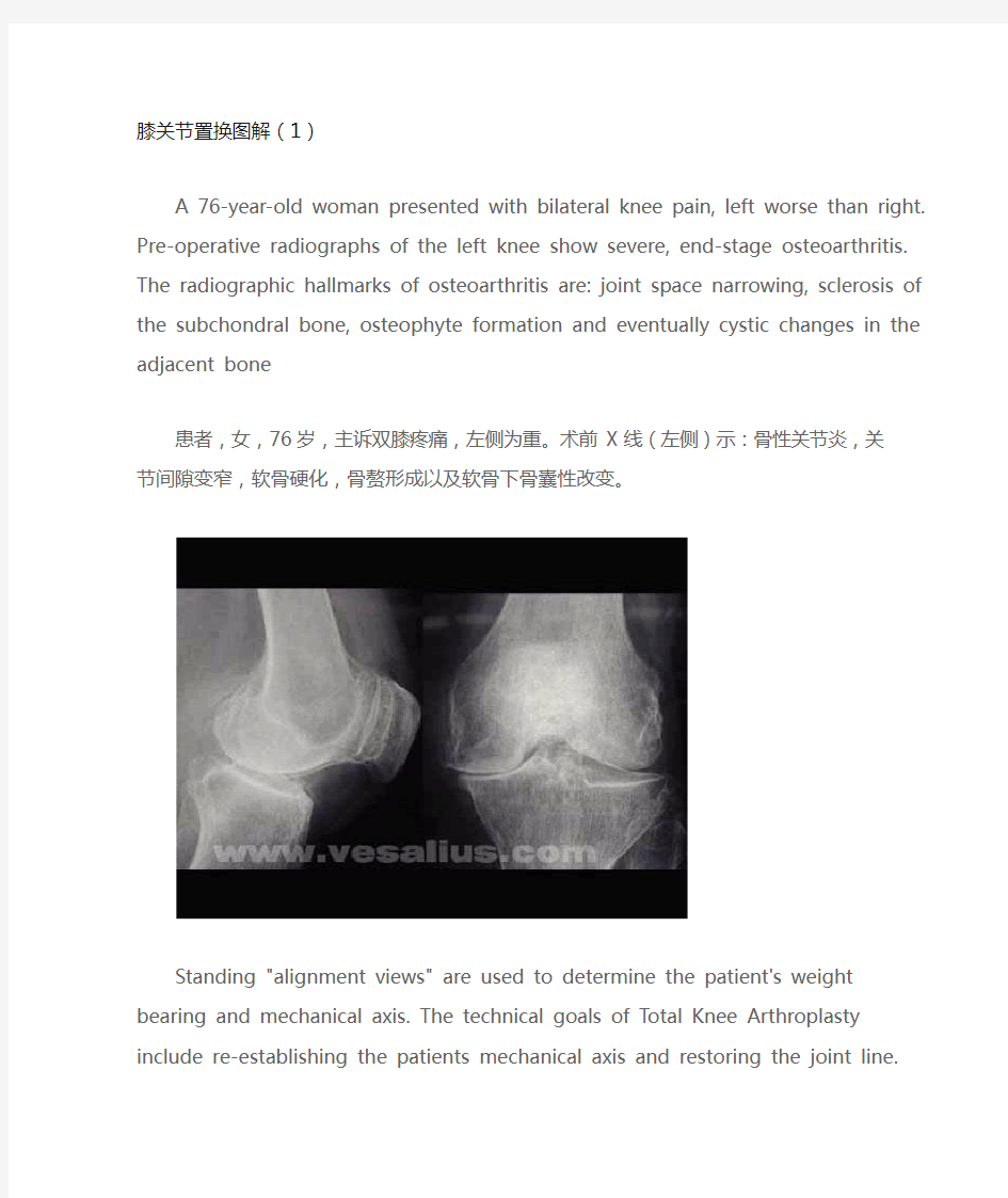
A 76-year-old woman presented with bilateral knee pain, left worse than right. Pre-operative radiographs of the left knee show severe, end-stage osteoarthritis. The radiographic hallmarks of osteoarthritis are: joint space narrowing, sclerosis of the subchondral bone, osteophyte formation and eventually cystic changes in the adjacent bone
患者,女,76岁,主诉双膝疼痛,左侧为重。术前X线(左侧)示:骨性关节炎,关节间隙变窄,软骨硬化,骨赘形成以及软骨下骨囊性改变。
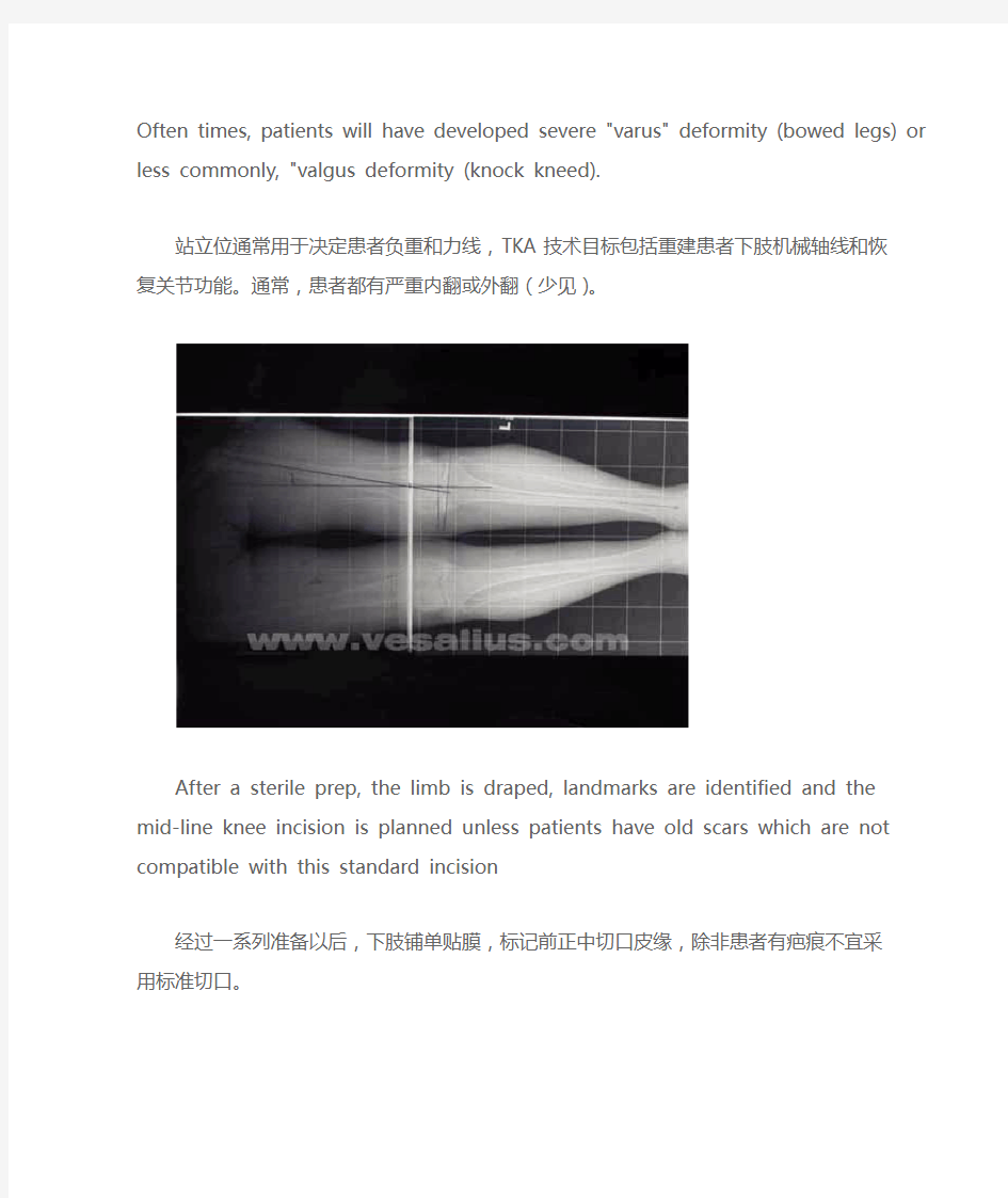
Standing "alignment views" are used to determine the patient's weight bearing and mechanical axis. The technical goals of Total Knee Arthroplasty include re-establishing the patients mechanical axis and restoring the joint line. Often times, patients will have developed severe "varus" deformity (bowed legs) or less commonly, "valgus deformity (knock kneed).
站立位通常用于决定患者负重和力线,TKA技术目标包括重建患者下肢机械轴线和恢复关节功能。通常,患者都有严重内翻或外翻(少见)。
After a sterile prep, the limb is draped, landmarks are identified and the mid-line knee incision is planned unless patients have old scars which are not compatible with this standard incision 经过一系列准备以后,下肢铺单贴膜,标记前正中切口皮缘,除非患者有疤痕不宜采用标准切口。
The leg is exsanguinated and a tourniquet is used to maintain hemostasis throughout the case. 用下肢驱血带驱血,保持整个手术过程清晰。
Once the incision is made, the quadriceps tendon, the patella and the patellar tendon are identified. A medial para-patellar arthrotomy is made and the soft tissues are elevated from the tibia. Great care must be taken not to strip to much medially or laterally as this may result in disruption of the medial collateral ligament or the patellar tendon, respectfully. Both are disastrous complications.
切开皮肤,显露股四头肌腱,髌骨,髌腱。从髌骨内侧缘(保留0.5cm软组织以利用缝合)切开软组织致胫骨,注意不要向内侧或外侧剥离过多,以防损伤内侧副韧带或髌韧带,以免引起严重并发症。
The patella and patellar tendon are released from the underlying fat pad and other soft tissues so the patella may be everted laterally to expose the distal femur and proximal tibia.
分离髌骨和髌韧带下方的脂肪垫及其他软组织,以便髌骨向外翻,以显露股骨远端和胫骨近端。
After the patella and tendon are everted (under rake in photo), remaining capsular tissues are released. The patellar-femoral ligament above the clamp is about to be divided.
把髌骨和髌韧带翻向一侧后,清除剩余软组织,分离髌股韧带(下图弯钳上方组织)
Only a single cut is made to prepare the tibia. An extramedullary alignment guide is placed and secured with pins in the proximal tibia. This guide is used to resect the proper amount of bone and create the proper surface angulation for the new tibial joint line
胫骨下方切一个单独切口,安装髓外导向器并用克氏针固定在胫骨近端,导向器是为了切除适量骨关节面,为新胫骨关节线创造一个恰当面。
Several pins are placed to secure the guide.
几枚克氏针加强固定。
Once the guide is secure, the arthritic articulating surface of the tibia is resected using an oscillating saw
当导向器安装稳定,使用摆锯切除胫骨关节面
After the cut is made with the oscillating saw, the section of tibia is removed.
切除完成后,移除关节面。
The resected arthritic articular surface of proximal tibia is shown
After the tibial bone is resected, edges and any remaining bone are removed.