概述髋关节是躯干与下肢的重要连接装置与承重结构,主要由股骨头、颈与髋臼共同构成。股骨颈的长轴线与股骨干纵轴线之间形成颈干角,为110°-140°,平均为127°儿童颈干角大于成人。成人股骨头的血液供应有多种来源:①股骨头圆韧带内的小凹动脉,提供股骨头凹部的血液循环;②股骨干滋养动脉升支,沿股骨颈进入股骨头;③旋股内、外侧动脉的分支,是股骨头、颈的重要营养动脉。股骨颈骨折(fracture of the fenoral neck)常发生于老年人,随着人的寿命延长,其发病率日渐增高。其临床治疗中存在骨折不愈合和股骨头缺血坏死两个主要问题。病因和发病机制中老年人发生骨折有两个基本因素,一是骨强度下降,二是老年人髋周肌群退变,不能有效地抵消髋部有害应力。而青壮年股骨颈骨折,往往由于严重损伤所致,且多见于不稳定型。一、按骨折部位分类 1、股骨头下骨折骨折线在股骨头下,损伤旋股内、外侧动脉发出的营养血管支,使股骨头的血液供应中断,仅有股骨头圆韧带内的小凹动脉提供是少量供血,导致股骨头缺血严重,发生股骨头缺血性坏死。2、经股骨颈骨折骨折线在股骨颈中部,呈斜形,多有一三角形骨块与股骨头相连。骨折损伤股骨干滋养动脉升支,导致股骨头供血不足,易发生股骨头缺血性坏死和骨折不愈合。3、股骨颈基底骨折骨折线位于股骨颈大、小转子之间连线处。旋股内、外侧动脉的分支提供血液循环,故对骨折部的血液供应影响不大,骨折容易愈合。二、按X线表现分类 1、内收骨折远端骨折线与两侧髂嵴连线的夹角(Pauwells角)大于50°,为内收骨折,属于不稳定型骨折,因为其骨折面接触较少,容易移位。Pauwells角越大,骨折端所遭受的剪切力越大,骨折越不稳定。2、外展骨折远端骨折线与两侧髂嵴连线的夹角小于30°,为外展骨折,属于稳定性骨折。若处理不当,如过度牵引,外旋、内收,或过早负重等,也可发生移位,成为不稳定性骨折。三、按移位程度分类 1、不完全骨折仅有部分骨完整性破坏,股骨颈的一部分出现裂纹,类似于骨折的裂纹骨折。2、完全性骨折骨折线贯穿股骨颈,骨结构完全破坏。可分为:(1)无移位的完全性骨折;(2)部分移位的完全骨折;(3)完全移位的完全骨折。临床表现一、畸形患肢多有轻度屈髋屈膝及外旋畸形,一般在45°-60°之间。二、疼痛移动患肢时髋部疼痛明显。在患肢足跟部或大粗隆部叩击时,髋部感疼痛。三、功能障碍移位骨折病人在伤后不能坐起或站立。有时伤后并不立即出现功能障碍,仍能行走,数天后髋部疼痛逐渐加重,才出现不能行走。实验室及其他检查一、肢体测量通过检查可发现患肢缩短。在平卧位,由髂前上嵴向水平划垂线,再由大转子与髂前上嵴的垂线画水平线,构成Bryant三角,股骨颈骨折时,此三角底边较健侧缩短。在平卧位,由髂前上嵴与坐骨结节之间画线,为Nélaton线。正常情况下,大转子在此线上,若大转子超过此线,表明大转子有向上移位。二、X线检查X线检查可同时发现骨折的部位、类型、移位情况,是选择治疗方法的重要依据。疾病概述病因病理临床表现检查检验诊断鉴别并发症治疗预后预防诊断和鉴别诊断根据中老年患者摔倒后出现髋部疼痛,下肢活动受限,不能站立和行走,应怀疑病人有股骨颈骨折。结合X线检查、肢体测量可明确骨折部位、类型和移位。一般诊断明确,不需要鉴别。并发症一、骨折不愈合。二、股骨头缺血性坏死是股骨颈骨折晚期最常见的并发症,发生率为20%-40%.当患者已恢复正常活动后患髋出现疼痛时应复查,若X线显示股骨变白、囊性变活股骨头塌陷,可认为是股骨头缺血性坏死的表现,但往往难以预测其发病趋势。治疗一、治疗时机早期治疗有利于尽快恢复骨折后血管受压或痉挛。股骨颈骨折手术原则上不超过2周。二、骨折复位准确良好的复位是骨愈合重要的条件。牵引患肢,同时在大腿根部加反牵引,待肢体原长度恢复后,行内旋外展复位。三、内固定目前内固定器材主要四类:①单钉类:三翼钉为代表,三刃钉内固定为众所熟悉的传统疗法。这种单根钉在骨的力学效能上不能持久,另外,此钉也不适于青少年及颈部粉碎性骨折者。②多钉固定类:包括史氏针、三角针和多根螺纹钉。此类固定对骨的损伤较小,利用多钉的布局在生物力学上的优势,疗效较好,缺点是钉退出后骨不愈合。③滑移式钉板固定装置
?股骨粗隆间骨折(intertrochanteric fracture) ?2009年03月04日电力医院骨科 ? 股骨近端解剖 定义:股骨粗隆间骨折系指股骨颈基底至小粗隆(小转子)下缘之间的骨折。 概述:股骨粗隆间骨折多见于老年人,男性多于女性,约为1.5:1,属于关节囊外骨折。美国每年发生超过25万例髋部骨折,总的的治疗费用估计超过80亿美元,其中股骨粗隆间骨折约占1/2,我国统计股骨粗隆间骨折患病年龄平均为70岁,比股骨颈骨折患者高5~6岁,高龄患者长期卧床引起并发症(肺炎;褥疮;泌尿系感染)较多,病死率为15~20%。 股骨粗隆部有许多肌肉附着,所以局部的血液供给丰富,加以骨折的接触面积大,因此,骨折后愈合连接一般不成问题。主要问题是有发生髋内翻的趋势,形成畸形连接,造成跛行,并由于承重线的改变,可能在后期引起患肢创伤性关节炎。 病因及危险因素: 1.直接暴力:大粗隆受到直接打击。 2.间接暴力:下肢突然扭转、跌倒时强力内收或外展。 3.骨质疏松:骨质疏松本身不是单独的危险因素,但绝经后妇女增加锻 炼、激素替代治疗并摄入足够的钙质能够降低股骨粗隆间骨折(髋部骨折)的发生率。 分类及分型: 1.按骨折线方向分型:此分型目的在于表示其稳定性。
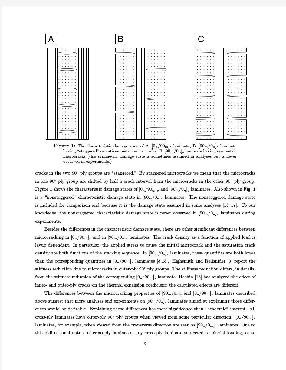
(1)顺粗隆间线型(骨折),即骨折线由大粗隆向内下至小粗隆,其走行与粗隆间线平行,称为稳定型。 (2)逆粗隆间线型(骨折):即骨折线由大粗隆下方向内上达小粗隆的上方,称为不稳定型。有时骨折线难以分辨走向,呈粉碎骨折,其稳定性亦差。 临床实践表现,以骨折的原始状态来判断其稳定性似乎更为重要。凡伤后髋内翻越严重,骨折越不稳定,反之,原始髋内翻越轻或无内翻者,骨折越趋稳定。因此,骨折的稳定性似与骨折走向方向无关。 A图为顺粗隆间线型(骨折) B图为逆粗隆间线型(骨折) 2. 改良Evans或Evans-Jensen分型系统: Jensen对于Evans分型进行了改进,基于大小粗隆是否受累及复位后骨折是否稳定而分为五型。Ⅰa型:两骨折片段,骨折无移位。Ⅰb型:两骨折片段,骨折有移位。Ⅱa型:三骨折片段,累及大粗隆,因为移位的大粗隆片段而缺乏后外侧支持。Ⅱb型:3骨折片段,累及小粗隆,由于小粗隆或股骨矩骨折缺乏后内侧支持。Ⅲ型:四骨折片段,骨折累及两个粗隆,缺乏内侧和外侧的支持,为Ⅱa型和Ⅱb型的结合。Jensen研究发现Ⅰa、Ⅰb型骨折94%复位后稳定;Ⅱa型骨折33%复位后稳定;Ⅱb 型骨折21%复位后稳定;Ⅲ型骨折8%复位后稳定。Jensen指出大小粗隆的粉碎程度与复位后骨折的稳定性成反比,改良的Evans分型为判断复位后的稳定性和骨折再次移位的风险提供了最为可靠的预测。
【创伤骨科常用汉英词汇】 A 凹陷骨折depressed fracture B 半脱位subluxation 半月板meniscus 闭合性骨折closed fracture 髌骨patella 髌骨骨折fracture of patella 髌骨脱位dislocation of patella 髌上囊suprapatellar bursa 髌下脂肪垫subpatellar fat pad 髌韧带patellar ligament 病理性骨折pathological fracture 爆裂骨折bursting fracture 鼻骨骨折fracture of nasal bone 不完全骨折no-complete fracture C 尺骨ulnar 尺骨茎突styloid process of ulna
尺骨鹰嘴骨折olecranon fracture of ulna 创伤性脱位traumatic dislocation 创伤性关节炎traumatic arthritis 创伤性滑膜炎traumatic synovitis 耻骨pubis 耻骨联合pubic symphysis 耻骨上支superior ramus of pubis 耻骨下支inferior ramus of pubis 陈旧性骨折old fracture 垂直压迫损伤vertical compression injuries 齿状突odontoid process 错位displacement D 骶骨sacrum 骶椎sacral vertebrae 骶髂关节cacroiliac joint 骶髂关节分离separation of cacroiliac joint 大多角骨trapezium bone 大结节greater tubercle 大转子greater trochanter 对位paratope 对线alignment
骨科相关词汇 abduction 外展 abrasion 擦伤 abscess 脓肿 acromegaly 肢端肥大症 adenoma 腺瘤 adolescent 青少年的 adduction 内收 afferent neuron 传入神经 ankylosing spondylitis 强直性脊柱炎 achilles tendon 跟腱 arthritis 关节炎 arthrogryposis 关节屈曲 Aseptic Necrosis 无菌坏死 Avascular necrosis of femoral head 股骨头坏死 axillary nerve 腋神经 bone 骨 brachial plexus 臂丛 Bunion拇囊炎 bursitis 粘液囊炎 calcaneus 跟骨 callus 胼胝 Cellulitis 蜂窝组织炎 cervical 颈椎的 bone cement 骨水泥 chiropody 足科 clubfoot 马蹄内翻足 corn 鸡眼 crescent 新月形 cubital 尺侧的 cyst 囊 cystic 囊性变 débridement 清创术 dislocation 脱位 dysplasia 发育不良 effusion 渗出 erosion 侵蚀 extension 伸展 flexion 屈曲 fracture 骨折 fungal 真菌的 fusion 融合 humeral 肱骨的 humerus 肱骨 hyperostosis 骨肥厚 hypertrophy 肥大 lumbar 腰椎的 idiopathic 特发的 Ingrown toenail 嵌甲
Fracture Toughness Fracture toughness is an indication of the amount of stress required to propagate(繁殖) a preexisting(先前的) flaw. It is a very important material property since the occurrence of flaws is not completely avoidable in the processing, fabrication, or service of a material/component. Flaws may appear as cracks, voids, metallurgical inclusions, weld defects, design discontinuities, or some combination thereof. Since engineers can never be totally sure that a material is flaw free, it is common practice to assume that a flaw of some chosen size will be present in some number of components and use the linear elastic fracture mechanics (LEFM) approach to design critical components. This approach uses the flaw size and features, component geometry, loading conditions and the material property called fracture toughness to evaluate the ability of a component containing a flaw to resist fracture. A parameter called the stress-intensity factor (K) is used to determine the fracture toughness of most materials. A Roman numeral subscript indicates the mode of fracture and the three modes of fracture are illustrated in the image to the right. Mode I fracture is the condition in which the crack plane is normal to the direction of largest tensile loading. This is the most commonly encountered mode and, therefore, for the remainder of the material we will consider K I The stress intensity factor is a function of loading, crack size, and structural geometry. The stress intensity factor may be represented by the following equation: Where:K I is the fracture toughness in s is the applied stress in MPa or psi a i s the crack length in meters or inches B is a crack length and component geometry factor that is different for each specimen and is dimensionless.
骨折愈合(Fracture healing) General fracture healing schedule: Fracture site healing time (week), fracture site healing time (unit: week) Phalanx (metacarpal bone) 4-8 Pelvic 6-10 Phalanx (metatarsus) 6-8 Femoral neck 12-24 Common fracture healing time unit: Week Fracture type - spiral or long oblique: upper limb clinical healing 3, firm heal 6 Lower limb clinical healing 6, firm healing 12 Fracture type transverse section: upper limb clinical healing 6, firm union 12 Lower limb clinical healing 12, firm healing 24 Note: suitable for tubular bone; children heal half time How long is the fracture healing time? In order to promote fracture healing faster and better, fracture healing time is faster.