LETTERS
TRPC channel activation by extracellular thioredoxin
Shang-Zhong Xu 1*{,Piruthivi Sukumar 1*,Fanning Zeng 1,Jing Li 1,Amit Jairaman 1,Anne English 3,
Jacqueline Naylor 1,Coziana Ciurtin 1,Yasser Majeed 1,Carol https://www.doczj.com/doc/8f13459199.html,ligan 1,Yahya M.Bahnasi 1,Eman Al-Shawaf 1,Karen E.Porter 2,Lin-Hua Jiang 1,Paul Emery 3,Asipu Sivaprasadarao 1&David J.Beech 1
Mammalian homologues of Drosophila melanogaster transient receptor potential (TRP)are a large family of multimeric cation channels that act,or putatively act,as sensors of one or more chemical factor 1,2.Major research objectives are the identification of endogenous activators and the determination of cellular and tissue functions of these channels.Here we show the activation of TRPC5(canonical TRP 5)homomultimeric and TRPC5–TRPC1heteromultimeric channels 3–5by extracellular reduced thiore-doxin,which acts by breaking a disulphide bridge in the predicted extracellular loop adjacent to the ion-selectivity filter of TRPC5.Thioredoxin is an endogenous redox protein with established intracellular functions,but it is also secreted and its extracellular targets are largely unknown 6–9.Particularly high extracellular con-centrations of thioredoxin are apparent in rheumatoid arth-ritis 8,10–12,an inflammatory joint disease that disables millions of people worldwide 13.We show that TRPC5and TRPC1are expressed in secretory fibroblast-like synoviocytes from patients with rheumatoid arthritis,that endogenous TRPC5–TRPC1chan-nels of the cells are activated by reduced thioredoxin,and that blockade of the channels enhances secretory activity and prevents the suppression of secretion by thioredoxin.The data indicate the presence of a previously unrecognized ion-channel activation mechanism that couples extracellular thioredoxin to cell function.TRPC5is markedly activated by extracellular lanthanide ions 4,14,15.The effects of these ions depend on a glutamic acid residue at position 543(ref.14)in the predicted extracellular loop adjacent to the ion pore (Supplementary Figs 1and 2).This structural feature may there-fore have functional importance in enabling extracellular factors to activate the channels.Because lanthanides are unlikely to be physio-logical activators,we were interested in alternatives and developed a hypothesis based on amino acid sequence alignment,which showed two cysteine residues near glutamic acid 543that are conserved in TRPC5,TRPC4and TRPC1(Supplementary Fig.2),a subset of the seven TRPC channels 1–5.TRPC5and TRPC4have similar functional properties 4and both form heteromultimers with TRPC1(refs 3–5),a subunit that has weak targeting to the plasma membrane when expressed in isolation 3,16.
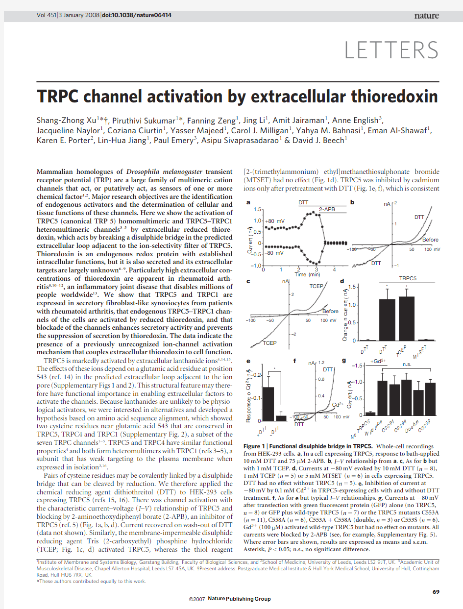
Pairs of cysteine residues may be covalently linked by a disulphide bridge that can be cleaved by reduction.We therefore applied the chemical reducing agent dithiothreitol (DTT)to HEK-293cells expressing TRPC5(refs 15,16).There was channel activation with the characteristic current–voltage (I –V )relationship of TRPC5and blocking by 2-aminoethoxydiphenyl borate (2-APB),an inhibitor of TRPC5(ref.5)(Fig.1a,b,d).Current recovered on wash-out of DTT (data not shown).Similarly,the membrane-impermeable disulphide reducing agent Tris (2-carboxyethyl)phosphine hydrochloride (TCEP;Fig.1c,d)activated TRPC5,whereas the thiol reagent
[2-(trimethylammonium)ethyl]methanethiosulphonate bromide (MTSET)had no effect (Fig.1d).TRPC5was inhibited by cadmium ions only after pretreatment with DTT (Fig.1e,f),which is consistent
*These authors contributed equally to this work.
1
Institute of Membrane and Systems Biology,Garstang Building,Faculty of Biological Sciences,and 2School of Medicine,University of Leeds,Leeds LS29JT,UK.3Academic Unit of Musculoskeletal Disease,Chapel Allerton Hospital,Leeds LS74SA,UK.{Present address:Postgraduate Medical Institute &Hull York Medical School,University of Hull,Cottingham Road,Hull HU67RX,
UK.
–100
–50
50
100 mV
–1
1
2
Before
DTT
DTT
nA 2+b
D T T
+D
T T N
o
T R P C
5
C 553A C 553S
D o u b l e C 558A W
i l d
t y p e –D T T D T T
T C E P
M T S E T
Figure 1|Functional disulphide bridge in TRPC5.Whole-cell recordings from HEK-293cells.a ,In a cell expressing TRPC5,response to bath-applied 10mM DTT and 75m M 2-APB.b ,I –V relationship from a .c ,As for b but with 1mM TCEP.d ,Currents at 280mV evoked by 10mM DTT (n 58),1mM TCEP (n 55)or 5mM MTSET (n 56)in cells expressing TRPC5.DTT had no effect without TRPC5(n 55).e ,Inhibition of current at
280mV by 0.1mM Cd 21in TRPC5-expressing cells with and without DTT treatment.f ,As for e but typical I –V relationships.g ,Currents at 280mV after transfection with green fluorescent protein (GFP)alone (no TRPC5,n 58)or GFP plus wild-type TRPC5(n 57)or the TRPC5mutants C553A (n 511),C558A (n 56),C553A 1C558A (double,n 53)or C553S (n 56).Gd 31(100m M)activated wild-type TRPC5but had no effect on mutants.All currents were blocked by 2-APB (see,for example,Supplementary Fig.5).Where error bars are shown,results are expressed as means and s.e.m.Asterisk,P ,0.05;n.s.,no significant difference.
Vol 451|3January 2008|doi:10.1038/nature06414
69
with the metal ions acting by re-engaging cysteine residues17.Other TRP channels lacking the cysteine pair in a similar position were unresponsive to DTT(Supplementary Figs2and3).The data sup-port the hypothesis that the cysteine pair in TRPC5normally engages in a disulphide bridge that constrains the channel in a state of limited opening probability,enabling enhanced channel activity when the bridge is broken.
To test the hypothesis further,we expressed TRPC5mutants con-taining alanine in place of cysteine.Such mutants were constitutively active and were not stimulated by reducing agent or lanthanide (Fig.1g and Supplementary Figs4and5).Ionic currents for the single mutants(C553A,C553S or C558A)and double mutant (C553A1C558A)were not significantly different,suggesting that the two cysteine residues have a joint role(Fig.1g).Expression of wild-type TRPC1together with the TRPC5double mutant led to smaller constitutive currents that were not affected by DTT or lan-thanide,which is consistent with TRPC1suppressing the current amplitude but not conferring a functional effect of reducing agents (Supplementary Fig.6).Dimers of TRPC5were not detected under non-reducing conditions,suggesting an intra-subunit rather than inter-subunit disulphide bridge(Supplementary Fig.7).
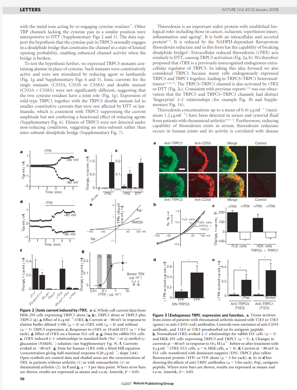
Thioredoxin is an important redox protein with established bio-logical roles including those in cancer,ischaemic reperfusion injury, inflammation and ageing8.It is both an intracellular and secreted protein6–9.It is reduced by the NADPH-dependent flavoprotein thioredoxin reductase and in this form has the capability of breaking disulphide bridges8.Extracellular reduced thioredoxin(rTRX)acts similarly to DTT,causing TRPC5activation(Fig.2a,b).We therefore proposed that rTRX is a previously unrecognized endogenous extra-cellular regulator of TRPC5.In taking this idea forward we also considered TRPC1because many cells endogenously expressed TRPC5and TRPC1together,leading to TRPC5–TRPC1heteromul-timers3,5,16,18.The TRPC5–TRPC1channel is also activated by rTRX or DTT(Fig.2c).Consistent with previous reports3,16was our obser-vation that the TRPC5and TRPC5–TRPC1channels had distinct ‘fingerprint’I–V relationships(for example Fig.3b and Supple-mentary Fig.14).
Thioredoxin concentrations up to a mean of0.41m g ml21(maxi-mum1.2m g ml21)have been detected in serum and synovial fluid from patients with rheumatoid arthritis8,10–12.Furthermore,reducing capability of thioredoxin exists in serum;thioredoxin reductase occurs in human joints and its activity is correlated with
disease D
T
T
B
o
i
l
e
d
D
T
T
B
u
f
f
e
r
R
e
s
i
n
r
T
R
X
B
o
i
l
e
d
r
T
R
X
o
T
R
X
[rTRX] (μg ml–1)
a
d
f g
C
u
r
r
e
n
t
(
n
A
)
C
u
r
r
e
n
t
(
n
A
)
C
h
a
n
g
e
i
n
c
u
r
r
e
n
t
(
n
A
)
Figure2|Ionic current induced by rTRX.a–c,Whole-cell current data from
HEK-293cells expressing TRPC5alone(a,b),TRPC1alone or TRPC5plus
TRPC1(c).a,Effect of4m g ml21rTRX.b,Current at280mV in response to
elution buffer diluted1:100(n54)or rTRX with(n58)and without
(n53)TRPC5expression.c,Responses to rTRX or10mM DTT(n55for
each).d,Effect of rTRX on a human FLS cell.e–g,Data for rabbit FLS cells.
e,rTRX induced I–V relationships in standard bath(Na1)or N-methyl-D-
glucamine(NMDG1)solution(see Supplementary Fig.9).f,Currents
evoked at280mV.g,Data for human rTRX with a fitted Hill equation
(concentration giving half-maximal response0.20m g ml21,slope2.64).
Open symbols are control data and shaded areas are the concentrations of
TRX in patients without arthritis(1)or with osteoarthritis(1)or
rheumatoid arthritis(2).In f and g,n55per data point.Where error bars
are shown,results are expressed as means and s.e.m.Asterisk,P,
0.05.
a
b
DN-TRPC5Anti-TRPC1
(T1E3)
Anti-TRPC5
(T5E3)
c
d e
C
h
a
n
g
e
i
n
c
u
r
r
e
n
t
(
%
)
TRPC5 + TRPC1
Anti-TRPC1Anti-CD55Merge Control
Anti-TRPC5Anti-CD55Merge Control
r
T
R
X
-
e
v
o
k
e
d
c
u
r
r
e
n
t
(
n
A
)
r
T
R
X
-
e
v
o
k
e
d
c
u
r
r
e
n
t
(
n
A
)
Figure3|Endogenous TRPC expression and function.a,Tissue sections
from joints of patients with rheumatoid arthritis stained with T1E3or T5E3
(green)or anti-CD55(red)antibodies.Controls were omission of anti-CD55
antibody,and T1E3or T5E3preadsorbed on its antigenic peptide.
b,Normalized rTRX-evoked I–V relationships for rabbit FLS cells(n53)
and HEK-293cells expressing TRPC5and TRPC1(n55).c,Changes in
currents at280mV in response to10m M La31before or after treatment with
4m g ml21rTRX(FLS cells,n56;HEK cells,n54).d,Current at280mV in
FLS cells transfected with dominant-negative(DN)TRPC5plus yellow
fluorescent protein(YFP)or YFP alone(n55for each).e,As in d but
showing the effects of anti-TRPC antibodies(n55for each).Pep.,antigenic
peptide.Where error bars are shown,results are expressed as means and
s.e.m.Asterisk,P,0.05.
LETTERS NATURE|Vol451|3January2008 70
severity10,19,20(see also Supplementary Results and Supplementary Discussion).We therefore considered whether the activation of TRPC5by rTRX is relevant to the cells that secrete synovial fluid, the CD55-positive fibroblast-like synoviocytes(FLS cells).CD55-positive FLS cells(Supplementary Fig.8)showed a non-selective cationic current in response to DTT or rTRX(Fig.2d–g and Supplementary Figs9and10).The mean current evoked by rTRX at280mV in FLS cells from the knee joint of patients with rheum-atoid arthritis was20.8560.42nA(mean6s.e.m.;n514). Oxidized TRX(oTRX)had no effect(Fig.2f).The effective concen-trations of rTRX indicate a possible relevance to rheumatoid arthritis (Fig.2g).Nitric oxide is an alternative endogenous regulator of cysteine residues21;however,it failed to evoke current in FLS cells, even at a concentration100-fold that required to evoke vasorelaxa-tion(Supplementary Fig.10,Supplementary Results and Supple-mentary Discussion).
There have been no previous reports on the expression of TRPC channels in synovial joints,so we explored synovial tissue biopsies from patients with rheumatoid arthritis.TRPC5and TRPC1proteins were detected and localized together with CD55(Fig.3a).Similarly, the FLS cells used in our electrophysiological experiments expressed messenger RNAs encoding TRPC5and TRPC1,western blotting indicated the presence of TRPC5and TRPC1proteins,and immuno-labelling revealed TRPC5and TRPC1at the cell surface(Supple-mentary Figs8,11and13).
The I–V relationship of the rTRX-evoked current in FLS cells was similar to that of the TRPC5–TRPC1heteromultimeric channel (Fig.3b).Furthermore,experiments with lanthanum ions showed unusual and striking similarity between the endogenous current and the current of overexpressed TRPC5–TRPC1:in the absence of a reducing agent,lanthanum ions stimulated current in both HEK-293cells(exogenously expressing TRPC5–TRPC1)and FLS cells, whereas after the induction of current by rTRX,lanthanum ions were inhibitory in both cases(Fig.3c and Supplementary Fig.9).Also consistent with the involvement of TRPC channels were the observa-tions that the rTRX-evoked current of FLS cells was blocked by 2-APB and that the inward current was suppressed when most of the extracellular Na1was replaced by the bulky and impermeant cation N-methyl-D-glucamine(Fig.2e and Supplementary Fig.9). As a further test of the involvement of TRPC5and TRPC1,FLS cells were transfected with a dominant-negative ion-pore mutant of TRPC5that inhibited native channels capable of interacting with TRPC5(refs16,22).The mutant suppressed current evoked by rTRX(Fig.3d).
Further evidence that TRPC5and TRPC1contribute to the endo-genous rTRX-responsive channel of FLS cells came from studies with anti-TRPC5(T5E3)and anti-TRPC1(T1E3)antibodies,which tar-get the predicted extracellular loop region and specifically block the functions of TRPC5and TRPC1,respectively23–25.T5E3and T1E3 antibodies labelled unpermeabilized FLS cells,unlike antibody tar-geted to the intracellular carboxy terminus of TRPC5,which labelled only permeabilized cells(Supplementary Figs8and11),indicating that TRPC5and TRPC1are transmembrane proteins with extracel-lular epitopes.Like dominant-negative mutant TRPC5,T5E3or T1E3suppressed rTRX-evoked current(Fig.3e).Antibody targeted to CD55,which is a membrane protein unrelated to TRP,had no significant effect(n57;data not shown).Gene expression,electro-physiology,pharmacology,recombinant DNA and antibody studies therefore yielded data consistent with the carrying of rTRX-evoked current in FLS cells by a channel containing TRPC5and TRPC1. One of the functions of FLS cells is to secrete matrix metallopro-teinases(MMPs),which are associated with tissue remodelling and the progression of arthritis26.The use of zymography to detect gela-tinase activities of MMP-2and MMP-9secreted from rabbit FLS cells (Supplementary Fig.12)revealed that T5E3and T1E3antibodies have large stimulatory effects(Fig.4a,b).Human FLS cells showed greater MMP-2secretion than that of MMP-9(compare Supplementary Fig.12with Fig.4a).Enzyme-linked immuno-sorbent assays(ELISAs)for human MMP-2enabled the quantifica-tion of the absolute concentration of total MMP-2secreted;again, either T5E3or T1E3antibody had a profound stimulatory effect (Fig.4c).Similarly,knockdown of expression of the genes encoding TRPC1and TRPC5by RNA-mediated interference enhanced the secretion of MMP-2(Supplementary Fig.13).Inhibition of MMP secretion by the addition of exogenous reducing TRX was lost in the presence of T5E3(Fig.4d).Similar data were obtained for pro-MMP-1secretion from human FLS cells(Fig.4e,f)and MMP-9measured by zymography in rabbit FLS cells(n56;data not shown).The data therefore reveal constitutive and rTRX-evoked activity of TRPC5and TRPC1channels that inhibits the secretion of MMP from FLS cells.
The data of this study indicate that secreted TRX is a type of ion channel agonist that acts through its reduced form to break a restraining intra-subunit disulphide bridge between cysteine residues in TRPC5,thereby stimulating the channel either as a homomeric assembly or as a heteromultimer with TRPC1.A transduction mech-anism is therefore revealed that can directly couple cell activity to extracellular reduced thioredoxin.This mechanism may have par-ticular relevance in conditions such as rheumatoid arthritis,in which TRX concentrations are strongly elevated,but the broad distribu-tions of TRX and the channels indicate that the mechanism could be widely
used.
a
cocktail [
M
M
P
-
2
]
(
n
g
m
l
–
1
)
[
M
M
P
-
1
]
(
p
g
m
l
–
1
)
c
e
Figure4|Relevance to secretion from FLS cells.a,Zymogram showing MMP-9(pro and active)and MMP-2from rabbit.Ab,antibody.b,As for a but mean data after normalization of rabbit MMP-9band intensity to the control group without antibody(n53for each).c,ELISA data for human MMP-2(n54).d,Effect of T5E3(n54)on inhibition of human MMP-2 secretion by exogenous TRX cocktail.For each group,secretion in TRX was normalized to that in its absence(control).e,f,As for c,d,but for secretion of human pro-MMP-1.Results are expressed as means and s.e.m.Asterisk, P,0.05;n.s.,no significant difference.
NATURE|Vol451|3January2008LETTERS
71
METHODS SUMMARY
Cells.Synovial tissue biopsies were obtained with informed consent from patients diagnosed with rheumatoid arthritis at the Academic Unit of Musculoskeletal Disease,Chapel Allerton Hospital,Leeds.Ethical approval was given by the local ethics committee.Human synovial tissue biopsies were washed with PBS and digested in0.25%type1A collagenase for4h at37u C,after which FLS cells were cultured in DMEM/F-121Glutamax(Gibco).HEK-293 cells were grown in DMEM-F12(Gibco)and rabbit FLS cells(HIG82;ATCC) were grown in Ham’s F12(Gibco).Culture media contained10%fetal calf serum,100IU ml21penicillin and100m g ml21streptomycin.Cells were main-tained at37u C in a humidified atmosphere of5%CO2in air and replated on coverslips or24-well plates before experiments.
Electrophysiology.Whole-cell patch-clamp recordings were performed15,16at 2162u C using patch pipette solution containing(in mM):115CsCl,10EGTA, 2MgCl2,5Na2ATP,0.1NaGTP,10HEPES,5.7CaCl2;the pH was adjusted to7.2 with CsOH.The standard bath solution contained(in mM):130NaCl,5KCl,8 D-glucose,10HEPES,1.2MgCl2and1.5CaCl2;the pH was adjusted to7.4with NaOH.
Data analysis.Ionic currents are shown as positive values when they increased in response to a treatment,and as negative values when they decreased.Data are expressed as means and s.e.m.,where n is the number of individual experiments. Data sets were compared by using paired or unpaired Student’s t-tests,with a significant difference indicated by P,0.05(asterisk)and no difference by n.s. All human tissue or cell data are derived from,or are representative of,at least three independent experiments on samples from three patients.
Full Methods and any associated references are available in the online version of the paper at https://www.doczj.com/doc/8f13459199.html,/nature.
Received6July;accepted24October2007.
1.Flockerzi,V.An introduction on TRP channels.Handb.Exp.Pharmacol.179,1–19
(2007).
2.Nilius,B.,Owsianik,G.,Voets,T.&Peters,J.A.Transient receptor potential cation
channels in disease.Physiol.Rev.87,165–217(2007).
3.Strubing,C.,Krapivinsky,G.,Krapivinsky,L.&Clapham,D.E.TRPC1and TRPC5
form a novel cation channel in mammalian brain.Neuron29,645–655(2001).
4.Plant,T.D.&Schaefer,M.TRPC4and TRPC5:receptor-operated Ca21-
permeable nonselective cation channels.Cell Calcium33,441–450(2003).
5.Beech,D.J.Canonical transient receptor potential5.Handb.Exp.Pharmacol.179,
109–123(2007).
6.Rubartelli,A.,Bajetto,A.,Allavena,G.,Wollman,E.&Sitia,R.Secretion of
thioredoxin by normal and neoplastic cells through a leaderless secretory
pathway.J.Biol.Chem.267,24161–24164(1992).
7.Arner,E.S.&Holmgren,A.Physiological functions of thioredoxin and thioredoxin
reductase.Eur.J.Biochem.267,6102–6109(2000).
8.Burke-Gaffney,A.,Callister,M.E.&Nakamura,H.Thioredoxin:friend or foe in
human disease?Trends Pharmacol.Sci.26,398–404(2005).
9.Schwertassek,U.et al.Selective redox regulation of cytokine receptor signaling by
extracellular thioredoxin-1.EMBO J.26,3086–3097(2007).10.Maurice,M.M.et al.Expression of the thioredoxin–thioredoxin reductase system
in the inflamed joints of patients with rheumatoid arthritis.Arthritis Rheum.42, 2430–2439(1999).
11.Yoshida,S.et al.Involvement of thioredoxin in rheumatoid arthritis:its
costimulatory roles in the TNF-a-induced production of IL-6and IL-8from
cultured synovial fibroblasts.J.Immunol.163,351–358(1999).
12.Jikimoto,T.et al.Thioredoxin as a biomarker for oxidative stress in patients with
rheumatoid arthritis.Mol.Immunol.38,765–772(2002).
13.Smolen,J.S.,Aletaha,D.,Koeller,M.,Weisman,M.H.&Emery,P.New therapies
for treatment of rheumatoid https://www.doczj.com/doc/8f13459199.html,ncet advance online publication
doi:10.1016/S0140-6736(07)60784-3(13June2007).
14.Jung,S.et https://www.doczj.com/doc/8f13459199.html,nthanides potentiate TRPC5currents by an action at extracellular
sites close to the pore mouth.J.Biol.Chem.278,3562–3571(2003).
15.Zeng,F.et al.Human TRPC5channel activated by a multiplicity of signals in a
single cell.J.Physiol.(Lond.)559,739–750(2004).
16.Xu,S.Z.et al.A sphingosine-1-phosphate-activated calcium channel controlling
vascular smooth muscle cell motility.Circ.Res.98,1381–1389(2006).
17.Elliott,D.J.et al.Molecular mechanism of voltage sensor movements in a
potassium channel.EMBO J.23,4717–4726(2004).
18.Riccio,A.et al.mRNA distribution analysis of human TRPC family in CNS and
peripheral tissues.Brain Res.Mol.Brain Res.109,95–104(2002).
19.Lemarechal,H.et al.High redox thioredoxin but low thioredoxin reductase
activities in the serum of patients with rheumatoid arthritis.Clin.Chim.Acta367, 156–161(2006).
20.Lemarechal,H.et al.Impairment of thioredoxin reductase activity by oxidative
stress in human rheumatoid synoviocytes.Free Radic.Res.41,688–698(2007).
21.Yoshida,T.et al.Nitric oxide activates TRP channels by cysteine S-nitrosylation.
Nature Chem.Biol.2,596–607(2006).
22.Strubing,C.,Krapivinsky,G.,Krapivinsky,L.&Clapham,D.E.Formation of novel
TRPC channels by complex subunit interactions in embryonic brain.J.Biol.Chem.
278,39014–39019(2003).
23.Xu,S.Z.&Beech,D.J.TrpC1is a membrane-spanning subunit of store-operated
Ca21channels in native vascular smooth muscle cells.Circ.Res.88,84–87
(2001).
24.Xu,S.Z.et al.Generation of functional ion-channel tools by E3targeting.Nature
Biotechnol.23,1289–1293(2005).
25.Xu,S.Z.,Boulay,G.,Flemming,R.&Beech,D.J.E3-targeted anti-TRPC5antibody
inhibits store-operated calcium entry in freshly isolated pial arterioles.Am.J.
Physiol.Heart Circ.Physiol.291,H2653–H2659(2006).
26.Burrage,P.S.,Mix,K.S.&Brinckerhoff,C.E.Matrix metalloproteinases:role in
arthritis.Front.Biosci.11,529–543(2006).
Supplementary Information is linked to the online version of the paper at https://www.doczj.com/doc/8f13459199.html,/nature.
Acknowledgements This work was supported by Wellcome Trust grants to D.J.B. and A.S.,and a Physiological Society Junior Fellowship to C.C.P.S.has an Overseas Research Scholarship and University Studentship,J.N.has a Biotechnology and Biological Sciences Research Council PhD studentship,Y.M.a university studentship and Y.B.a scholarship from the Egyptian Ministry of Higher Education. Author Information Reprints and permissions information is available at
https://www.doczj.com/doc/8f13459199.html,/reprints.Correspondence and requests for materials should be addressed to D.J.B.(d.j.beech@https://www.doczj.com/doc/8f13459199.html,).
LETTERS NATURE|Vol451|3January2008 72
METHODS
Complementary DNA clones,mutagenesis and cell transfection.HEK-293 cells stably expressing tetracycline-regulated human TRPC5have been described15. Expression was induced by1m g ml21tetracycline(Tet1;Sigma)for24–72h before recording.Non-induced cells without addition of tetracycline(Tet2)were con-trols.Human TRPC1cDNA was expressed transiently from the bicistronic vector pIRES EYFP16.Point mutations in human TRPC5were introduced by using QuikChange site-directed mutagenesis(Stratagene)and appropriate primer sets. Dominant-negative TRPC5is a triple alanine mutation of the conserved LFW sequence in the ion pore16,22(Supplementary Fig.2).The mutations were con-firmed by direct sequencing of the entire reading frame.cDNAs were transiently transfected into HEK-293cells or synoviocytes with FuGENE6transfection reagent(Roche)or Lipofectamine2000(Invitrogen)48h before recording. cDNA encoding GFP or YFP was cotransfected to identify transfected cells. Electrophysiology.A salt-agar bridge was used to connect the ground Ag–AgCl wire to the bath solution.Signals were amplified with an Axopatch200B patch clamp amplifier and controlled with pClamp software v.6.0(Axon)or Signal software v.3.05(CED).A1-s ramp voltage protocol from2100mV to1100mV was applied at a frequency of0.1Hz from a holding potential of260mV. Current signals were filtered at1kHz and sampled at3kHz.Patch pipettes were made from borosilicate tubing which,after fire-polishing and filling with pipette solution,had a resistance of3–5M V.The osmolarity of the pipette solution was adjusted to,290mosM with mannitol and the calculated free Ca21was200nM. ATP and GTP were omitted when recording from cells expressing TRPC5alone. When we were studying TRPC5in HEK-293cells,gadolinium chloride(Gd31, 1–5m M)was included in the bath solution to block background currents15, which evoked submaximal TRPC5current in some recordings before other agents were applied.The effect of reducing agents was not dependent on the presence of Gd31.The recording chamber had a volume of150m l and was perfused at a rate of about2ml min21.Recordings from human FLS cells used the Patchliner(Nanion)planar patch-clamp system with rapid bath solution exchange.For antibody treatment experiments,cells were treated with one of T1E3at1:500dilution(ref.23),T5E3at1:100dilution(refs24,25),boiled (10min)T1E3at1:500dilution,T5E3antibody at1:100dilution preabsorbed on its antigenic peptide(10m M)or anti-CD55antibody(see below),which were diluted in F12Ham’s medium and incubated with cells for2–3h at37u C before patch-clamp recording.
Immunostaining.Sections4m m thick were obtained from snap-frozen synovial tissue biopsy samples of patients suffering from rheumatoid arthritis,fixed with acetone and stored at280u C until use.Staining was in accordance with standard protocols.In brief,sections were incubated with primary antibody overnight at 4u C and with secondary antibody(goat anti-rabbit IgG conjugated with fluor-escein isothiocyanate(FITC)(Sigma)and donkey anti-mouse IgG conjugated with Cy3(Jackson))for1h at2162u C.For cell labelling,FLS cells adhered to coverslips were fixed for13min in4%paraformaldehyde and,unless indicated, permeabilized for2h with0.1%Triton X-100in1%BSA.Incubation in primary antibody was overnight at4u C and with secondary antibody(goat anti-rabbit IgG-FITC)for2h at room temperature.For control experiments,antibodies were preabsorbed on their antigenic peptide(10m M)or omitted,as specified. Slides were mounted with4,6-diamidino-2-phenylindole hardest mounting medium(Vector Labs)and analysed with a Zeiss confocal microscope.T1E3, T5E3,T5C3,anti-CD55(Serotec)and CD68(Dako)antibodies were used at 1:500,1:100,1:500and1:200dilutions,respectively.
Secretion assays.FLS cells were cultured for24h in24-well plates and serum-starved for24h;fresh serum-free medium was then added that contained a TRX cocktail,which included TRX(0.4m g ml21),NADPH-dependent flavoprotein TRX reductase(0.5m g ml21)and NADPH(2m g ml21)for12h.Omission of TRX was the control.Incubations with antibodies occurred for2h before addi-tion of the TRX cocktail and were maintained in the presence of the TRX cocktail.Supernatants were collected,frozen and analysed by zymography or ELISA.For zymography the supernatant was mixed with23non-reducing SDS–PAGE sample buffer and resolved through a7.4%polyacrylamide gel impreg-nated with1.5mg ml21gelatin.After electrophoresis,gels were washed,incu-bated and stained as described previously27.The relative density of gelatinolytic bands was determined from scanned images of gels by using ImageQuant soft-ware(Amersham).MMP-2or MMP-1concentrations in supernatants from human cells were quantified with Quantikine Human total MMP-2and pro-MMP-1ELISA kits in accordance with the manufacturer’s instructions(R&D Systems).
Chemicals.All salts and reagents were from Sigma or BDH.Gadolinium(Gd31) chloride,lanthanum(La31)chloride,cadmium(Cd21)chloride,DTT,2-APB, NADPH and NADPH-dependent flavoprotein thioredoxin reductase (Escherichia coli)were from Sigma.Recombinant thioredoxin(TRX;Sigma) was from E.coli(unless specified)or human(no differences in effect were observed compared with E.coli TRX)and purchased from Sigma.MTSET was from Toronto Research Chemicals and TCEP was from Pierce Biotech.MTSET, TCEP and NADPH were prepared fresh for each experiment.Collagenase was from StemCell Technologies Inc.2-APB(75mM)stock solution was in100% dimethylsulphoxide.To prepare reduced thioredoxin(rTRX),TRX(1mg)was dissolved in1ml of the binding buffer(10mM HEPES,1mM EDTA,50mM NaCl,pH7.0)and0.25ml of this was mixed with2.5m l of1M DTT,incubated at room temperature for30min and then added to0.25ml of pre-equilibrated resin (DEAE-Sephadex;Sigma).The mixture was centrifuged for30s and then washed three times with binding buffer to remove DTT completely.Elution buffer (0.25ml;10mM HEPES,1mM EDTA,1M NaCl,pH7.0)was added and cen-trifuged to harvest the supernatant.The final TRX concentration was deter-mined by Bradford assay.rTRX was diluted from cold stocks(on ice) immediately before use.
27.Porter,K.E.et al.Simvastatin inhibits human saphenous vein neointima formation
via inhibition of smooth muscle cell proliferation and migration.J.Vasc.Surg.36, 150–157(2002).
doi:10.1038/nature06414