
Dual Microelectrodes for Distance Control and Detection of Nitric Oxide from Endothelial Cells by Means of Scanning Electrochemical Microscope Sonnur Isik,?Mathieu Etienne,?,§Joshua Oni,?Andrea Blo1chl,?Sabine Reiter,?and
Wolfgang Schuhmann*,?
Analytische Chemie,Elektroanalytik und Sensorik,Lehrstuhl fu¨r Molekulare Neurobiochemie,
Ruhr-Universita¨t Bochum,Universita¨tsstrasse150,D-44780Bochum,Germany
Dual Pt disk microelectrodes consisting of a10-μm distance sensor and a50-μm nitric oxide sensor were prepared.The50-μm electrode was modified with Ni(4-N-tetramethyl)pyridyl porphyrin enclosed in the polymer network of a negatively charged electrodeposition paint. This paint prevented the dissolution of the otherwise soluble porphyrin in the aqueous test medium due to charge interactions.It also denied negatively charged ions in the analyte solution access to the electrode surface by electrostatic repulsion,thereby preventing interference from anions such as nitrite,nitrate,and ascorbate.With the aid of a scanning electrochemical microscope,it was possible to use the distance sensor by recording the negative feedback effect on the reduction of molecular oxygen to“guide”the nitric oxide sensor to various known distances from a layer of adherently growing human umbilical vein endothelial cells for the detection of nitric oxide released from the cells upon stimulation with bradykinin.The use of the distance sensor made it possible to preserve the integrity of the adherently growing cells concomitantly with the modified electrode by pre-venting the deterioration of the modifying layer during the distance adjustment step.
Events and activities taking place in biological systems have been extensively investigated using electrochemical techniques.1-7 The signals obtained,however,depend on the size of the active surface area of the sensor,8the separation between the sensor tip9,10and the source of secretion,the amount of secretion,and the diffusion profile of the secreted substance and its possible reaction with its immediate environment.To obtain quantitative and reproducible information from the secretion process,an accurate knowledge of the separation of the electrode from the secreting substrate is indispensable.Often,manual approaches (that are difficult to use under realistic circumstances)are adopted for the control of the sensor tip-substrate separation with the attendant effects of a possible contamination of the electrode surface due to its contact with the cell membrane,the eventual deterioration of the cell at the point of contact or mechanical depolarization of the cell,insufficient reproducibility of the tip-to-sample distance,and the impossibility of investigating sequentially different spots on the same cell.3
A reproducible and accurate positioning of a microsensor over a substrate can be achieved with the aid of scanning electrochemi-cal microscopy(SECM)by monitoring changes in the diffusion-limited current flowing through the tip of a microsensor due to the reaction of a free-diffusing redox species as the tip travels from a position in the bulk solution toward an interface.11-15Since there is simultaneously the possibility of carrying out ampero-metric measurements,this technique has been employed for the investigation of cellular activities.16-22The approach is simple if
*Corresponding author.Phone:+492343226200.Fax:+492343214683. E-mail:wolfgang.schuhmann@rub.de.
?Analytische Chemie,Elektroanalytik und Sensorik,Ruhr-Universita¨t Bochum.
?Lehrstuhl fu¨r Molekulare Neurobiochemie,Ruhr-Universita¨t Bochum.
§Present address:Laboratoire de Chimie Physique pour L’Environment, CNRS-Universite de Nancy1,405rue de Vandoeuvre,54600Villers-de-Nancy, France.
(1)Yasukawa,T.;Kaya,T.;Matsue,T.Anal.Chem.1999,71,4637-4641.
(2)Pailleret,A.;Oni,J.;Reiter,S.;Isik,S.;Etienne,M.;Bedioui,F.;Schuhmann,
https://www.doczj.com/doc/6015595371.html,mun.2003,5,847-852.
(3)Hengstenberg,A.;Blo¨chl,A.;Dietzel,I.D.;Schuhmann,W.Angew.Chem.,
Int.Ed.2001,40,905-908.
(4)Xim,Q.;Wightman,R.M.Anal.Chem.1998,70,1677-1681.
(5)Amatore,C.;Bouret,Y.;Travis,E.R.;Wightman,R.M.Biochemie2000,
82,481-496.
(6)Venton,B.J.;Michael,D.J.;Wightman,R.M.J.Neuro.Chem.2003,84,
373-381.
(7)Arbault,S.;Sojic,N.;Bruce,D.;Amatore,C.;Sarasin,A.;Villaume,M.
Carcinogenesis2004,25,1-7.
(8)Cahill,P.S.;Walker,Q.D.;Finnegan,J.M.;Mickelson,G.E.;Travis,E.R.;
Wightman,R.M.Anal.Chem.1996,68,3180-3186.
(9)Malinski,T.Electrochemical measurements of nitric oxide in biological
systems.In Encyclopedia of Electrochemistry;Bard,A.J.,Stratmann,M.Eds.;
Wiley-VCH:Weinheim,2002;Vol.9,pp229-256.
(10)Kashyap,R.;Gratzel,M.Anal.Chem.1999,71,2814-2820.
(11)Kwak,J.;Bard,A.J.Anal.Chem.1989,61,1221-1227
(12)Bard,A.J.;Fan,F.-R.F.;Kwak,J.;Lev,O.Anal.Chem.1989,61,132-
138.
(13)Mirkin,M.V.;Horrocks,B.R.Anal.Chim.Acta2000,406,119-146.
(14)Gonsalves,M.;Barker,A.L.;Macpherson,J.V.;Unwin,P.R.;O’Hare,D.;
Winlove,C.P.Biophys.J.2000,78,1578-1588.
(15)Barker,A.L.;Gonsalves,M.;Macpherson,J.V.;Slevin,C.J.;Unwin,P.R.
Anal.Chim.Acta1999,385,223-240.
(16)Liebetrau,J.M.;Miller,H.M.;Baur,J.E.;Takacs,S.A.;Anupunpisit,V.;
Garris,P.A.;Wipf,D.O.Anal.Chem.2003,75,563-571
(17)Torisawa,Y.-S.;Kaya,T.;Takii,Y.;Oyamatsu,D.;Nishizawa,M.;Matsue,
T.Anal.Chem.2003,75,2154-2158.
(18)Cai,C.;Liu,B.;Mirkin,M.V.;Frank,H.A.;Rusling,J.F.Anal.Chem.2002,
74,114-119.
(19)Shiku,H.;Ohya,H.;Matsue,T.Scanning electrochemical microscopy applied
to biological systems.In Encyclopedia of Electrochemistry;Bard, A.J., Stratmann,M.,Eds.;Wiley-VCH:Weinheim,2002;Vol.9,pp257-275.
Anal.Chem.2004,76,6389-6394
10.1021/ac049182w CCC:$27.50?2004American Chemical Society Analytical Chemistry,Vol.76,No.21,November1,20046389 Published on Web09/23/2004
an unmodified disk-shaped microelectrode is used,keeping in mind that disk-shaped microelectrodes are required because they are capable of rendering localized information with good spatial resolution.With the use of chemically modified microelectrodes, however,some measure of complications can be introduced. Electrode surface modification is imperative(in certain cases)to increase the sensitivity of the microelectrodes used because of the small active surface area so that measurable current signals can be generated.One possible problem is,hence,related to the disintegration of the modifying layer by mechanical interactions due to touching of the surface during z-positioning of the sensor.
In this report,we describe the combination of the precision of the SECM for the control of the distance of disk-shaped micro-electrodes from an interface with the use of a specifically fabricated dual platinum disk microelectrode(consisting of a10-μm distance sensor and a50-μm nitric oxide sensor)for the detection of nitric oxide released by a population of human umbilical vein endothelial cells(HUVEC).Nitric oxide has recently been shown to be a bioregulatory molecule that plays a number of important roles in several physiological and pathological processes.23-27The nitric oxide sensor was modified with Ni(4-N-tetramethyl)pyridyl porphyrin entrapped in a layer of a negatively charged elec-trodeposition paint.28,29The active surface areas of the distance sensor and the nitric oxide sensor were carefully selected to minimize“electrochemical cross-talking”in order to preserve the integrity of the modifying layer on the nitric oxide sensor.
EXPERIMENTAL SECTION
Reagents.Hank’s buffer,30required for the stability and survival of the living cells,was prepared from NaOH,KOH,NaCl, KCl,NaH2PO4,MgCl2,CaCl2,NaHCO3,glucose,and HEPES (Riedel-de-Haen,Seelze,Germany),and its pH-value was adjusted to7.4.(4-N-Tetramethyl)pyridyl porphyrin(TmPyP)free base and bradykinin were purchased from(Aldrich,Steinheim,Germany). Metalation of the free base was accomplished following a procedure described in the literature,31yielding Ni(4-N-tetra-methyl)pyridyl porphyrin(NiTmPyP).Acrylic acid,butyl acrylate, cyclohexyl methacrylate,and di-tert-butyl peroxide,used for the synthesis of the electrodeposition paint,were purchased from Fluka(Seelze,Germany).
Synthesis of Acrylic Acid Resin.Acrylic resin was synthe-sized32through the polymerization of an aqueous suspension of 3mM butyl acrylate,6mM cyclohexyl methacrylate,and1mM acrylic acid in NaOH(3mL,1M)in a glass reactor in the presence of di-tert-butyl peroxide as the polymerization initiator.The mixture was heated to100°C and maintained at this temperature for5h, stirring continuously.On cooling to room temperature,the pH was adjusted to8.5with1M NaOH,and the mixture was stirred for24h.A stable emulsion was obtained thereafter.
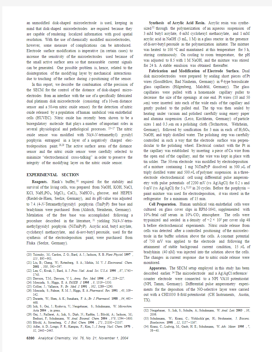
Fabrication and Modification of Electrode Surface.Dual disk microelectrodes were prepared by sealing short pieces of Pt wires(Goodfellow,Bad Nauheim,Germany)inθ-type borosilicate glass capillaries(Hilgenberg,Malsfeld,Germany).The glass capillaries were pulled with a homemade capillary puller to decrease the size of the openings at one end.Pt wires(10and50μm)were inserted into each of the wide ends of the capillary and gently pushed to the pulled end.The tip was then sealed by heating under vacuum and polished carefully using emery paper and alumina suspension(Leco,Kirchheim,Germany)of particle sizes1and0.3μm on a polishing cloth(Technotron,Wehrheim, Germany),followed by sonification for3min in each of H2SO4, NaOH,and triply distilled water.The polishing step was carefully controlled in such a way that the electrode surface was perpen-dicular to the polishing wheel.Electrical contact with the Pt in the capillary was established by inserting a piece of Cu wire from the open end of the capillary,and the wire was kept in place with tin solder.The50-μm electrode was modified by electrodeposition of a mixture containing1mg NiTmPyP dissolved in500μL of triply distilled water and500μL of polymer suspension in a three-electrode electrochemical cell using differential pulse amperom-etry with pulse potentials of2200mV(vs Ag|AgCl)for0.2s and 0mV(vs Ag|AgCl)for5s,32,33in20cycles.Before the porphyrin-paint mixture was used for electrodeposition,it was stored in the refrigerator for a minimum of15min.
Cell Preparation.Human umbilical vein endothelial cells were cultured on glass cover slips in RPM-1641,supplemented with 10%fetal calf serum in10%CO2atmosphere.The cells were trypsinized and seeded in a density of~2×10⁵per cover slip48 h before electrochemical experiments.Nitric oxide release from cells was detected after a controlled positioning of the microelec-trode in the buffer solution above the cells.A constant potential of750mV was applied to the electrode and following the attainment of stable background current condition,15μL of bradykinin(40nM)was injected into the solution above the cells. The changes in current response due to nitric oxide release were monitored.
Apparatus.The SECM setup employed in this study has been described earlier.34The microelectrode and a Ag|AgCl reference/ counter electrode were connected to a NPI VA10potentiostat (NPI,Tamm,Germany).Differential pulse amperometry experi-ments for the deposition of the NO-selective layer were carried out with a CHI10308-fold potentiostat(CH Instruments,Austin, TX).
(20)Tsionsky,M.;Cardon,Z.G.;Bard,A.J.;Jackson,R.B.Plant Physiol.1997,
113,895-901.
(21)Liu,B.;Cheng,W.;Rotenberg,S.A.;Mirkin,M.V.J.Electroanal.Chem.
2001,500,590-597.
(22)Lee,C.;Kwak,J.;Bard,A.J.Proc.Natl.Acad.Sci.U.S.A.1990,87,1740-
1743.
(23)Dawson,T.M.;Dawson,V.L.Annu.Rev.Med.1996,47,219-227.
(24)Moncada,S.;Higgs,E.A.FASEB J.1995,9,1319-1330.
(25)Collier,J.;Vallance,P.;Br.Med.J.1991,302,1289-1290.
(26)Moncada,S.;Palmer,R.M.J.;Higgs,E.A.Pharmacol.Rev.1991,43,109-
142.
(27)Durante,W.;Shen,A.K.;Sunahara,F.A.;Br.J.Pharmacol.1988,94,463-
468.
(28)Isik,S.;Oni,J.;Ryabova,V.;Neugebauer,S.;Schuhmann,W.Microchim.
Acta2004,in press.
(29)Oni,J.;Pailleret,A.;Isik,S.;Diab,N.;Radtke,I.;Blo¨chl,A.;Jackson,M.;
Bediuoi,F.;Schuhmann,W.Anal.Bioanal.Chem.2004,378,1594-1600.
(30)Blo¨chl,A.;Sirrenberg,C.J.Biol.Chem.1996,271,21100-21107.
(31)Adler,A.D.;Longo,F.R.;Kampas,F.;Kim,J.;J.Inorg.Nucl.Chem.1970,
32,2443-2445.(32)Neugebauer,S.;Isik,S.;Schulte,A.;Schuhmann,W.Anal.Lett.2003,36,
2006.
(33)Schuhmann,W.;Kranz, C.;Wohlschla¨ger,H.;Strohmeier,J.Biosens.
Bioelectron.1998,12,1157-1167.
(34)Kranz,C.;Ludwig,M.;Gaub,H.E.;Schuhmann,W.Adv.Mater.1995,7,
38-40.
6390Analytical Chemistry,Vol.76,No.21,November1,2004
RESULTS AND DISCUSSION
A chemical substance released from cells needs to travel from the secreting cell to the surface of an electrode to be detected.In the particular case of NO,a stimulant is required to effect its production by nitric oxide synthase (NOS).The NO so produced diffuses through the cell membrane into the bulk of the buffer solution above the cell,thus showing a concentration gradient from the release site toward the bulk of the solution.In addition,due to the high reactivity of the radical species NO,there is the possibility on its diffusion path to the electrode surface that it will react with molecular oxygen,leading to an additional depletion of NO with increasing distance from the release site.Therefore,the amount of NO that eventually gets to and undergoes oxidation at the electrode surface will highly depend on the separation
between the electrode and the source of secretion.Thus,NO concentration values as often found in the literature represent an average NO concentration over the length of the cylindrical electrode often used,in relation to its position with respect to the NO source.An overview of the different complex processes taking place around the cell is presented as Scheme 1.
Thus,to have realistic localized information with good resolu-tion for the quantitative and dynamic treatment of release processes from cells,the use of accurately positioned disk microelectrodes is indispensable.In addition,the exact distance between the electrode surface and the secreting cell has to be known in order to elucidate the concentration profile of the analyte in its diffusion path from the source to the electrode surface.For achieving the goal of accurately positioning a microsensor at known distances above a population of cells for the detection of nitric oxide release without adversely affecting the properties of the nitric oxide sensor during the positioning step,dual disk-shaped microelectrodes were fabricated.The dual microelectrode consists of a 10-μm distance sensor and a 50-μm nitric oxide sensor.A schematic representation of the dual disk-shaped microelectrode is shown in Figure 1a;the scanning electron microscope (SEM)image of the electrode surface,showing the relative dimensions of the distance sensor (10μm)and the nitric oxide sensor (50μm),is presented as Figure 1b.
The size of the active surface of the individual electrodes in the dual microelectrode was carefully chosen to minimize elec-trochemical cross-talk between the two electrodes.This is important so that electrochemical events taking place on the surface of one of the electrodes do not significantly affect the other electrode,particularly the chemically modified nitric oxide sensor.Monitoring the changes in the reduction current of molecular oxygen in the buffer solution above the cells is intended for the accurate positioning of the nitric oxide sensor close to the cells.Preliminary investigations suggested that the reduced oxygen
Scheme 1.Representation of the Likely Processes a That NO Released into a
Microvolume of Buffer Solution May
Undergo
a
Diffusion from the cell into the microvolume of buffer solution,reaction with dissolved oxygen in buffer solution,diffusion toward the electrodes surface,reaction with atmospheric oxygen,diffu-sion out of the buffer
solution.
Figure 1.(a)Schematic representation of the dual disk-shaped microelectrode.(b)SEM image of the tip of the dual disk-shaped microelectrode showing the relative sizes of the distance sensor and the nitric oxide sensor.
Analytical Chemistry,Vol.76,No.21,November 1,20046391
species (H 2O 2,O 2-)generated during this procedure are capable of altering the properties of the modifying layer on the nitric oxide sensor,thereby adversely affecting the sensitivity of the electrode toward the detection of nitric oxide.The absence of cross-talk between the two electrodes,or at least the influence of the “guide”electrode on the NO sensor,is hence an important issue in order to preserve the integrity of the modifier on the latter.
The choice of the relative sizes and the distance between the “guide”electrode and the nitric oxide sensor was determined by the absence of electrochemical cross-talking between the two electrodes when the distance between the centers of the two electrodes was kept constant at 70μm.Initially,both electrodes in the assembly had diameters of 50μm.The extent of electro-chemical cross-talking between the two electrodes was evaluated by recording SECM approach curves in a solution of 5mM [Ru(NH 3)6]Cl 3in 0.1M KCl when one of the electrodes in the assembly was poised at -500mV and the other was at open-circuit potential.This approach curve is shown in Figure 2a,curve i.Then a potential of -500mV was applied to both electrodes and the approach curve was recorded again,Figure 2,curve ii.
As expected,it was observed that the steady-state diffusion layers of the two electrodes overlap considerably and the electrochemical process taking place in the vicinity of one of the electrodes disturbed the response at the other,causing the significant difference seen in the approach curves in Figure 2a.This suggests that a dual electrode in which the size of each of the individual electrodes is 50μm cannot be used to achieve the goal of this study.The dual electrode was then prepared such that one of the electrodes had a diameter of 10μm,and the other,50μm.Again,approach curves were used to evaluate the extent of electrochemical cross-talk between the electrodes.Figure 2b,curve i,shows an approach curve recorded with the 10-μm electrode at -500mV,when there was no net potential applied to the 50-μm electrode,whereas curve ii,shows the same experiment repeated but with a potential of -500mV applied to the 50-μm electrode.Although having both electrodes at the potential for [Ru(NH 3)6]Cl 3reduction is the most demanding situation due to an expected competition for the same species,the curves are very similar.Obviously,there was no significant overlap of the steady-state diffusion layers of the two electrodes,
which suggests a negligible electrochemical cross-talk between the two electrodes.An observation similar to Figure 2b was made when the approach curve was recorded with the 50-μm electrode.The similarity of these approach curves indicates that the 50-μm electrode is “unaware”of the presence of the 10-μm electrode in its vicinity and can therefore be used to position the electrode reliably.Thus,it was expected that species generated at the 10-μm electrode will not significantly affect the steady-state diffusion layer in front of the 50-μm electrode,and the integrity of the modifier on the electrode will be preserved.
Further evidence in favor of the absence of electrochemical cross-talk between both electrodes when the diameters of the individual electrodes are 10and 50μm was obtained by carrying out generation-collection experiments to evaluate the generation/collection efficiencies of the 50-μm electrode vis-a `-vis the 10-μm electrode using the redox currents of Ru 3+/Ru 2+of a solution containing 5mM [Ru(NH 3)6]Cl 3in 0.1M KCl.A potential ramp in the cyclic voltammetry mode was applied to one electrode while a constant potential was applied to the other to cause a reversal of the process taking place on the electrode performing the cyclic voltammetry.Figure 3a,curve i shows the cyclic voltammogram of a solution of [Ru(NH 3)6]Cl 3recorded with the 50-μm electrode.The 10-μm electrode was poised at 0V for the collection of the Ru 2+generated at the 50-μm electrode.Figure 3b,curve i shows the collection current due to the reoxidation of Ru 2+to Ru 3+at the 10-μm microelectrode.
The magnitude of the collection current (~5nA)at the 10-μm electrode represents only one-half of the maximum current obtainable from the electrode in this solution.When the 10-μm disk electrode was used to generate Ru 2+(Figure 3a,curve ii)and the 50-μm electrode was used for collection at 0V (Figure 3b,curve ii),the collection current recorded on the 50-μm electrode was only ~0.8nA.These results indicate that the steady-state diffusion layer of the 10-μm electrodes does not significantly overlap with that on the 50-μm electrode,particularly when the 10-μm electrode is the one generating the species being collected at the 50-μm electrode.It means that the 10-μm electrode can be used safely in the vicinity of the 50-μm electrode without a fear of the species being generated affecting this electrode and,conse-quently,the modifying layer on its
surface.
Figure 2.(a)SECM approach curves recorded with a dual microelectrode of diameter 50μm,each in a solution of 5mM [Ru(NH 3)6]Cl 3in 0.1M KCl when one electrode was poised at -500mV and there was no net potential applied to the second (curve i)and when a potential of -500mV was applied to both electrodes (curve ii).(b)SECM approach curves recorded with the 10-μm electrode in a dual microelectrode in a solution of 5mM [Ru(NH 3)6]-Cl 3in 0.1M KCl at -500mV when there was no net potential applied to the second electrode (50-μm diameter,curve a),and when a potential of -500mV was applied to both electrodes (curve
b).
Figure 3.Plots of (a)generation and (b)collection curves obtained with the dual disk-shaped microelectrodes.
6392Analytical Chemistry,Vol.76,No.21,November 1,2004
It was reported earlier that electrochemical cross-talk at dual disk microelectrodes can be minimized by fabricating electrodes with open L-shaped tips whose active surface areas are located on two planes at an angle(≈135°)to each other.1This approach, while it was reported to effectively decrease electrochemical cross-talk by about94%,is not suitable for obtaining the exact distance of the electrode tip from the substrate because the two electrodes were located on different planes.For the“guide electrode”or distance sensor of the dual microelectrode to provide the exact distance of the other electrode(on whose surface the electro-chemical reaction of interest occurs)from the substrate,the tips of both electrodes have to lie on the same plane.The approach to controlling electrochemical cross-talk effects in this study is, hence,through a careful selection of the sizes of the“guide”electrode or distance sensor relative to the electrode intended for the detection of the product of cellular activity.
The sensitivity of the50-μm electrode in the dual microelec-trode toward the detection of nitric oxide was improved by modifying its surface with Ni(4-N-tetramethyl)pyridyl porphyrin entrapped in a negatively charged electrodeposition paint,as reported previously.28,29The charged side chains on this porphyrin moiety make it highly soluble in both organic and inorganic solvents,which consequently places a limitation on its use for the preparation of chemically modified electrodes,since it leaks away from the electrode surface upon contact of the modified electrode with a solvent.An approach to overcoming this leakage problem is by enclosing the porphyrin in a stable and insoluble“capsule”of an anodic electrodeposition paint on the electrode surface.The chemically modified electrodes obtained could be successfully applied for the sensitive determination of nitric oxide,and they displayed a satisfactory stability and sensitivity for the determi-nation of nitric oxide.28,29
The controlled and accurate positioning of the microelectrode at known distances from the population of human umbilical vein endothelial cells was achieved with the aid of the SECM.A schematic representation of the experimental setup is shown in Figure4.
A glass coverslip on which cells were grown was placed on the platform of the SECM,and a droplet of100μL of buffer
solution was placed on the cells.The tip of the dual microelectrode was then carefully placed into the bulk of the droplet of electrolyte over the cells.SECM approach curves were recorded with the 10-μm microelectrode above the cell culture by monitoring the reduction current of oxygen at-600mV vs Ag/AgCl at this electrode.Series of SECM approach curves were recorded from which a correlation between the percentage decrease in the steady state value of the electrode tip current and the distance of the tip from the layer of the cell population was established.It then became straightforward to fix the electrode at a known percentage value of the steady state current obtained far away from the surface.The corresponding distance of the electrode from the cell population can be deduced from the previously recorded approach curves.A typical SECM approach curve above the cell culture obtained with the10-μm electrode and the diffusion-limited reduction of molecular oxygen is presented in Figure5.
Although the cell metabolism may locally decrease the O2 concentration or the permeability of O2through the cells may lead to a decrease in the diffusion blocking the observed negative feedback,approach characteristics could be successfully applied for the correct positioning of the dual electrode,since the positioning was always done over the adherently growing cells. The datapoints for distances<0were obtained when the elec-trodes already pressed the cells.However,for the measurements, a predefined decrease in the O2reduction current derived from the approach curve was used to position the electrodes during the z approach.
As a matter of fact,the possible cross-talk is even less important if during z positioning the50-μm-diameter NO sensor electrode is kept at open circuit potential,thus avoiding any disturbance of the z approach characteristics at the“guide”electrode.Following the successful positioning of the electrode tip above the cell culture,the50-μm modified electrode was then used to detect the release of NO by the cells upon stimulation with bradykinin. Typically,a constant potential of750mV was applied to the modified50-μm NO sensor accurately positioned above the cell population.After the establishment of a stable background current, the release of NO was stimulated by the injection of15μL
of Figure4.Schematic representation of the experimental setup for the detection of NO release from a layer of adherently growing endothelial cells upon stimulation with
bradykinin.
Figure5.Typical SECM approach curve recorded above a popula-tion of HUVEC with the10-μm microelectrode used for guidance. Arrows are used to indicate the points corresponding to the distances away from the cell where the NO sensor was finally positioned to detect NO release from the cells upon stimulation.
Analytical Chemistry,Vol.76,No.21,November1,20046393
bradykinin (80nM),and the changes in the current at the modified electrode were monitored.A representative plot of the relative magnitudes of the currents due to the oxidation of NO released when the electrode was 5,10,15,and 25μm away from the cells is shown in Figure 6.
The trend observed here was confirmed by repeating the experiments,and the results obtained are summarized in Table 1.
The smaller magnitude of the current response obtained when the electrode was positioned directly on the surface of the cell is
due to the blockage of the diffusion of NO produced from neighboring cells to the electrode surface and corresponds to the well-established negative feedback effect of SECM.These results clearly indicate that the distance of the sensor from the source of secretion plays an extremely important role in determining the magnitude of the signals obtained.Thus,the quantitative and dynamic treatment of cellular activities such as release and reuptake process requires a precise knowledge of the separation of the electrodes from the substrate.
When the current -time plots were normalized for the various distances of the electrode from the cell population by dividing them by the peak current,very similar shapes of the current/time curves were obtained.This clearly indicates that the differ-ences observed in the current -time plots are due to the distance of the electrode from the cell,showing that the diffusion of NO from the source to the electrode tip plays a major role in determining the magnitude of the signal obtainable from the electrode during the electron-transfer step.
CONCLUSION
The possibility of an accurate positioning of modified disk-shaped microelectrodes at known distances above a population of living cells for the detection of the products of cellular activity was demonstrated.Dual disk-shaped microelectrodes were fab-ricated in which one of the electrodes was used as a “guide”for positioning the other modified electrode for the detection of nitric oxide release from human umbilical vein endothelial cells.The magnitude of the signals obtained was found to be dependent on the separation between the electrode and the cell population,thereby underlining the need for a precise and accurate knowledge of the distance of the microsensor from the substrate for the qualitative and dynamic treatment of the release process.ACKNOWLEDGMENT
This work was supported by the European Commission in the framework of the project “CellSens”(QLK3-2001-00244)and the Ministry of Education and Research,Germany (BMBF)in the framework of its program “Nanobiotechnologie”(AZ NBT066).Received for review June 4,2004.Accepted July 30,2004.
AC049182W
Figure 6.Representative plots of the relative magnitudes of the current signals due to the oxidation of NO recorded at (a)5,(b)10,(c)15,(d)25μm away from the cells.
Table 1.Statistical Summary of NO Measurements Made above Endothelial Cell Culture d a (μm)N b mean (pA)SD c (pA)min d (pA)max e (pA)06120.368.64625058448.6194.11827601011200.068.6108327151380.057.12120420534.818.045125522.616.5115130
2
12.0
4.2
9
15
a
Distance between the electrode and the cells.b Number of mea-surements.c Standard deviation.d Minimum value of current measured with the NO sensor.e Maximum value of current measured with the NO sensor.
6394Analytical Chemistry,Vol.76,No.21,November 1,2004