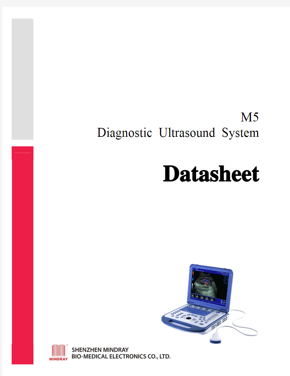
M5 Diagnostic Ultrasound System
Datasheet
General information
Dimensions and weight
?Height: 75 mm (2.95 inch)
?Width: 361 mm (14.21 inch)
?Depth: 357 mm (14.06 inch) with handle
?Weight (main unit, without battery): less than 6 kg (13.23 lb.)
Electrical power
?AC adapter input
–Voltage: 100VAC~240VAC
–Frequency: 50/60 Hz
–Input current: 2A (maximum)
?AC adapter output
–Voltage: 12VDC
–Output current: 10A (maximum)
?Battery
–Exchangeable li-ion batteries: 11.1VDC, 4500mAh
–Continuous scanning for more than 1h (normal condition) User interface
Operator keyboard
?Soft keys for rapid and convenient control
?Home based and grouped design
?Alphanumeric keyboard
?8-segment TGC, with remapping functionality at any depth ?Interactive backlit keys
?User-centric control panel with homebase design
?Blank keys for user-define functions
?Programmable softkeys
Display screen
?High-resolution color LCD
–Diagonal dimension: 15 inch
–Resolution: 1024X768
–Brightness adjustment
?Integrated speakers
–Volume adjustment
Ergonomic design
?Operation navigation: logical instructions for most operations
?System hibernation: switch off transducer transmitting, and launch screensaver ?Q-Click: icons to activate most frequently used functions
?Thumbnails for acquired images and cineloops
?Programmable two-pedal footswitch
System overview
Application
?Abdomen
?Cardiology
?Gynecology
?Obstetrics
?Urology
?Small Part
?Pediatrics
?Musculoskeletal
?Orthopaedics
?Intraoperative
?Peripheral Vascular
?Transcranial Doppler
Scanning method
?Electronic convex
?Electronic linear
?Electronic sector
Transducer types
?Convex array
?Microconvex array
?Linear array
?Phased array*
Imaging modes
? B mode
?M mode
?CDFI (Color Doppler Flow Imaging, Color)
?Power (Power Doppler Flow Imaging, including DirPower, directional power Doppler)
?Pulse Wave Doppler (PW)
?Continuous Wave Doppler (CW, option)*
Special imaging features
?Tissue harmonic imaging
?Steer scanning for linear transducers (B, color/power, PW independent) ?Trapezoid imaging for linear transducers
?HPRF for PW
?Multi-frequency in both 2D and Doppler imaging
Display mode
?Quad/dual display (for B, color and power modes)
?B/C Live (B and color simultaneous comparison display)
?Duplex for simultaneous B and PW/CW
?Triplex mode for simultaneous B, color/power, and PW/CW
?Time line display: left/right and top/bottom (1:1, 1:2, full)
Standard configuration
?High resolution 15 inch LCD display
?Pulse Wave Doppler
?HPRF
?Color Doppler Flow Imaging
?Power Doppler Flow Imaging
?Directional Power Doppler Flow Imaging
?Tissue Harmonic Imaging
?Trapezoidal Imaging
?iBeam (Spatial compounding imaging for linear probe)
?iTouch? (Automatic image optimization by pressing one button)
?80G integrated hard disk
?iStation?
?USB ports
?Ethernet port
?S-video out port and cable
?Measurement & calculation software packages
?Multi-language screen display
?Convex array transducer 3C5s (2.5/3.5/5.0/H5.0/H6.0MHz)
?Trolley case
Software options
?DICOM 3.0 software
?Free Xros? Imaging (Anatomical M mode)*
?iScape? View (Panoramic imaging, or extended field of view)
?Smart3D? (Freehand 3D)
Hardware options
?Additional transducer connector
?CW*
?Transducers
?Needle guide brackets
?I/O module for data transportation
?USB V/A module for VCR connection
?USB ECG module with electrodes and cables (AHA/IEC)
?External USB DVD-R/W
?Spare battery
?Foot switch with programmable functionality
?Trolley
Peripherals
?Thermal B/W video printer
?Thermal color video printer
?Digital video B/W or color printer
?USB text/graph printer
?VCR
?DVD recorder
Imaging processing and presentation
System architecture
?Powerful Multi-beam Parallel Imaging (MBP)
?Fine Tissue Optimization (FTO)
?Transmitting Spectrum Focusing (TSF)
?Innovative Transmitting Apodization (ITA)
?Accurate Vessel Imaging (AVI)
Intelligent digital imaging process
?iTouch?: automatic image optimization
?IP(image processing): one key for B and color image fast optimization ?Q-click?: quick adjustment for parameters displayed on screen Imaging platform
?All-digital broadband beam-former
?1024 digital processing channel technology
?Displayed depth
–Minimum: 26 mm, transducer dependent
–Maximum: 308mm, transducer dependent
?Focus
–1~4 focus points selectable (depth dependent)
–Up to 8 focal positions selectable (depth dependent) ?Wideband processing technology
–Fundamental frequencies: 3 steps
–Harmonic frequencies: 2 steps
–Doppler frequencies: 2 steps
?Gray scale: 256 levels
?Total system dynamic range: 160 dB
?Zoom
–RAZ (regional acoustic zoom)* and pan zoom
–PIP (picture in picture)
–Zoom ratio: up to 400%
–For real-time or frozen images
B mode
?Acoustic power (10~100%,6% step)
?Gain (0-100%)
?TGC (8 segments, with re-mapping functionality at any depth) ?Frame Rate (up to 396f/s, transducer dependent)
?Focus number (1~4)
?Focus position (8 steps)
?Scan range (N, M1, M2, W)
?Line density (L, H)
?Steer (-6°,0,6)
?TSI (Tissue, Muscle, Fat, Fluid) tissue specific imaging
? 6
?Display dynamic range (up to 100dB)
?Frame average (0~7)
? 6
?Noise rejection (0~3)
? 6
?Image enhancement (off, 1~4)
?IP (1~8) image processing
?Colorize (7 maps)
?Gray map (1~8)
?Gray Transform
?Gray Rejection
?γ correction (0~3)
?Rotate (0°, 90°, 180°, 270°)
?Reverse (left/right, up/down)
M mode
?Display mode: scroll
?Sweep speed (1, 2, 4, 8 s/screen)
?Gain (0-100%)
?Display dynamic range (up to 100dB)
?M soften (0~4)
?Gray map (1~8)
?Colorize (7 maps)
?Time mark (on, off)
Color mode
?Gain (up to 29dB)
?Frequency (2 frequencies)
?Frame Rate (up to 448f/s, transducer dependent)
?Steer (-12o, -6o, 0o, 6o, 12o)
?PRF (1.3kHz~14.5kHz)
?Scale (±2.3cm/s~±246cm/s, up to 492cm/s in one direction, transducer dependent) ?Baseline (17 levels)
?Color map (1~11)
?Wall filter (0~7)
?Line density (L, H)
?Packet size (0-4)
?Flow state(L, M, H)
?Smooth (0~4)
?Persistence (0~4)
?Contrast (0~3)
?Priority (0~100%)
?Map reverse
?Focus position (10 levels)
?B/C wide (on, off) automatically adjust the 2D image size according to the color ROI
?ROI color (off, red, green, blue, cyan, MAG, yellow, white)
?B/C dual live (on, off)
?Image display (on, off)
PW/CW* mode
*CW mode is available only with phased array transducers.
?PW frequency (2 frequencies)
?PRF (1.3kHz~11.4kHz)
?PW Scale (±6.1cm/s~±291.7cm/s, up to 583.4cm/s in one direction, transducer dependent)
?CW Scale (±0.61m/s~±15.04m/s, up to 30.08m/s in one direction, transducer dependent)
?Baseline (9 levels)
?Sweep speed (1, 2, 4, 8s/screen)
?Sample volume (0.5~15.0mm)
?Sample depth (up to 308mm)
?Steer (-12o, -6o, 0o, 6o, 12o)
?Angle correlation (-80°~80°)
?Colorize (7 maps)
?Wall filter (7 levels, scale dependent)
?Auto Trace (Vmax, Vmean, Vmode)
?Triplex (on, off)
?Threshold (0~5)
?Trace Area (Below, Above, All)
?Trace smooth (off, 1~4)
?Trace sensitivity (0~5)
?Audio (on, off)
?Full screen(on, off)
?Time mark (on, off)
ECG
?Gain
?Position
?Trigger mode (single, dual, AT, timer)
?HR display
Power/ DirPower mode
?Display dynamic range (up to 70dB)
?Power Map (1~8)
?Line density (L, H)
?Packet size (L, M, H)
?Smooth (0~4)
?Persistence (0~4)
?Contrast (0~3)
?Priority (0~4)
?Reverse (on, off)
?B/C wide (on, off) automatically adjust the 2D image size according to the ROI ?LVR (Low velocity resistance) (off, 1~7)
?Focus position (10 levels)
?ROI color (off, red, green, blue, cyan, MAG, yellow, white)
?B/C live (on, off)
?Image display (on, off)
Free Xros? Imaging (option)*
?Free Xros imaging is also called anatomical M mode.
?Available on all convex, linear and phased array transducers
?Based on real-time imaging or cineloop of 2D mode
?Sweep speed (1, 2, 4, 8 s/screen)
?Image enhance (off, 1~3)
?Gray map (1~8)
?Colorize (7 maps)
?Time mark (on, off)
?Store and review Free Xros? images
?Store and review cineloop
?All M measurement items available
iScape? View (option)
?iScape? view is also called extended field of view imaging, or panoramic imaging.
?Available on all linear array transducers
?Based on real-time imaging of 2D mode (not available in Color or Power mode) ?Displays up to 40cm in length (frame rate and scanning speed dependent)
?Rotate (1o/step)
?Zoom (100%~400%, actual size, fit size)
?Colorize (7 maps)
?Store and review iScape? images
?Store and review cineloop
?All 2D measurement items available, except depth, profile and histogram
Smart3D? (option)
?Smart3D? is also called freehand 3D.
?Available on all convex, linear and phased array transducers without sensor ?Method (linear, fan)
?Distance (1~50cm)
?Angle (10o~80o)
?Render (surface, max, min, X-ray)
?Threshold (0~63)
?Contrast (0~39)
?Brightness (0~39)
?Colorize (7 maps)
?Single or quad display
?Rotate
?Store and review Smart3D? images
?Cut
Cineloop
?Support 2D, M, Spectral Doppler, Color, Power, DirPower
?Simultaneous and independent review in duplex/triplex mode
?ECG wave for retrospective review
?Capacity:
–2D, Color, Power, DirPower: Maximum >1200 frames
–M, Spectral Doppler: Over 131s
?Variable cine playback speed
?User-define start and end frame of cine storage
?Permanent storage in hard disk and display in real-time and duplex modes
?iVision:Slides show function
iStation?
Intelligent patient information management platform
?Quick image and cine storage
?Auto image review automatic browser, icon review
?Offline analysis system
?Professional clinical reports with images embedded
?Integrated search engine for patient information
?Intelligent data backup
?Support DICOM worklist from server and file transportation in DICOM format on internet (option)
Storage
?80 GB integrated hard drive
?External DVD-R/W (option)
?USB ports
?Still images storage format: BMP, JPG, DCM and FRM (defined by Mindray, support offline analysis function)
?Cine loops storage format: AVI, DCM and CIN (defined by Mindray, support offline analysis function)
Measurement and calculation
?Software packages for various specific clinical use
?Comprehensive analysis methods
?Clinical analysis reports
General Measurement package
General B mode measurement
?Depth
?Distance
?Angle
?Area
?Volume
?Cross Line
?Parallel Line
?Trace Length
?Ratio
? B Profile
? B Histogram
General M mode measurement
?Distance
?Time
?Slope
?Heart Rate
?General Color mode measurement
?Color velocity
General PW/CW mode measurement
?Velocity
?Acceleration
?Resistance index
?Spectrum trace
?Heart rate
Clinical Analysis Packages
Obstetrics
?Fetal measurement
?Fetal weight calculation
?Calculation items, such as HC/AC, FL/AC, FL/BPD, AXT
?Amniotic fluid index
?Fetal biophysical profile
?Fetus Doppler measurement
?Multi-fetus exam
?Estimated delivery date display
?Growth Curve: Four curves display for comparison
(GA calculation formulas include but may not be limited to the following: Tokyo, Hadlock, Jeanty, Hohler, Merz, Kurtz, Sabbagha, Hansmann, Rempen, Osaka,
Chitty, O”Brien and Warda. EFW formulas include Hadlock1, Hadlock2,
Hadlock3, Hadlock4, Shepard, Merz1, Merz2, Hansmann, Tokyo, Osaka and
Campell.)
Cardiac
?Left ventricular function measurement
–Single Plane Ellipse method
–Biplane Ellipse method
–Bullet method
–Simpson’s method
–Simpson’s Single Plane Ellipse method
–Simpson’s Biplane Ellipse method
–Cube method
–Teichholz method
–Gibson method
?Left ventricular
?Right ventricular
?Aortic
?Main pulmonary artery
?Mitral valve
?Tricuspid valve
?Pulmonary valve
?Pulmonary vein valve
?Volume flow
?Heart rate
(Cardiac calculation results include but may not be limited to the following items: HR,EDV, ESV, SV, CO, EF, CI, SI, LV Mass, LVMWI, FS, MVCF, ET, PHT,
MV-Area, VTI, MPG, MV-IRT, MV-DcT, RVSP, AoV-Area, RV-ET, RV-AcT, RV-PEP, RV-AcT/ET, RV-STI, PV DcT, PV-SF and Volume Flow.) Gynecology
?Endometrium
?Uterus
?Uterine cervix
?Uterus/cervix
?Ovary
?Follicle
Small Parts
?Thyroid
Urology
?Prostate
?Left/right Seminal Vesicle
?Left/right Renal
?Left/right Adrenal
?Residual Volume
?Left/right Testicular
Orthopaedics
?Hip angel:BL, IL, ARL, Angle between BL/ARL, Angle between BL/IL
Peripheral Vascular
?Left/right Distal Common Carotid Artery
?Left/right Middle Common Carotid Artery
?Left/right Proximal Common Carotid Artery
?Left/right Distal Internal Carotid Artery
?Left/right Middle Internal Carotid Artery
?Left/right Proximal Internal Carotid Artery
?Left/right Distal External Carotid Artery
?Left/right Middle External Carotid Artery
?Left/right Proximal External Carotid Artery
?Left/right Distal Vertebral Artery
?Left/right Middle Vertebral Artery
?Left/right Proximal Vertebral Artery
?Left/right Distal Subclavian Artery
?Left/right Middle Subclavian Artery
?Left/right Proximal Subclavian Artery
?Left/right Distal Subclavian Vein
?Left/right Middle Subclavian Vein
?Left/right Proximal Subclavian Vein
?Left/right Bulbillate
?Innominate Artery
?Left/right Upper Extremity
?Left/right Lower Extremity
?Volume flow
?Stenosis
System setup
User‐define functions
By user-define function, users could
?Customize twenty four user-define exam modes, including but not limited to –Exam mode name
–Imaging parameters
–General measurement items for each imaging mode
–Measurement packages
–Obstetric formula
–Comment library
–Body mark library
?Create new measurement items, or new calculations based on measurement results
?Set volume calculating index
?Assign frequently used functions to user-define buttons on control panel and foot switch
?Adjust key volume, key lightness and trackball speed
Multi‐language*
Screen display, keyboard layout and user manuals support
?English
?German
?French
?Spanish
?Portuguese
?Italian
?Russian
?Chinese
Operation system
?Windows? XP Embedded system
Windows is a registered trade mark of Microsoft Corporation.
Transducers
Sockets
?One universal array transducer socket
?One dedicated CW Doppler pencil socket
?Optional three universal array transducer sockets (additional transducer connector)
Models
?Convex array transducer 3C5s (2.5/3.5/5.0/T5.0/T6.0MHz)
?Linear array transducer 7L4s (5.0/7.5/10.0MHz)
?Linear array transducer 10L4s (8.0/10.0/12.0MHz)
?Linear array transducer 7L6s (5.0/7.5/10.0MHz)
?Biplanar transducer 6LB7s (5.0/6.5/8.0MHz)
?Convex array transducer 6CV1s (5.0/6.5/8.0MHz)
?Endorectal array transducer 6LE7s (5.0/6.5/8.0MHz)
?Convex array transducer 6C1s (5.0/6.5/8.0MHz)
Inputs and outputs
Main unit
?USB (2)
?Ethernet
?S-video out
?I/O module connector
I/O module
?USB (2)
?Parallel port
?Serial port
?Composite video out
?Audio out (L/R)
?VGA out
?Microphone in
V/A module
?S-video in
?Composite video in
?Audio in (L/R)
*Not available yet.