A PLETHORA-Auxin Transcription Module Controls Cell Division Plane Rotation through MAP65and CLASP
Pankaj Dhonukshe,1,*Daan A.Weits,1Alfredo Cruz-Ramirez,1Eva E.Deinum,2Simon H.Tindemans,2,6
Klementina Kakar,1Kalika Prasad,1,7Ari Pekka Ma¨ho¨nen,1,8Chris Ambrose,3Michiko Sasabe,4Guy Wachsmann,1 Marijn Luijten,1Tom Bennett,1,9Yasunori Machida,4Renze Heidstra,1Geoffrey Wasteneys,3Bela M.Mulder,2,5
and Ben Scheres1,*
1Department of Biology,Utrecht University,Padualaan8,3584CH Utrecht,The Netherlands
2FOM Institute AMOLF,Science Park104,1098XG Amsterdam,The Netherlands
3Department of Botany,University of British Columbia,6270University Boulevard,Vancouver,British Columbia V6T1Z4,Canada
4Division of Biological Science,Graduate School of Science,Nagoya University,Nagoya,464-8602,Japan
5Laboratory of Plant Cell Biology,Wageningen University,Droevendaalsesteeg1,6708PB Wageningen,The Netherlands
6Present address:Department of Electrical and Electronic Engineering,Imperial College,South Kensington Campus,London SW72AZ,UK 7Present address:School of Biology,Indian Institute of Science Education and Research,Thiruvananthapuram695016,Kerala,India
8Present address:Institute of Biotechnology,University of Helsinki,Helsinki FIN-00014,Finland
9Present Address:Department of Plant Sciences,University of Cambridge,Downing Site Downing Street,Cambridge CB23EA,UK
*Correspondence:p.b.dhonukshe@uu.nl(P.D.),b.scheres@uu.nl(B.S.)
DOI10.1016/j.cell.2012.02.051
SUMMARY
Despite their pivotal role in plant development,
control mechanisms for oriented cell divisions have
remained elusive.Here,we describe how a precisely
regulated cell division orientation switch in an
Arabidopsis stem cell is controlled by upstream patterning factors.We show that the stem cell regula-
tory PLETHORA transcription factors induce division
plane reorientation by local activation of auxin sig-
naling,culminating in enhanced expression of the
microtubule-associated MAP65proteins.MAP65up-
regulation is suf?cient to reorient the cortical microtu-
bular array through a CLASP microtubule-cell cortex
interaction mediator-dependent mechanism.CLASP
differentially localizes to cell faces in a microtubule-
and MAP65-dependent https://www.doczj.com/doc/5a11547416.html,putational
simulations clarify how precise90 switches in cell
division planes can follow self-organizing properties
of the microtubule array in combination with biases
in CLASP localization.Our work demonstrates how
transcription factor-mediated processes regulate
the cellular machinery to control orientation of forma-
tive cell divisions in plants.
INTRODUCTION
The orientation of cell division plane is key to the generation of multicellular organisms as their randomization often leads to morphogenetic defects(Baena-Lo′pez et al.,2005;Torres-Ruiz and Ju¨rgens,1994;Traas et al.,1995).In plants,neighboring cells cannot relocate due to shared cell walls,and cell divisions have to be oriented parallel to the surface(‘‘periclinal’’)to create new layers.Asymmetric periclinal cell divisions,where daughter cells acquire distinct identities,have been termed‘‘formative divisions’’(Gunning et al.,1978).Most formative divisions occur at early embryo stages when the body plan is established(Ju¨r-gens,1995),but others take place when lateral organs are gener-ated(De Smet and Beeckman,2011).New layers are repeatedly established in the ground tissue and epidermis/lateral root cap (LRC)stem cells of Arabidopsis roots(Dolan et al.,1993).Several transcription factors required for these divisions have been iden-ti?ed(Di Laurenzio et al.,1996;Helariutta et al.,2000;Willemsen et al.,2008)but mechanisms by which the orientation of cell divi-sion planes are controlled have remained unknown.
Plant cell division planes are speci?ed prior to mitosis by formation of a cortical microtubular band called preprophase band(PPB)(Pickett-Heaps and Northcote,1966).The cortical division site remains marked throughout mitosis and cytokinesis after the PPB has disassembled(Smith,2001),with negative and positive markers of the cortical division site memorizing PPB position to guide the cell plate(Mu¨ller et al.,2009).Most of those proteins follow the localization of PPB microtubules and seem to operate downstream(Rasmussen et al.,2011a;Rasmussen et al.,2011b).These observations indicate how the microtubular PPB can be coupled with cytokinesis but do not reveal how the PPB is oriented.
Cell divisions associated with the Arabidopsis root stem cell niche are sustained by the activity of PLETHORA(PLT)proteins, members of the AP2transcription factor family(Aida et al.,2004; Galinha et al.,2007).Initial induction of PLT expression is regu-lated by distal accumulation of the plant growth regulator auxin(Aida et al.,2004;Blilou et al.,2005).Auxin distribution patterns have been linked with altered cell division planes during Cell149,383–396,April13,2012a2012Elsevier Inc.383
embryo development(Petricka et al.,2009),lateral root initiation (Pe′ret et al.,2009),and in primary roots(Sabatini et al.,1999). In addition,auxin accumulation in cultured cells alters PPB orien-tation and cell division planes(Dhonukshe et al.,2005).How auxin in?uences cell division planes and whether this directs stem cells and their daughters to divide in speci?c orientations has remained unknown.
Here,we show that PLT proteins induce root epidermal cells to orient cell division planes through TIR1-dependent auxin signaling(Dharmasiri et al.,2005a;Kepinski and Leyser,2005), which enhances expression of microtubule-associated MAP65 proteins(Chan et al.,1999;Smertenko et al.,2000).MAP65 guides localization of CLASP,a microtubule cortex interaction mediator(Ambrose et al.,2011),and we postulate a mechanism by which this precisely orients cell division planes.Our results provide a paradigm for plant transcription factor control of cell division planes.
RESULTS
PLT1,PLT2,and PLT3Are Required for LRC-Generating Periclinal Cell Divisions in the Root Stem Cell Niche Arabidopsis root epidermis/LRC stem cells divide periclinally to generate new LRC layers and extend the epidermis by divisions perpendicular to the cell surface(anticlinal)(Figures1A and1B; Dolan et al.,1993).In roots of plt1plt2but not plt1plt3and plt2plt3 mutants,periclinal cell division frequency was reduced in the epidermis/LRC stem cell domain,whereas anticlinal divisions appeared normal(Figures1C and1L,and Figure S1A available online).Consistent with periclinal cell division defects,plt1plt2 roots possessed single or double LRC layers compared to three LRC layers in wild-type(WT)(compare Figure1B with Figure1C and compare the panels of Figure S1A),which did not occur in unrelated stem cell maintenance mutants(Figure S1F).In plt1plt2 roots with a single outer layer,epidermis marker GL2::ER-GFP and epidermis/LRC marker WER::ER-CFP labeled the outer layer (Figures1D and1E and Figures S1B–S1E)indicating a mixed identity.Mature plt1plt2but not plt1or plt2embryos revealed periclinal cell division defects(Figures1F–1H)suggesting redun-dant roles for PLT1and PLT2in this process.The absence of peri-clinal epidermis/LRC divisions in plt1plt2plt3embryos(Galinha et al.,2007)indicated a residual role for PLT3in this process. Indeed,PLT3coding region fused to the glucocorticoid receptor (GR)under the PLT2promoter restored periclinal cell divisions after DEX induction in the epidermis/LRC stem cell domain of plt1plt2roots,albeit to a lesser extent than the PLT2coding region(Figures1I–1J,1L and Figure S1G).
Ectopic Induction of PLT1and PLT2Triggers
LRC-Generating Periclinal Cell Divisions
in the Root Epidermis
Induction of PLT2-GR or PLT1-GR from the constitutive35S promoter in plt1plt2rescued periclinal divisions and,in addition, triggered periclinal cell divisions in the epidermis shootward from the stem cell niche(Figures1K and1L).In WT,constitutive induc-tion of PLT1or PLT2triggered periclinal cell divisions throughout the root epidermis,leading to an extra layer(Figures1M–1P and Figures S1H–S1I and S1O–S1P).Although cortex and endo-dermis identity markers were unaltered after PLT2induction, both daughter cells of periclinal cell divisions retained epidermal identity(Figure S1N).The LRC marker SMB(Figure1Q),required for LRC differentiation(Bennett et al.,2010;Willemsen et al., 2008),appeared in the epidermis upon induction of PLT2 (Figures1Q and1R)before periclinal cell division and asymmet-rically segregated into outer daughter cells(see inset in Fig-ure1R)as in epidermis/LRC stem cells(see inset in Figure1Q); these outer cells detached like typical LRC cells(Figure1S). We concluded that PLT-induced ectopic periclinal cell divisions switch division plane and segregate cell fates similar to epidermis/LRC stem cells.
Dosage-Dependent and Cell-Autonomous PLT2Action Switches Cell Division Planes
PLT proteins form gradients(Galinha et al.,2007),and periclinal divisions occur in the stem cell niche where PLT levels are elevated.The frequency of periclinal cell divisions increased with longer PLT2induction times(Figures S1J–S1L)and PLT2::PLT2-YFP fusion proteins displayed strongest?uores-cence in the stem cell niche region(Figure S1Q),indicating that high levels of PLT2trigger periclinal cell divisions. Epidermal cells expressing higher PLT2levels preferably under-went periclinal cell divisions,whereas neighboring cells with lower levels underwent anticlinal cell divisions(Figures1T–1U and1Y).We induced PLT2-YFP using the WER::XVE epi-dermis-speci?c induction system(A.P.Ma¨ho¨nen et al.,in prep-aration),which triggered epidermal periclinal cell divisions (Figures S1R and S1S)strictly correlated with?uorescence-in-ferred expression strength based on serial scans(Figures1V, 1W,and1Z and Figures S1T and S1U).In six cell pairs within different roots,cells with higher PLT2-YFP levels before division underwent periclinal cell divisions,whereas neighboring cells with lower PLT2-YFP levels executed anticlinal cell divisions (Figure1X).Together,our results indicate that PLT2action promotes periclinal divisions in a dose-dependent and cell-autonomous manner.
Auxin and PLETHORA Together Trigger Periclinal Cell Divisions
In WT,auxin activity sensor DR5built up in the epidermis/LRC stem cell prior to periclinal division and segregated asymmetri-cally in the outer daughter cell adopting LRC fate(Figures2A–2C).After PLT2induction,DR5signal appeared in epidermal cells prior to periclinal cell division(Figures2D and2E).To address whether buildup of auxin levels was suf?cient for pericli-nal divisions,we performed single-cell laser ablations(Sabatini et al.,1999;Xu et al.,2006),which block polar auxin transport. In the epidermis,cells rootward but not shootward of the ablated cell gradually accumulated DR5signal(Figures2F and2G),and these cells divided periclinally(Figures2F–2H).
The auxin ef?ux inhibitor1-N-Naphthylphthalamic acid(NPA) also triggered DR5increase and periclinal cell divisions mainly in the epidermal cells(Figures2I and2J and Figure S2). After NPA treatment,PLT expression increased in epidermal cells undergoing periclinal cell divisions(Figures2K–2M). The ratio of NPA treated to untreated periclinal cell division frequency was higher in WT than that in plt1plt2mutant
384Cell149,383–396,April13,2012a2012Elsevier Inc.
(Figure 2N;compare with Figure 1L)indicating that NPA action to induce periclinal divisions requires PLT activity.The residual induction of NPA-triggered periclinal divisions in plt1plt2may either result from auxin-independent action on periclinal division or from enhanced activity of auxin-inducible PLT3(De Smet et al.,2008
).
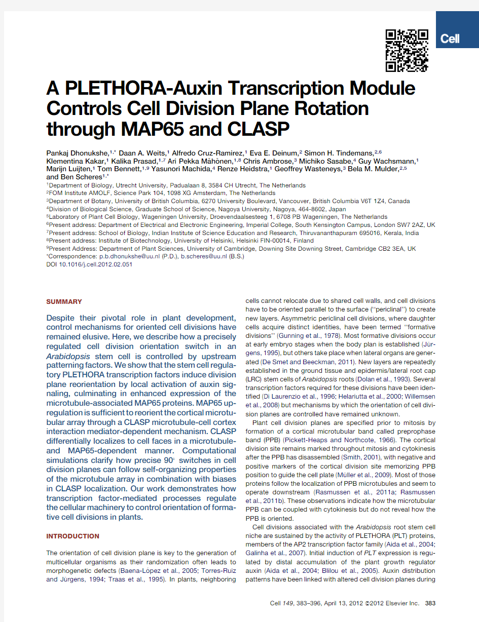
Figure 1.PLT Proteins Promote Periclinal Cell Divisions of Epidermis/LRC Stem Cells
(A)First periclinal cell division (white arrowhead)generates LRC and second anticlinal cell division (white arrow)extends epidermis in WT.
(B and C)plt1plt2mutant with anticlinal cell division at position of periclinal cell division in WT (compare C with B).Note three LRC layers in WT (B)compared to one LRC layer in plt1plt2.
(D and E)Epidermis marker GL2::ER-GFP labels epidermis in WT (covered by three LRC layers D),and outermost layer in plt1plt2(white asterisk in E).
(F–H)Aniline-blue staining of WT (F)and plt1plt2(G)mature embryos.WT with epidermis and LRC layers formed by periclinal cell divisions and single layer (white asterisk)in plt1plt2.Frequency of periclinal cell divisions in plt1plt2(H).
(I–K)Periclinal cell divisions after induction of PLT2(20hr DEX)(compare I and J)and PLT3(20hr DEX;L and Figure S1G)in PLT2domain.Ubiquitous PLT2induction (20hr DEX)in plt1plt2induces periclinal cell divisions in extended epidermal regions (K).(L)Periclinal cell division frequency in various analyzed backgrounds.
(M–P)Ubiquitous induction of PLT2(24hr DEX)triggers periclinal cell divisions in epidermis.Cross sections display the resulting extra cell layer (Compare N and P).(Q and R)LRC marker SMB (Q)appears in the epidermis at the onset of periclinal cell divisions (R)and after cell division segregates in the outer layer (inset in R)adapting LRC fate.
(S)Detachment of outer layer expressing SMB (white arrows).
(T–Z)Amount of functional PLT2::PLT2-YFP in plt1plt2correlates with periclinal cell division (?uorescence intensity quanti?cation in Y and intensity pro?le analysis in T and U).Fluorescence intensity quanti?cation in (Z)and intensity pro?le analysis in (V),(W),(X)correlate with higher PLT2levels before periclinal divisions.White arrowheads depict periclinal cell divisions;white arrows mark anticlinal cell divisions.The following abbreviations are used throughout all ?gure legends:c,cortex;e,epidermis;and l,LRC.Red,propidium iodide (PI)staining;green,GFP;and cyan,CFP.Columns in graphs display means ;error bars,standard deviations;asterisk (*),statistically signi?cant p values at <0.05.n =38embryos for (H),n =38roots for (L),n =23cells from six roots for (Y)and n =42cells from nine roots for (Z)from three independent experiments.See also Figure S1.
Cell 149,383–396,April 13,2012a2012Elsevier Inc.385
We tested the role of local auxin abundance by expressing the auxin conjugating enzyme GH3.5(Staswick et al.,2005)and the bacterial auxin synthesis gene iaaH (Kares et al.,1990)in the epidermis using the WER::XVE system.Coinduction of GH3.5and PLT2lowered DR5signal in the epidermis,consistent with increased auxin conjugation,and reduced the frequency of periclinal cell divisions (Figures 2O and 2Q).In contrast,simulta-neous iaaH and PLT2induction enhanced DR5signal in the epidermis and increased the frequency of periclinal cell divisions (Figures 2P and 2Q).Together,these results indicate that auxin levels in?uence cell division plane switch both through and in parallel to PLT action.
PLT2Induces Periclinal Cell Division through TIR1-Dependent Auxin Signaling
The tir1-1afb2-1afb3-1triple auxin signaling mutant (Dharma-siri et al.,2005b )displayed periclinal cell division de?ciencies
and abnormal cell division planes in the region where epi-dermis/LRC periclinal cell divisions normally occur (Figures 3A–3D).Strikingly,induction of PLT2in tir1-1afb2-1afb3-1mutant increased root meristem size by triggering anticlinal cell divisions (Figures 3E,3F,and 3O and Figures S3A–S3B).However,periclinal divisions were drastically reduced (Figures 3E,3F,and 3N).We concluded that the TIR1signaling pathway operates upstream of the PLT proteins for the general stimulation of cell division but downstream of the PLT proteins for triggering periclinal cell divisions.PLT2induction in plants treated with the TIR1signaling antagonist a -(phenyl ethyl-2-one)-indole-3-acetic acid (PEO-IAA)(Hayashi et al.,2008)also enhanced root meristem cell number,yet no DR5appeared in the epidermis and very few periclinal cell divisions occurred (Figures 3H–3J and 3N and Figures S3E–S3F)in contrast to treatment with an inactive PEO-IAA analog (Figures 3K–3N and Figures S3C–S3D).
NPA-induced
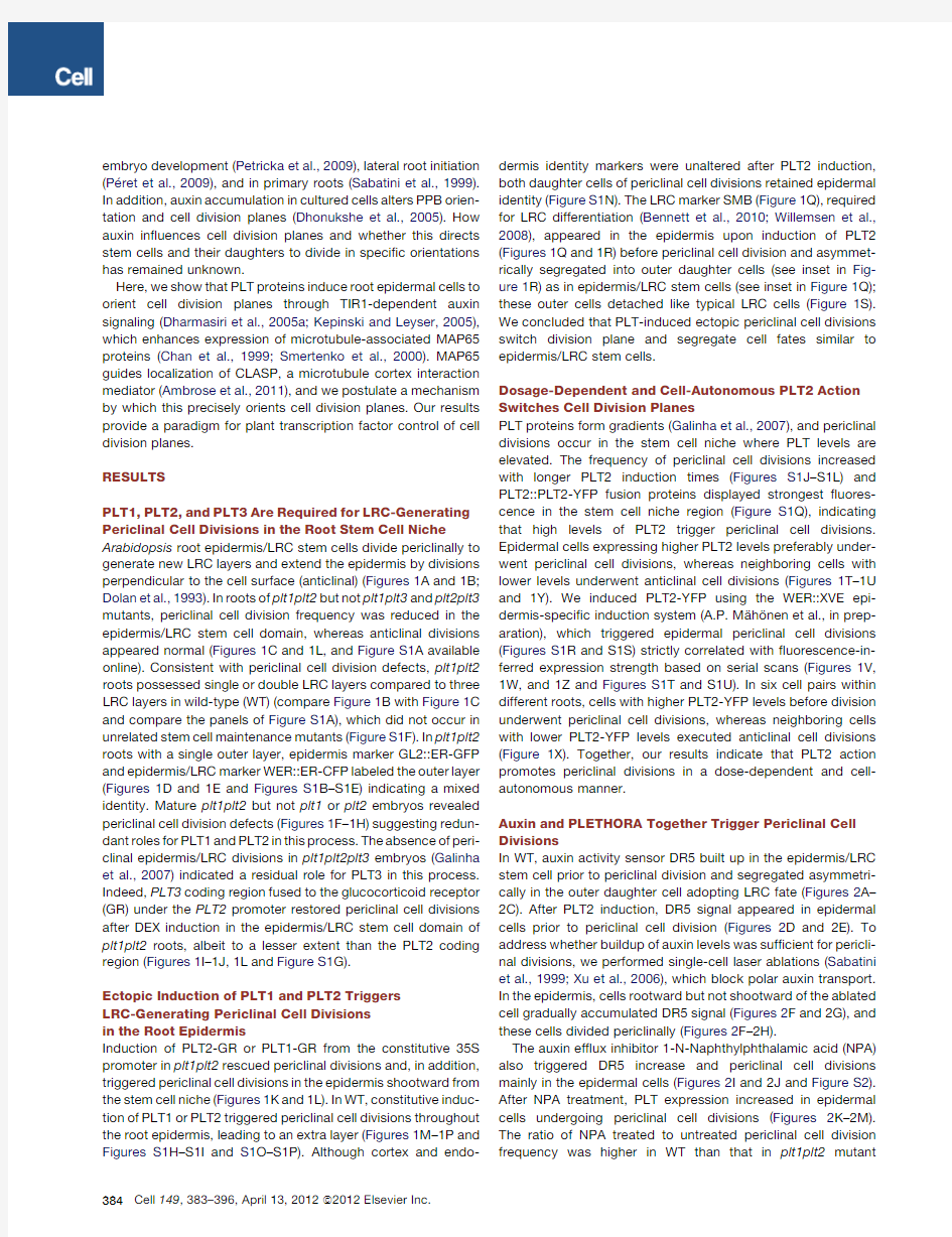
Figure 2.PLT2Induced Periclinal Cell Divisions Are Forecasted by Auxin Activity and Require an Auxin Threshold
(A–C)DR5::YFP-NLS segregation after periclinal epidermis/LRC stem cell division (white arrows in B;n =9roots)and its restricted localization to LRC (C).(D and E)Post-PLT2induction (12hr DEX treated [white arrows in D])DR5::YFP-NLS appearance in epidermis prior to periclinal cell divisions (22hr DEX treated [white arrowheads in E]).
(F–H)Laser ablation of epidermal cell (white circle in F and 24hr after ablation G)triggers DR5::YFP-NLS appearance in epidermal cells below the ablated cell followed by periclinal cell division (G and H).(n =12ablated roots followed in three independent experiments).
(I and J)DR5::YFP-NLS appearance (white arrow in I)and periclinal cell divisions (white arrowheads in I)in the epidermis after NPA treatment.DR5::YFP-NLS in a premitotic cell (white arrow in I).Buildup of DR5::YFP-NLS from the root tip toward the root base correlating with periclinal cell divisions (J).
(K–N)Expression of PLT2-YFP in control (K).Induced expression of PLT2-YFP and periclinal cell divisions (white arrowheads in L and M)after NPA-treatment.Frequency of NPA induced periclinal cell divisions in plt1plt2mutant (N).
(O and P)GH3-mediated auxin conjugation in epidermis (O)reduces PLT2mediated periclinal cell divisions.Auxin overproduction by epidermal expression of iaaH (P)enhances PLT2mediated periclinal cell divisions.After auxin overproduction some cortex cells also divide periclinally (P).DR5::ER-GFP levels correlate with ef?ciency of periclinal cell divisions (O and P).
(Q)Quanti?cation of periclinal cell division frequency after PLT2induction and auxin level manipulations.
Unless stated otherwise,white arrowheads depict periclinal cell divisions and white arrows auxin activity as visualized by the DR5reporter.Red,propidium iodide (PI)staining;green,GFP or YFP.Columns in graphs display means and error bars depict standard deviations.n =29roots for (N)and n =28roots for (Q)from three independent experiments.See also Figure S2.
386Cell 149,383–396,April 13,2012a2012Elsevier Inc.
periclinal cell divisions were also drastically reduced in tir1-1afb2-1afb3-1mutant (Figure 3G)and after active PEO-IAA treatment (Figure S3G).Together our results reveal that TIR1-mediated auxin signaling is critical for stimulation of periclinal cell divisions by the PLT-auxin module (Figure 3
P).
Figure 3.TIR1-Dependent Auxin Signaling Is Critical for PLT2Induced Periclinal Cell Divisions
(A–D)tir1-1afb2-1afb3-1auxin signaling mutant with reduced meristem size (A and B),cell division plane defects (C),and reduced periclinal cell divisions (D)in epidermis/LRC stem cells.
(E–O)PLT2induction rescues anticlinal (O)but not periclinal (N)cell divisions in tir1-1afb2-1afb3-1with increased root meristem size (compare E with A and F with B).NPA induced anticlinal but not periclinal cell divisions in tir1-1afb2-1afb3-1(G and N).Reduction of PLT2-mediated periclinal cell divisions in presence of auxin signaling antagonist PEO-IAA (H–J and N)but not in presence of the inactive analog PEO-IAA (K–N).Absence of DR5signal in epidermis after PEO-IAA treatment (H–J)correlates with speci?c inhibition of periclinal cell divisions.Quanti?cation of periclinal cell division frequencies and percentage periclinal and percentage anticlinal cell divisions (N).
(P)Summary of regulatory interactions.PLT induced periclinal cell divisions require auxin threshold and act through TIR1-dependent auxin signaling.PLT regulates auxin abundance and is auxin responsive.PLT expression is TIR1-auxin signaling pathway dependent.Blue arrows indicate interactions identi?ed in this work,green arrows,previously published interactions.
White arrowheads depict periclinal cell divisions;white arrows mark anticlinal cell divisions.Red:propidium iodide (PI)staining and green:YFP.Columns in graphs display means and error bars depict standard deviations.n =28roots for (D),n =29roots for (N)and (O)from three independent experiments.See also Figure S3.
Cell 149,383–396,April 13,2012a2012Elsevier Inc.387
388Cell149,383–396,April13,2012a2012Elsevier Inc.
PLT Proteins Induce Premitotic Microtubule Reorganization and Cell Division Plane Switch through Transcriptional Regulation of MAP65-1and MAP65-2
In WT epidermis,the PPB visualized by the GFP-microtubule binding domain(MBD)marker(Granger and Cyr,2001)formed anticlinally and cells divided in that plane to extend the epidermis (Figures4A–4C).However,after PLT2induction,premitotic microtubules reorganized longitudinally and formed periclinal PPBs(Figures4D–4F),forecasting the periclinal cell division plane(Figure4G).As a transcription factor,PLT2should switch cell division plane through its transcriptional targets.We pursued downstream targets of PLT2by a genome-wide microarray anal-ysis that distinguished between direct and indirect targets (R.Heidstra and B.Scheres,in preparation).This analysis sug-gested that plant microtubule-associated protein MAP65-2 (Li et al.,2009)was upregulated by PLT2.qRT-PCR analysis con?rmed that MAP65-2is upregulated by PLT2prior to PLT2-mediated induction of periclinal cell division(Figure4H).The closely related MAP65-1(Smertenko et al.,2008)was also induced by PLT2(Figure4H).In tir1-1afb2-1afb3-1mutants, PLT2did not ef?ciently induce MAP65-1and MAP65-2upregu-lation(Figure4H),indicating that their induction requires TIR1-dependent auxin signaling.Transcriptional and translational MAP65-2fusions were strongly expressed in regions with longi-tudinal microtubules(Figures4I–4M)where LRC-generating periclinal cell divisions occur.Our data match with the MAP65-2mRNA pro?le of the Arabidopsis root(Brady et al., 2007),and indicate that MAP65-2transcript levels are regulated by the PLT gradient in the root.
Single map65-2and map65-1T-DNA insertion mutants(Fig-ure S4A)did not display cell division plane defects in the epidermis/LRC domain(not shown).We obtained map65-1map65-2double mutants(Figure S4B)and repressed the expression of both genes by RNAi and amiRNAi approaches (Figure4N).All lines exhibited similar decreased periclinal divi-sions and cell division plane alterations in the epidermis/LRC stem cell region,creating fewer LRC layers(Figure4O).We focused our analysis on MAP65RNAi lines and found that PLT2induction or NPA treatment in this background led to reduced periclinal cell division induction(Figures4P,4R,and 4W).Overexpression of MAP65-2was suf?cient to trigger cell division plane switches in epidermal cells proximal to the root stem cell niche(Figures4S and4W)and occasionally created an extra LRC layer(Figure4T).Subsequently,we visualized the microtubule conformation in MAP65-2-overexpressing lines using MAP65-2-Cherry(Figures4U and4V),GFP-MBD(Fig-ure4X),or GFP-tubulin(Ueda et al.,1999;Figure S4C).Overex-pression of MAP65-2induced microtubule bundling(Figures S4C–S4E)and premitotic microtubules organized longitudinally in several MAP65-2-overexpressing epidermal cells,resulting in the formation of periclinal PPBs(Figures4V and4X).Induced PLT2was unable to switch PPB position from anticlinal to peri-clinal in MAP65RNAi lines(Figure4Y),consistent with the notion that MAP65-1and MAP65-2operate downstream of PLT genes. In addition,auxin-signaling-independent constitutive MAP65-2 expression allowed PPB relocation when PLT2was induced with impaired auxin signaling(Figure4Z).We concluded that MAP65-2bypasses the requirement of TIR1auxin signaling for PPB reorientation and acts downstream of TIR1auxin signaling. Our results show that MAP65-2is a downstream effector of PLT2 and auxin signaling action with the capacity to alter microtubule conformation,change PPB placement and reorient cell division planes.
PLT2Induced Premitotic Microtubule Reorganization Depends on CLASP Function and CLASP Localization
Is MAP65Dependent
The clasp-1mutation in the microtubule bypass mediator CLASP (Ambrose et al.,2011)revealed cell division plane abnormalities in the epidermal/LRC stem cell division region(Figures5A and 5C).PLT2was unable to ef?ciently induce epidermal periclinal cell divisions in the clasp-1mutant(Figures5B and5D).A func-tional GFP-CLASP fusion expressed under the CLASP promoter that rescued the clasp-1mutant phenotype(Ambrose et al.,
Figure4.PLT2Triggers Premitotic Microtubule Organization Switch through TIR1-Dependent Transcriptional Regulation of MAP65-1and MAP65-2
(A–C)GFP-MBD labeled microtubules in WT display transverse orientation(B)along with transverse PPBs(C)and anticlinal cell divisions(A).
(D–G)PLT2induction triggers transverse to longitudinal pre-mitotic microtubule reorganization in epidermis(D and E),along with PPB orientation switch by90 (F and G)and periclinal cell division(G).
(H)MAP65-1and MAP65-2levels after PLT2induction in WT and tir1-1afb2-1afb3-1mutant(16hr DEX).qRT-PCR expression values are from three independent experiments.
(I–M)MAP65-2expression in stem cell niche.MAP65-2expression is high in cells that can undergo periclinal cell divisions(white arrow in I,J,K,and L)where the microtubules localize to apical and basal cell sides(K)and form a periclinal PPB(white arrowheads in M).Zone marked by white brackets reveals gradual reduction in apical-basal microtubules marked by GFP-MBD correlated with reduced capacity to undergo periclinal cell divisions.
(N)RT-PCR analysis of MAP65-1and MAP65-2transcripts in MAP65-1MAP65-2RNAi(‘MAP65RNAi’)and MAP65-1MAP65-2amiRNA(‘MAP65amiRNA’)lines. (O)Division plane changes and LRC reduction in map65-1-1map65-2-1,MAP65RNAi,and MAP65amiRNA lines.White arrows indicate cell division orientations. (P–R)MAP65-1MAP65-2silencing reduces NPA-triggered(compare Q and R)or PLT2-triggered periclinal cell divisions(P).
(S–V)MAP65-2overexpression induces periclinal cell divisions in epidermis proximal to the stem cell niche.Note recent periclinal divisions(S)or extra LRC layer indicating embryonic periclinal division(T).MAP65-2labeled microtubules reorient in epidermal cells prior to periclinal cell division(V)but not in LRC cells(U). White arrows indicate periclinal cell divisions and bidirectional white arrows depict microtubule orientation.
(W)Quanti?cation of periclinal and anticlinal cell division frequencies and number of cell divisions in MAP65-1-and MAP65-2-related manipulations.The columns in graphs display means and error bars represent standard deviations.n=28roots from three independent experiments.
(X–Z)GFP-MBD labeled microtubules mark periclinal PPB in MAP65-2overexpression line(X).PLT2induction after MAP65repression does not switch PPB orientation from anticlinal to periclinal(Y).TIR1-auxin signaling-independent expression of MAP65-2after PLT2induction and PEO-IAA treatment switches PPBs to periclinal orientation(Z).White arrows indicate periclinal cell divisions,white arrowheads,PPBs.
Red,propidium iodide(PI)staining and green,GFP or YFP.See also Figure S4.
Cell149,383–396,April13,2012a2012Elsevier Inc.389
2011)was expressed in the root region encompassing formative cell divisions (Figure 5E).CLASP localized predominantly to apical and basal cell sides within the epidermal/LRC domain prior to periclinal cell divisions,in contrast to its lateral localiza-tion after periclinal division and in many other cell types under-going anticlinal cell divisions (Figures 5E–5G).Furthermore,CLASP was enriched at sharp radial cell edges (Figures 5H–5J).This CLASP localization typically occurred within cells competent to undergo periclinal cell divisions (Campilho et al.,2006),but not in cells or cell layers where anticlinal cell divisions take place (Figures 5E–5G).PLT2induction and NPA treatment gradually shifted CLASP localization from lateral to apical-basal
cell sides,consistent with the capacity of these manipulations to relocate PPBs (Figures 5K–5N and Figures S5A–S5F).CLASP abundance was not altered after PLT2induction and auxin application (Figure S5G).
GFP-CLASP and mRFP-tubulin coexpression revealed CLASP colocalization with microtubules (Figures 5O and 5Q).Interestingly,CLASP lost its cell-edge-related localization after oryzalin-induced microtubule depolymerization (Figure 5P),demonstrating that the maintenance of subcellular CLASP local-ization requires intact microtubules.
PLT2induction increased MAP65-1and MAP65-2expression in the clasp-1mutant (Figure S6E)but failed to induce
periclinal
Figure 5.PLT2-Triggered Switch in Premitotic Microtubule Organization Requires CLASP Action and CLASP Localization Is Microtubule Dependent
(A–D)Abnormal cell division orientation in epidermis/LRC stem cell in clasp-1mutant (A)and reduced frequency of periclinal cell divisions (C).PLT2induced periclinal cell divisions are reduced in clasp-1(B and D).
(E–J)GFP-CLASP labels apical-basal cell sides (white arrowheads)prior to division only in epidermis/LRC stem cells prone to undergo periclinal cell division,and lateral cell sides (white arrows)in divided epidermis/LRC stem cells and all other cells (E).3D assembly of 300.5m m equidistant CLSM scans depicts differential GFP-CLASP localization to apical-basal cell sides in epidermis/LRC domain cells prone to undergo periclinal cell division (F).Image is color coded to highlight different cell sides (G).Cross-section (H)with GFP-CLASP enrichment on radial cell sides (the cell sides on which a PPB assembles to mark a periclinal cell division)as evident by quanti?cation of GFP-CLASP intensity at radial and peripheral cell edges (J).
(K–N)After PLT2induction GFP-CLASP labels apical-basal cell sides (white arrowheads)prior to division (K).During reorientation,GFP-CLASP displays transient nonpolar localization (L–N).
(O–Q)CLASP colocalization with tubulin-labeled microtubules (see the overlap coef?cient in Q)and aberrant CLASP localization after oryzalin induced micro-tubule depolymerization (P).
White arrowheads depict periclinal cell divisions,white arrow anticlinal cell divisions in (A)and (B).Red,propidium iodide (PI)staining or mRFP and green,GFP.White arrowheads depict GFP-CLASP localization in (E)–(N).Graph columns depict means,error bars indicate standard deviation.n =26roots for (C),n =29roots for (D),n =42cells from 6roots for (J)and n =22cells from ?ve roots for (Q)from three independent experiments.See also Figure S5.
390Cell 149,383–396,April 13,2012a2012Elsevier Inc.
cell divisions indicating that CLASP and MAP65are both required for microtubule array reorientation and cell division plane switch.To probe the nature of this interdependency we ?rst analyzed whether localization and function of CLASP depends on MAP65levels.MAP65-2overexpression induced transverse microtubule bundles in cells within the epidermal/ LRC region,which were colabeled with CLASP especially at the apical-basal cell edges(Figure6A and Figures S4C–S4E). Quantitative?uorescence intensity pro?ling revealed selective enrichment of CLASP at microtubule bundles possessing higher MAP65levels and contacting top-down cell edges(Figures6C and6F)and CLASP colocalization with MAP65at these edges (Figure6E).MAP65-positive microtubule bundles were relatively resistant to oryzalin-induced microtubule depolymerization (Figures S6A–S6D).CLASP retained its cell-edge-related locali-zation where MAP65-positive microtubule bundle resisted microtubule depolymerization(Figures6B,6D,and6G),sug-gesting that MAP65reinforces CLASP persistence at cell edges. Indeed,CLASP did not ef?ciently load on microtubules and on cell edges in the MAP65RNAi line and instead remained largely cytosolic(Figures6L and6M).The localization of CLASP at apical-basal cell sides was more severely affected than at lateral cell sides(Figures6L and6M and compare Figure5E with Fig-ure6L).Our data reveal that MAP65has a role in recruiting CLASP on microtubules and at apical-basal sharp cell edges that promote microtubule passage at those edges to favor peri-clinal PPB and periclinal cell divisions.Conversely,MAP65 induced many transverse and a few longitudinal microtubule bundles in the absence of CLASP(Figures6H–6J).Overex-pressed CLASP induced microtubule bundling in nonroot cell types(Kirik et al.,2007),and in our hands it also induced spaghetti-shaped microtubule bundles in root cells(Figure6O) but did not consistently reorient cell division planes(Figures 6K–6N),although randomized cell division planes were occa-sionally observed(data not shown).Overexpressed CLASP was unable to load on to microtubules and induce microtubule bundling in the MAP65RNAi line(Figures6P and6Q).Together our results show that MAP65function is required for CLASP localization to microtubules,for CLASP recruitment to the edges of apical-basal cell sides,and for CLASP function.
CLASP-Facilitated Crossing of Apical and Basal Cell Edges Is Suf?cient for90 Rotation of the Microtubular Array
In switching from an anticlinal to a periclinal cell division,the pre-mitotic cortical microtubule array reorients and the orientation of the PPB changes by90 .How can this orientation be so precisely controlled?It was recently shown that microtubule organization in nondividing cells is in?uenced by the ease with which microtubules can traverse edges between adjacent cell faces(Ambrose et al.,2011).We tested whether CLASP-induced changes in microtubule crossing rates at the cell edges bound-ing the apical and basal cell faces are suf?cient to reliably switch the orientation of the microtubule array for rotating the cell divi-sion plane.To that end we performed simulations of interacting microtubules on cubical surfaces using a previously developed algorithm(Tindemans et al.,2010);see Extended Experimental Procedures for details).
The barrier presented by a cell edge for microtubule crossing
to an adjacent cell face was modeled as a probability of under-
going a catastrophe upon reaching the edge,chosen differently
for the periclinal edges(P PC)and anticlinal edges(P AC)(Fig-
ure7A).Ambrose et al.(2011)have shown that in the absence
of CLASP,anticlinal edges present a strong barrier to microtu-
bule crossing,so we associated this with high values of P AC.
When CLASP localizes to anticlinal edges,microtubules readily
cross to and from the apical and basal faces,which we associ-
ated with small values of P AC.To quantify the orientation of the
aligned array on the cubical surface,we introduced an order
parameter C2with valueà0.5for a perfectly ordered array in
an anticlinal orientation and value1for a perfectly ordered array
in one of the two equivalent periclinal orientations.Figure7A
displays average C2values as a function of the catastrophe
probability on impinging an anticlinal edge P AC for systems
with and without bundling.When P AC>P PC(low density of
CLASP at the anticlinal edges),microtubules attempting to cross
experience a high rate of edge-induced catastrophes and hence
have a diminished lifespan.In this case we?nd C2$1,indicating that the systems are almost exclusively ordered with an anticlinal
orientation.When P AC
clinal edges),the lifespan of microtubules entering the apical and
basal faces is enhanced with respect to those attempting to
cross over between periclinal faces,and we?nd C2$à0.5,indi-cating predominant periclinal ordering.This is illustrated by two characteristic snapshots taken at a high value of P AC(Figure7B) and a low value of P AC(Figure7C).In the intermediate regime where P AC$P PC the ability of the system to choose a speci?c orientation was impaired.This resulted in bimodal distributions for the order parameter C2(see Figure S7),indicating that the system randomly chooses one of the three possible orientations dictated by the symmetry of the cell.We also addressed the role that MAP65-mediated bundling could play in this process.Histo-grams of the order parameter C2for the four possible situations in presence or absence of CLASP,and presence or absence of bundling,revealed that in all cases a unique anticlinal(Figures 7D and7F)or periclinal(Figures7E and7G)orientation is ob-tained.Our simulations reveal that changes in CLASP posi-tioning are suf?cient to reliably determine emergent90 switches in orientation of the microtubule array.The simulations further suggest that the role of MAP65in cell division plane orientation is primarily through its contribution to CLASP localization rather than microtubule bundling.
DISCUSSION
Spatiotemporal Control of Formative Divisions
In this study we show that PLT transcription factors and auxin together control the division plane reorientation and asymmetric cell division that de?nes a formative division in plants.The PLT proteins and the auxin response machinery upregulate members of the MAP65family of microtubular cytoskeleton regulators, which we show to be essential for premitotic microtubule array reorientation and cell division plane rotation through a hitherto unexpected role in CLASP localization.Our work thus addresses the long-standing issue of how patterning is connected to the mechanistic control of precisely oriented cell divisions in plants.
Cell149,383–396,April13,2012a2012Elsevier Inc.391
Figure6.CLASP Localization to Microtubules and to Apical-Basal Cell Sides Require MAP65
(A–G)CLASP colocalizes with MAP65on microtubules(overlap coef?cient in E).Overexpressed MAP65induces microtubule bundling and retains CLASP on oryzalin resistant longitudinal microtubule bundles.Full intensity pro?le landscapes of whole images shown in C and D.CLASP intensity(green peaks highlighted by green arrowheads in C)on transversal microtubules in MAP65-2mCherry nonexpressing cell is reduced compared to longitudinal microtubules in neighboring cell expressing MAP65-2mCherry(yellow arrowheads in C).After oryzalin treatment green-colored CLASP intensity in the cell without overexpressed MAP65 decreases(green arrowhead in D),but colocalization with red MAP65-2intensity peaks(yellow arrowhead in D)remains in cell overexpressing MAP65-2mCherry. GFP-CLASP intensity quanti?cation shown in(F)and(G).
(H–J)MAP65microtubule localization and bundling capacity is CLASP-independent both after constitutive(H)and induced expression(J).In WT,induced MAP65 triggers formation of more longitudinal microtubule bundles and transverse-to-longitudinal microtubule array switch(I),whereas in absence of CLASP its ef?-ciency to induce longitudinal microtubule bundles(compare J with I)and microtubule array reorientation(H)is hampered.
(K)Quanti?cation of periclinal and anticlinal cell division frequencies in case of MAP65and CLASP overexpression.
(L and M)CLASP localization to microtubules,especially to longitudinal microtubules(compare L with Figure5E),is impaired upon MAP65repression.
(N–Q)CLASP promotes microtubule bundles but less after MAP65repression(compare N and O with P and Q),which promotes predominant cytosolic local-ization of CLASP.
Bold bidirectional arrows show direction of microtubule orientation.White or green arrowheads indicate GFP-CLASP localization and red arrowheads depict MAP65-mCherry.The columns in graphs display means,and error bars represent standard deviation.n=34cells from?ve roots for(E),n=45cells from eight roots for(F),n=37cells from eight roots for(G)and n=29roots for(K)from three independent experiments.See also Figure S6.
392Cell149,383–396,April13,2012a2012Elsevier Inc.
The epidermis/LRC stem cell division takes place repeatedly in the stem cell niche of the Arabidopsis root,where PLT proteins are abundant (Galinha et al.,2007)and where the growth regu-lator auxin reaches maximum levels (Grieneisen et al.,2007;Petersson et al.,2009).Auxin signaling is required for the initia-tion of PLT transcription (Aida et al.,2004),but PLT transcrip-tional activation also induces increased auxin response (Galinha et al.,2007;this manuscript).Both high PLT activity and threshold auxin levels promote the epidermis/LRC formative division.This synergy between high PLT levels and auxin action on the epidermis/LRC division may serve to precisely specify the position of formative divisions.The auxin signaling TIR1module is critically required for the execution of division plane rotation downstream of PLT gene action but upstream of MAP65activa-tion.This pathway suggests that PLT action activates speci?c auxin responsive transcription factors (ARFs)or represses their repressors (AUX/IAAs)(Guilfoyle and Hagen,2007;Lau et al.,2011)to allow a speci?c change in auxin response leading to
MAP65transcription.Similar adaptive changes in auxin response factors have been demonstrated for the progression of lateral root initiation (De Smet et al.,2010).The notion of specialized auxin response modules for cell division plane regu-lation is consistent with reports on precise alterations in cell division planes upon reduction of ARF function in the embryo (Hamann et al.,1999;Hardtke and Berleth,1998).Intriguingly,PLT expression in the tir1-1afb2-1afb3-1auxin signaling mutants uncouples control of cell division orientation from the general stimulatory effect of auxin on cell division.In contrast,PLT induc-tion in lines with reduced ABP1activity,which represents another auxin signaling pathway,could not rescue general cell cycle control (Tromas et al.,2009).
Microtubule-Based Division Plane Control in Multicellular Context
We demonstrate that MAP65and CLASP proteins,involved in microtubule dynamics,are relevant players in the control of
the
Figure 7.A Model for Transcription-Factor-Mediated Precise Rotation of Cell Division Plane
(A–E)Impact of CLASP-based differences in catastrophe probabilities when crossing cell edges determined by stochastic simulations of interacting cortical microtubules.Spontaneous catastrophe probability for microtubules crossing an edge given by P AC (anticlinal edges)and P PC (periclinal edges),see inset.P AC is decreased from 1to 0with P PC =0.26kept constant.When the anticlinal edges are hard or impossible to cross,the transverse orientation prevails (Alignment order parameter C 2>0,with a maximum of C 2=1when all microtubules are transversely aligned).When the anticlinal edges are easiest to cross,the longitudinal alignment dominates (C 2<0,with a minimum of C 2=à0.5when all microtubules are longitudinally aligned).This holds true both with (red solid curve)and without (cyan dotted curve)microtubule bundling.Both curves cross C 2=0when P AC $P PC ,i.e.,when there is no appre-ciable difference between anticlinal and periclinal edges.
Bottom panels present two speci?c cases:without CLASP located at the anticlinal edges (left;P AC =0.9)and with CLASP at the anticlinal edges (right;P AC =0.1).Snapshots (B)and (C)show repre-sentative microtubule arrays for WT cells,i.e.,with bundling.The superimposed green lines show the ease of crossing the edge:easy (dotted,P AC =0.1),intermediate (dashed,P PC =0.26)and hard (long dashed /almost solid,P AC =0.9).
Histograms of array orientations (N =200simula-tions each)for the same parameters are shown in (D)and (E),respectively.Histogram (F)corre-sponds to the MAP65mutant,which has neither bundling nor CLASP at the anticlinal edges.(G)represents a hypothetical MAP65mutant de?cient in bundling,but allowing proper CLASP localiza-tion at the anticlinal edges.
(H)Summary of regulatory interactions:The PLT2-auxin pathway changes abundance of MAP65,which facilitates CLASP relocalization for cell division plane switching.Blue arrows indicate interactions identi?ed by this work;green arrows,previously published interactions.See also Figure S7.
Cell 149,383–396,April 13,2012a2012Elsevier Inc.393
epidermis/LRC stem cell formative division and the associated shift in PPB positioning.MAP65-1and MAP65-2localize to regions of microtubule overlap and promote crosslinking of anti-parallel microtubules and their stabilization(Gaillard et al.,2008; Li et al.,2009;Van Damme et al.,2004).Recent dynamic coloc-alization of MAP65-1and MAP65-2with polymerizing microtu-bules indicate that plant cortical microtubules bundle through a microtubule-microtubule templating mechanism(Lucas et al., 2011).Another member of the same MAP65protein family, MAP65-4,promotes microtubule bundle elongation(Fache et al.,2010).However,we show that the role of MAP65in division plane reorientation may be separable from microtubule bundling and instead largely relies on its role in CLASP localization. Plant CLASP and MAP65proteins have both been implicated as regulators of general microtubular array stability(Ambrose et al.,2011;Kirik et al.,2007;Li et al.,2009).In addition,there is evidence that CLASP increases the attachment strength of microtubules to the cell cortex(Ambrose and Wasteneys, 2008).CLASP levels are not regulated by PLTs or auxin and CLASP is expressed ubiquitously in mitotic root cells(Kirik et al.,2007).CLASP’s involvement in selective microtubule passage at sharp cell edges(Ambrose et al.,2011)and its typical localization at those edges during the cell division plane switch (this study)suggest that localized CLASP guides directional microtubule reorganization.How CLASP is recruited to selective cell edges remains unclear,but MAP65plays a role in either delivery by microtubules or stabilization of CLASP at selected cell edges and then,through its association with transfacial microtubule bundles,enables the passage of microtubules. Our modeling efforts support a scenario in which localization of CLASP by MAP65,rather than MAP65bundling activity,contrib-utes to cell division plane switches.The simulations reveal that CLASP localization to anticlinal edges,enabling microtubules to freely pass,is a robust mechanism for precisely switching the preferred orientation of the cortical array.As presence or absence of microtubule bundling without considering CLASP function has little effect on this mechanism,MAP65likely facili-tates this process through its role in CLASP localization.
It is broadly recognized that the cortical microtubule array is a self-organizing network where microtubule nucleation,dynamic microtubule instability,and microtubule-microtubule encounters determine spatial ordering(Wasteneys and Ambrose,2009).Our results demonstrate how transcription factors feed into cytoskel-etal dynamics through MAP65-mediated CLASP localization. The precise cellular mechanisms by which CLASP is differentially localized and how this affects microtubule dynamics will have to be elucidated in future studies.
EXPERIMENTAL PROCEDURES
Plant Material and Microscopy
Details of plant lines and growth conditions,constructs,molecular cloning, plant transformation,and expression pro?ling are described in Supplemental Information.Confocal laser-scanning microscopy(CLSM)(Dhonukshe et al., 2006;Dhonukshe et al.,2008)and cell ablations(Xu et al.,2006)were per-formed as previously described.Fluorescence signal intensity was analyzed with Leica(Live)and Zeiss(ZEN)confocal softwares.Overlap coef?cients were calculated based on Manders et al.(1992).Data were statistically evalu-ated with Excel2003(Microsoft).Cell surface and median confocal sections displaying microtubules were obtained with slightly widened pin-holes in the CLSM setup that allows visualizing microtubule conformations in cells within the same confocal section.
Chemical Treatments
NPA(Duchefa),Oryzalin(Sigma),Dexamethasone(Sigma),Estradiol(Sigma), and PEO-IAA(a gift from Prof.Hayashi)were used from DMSO stock solutions at25m M NPA,2m M Oryzalin,10m M Dexamethasone(Dex),5m M Estradiol, and20m M PEO-IAA working concentrations for indicated periods.
Cell Division Plane Frequency Analysis
Periclinal cell division frequency in the epidermal layer including the epidermis/ LRC stem cell region(the colored region in Figure S1M)was quanti?ed by counting periclinal cell divisions in comparable CLSM root scans.Periclinal and anticlinal cell division ratios were obtained by counting the number of peri-clinal and anticlinal cell divisions and dividing by total division number from the colored regions as shown in Figure S1M.plt1plt2and tir1-1afb2-1afb3-1 mutants had very short roots,so only the periclinal cell division was quanti?ed. However,rescue of cell divisions after PLT2induction in those mutants al-lowed quanti?cation of periclinal and anticlinal cell division ratios.Data were statistically evaluated with Excel2003(Microsoft).
Computer Simulations
The simulations of the cortical microtubule array were performed using the event-based algorithm also employed in Tindemans et al.(2010).Details are described in the Supplemental Information.
SUPPLEMENTAL INFORMATION
Supplemental Information includes Supplemental Experimental Procedures, seven?gures,and one table and can be found with this article online at doi:10.1016/j.cell.2012.02.051.
ACKNOWLEDGMENTS
We thank Mark Estelle,Kenichiro Hayashi,Martine Pastuglia,Marcus Heisler, and Takashi Hashimoto for sharing published materials;Anirban Baral for help with experiments;and Frits Kindt and Ronald Leito for photography and image processing.This work was supported by:Netherlands Scienti?c Organiza-tion’s(NWO’s)-VENI Grant(P.D.),Utrecht University Independent Investigator Grant(P.D.),European Research Council Advanced Investigator Grant(B.S.), HFSP fellowship(A.P.M.),EMBO fellowship(K.P.),Netherlands Genomics Initiative(NGI)and NWO-Horizon grants(M.L.and R.H.),Natural Sciences and Engineering Research Council of Canada Grant(G.W.and C.A.), Netherlands Consortium for System Biology’s grant(B.S.and E.E.D.),and Foundation for Fundamental Research on Matter(FOM)-NWO(B.M.M.).
Received:July4,2011
Revised:November15,2011
Accepted:February28,2012
Published:April12,2012
REFERENCES
Aida,M.,Beis, D.,Heidstra,R.,Willemsen,V.,Blilou,I.,Galinha, C., Nussaume,L.,Noh,Y.S.,Amasino,R.,and Scheres, B.(2004).The PLETHORA genes mediate patterning of the Arabidopsis root stem cell niche. Cell119,109–120.
Allard,J.F.,Wasteneys,G.O.,and Cytrynbaum,E.N.(2010).Mechanisms of self-organization of cortical microtubules in plants revealed by computational simulations.Mol.Biol.Cell21,278–286.
Ambrose,J.C.,and Wasteneys,G.O.(2008).CLASP modulates microtubule-cortex interaction during self-organization of acentrosomal microtubules. Mol.Biol.Cell19,4730–4737.
394Cell149,383–396,April13,2012a2012Elsevier Inc.
Ambrose,J.C.,Shoji,T.,Kotzer,A.M.,Pighin,J.A.,and Wasteneys,G.O. (2007).The Arabidopsis CLASP gene encodes a microtubule-associated protein involved in cell expansion and division.Plant Cell19,2763–2775.
Ambrose,C.,Allard,J.F.,Cytrynbaum,E.N.,and Wasteneys,G.O.(2011).
A CLASP-modulated cell edge barrier mechanism drives cell-wide cortical microtubule organization in https://www.doczj.com/doc/5a11547416.html,mun.2,430.
Baena-Lo′pez,L.A.,Baonza,A.,and Garc?′a-Bellido,A.(2005).The orientation of cell divisions determines the shape of Drosophila organs.Curr.Biol.15, 1640–1644.
Bennett,T.,van den Toorn,A.,Sanchez-Perez,G.F.,Campilho,A.,Willemsen, V.,Snel,B.,and Scheres,B.(2010).SOMBRERO,BEARSKIN1,and BEAR-SKIN2regulate root cap maturation in Arabidopsis.Plant Cell22,640–654.
Blilou,I.,Xu,J.,Wildwater,M.,Willemsen,V.,Paponov,I.,Friml,J.,Heidstra, R.,Aida,M.,Palme,K.,and Scheres,B.(2005).The PIN auxin ef?ux facilitator network controls growth and patterning in Arabidopsis roots.Nature433, 39–44.
Brady,S.M.,Orlando,D.A.,Lee,J.Y.,Wang,J.Y.,Koch,J.,Dinneny,J.R., Mace,D.,Ohler,U.,and Benfey,P.N.(2007).A high-resolution root spatiotem-poral map reveals dominant expression patterns.Science318,801–806.
Camilleri,C.,Azimzadeh,J.,Pastuglia,M.,Bellini,C.,Grandjean,O.,and Bouchez,D.(2002).The Arabidopsis TONNEAU2gene encodes a putative novel protein phosphatase2A regulatory subunit essential for the control of the cortical cytoskeleton.Plant Cell14,833–845.
Campilho,A.,Garcia,B.,Toorn,H.V.,Wijk,H.V.,Campilho,A.,and Scheres,B. (2006).Time-lapse analysis of stem-cell divisions in the Arabidopsis thaliana root meristem.Plant J.48,619–627.
Chan,J.,Jensen,C.G.,Jensen,L.C.,Bush,M.,and Lloyd,C.W.(1999). The65-kDa carrot microtubule-associated protein forms regularly arranged ?lamentous cross-bridges between microtubules.Proc.Natl.Acad.Sci. USA96,14931–14936.
De Smet,I.,and Beeckman,T.(2011).Asymmetric cell division in land plants and algae:the driving force for differentiation.Nat.Rev.Mol.Cell Biol. 12,177–188.
De Smet,I.,Vassileva,V.,De Rybel,B.,Levesque,M.P.,Grunewald,W.,Van Damme,D.,Van Noorden,G.,Naudts,M.,Van Isterdael,G.,De Clercq,R., et al.(2008).Receptor-like kinase ACR4restricts formative cell divisions in the Arabidopsis root.Science322,594–597.
De Smet,I.,Lau,S.,Voss,U.,Vanneste,S.,Benjamins,R.,Rademacher,E.H., Schlereth,A.,De Rybel,B.,Vassileva,V.,Grunewald,W.,et al.(2010).Bimod-ular auxin response controls organogenesis in Arabidopsis.Proc.Natl.Acad. https://www.doczj.com/doc/5a11547416.html,A107,2705–2710.
Dharmasiri,N.,Dharmasiri,S.,and Estelle,M.(2005a).The F-box protein TIR1 is an auxin receptor.Nature435,441–445.
Dharmasiri,N.,Dharmasiri,S.,Weijers,D.,Lechner,E.,Yamada,M.,Hobbie, L.,Ehrismann,J.S.,Ju¨rgens,G.,and Estelle,M.(2005b).Plant development is regulated by a family of auxin receptor F box proteins.Dev.Cell9,109–119.
Dhonukshe,P.,Mathur,J.,Hu¨lskamp,M.,and Gadella,T.W.,Jr.(2005). Microtubule plus-ends reveal essential links between intracellular polarization and localized modulation of endocytosis during division-plane establishment in plant cells.BMC Biol.3,11.
Dhonukshe,P.,Baluska,F.,Schlicht,M.,Hlavacka,A.,Samaj,J.,Friml,J.,and Gadella,T.W.,Jr.(2006).Endocytosis of cell surface material mediates cell plate formation during plant cytokinesis.Dev.Cell10,137–150.
Dhonukshe,P.,Tanaka,H.,Goh,T.,Ebine,K.,Ma¨ho¨nen,A.P.,Prasad,K., Blilou,I.,Geldner,N.,Xu,J.,Uemura,T.,et al.(2008).Generation of cell polarity in plants links endocytosis,auxin distribution and cell fate decisions.Nature 456,962–966.
Di Laurenzio,L.,Wysocka-Diller,J.,Malamy,J.E.,Pysh,L.,Helariutta,Y., Freshour,G.,Hahn,M.G.,Feldmann,K.A.,and Benfey,P.N.(1996).The SCARECROW gene regulates an asymmetric cell division that is essential for generating the radial organization of the Arabidopsis root.Cell86,423–433.Dolan,L.,Janmaat,K.,Willemsen,V.,Linstead,P.,Poethig,S.,Roberts,K., and Scheres, B.(1993).Cellular organisation of the Arabidopsis thaliana root.Development119,71–84.
Eren,E.C.,Dixit,R.,and Gautam,N.(2010).A three-dimensional computer simulation model reveals the mechanisms for self-organization of plant cortical microtubules into oblique arrays.Mol.Biol.Cell21,2674–2684.
Fache,V.,Gaillard,J.,Van Damme,D.,Geelen,D.,Neumann,E.,Stoppin-Mellet,V.,and Vantard,M.(2010).Arabidopsis kinetochore?ber-associated MAP65-4cross-links microtubules and promotes microtubule bundle elonga-tion.Plant Cell22,3804–3815.
Gaillard,J.,Neumann, E.,Van Damme, D.,Stoppin-Mellet,V.,Ebel,C., Barbier,E.,Geelen,D.,and Vantard,M.(2008).Two microtubule-associated proteins of Arabidopsis MAP65s promote antiparallel microtubule bundling. Mol.Biol.Cell19,4534–4544.
Galinha,C.,Hofhuis,H.,Luijten,M.,Willemsen,V.,Blilou,I.,Heidstra,R.,and Scheres,B.(2007).PLETHORA proteins as dose-dependent master regulators of Arabidopsis root development.Nature449,1053–1057.
Granger,C.L.,and Cyr,R.J.(2001).Spatiotemporal relationships between growth and microtubule orientation as revealed in living root cells of Arabidop-sis thaliana transformed with green-?uorescent-protein gene construct GFP-MBD.Protoplasma216,201–214.
Grieneisen,V.A.,Xu,J.,Mare′e,A.F.,Hogeweg,P.,and Scheres,B.(2007). Auxin transport is suf?cient to generate a maximum and gradient guiding root growth.Nature449,1008–1013.
Guilfoyle,T.J.,and Hagen,G.(2007).Auxin response factors.Curr.Opin.Plant Biol.10,453–460.
Gunning,B.,Hughes,J.,and Hardham,A.(1978).Formative and Proliferative Cell Divisions,Cell Differentiation,and Developmental Changes in the Meri-stem of Azolla Roots.Planta143,121–144.
Hamann,T.,Mayer,U.,and Ju¨rgens,G.(1999).The auxin-insensitive bodenlos mutation affects primary root formation and apical-basal patterning in the Arabidopsis embryo.Development126,1387–1395.
Hardtke,C.S.,and Berleth,T.(1998).The Arabidopsis gene MONOPTEROS encodes a transcription factor mediating embryo axis formation and vascular development.EMBO J.17,1405–1411.
Hayashi,K.,Tan,X.,Zheng,N.,Hatate,T.,Kimura,Y.,Kepinski,S.,and Nozaki,H.(2008).Small-molecule agonists and antagonists of F-box protein-substrate interactions in auxin perception and signaling.Proc.Natl. https://www.doczj.com/doc/5a11547416.html,A105,5632–5637.
Heidstra,R.,Welch,D.,and Scheres,B.(2004).Mosaic analyses using marked activation and deletion clones dissect Arabidopsis SCARECROW action in asymmetric cell division.Genes Dev.18,1964–1969.
Heisler,M.G.,Ohno,C.,Das,P.,Sieber,P.,Reddy,G.V.,Long,J.A.,and Meyerowitz,E.M.(2005).Patterns of auxin transport and gene expression during primordium development revealed by live imaging of the Arabidopsis in?orescence meristem.Curr.Biol.15,1899–1911.
Helariutta,Y.,Fukaki,H.,Wysocka-Diller,J.,Nakajima,K.,Jung,J.,Sena,G., Hauser,M.T.,and Benfey,P.N.(2000).The SHORT-ROOT gene controls radial patterning of the Arabidopsis root through radial signaling.Cell101,555–567.
Ju¨rgens,G.(1995).Axis formation in plant embryogenesis:cues and clues. Cell81,467–470.
Kares,C.,Prinsen,E.,van Onckelen,H.,and Otten,L.(1990).IAA synthesis and root induction with iaa genes under heat shock promoter control.Plant. Mol.Biol.15,225–236.
Kepinski,S.,and Leyser,O.(2005).The Arabidopsis F-box protein TIR1is an auxin receptor.Nature435,446–451.
Kirik,V.,Herrmann,U.,Parupalli,C.,Sedbrook,J.C.,Ehrhardt,D.W.,and Hu¨lskamp,M.(2007).CLASP localizes in two discrete patterns on cortical microtubules and is required for cell morphogenesis and cell division in Arabidopsis.J.Cell Sci.120,4416–4425.
Lau,S.,De Smet,I.,Kolb,M.,Meinhardt,H.,and Ju¨rgens,G.(2011).Auxin trig-gers a genetic switch.Nat.Cell Biol.13,611–615.
Cell149,383–396,April13,2012a2012Elsevier Inc.395
Li,H.,Zeng,X.,Liu,Z.Q.,Meng,Q.T.,Yuan,M.,and Mao,T.L.(2009). Arabidopsis microtubule-associated protein AtMAP65-2acts as a microtubule stabilizer.Plant Mol.Biol.69,313–324.
Lucas,J.R.,Courtney,S.,Hassfurder,M.,Dhingra,S.,Bryant,A.,and Shaw, S.L.(2011).Microtubule-associated proteins MAP65-1and MAP65-2 positively regulate axial cell growth in etiolated Arabidopsis hypocotyls.Plant Cell23,1889–1903.
Manders,E.M.,Stap,J.,Brakenhoff,G.J.,van Driel,R.,and Aten,J.A.(1992). Dynamics of three-dimensional replication patterns during the S-phase, analysed by double labelling of DNA and confocal microscopy.J.Cell Sci. 103,857–862.
Mu¨ller,S.,Wright,A.J.,and Smith,L.G.(2009).Division plane control in plants: new players in the band.Trends Cell Biol.19,180–188.
Nakajima,K.,Sena,G.,Nawy,T.,and Benfey,P.N.(2001).Intercellular move-ment of the putative transcription factor SHR in root patterning.Nature413, 307–311.
Pe′ret,B.,De Rybel,B.,Casimiro,I.,Benkova′,E.,Swarup,R.,Laplaze,L., Beeckman,T.,and Bennett,M.J.(2009).Arabidopsis lateral root development: an emerging story.Trends Plant Sci.14,399–408.
Petersson,S.V.,Johansson,A.I.,Kowalczyk,M.,Makoveychuk,A.,Wang, J.Y.,Moritz,T.,Grebe,M.,Benfey,P.N.,Sandberg,G.,and Ljung,K.(2009). An auxin gradient and maximum in the Arabidopsis root apex shown by high-resolution cell-speci?c analysis of IAA distribution and synthesis.Plant Cell21,1659–1668.
Petricka,J.J.,Van Norman,J.M.,and Benfey,P.N.(2009).Symmetry breaking in plants:molecular mechanisms regulating asymmetric cell divisions in Arabidopsis.Cold Spring Harb Perspect Biol1,a000497.
Pickett-Heaps,J.D.,and Northcote,D.H.(1966).Organization of microtubules and endoplasmic reticulum during mitosis and cytokinesis in wheat meri-stems.J.Cell Sci.1,109–120.
Rasmussen,C.G.,Humphries,J.A.,and Smith,L.G.(2011a).Determination of symmetric and asymmetric division planes in plant cells.Annu.Rev.Plant Biol. 62,387–409.
Rasmussen,C.G.,Sun,B.,and Smith,L.G.(2011b).Tangled localization at the cortical division site of plant cells occurs by several mechanisms.J.Cell Sci. 124,270–279.
Sabatini,S.,Beis,D.,Wolkenfelt,H.,Murfett,J.,Guilfoyle,T.,Malamy,J., Benfey,P.,Leyser,O.,Bechtold,N.,Weisbeek,P.,and Scheres,B.(1999). An auxin-dependent distal organizer of pattern and polarity in the Arabidopsis root.Cell99,463–472.
Smertenko,A.,Saleh,N.,Igarashi,H.,Mori,H.,Hauser-Hahn,I.,Jiang,C.J., Sonobe,S.,Lloyd,C.W.,and Hussey,P.J.(2000).A new class of microtu-bule-associated proteins in plants.Nat.Cell Biol.2,750–753.Smertenko,A.P.,Kaloriti,D.,Chang,H.Y.,Fiserova,J.,Opatrny,Z.,and Hussey,P.J.(2008).The C-terminal variable region speci?es the dynamic properties of Arabidopsis microtubule-associated protein MAP65isotypes. Plant Cell20,3346–3358.
Smith,L.G.(2001).Plant cell division:building walls in the right places.Nat. Rev.Mol.Cell Biol.2,33–39.
Staswick,P.E.,Serban, B.,Rowe,M.,Tiryaki,I.,Maldonado,M.T., Maldonado,M.C.,and Suza,W.(2005).Characterization of an Arabidopsis enzyme family that conjugates amino acids to indole-3-acetic acid.Plant Cell17,616–627.
ten Hove,C.A.,Willemsen,V.,de Vries,W.J.,van Dijken,A.,Scheres,B.,and Heidstra,R.(2010).SCHIZORIZA encodes a nuclear factor regulating asym-metry of stem cell divisions in the Arabidopsis root.Curr.Biol.20,452–457.
Tindemans,S.H.,Hawkins,R.J.,and Mulder,B.M.(2010).Survival of the aligned:ordering of the plant cortical microtubule array.Phys.Rev.Lett. 104,058103.
Torres-Ruiz,R.A.,and Ju¨rgens,G.(1994).Mutations in the FASS gene uncouple pattern formation and morphogenesis in Arabidopsis development. Development120,2967–2978.
Traas,J.,Bellini,C.,Nacry,P.,Kronenberger,J.,Bouchez,D.,and Caboche, M.(1995).Normal differentiation patterns in plants lacking microtubular pre-prophase bands.Nature375,676–677.
Tromas,A.,Braun,N.,Muller,P.,Khodus,T.,Paponov,I.A.,Palme,K.,Ljung, K.,Lee,J.Y.,Benfey,P.,Murray,J.A.,et al.(2009).The AUXIN BINDING PROTEIN1is required for differential auxin responses mediating root growth. PLoS ONE4,e6648.
Ueda,K.,Matsuyama,T.,and Hashimoto,T.(1999).Visualization of microtu-bules in living cells of transgenic Arabidopsis thaliana.Protoplasma206, 201–206.
Van Damme,D.,Van Poucke,K.,Boutant,E.,Ritzenthaler,C.,Inze′,D.,and Geelen, D.(2004).In vivo dynamics and differential microtubule-binding activities of MAP65proteins.Plant Physiol.136,3956–3967.
Wasteneys,G.O.,and Ambrose,J.C.(2009).Spatial organization of plant cortical microtubules:close encounters of the2D kind.Trends Cell Biol.19, 62–71.
Willemsen,V.,Bauch,M.,Bennett,T.,Campilho,A.,Wolkenfelt,H.,Xu,J.,Ha-seloff,J.,and Scheres,B.(2008).The NAC domain transcription factors FEZ and SOMBRERO control the orientation of cell division plane in Arabidopsis root stem cells.Dev.Cell15,913–922.
Xu,J.,Hofhuis,H.,Heidstra,R.,Sauer,M.,Friml,J.,and Scheres,B.(2006).
A molecular framework for plant regeneration.Science311,385–388.
396Cell149,383–396,April13,2012a2012Elsevier Inc.
减数分裂和受精作用练习题 1.下列选项中,属于形成配子多样性原因的一组是() ①交叉互换②同源染色体联会③同源染色体分离④非同源染色体随机组合 A.①②B.③④C.①④D.②③ 2.按自由组合定律遗传,能产生4种类型配子的基因型是() A.YyRR B.MmNnPP C.BbDdEe D.Aabb 3.人类的卵细胞比精子大得多,下列说法正确的是() A.卵细胞含有更多的染色体B.形成合子时,卵细胞起主导作用 C.卵细胞为受精卵的发育贮存着营养物质 D.卵细胞中含有大量的遗传物质 4.下图是部分同学在“建立减数分裂中染色体变化的模型”实验中制作的细胞分裂模型,其中错误的是() 5.下列关于受精作用的说法,不正确的是() A.受精作用也是精子和卵细胞相互识别的过程 B.受精卵的绝大部分细胞质来自于卵细胞 C.受精过程使卵细胞的生命活动变得活跃 D.同一双亲后代遗传的多样性只与卵细胞和精子的随机结合有关 6.某动物精原细胞中有3对同源染色体,经减数分裂产生的配子中,同时含有3个母方染色体的配子占() A.1/2 B.1/4 C.1/8 D.1/16 7.图中a、b、c、d分别是一些生物细胞某个分裂时期的示意图,下列有关描述正确的是() A.a图表示植物细胞有丝分裂中期 B.b图表示人的成熟的红细胞分裂的某个阶段 C.c图细胞分裂后将产生1个次级卵母细胞和1个极体 D.d图细胞中含有8条染色单体 8.动物的卵细胞与精子形成过程中的不同点是() ①次级卵母细胞将进行普通的有丝分裂 ②一个卵原细胞最终只形成一个卵细胞 ③一个卵原细胞经染色体复制后形成一个初级卵母细胞 ④卵细胞不经过变形阶段 A.①③B.②④C.②③D.①④ 9.下列关于由精子和卵细胞结合成受精卵的过程的叙述,正确的是()
三、细胞分裂素的发现和种类 一、细胞分裂素的发现和种类 生长素和赤霉素的主要作用都是促进细胞的伸长,虽然它们也能促进细胞分裂,但是次要的,而细胞分裂素类则是以促进细胞分裂为主的一类植物激素。 (一)细胞分裂素的发现 斯库格(F.Skoog)和崔氵山王攵(1948)等在寻找促进组织培养中细胞分裂的物质时,发现生长素存在时腺嘌呤具有促进细胞分裂的活性。1954年,雅布隆斯基(J.R.Jablonski)和斯库格发现烟草髓组织在只含有生长素的培养基中细胞不分裂而只长大,如将髓组织与维管束接触,则细胞分裂。后来他们发现维管组织、椰子乳汁或麦芽提取液中都含有诱导细胞分裂的物质。1955年米勒 (https://www.doczj.com/doc/5a11547416.html,ler)和斯库格等偶然将存放了4年的鲱鱼精细胞DNA加入到烟草髓组织的培养基中,发现也能诱导细胞的分裂,且其效果优于腺嘌呤,但用新提取的DNA却无促进细胞分裂的活性,如将其在pH<4的条件下进行高压灭菌处理,则又可表现出促进细胞分裂的活性。他们分离出了这种活性物质,并命名为激动素(kinetin,KT)。1956年,米勒等从高压灭菌处理的鲱鱼精细胞DNA分解产物中纯化出了激动素结晶,并鉴定出其化学结构(图7-15)为6-呋喃氨基嘌呤 (N6-furfurylaminopurine),分子式为C 10H 9 N 50 ,分子量为215.2,接着又人工合 成了这种物质。激动素并非DNA的组成部分,它是DNA在高压灭菌处理过程中发生降解后的重排分子。激动素只存在于动物体内,在植物体内迄今为止还未发现。 尽管植物体内不存在激动素,但实验发现植物体内广泛分布着能促进细胞分裂的物质。1963年,莱撒姆(D.S.Letham)从未成熟的玉米籽粒中分离出了一种类似于激动素的细胞分裂促进物质,命名为玉米素(zeatin,Z,ZT),1964年确定其化学结构为6-(4-羟基-3-甲基 -反式-2-丁烯基氨基)嘌呤 〔6-(4-hydroxyl-3-methy-trans-2-butenylamino)purine〕,分子式为C 10H 13 N 50 , 分子量为129.7(图7-15)。玉米素是最早发现的植物天然细胞分裂素,其生理活性远强于激动素。 图 7-15 常见的天然细胞分裂素和人工合成的细胞分裂素的结构式 1965年斯库格等提议将来源于植物的、其生理活性类似于激动素的化合物统称为细胞分裂素(cytokinin, CTK,CK),目前在高等植物中已至少鉴定出了30多种细胞分裂素。
《生长素的生理作用》教学设计 一、教学分析 1、教材分析 《生长素的生理作用》是人教版必修3《稳态与环境》第3章的第2节,是第1节《植物生长素的发现》的一个延伸和拓展,阐述生长素生理作用及生产实践应用的关键一节,也为第3节的学习做了知识储备。 本节课通过对数据、曲线、图形等材料的分析,介绍生长素作用的两重性,引导学生对生产、生活中的现象及问题的思考和分析,加深对生长素的生理作用特点的了解,把理论和实际相结合,树立学以致用的思想。 同时,本节的能力目标也在“探索生长素类似物促进插条生根的最适浓度”的这一探究活动得到体现,此项探究活动能够训练学生实验设计及得出结论时逻辑上的严密性,是体验科学研究的一般过程、领悟预实验意义的良好载体。合理有序地组织好此探究活动,对于进一步发展学生的科学探究能力,并将科学发现在生产实践中进行应用,有着重要意义。 2、学情分析 (1)经过上一节的学习,学生已了解了什么是植物激素,并对植物产生向光性的原因以及生长素的产生、极性运输和分布特点有了相应的知识准备。 (2)高一的学生的学习兴趣浓厚、思维较活跃。在初中阶段已初步了解过探究实验的一般过程,并在必修一的学习中系统的学习过实验设计的方法和原则,具有一定的实验设计、分析能力。但对于探究的目的性、过程和结论形成缺乏系统的思考,因此在探究实验设计的过程中应加强引导,细化各步骤的问题,做好知识的铺垫。 二、设计思路 由于生长素与生产实践联系紧密,学生对此有一定的感性认识,比较容易建立生长素生理作用两重性与其浓度之间的关系。通过分析坐标曲线、教师呈现图片和实物等教学手段,学生掌握课程标准中的本节内容标准“概述植物生长素的作用”并不难。 较为复杂的是本节教学难点“探索生长素类似物促进插条生根的最适浓度”探究活动的处理。由于该项探究活动跨越周期较长,材料、试剂不同也容易导致实验结果差异大,同时对学生探究能力要求相对较高,在1课时内无法完成。在设计本节课时,事先组织部分学生进行“生长素类似物浓度对插条生根的影响”的实验。通过实验的照片和数据记录,将其作为探讨生长素作用的两重性情景创设的材料,使学生对生长素的两重性有了直观印象,同时也为课堂探究奠定基础。同时,将课堂上的探究目标确立为对实验原理、方法的探究,让学生们在问题引导下积极思考,在合作讨论中有效学习,在整个探究性学习中提高能力。
高中生物专题练习:减数分裂与受精作用 一、单选题 1.下列有关玉米(2n=20)减数分裂的叙述,正确的是() A.减Ⅰ前的间期由于DNA复制,染色体数目加倍 B.细胞分裂的过程中,一对同源染色体就是一个四分体 C.减Ⅱ过程中发生染色单体分离,使染色体数目减半 D.减数分裂形成的配子中有10种形态、功能不同的染色体 2.下列关于某二倍体哺乳动物细胞有丝分裂和减数分裂的叙述,错误的是() A.有丝分裂前的间期和减数分裂前的间期,都进行1次染色质DNA的复制 B.减Ⅰ前期同源染色体配对,每条染色体含有4条染色单体,叫做四分体 C.减Ⅱ中期染色体数是染色体上DNA分子数的一半 D.有丝分裂后期与减数第二次分裂后期都发生着丝点断裂、姐妹染色单体分离 3.三体的产生多源于亲代减数分裂异常。现对一位21-三体综合征患者进行染色体检查,得知其中有两条21 号染色体分别来自外祖父和外祖母,则该患者患病最可能的原因是()A.母亲产生卵细胞时减数第一次分裂异常 B.父亲产生精子时减数第一次分裂异常 C.母亲产生卵细胞时减数第二次分裂异常 D.父亲产生精子时减数第二次分裂异常 4.某二倍体哺乳动物的睾丸中,有些细胞进行有丝分裂,也有些细胞进行减数分裂。下列关于有丝分裂和减数分裂的叙述,不正确的是() A.在细胞的有丝分裂与减数分裂过程中染色体都只复制一次 B.有丝分裂前期与减数第一次分裂前期细胞中都有同源染色体 C.有丝分裂中期与减数第二次分裂中期染色体都排列在细胞中央 D.有丝分裂后期与减数第一次分裂后期细胞中染色体数目相同 5.下列有关曲线分析正确的几项()
①图1中c对应的值是1②图2中b为有丝分裂③图2中OP段位于图1中ef段④图2中Q点后所对应的时期在图1中cd段上⑤图1中de段与图2中PQ段变化原因相同A.1项B.4项C.3项D.2项 6.现有女性红绿色盲基因携带者体内一个处于有丝分裂时期的细胞a和男性红绿色盲患者体内一个处于减数分裂时期的细胞b,在不考虑变异的情况下,下列说法正确的是() A.a细胞有丝分裂中期与b细胞减数第二次分裂后期的色盲基因数目可能相同 B.a细胞有丝分裂前期与b细胞减数第二次分裂中期的染色单体数目相同 C.a细胞有丝分裂中期与b细胞减数第二次分裂后期的染色体组数目不同 D.a细胞有丝分裂后期与b细胞减数第一次分裂前期的核DNA数目不同 7.下图是细胞分裂和受精作用过程中核DNA含量和染色体数目的变化,据图可得出() A.MN段染色体复制使核DNA含量加倍 B.CD段同源染色体会发生两两配对现象 C.a、b阶段为减数分裂、c阶段为有丝分裂 D.GH段和OP段,细胞中含有的染色体数目是相等的 8.如图为某动物体内细胞分裂的一组图像,则有关叙述正确的是() A.上述①②③细胞中染色体与DNA比例为1:2 B.细胞①②③⑤产生的子细胞中均有同源染色体 C.上图中表示有丝分裂的细胞及分裂的顺序是③→②→① D.④细胞分裂前,细胞中染色体与DNA分子数目比例为1:2 9.下列关于减数分裂的叙述,正确的是() ①减数分裂包括两次连续的细胞分裂②在次级精母细胞中存在同源染色体③着丝粒在第
《减数分裂和受精作用》教学设计 一、教材分析: 1.教材的地位与作用: 本节课的教学内容是以精子形成过程为例学习减数分裂过程,由此总结减数分裂的基本概念。这节课的内容是学生学习了细胞的基础知识。学生已经初步掌握了动植物细胞的结构和功能,明确了有性生殖的重要意义,理解了有性生殖是生物界普遍存在的一种生殖方式,对于生物的遗传、变异具有十分重要的意义。减数分裂这一内容还与以后学习遗传学知识密切相关,特别是与生物的遗传规律、变异中的基因重组以及染色体组、多倍体、单倍体等内容都有非常密切的联系。由于本节课的内容既是对有丝分裂知识的延伸和扩展,也是学习遗传学知识的基础,因此有着承前启后的作用。 2.教学的重点与难点: (1)教学重点:减数分裂的过程和概念。这是生殖细胞形成的基础,又是遗传和变异的细胞学基础。 (2)教学难点:同源染色体,四分体的概念,以及染色体行为的变化规律。其中染色体行为的变化规律既是重点又是难点,它是学生理解减数分裂的关键。 二、教学目标分析: 1.知识与技能: (1)通过本节课的学习要求学生理解减数分裂的基本概念、过程和特点,明确减数分裂是生殖细胞形成过程中的一种特殊的有丝分裂; (2)掌握同源染色体的基本概念; (3)理解和掌握减数分裂的两次分裂过程中染色体行为特征,特别是染色体数目、染色单体数目和DNA含量的变化规律。 2.过程与方法: 通过对比减数分裂和有丝分裂、精子和卵细胞形成过程的异同点,培养学生比较、分析问题并认识事物实质的思维能力;通过对减数分裂过程中染色体和DNA变化规律的认识,培养学生观察、分析、综合等方面的思维能力;通过分析讨论、巩固练习,培养学生解决问题的能力。 3.情感态度与价值观: 通过减数分裂过程中染色体和DNA变化规律的认识,使学生明确减数分裂和受精作用,是维持亲、子代之间体细胞染色体数目一致,保证物种稳定的重要条件;减数分裂产生遗传内容不同的配子,是有性生殖后代具有丰富变异性的重要原因。从而明确有性生殖在生物的生殖方式上有着更高级的进化地位。 三、学情分析: 1. 学生学习本课内容的基础: 根据本节课的知识体系、重点难点和学生的认识水平,采用“启发与探究”的教学模式实施教学,即:目标→观察→思考→总结→应用,根据教学目标设疑,引导学生观察和思考,通过归纳和总结来解决问题并形成知识体系。然后加以应用,目的在于培养学生的思维能力。教学过程尽量体现以教师为主导,学生为主体的教学原则。 2. 学生学习本课内容的能力: 借助多媒体辅助教学,增加教学的直观性、科学性和课堂内容容量。以此来突出本节课的重点,突破难点。
噻苯隆(TDZ):一种新型高效的细胞分裂素—荷兰Duchefa Biochemie 名称:Thidiazuron(TDZ) 货号:T0916 CAS号: 51707-55-2 分子量:220.2 噻苯隆是一种新型高效的细胞分裂素用于组织培养能更好的促进植物的芽分化。噻苯隆在棉花种植上作落叶剂使用。噻苯隆被植物吸收后,可促进叶柄与茎之间的分离组织自然形成而脱落,是很好的脱叶剂。 噻苯隆(TDZ),脲基细胞分裂素,是Dropp?的活性分成。它是N,N-diphenylurea(DPU)的衍生物,它属于取代苯基脲类化合物展现出类似具有腺嘌呤基细胞分裂素的活性。取 1/10-1/100 N6-取代的腺嘌呤基细胞分裂素的浓度时噻苯隆能达到同样的效应。不像天然的细胞分裂素,噻苯隆不含嘌呤环。另外启维益成代理的噻苯隆具有高度稳定性,且具有两个功能团:苯基和噻二唑,用其它的环状结构取代这两个功能基团的任何一个,TDZ都表现出活性下降。 TDZ的工作浓度随着物种的不同而不同。在栽培品种棉花’ Stoneville 519’的(Gossypium hirsutum L.)研究中,使用浓度范围为10-100uM的噻苯隆来诱导幼叶的脱落。噻苯隆的功能是通过诱导内源性乙烯的产生从而调节棉花叶子的脱落。 在植物组织培养中的应用: 在形态发生的早期阶段,称为愈伤组织的未分化细胞团开始发育。人们通常把生长素加入到细胞培养基中以诱导细胞增殖和愈伤组织生长,人工合成生长素类物质(NAA等)和其他具有生长素活性的植物生长调节物质(2, 4-D)已被广泛地用于这一目的。在多种植物培养体系中添加TDZ都表现出能诱导愈伤组织形成,而且大大高于其他植物生长调节物质的细胞增殖速率。 Capelle等(1983)的研究表明TDZ诱导愈伤组织生长的速率是其他植物生长调节剂的
高一生物减数分裂和受精作用知识点复习 高一生物减数分裂和受精作用知识点 一、减数分裂 1、减数分裂概念 1条件:进行有性生殖的生物,产生成熟生殖细胞时。 2主要特点:染色体只复制一次,细胞分裂两次。 3结果:成熟生殖细胞中染色体数目比原始生殖细胞减少一半。 4理解如下表: 范围进行有性生殖的生物 时期从原始生殖细胞→成熟生殖细胞 特点染色体只复制一次,二细胞分裂两次 结果染色体数目减少一半 场所有性生殖器官 2、减数分裂的过程 1间期:DNA复制,有关蛋白质合成 染色体复制、每条染色体含两条姐妹染色单体 3点拨:细胞的减数分裂是一个动态的渐变过程,每一个时期的转变都需要一定时间,所以判断不同时期时要根据该时期的主要特征。 减数分裂的结果是核DNA随染色体数目减半而减半,但质DNA不一定平分,因此质DNA不一定减半。 二、受精作用 1、概念:卵细胞和精子相互识别、融合成为受精卵的过程。 2、过程 1精子的头部进入卵细胞,尾部留在外面。 2卵细胞的细胞膜会发生复杂的生理反应,以阻止其他精子再进入。
3精子的细胞核与卵细胞的细胞核相融合,使彼此的染色体会合在一起。 3、结果:受精卵中的染色体数目又恢复到体细胞中的数目,其中一半染色体来自精子父方,一半来自卵细胞母方。 4、意义 减数分裂和受精作用对于维持每种生物前后代体细胞中染色体数目的恒定,对于生物的遗传和变异,都是十分重要的。 5、配子中染色体组合多样性的原因 1同源染色体的分离和非同源染色体的自由组合。 2同源染色体上非姐妹染色单体的交叉互换。 三、观察蝗虫精母细胞减数分裂固定装片的步骤 1 低倍显微镜观察蝗虫精母细胞减数分裂固定装片,识别初级精母细胞、次级精母细胞和精细胞 2 根据染色体的形态、位置和数目,利用低倍镜和高倍镜识别不同时期的细胞。 3 根据观察结果绘制不同时期的细胞简图。 感谢您的阅读,祝您生活愉快。
生长素的生理作用 一.教材分析 《生长素的生理作用》是人教版必修三第三章第二节内容,本节属于高考的范畴,占有一定地位;在农业生产实践中具有一定作用。 本课题是在前面讲过的植物生长素的发现和植物的向光性等知识基础上,进一步以生长素的生理作用为例讲诉植物的激素调节问题。 生长素的生理作用的讲解有利于学生对其他植物激素的理解,从而掌握植物的激素调节。 二.学情分析 经过前面的学习学生已有一定的生物学基础知识、一定的观察思维能力、逻辑推理能力及实验现象的分析能力,因此理解和掌握本节内容不难。其次对于生物知识学生有一定的好奇心理,有利于课程的进行。 三.教学目标 1、知识目标 (1)概述植物生长素的生理作用,通过实例让学生理解生长素作用的两重性; (2)理论联系实际,简述生长素及类似物在农业生产上的应用,加深学生理解; 2、能力目标 能利用现有实验条件,设计并完成实验; 3、情感目标 探讨了生长素在生活中的应用,培养学生将所学知识运用于生产实践和日常生活中的能力 四.教学重难点 1.教学重点 (1)植物生长素的两重性; (2)植物不同部位对生长素不同的敏感程度。 2.教学难点:顶端优势、根的向地性、茎的背地性产生的原因的分析 五、教学策略 替代式教学策略、竞争与合作学习策略 六、教具 多媒体教具 七、教学过程
“人体垂体分泌生长激素促进人体生长,那么植物呢?” .引入植物生长素。(2分钟) 提问:生长素在植物体内发挥生理作用时有什么特点?
解读生长素生理作用两重性曲线:最适浓度,根芽茎对生长素的敏感程度对比;“根据刚刚所学内容,结合生活中现象:农民会适时摘除棉花的顶芽,同学们思考下面这个问题” 提问:什么是顶端优势?
高考生物习题精选细胞分裂和受精作用2 一、选择题 1.下列各图是减数分裂过程中各阶段的示意图,下列相关叙述正确的是() A.减数分裂的先后顺序排列为甲→丙→戊→丁→乙→己 B.含有同源染色体的细胞有甲、丙、丁和戊 C.若在戊细胞中有一对同源染色体未分开,则形成的4个子细胞都异常 D.戊细胞的名称为初级精母细胞或初级卵母细胞,在减数分裂过程中可能存在2条Y染色体 2.如图为果蝇精巢中细胞在某一时期的分类统计结果。下列分析正确的是() A.甲组细胞的形态大小都相同,属于次级精母细胞 B.乙组的细胞中可能含有1个或2个染色体组 C.丙组细胞中正在进行染色体数目加倍的过程 D.丁组细胞中均能发生同源染色体联会的现象 3.图A、B、C是动物细胞分裂示意图,下列有关说法正确的是() A.A细胞处于有丝分裂后期,此时细胞中含有2个染色体组 B.B细胞可能为次级精母细胞,其体细胞中染色体数为8 C.染色体①上同时出现基因B、b的原因一定是交叉互换 D.在同一个生物体的睾丸内,可观察到A、B、C三种细胞 4.观察到的某生物(2n=6)减数第二次分裂后期细胞如图所示。下列解释合理的是() A.减数第一次分裂中有一对染色体没有相互分离 B.减数第二次分裂中有一对染色单体没有相互分离 C.减数第一次分裂前有一条染色体多复制一次 D.减数第二次分裂前有一条染色体多复制一次 5.如图表示某种动物不同个体的某些细胞分裂过程,相关说法不正确的是()
A.甲、丙两细胞都发生了基因重组 B.图中的细胞均处于细胞分裂后期 C.可属于卵原细胞分裂过程的是甲、乙、丁 D.乙、丁的染色体数都是体细胞的一半 6.下图①~⑤是用某种方法在相差显微镜(不用染色)下拍到的二倍体百合(2n=24)减数分裂不同时期的细胞图像。相关叙述不正确的是() A.图中按细胞分裂时序排列为①→③→②→⑤→④ B.图①细胞内染色体复制后其数目不变 C.图②③细胞中均可能发生基因重组 D.图②③⑤中都只有两个染色体组 7.图1表示某动物细胞分裂过程中染色体与核DNA数目比的变化;图2表示该动物的某组织切片显微图像。下列叙述正确的是() A.图1中DE段的一个细胞中含有8条染色单体 B.图2中的细胞①正在发生交叉互换 C.图1中BC段的一个细胞中可能含有1或2条X染色体 D.图2中细胞②分裂结束后形成极体和次级卵母细胞 8.图1表示某二倍体动物细胞分裂过程中同源染色体对数的变化曲线,图2为细胞分裂某时期模式图。下列叙述正确的是() A.图1中CD段细胞中有两个染色体组 B.图1中的FH段可能会发生突变和基因重组 C.图2所示细胞进入分裂后期不会发生等位基因分离 D.图2所示细胞进入分裂后期对应图1中的FG段 9.下列有关动物细胞减数分裂和受精作用的叙述,错误的是()
五大植物激素的生理作用及应用
生长素类:是和内源生长素(吲哚乙酸)具有相同或相似作用的合成或天然物质的统称. 生长素生理作用 1、促进或抑制植物生长 两重性决定于:IAA浓度、植物年龄、器官种类最适IAA浓度:根 10 –10 M,芽 10 –8 M,茎 10 – 4 M 2、促进细胞分裂和分化 3、延迟离层形成、防脱落 4、促进单性结实,形成无籽果实 5、诱导雌花形成 6、维持顶端优势 7、高浓度诱导乙烯产生 8、调节物质运输方向 9、延长休眠期 人工合成的生长素及其应用 1、种类:吲哚丙酸IPA,吲哚丁酸IBA,萘乙酸NAA,2,4- D、2,4,5- T,萘氧乙酸NOA 抗生长素:与生长素竞争受体,对生长素有专一抑制效应,如PCIB 2、结构与功能的关系 3、农业上的应用 *促进插枝生根 * 防止器官脱落 * 延长休眠 * 促进菠萝开花 * 性别分化控制 * 促进单性结实 赤霉素类 合成部位:发育的种子果实、根尖、茎尖 细胞内的部位:质体、内质网、细胞质。 赤霉素生理作用及应用 (一)组织、器官水平的作用 1 、促进茎、叶的伸长:显著,水稻“三系”制种,喷施GA减少包穗程度,提高制种产量。 2 、侧芽:抑制侧芽生长,加强顶端优势。 3 、种子:打破休眠,促进萌发,诱导a-淀粉酶的合成 4、花芽:代替长日照、低温促进抽苔开花、诱导雄花 5 、果实:诱导单性结实,形成无籽果实(葡萄) 6、离体器官、根:作用小,与IAA区别 7、克服遗传上的矮生性状 (二)细胞水平的作用:细胞分裂、伸长 GA诱发细胞伸长是在诱发细胞分裂之前,GA不能象IAA使细胞壁酸化而松弛,也没有刺激质子排除的现象,GA刺激伸长的滞后期比IAA长。说明两者刺激细胞生长机制不同,但不矛盾,有相加作用。均可提高细胞可塑性。 (三)分子水平的作用 GA增加细胞壁伸展性与它提高木葡聚糖内转糖基酶XET活性有关。木葡聚糖是初生壁的主要成分,XET把木葡聚糖切开,重新形成另一个木葡聚糖分子,再排列为木葡聚-纤维素网。XET利于伸展素穿入细胞壁,因此伸展素和XET是GA促进细胞延长所必需的。 1.增加核酸的含量 GA3对胚轴生长和细胞核酸含量的影响 2、诱导水解酶如α-淀粉酶的合成:啤酒生产* 大麦种子发芽时GA诱发酶的释放和糖类的移动GA3诱导糊粉层释放淀粉酶和蛋白酶 细胞分裂素 CTK生理作用及应用 (一)促进细胞分裂与扩大
减数分裂和受精作用--练习题
减数分裂和受精作用练习题 1.下列选项中,属于形成配子多样性原因的一组是() ①交叉互换②同源染色体联会③同源染色体分离④非同源染色体随机组合 A.①②B.③④C.①④D.②③ 2.按自由组合定律遗传,能产生4种类型配子的基因型是() A.YyRR B.MmNnPP C.BbDdEe D.Aabb 3.人类的卵细胞比精子大得多,下列说法正确的是() A.卵细胞含有更多的染色体B.形成合子时,卵细胞起主导作用 C.卵细胞为受精卵的发育贮存着营养物质 D.卵细胞中含有大量的遗传物质 4.下图是部分同学在“建立减数分裂中染色体变化的模型”实验中制作的细胞分裂模型,其中错误的是()
5.下列关于受精作用的说法,不正确的是() A.受精作用也是精子和卵细胞相互识别的过程 B.受精卵的绝大部分细胞质来自于卵细胞 C.受精过程使卵细胞的生命活动变得活跃 D.同一双亲后代遗传的多样性只与卵细胞和精子的随机结合有关 6.某动物精原细胞中有3对同源染色体,经减数分裂产生的配子中,同时含有3个母方染色体的配子占() A.1/2 B.1/4 C.1/8 D.1/16 7.图中a、b、c、d分别是一些生物细胞某个分裂时期的示意图,下列有关描述正确的是() A.a图表示植物细胞有丝分裂中期 B.b图表示人的成熟的红细胞分裂的某个阶段 C.c图细胞分裂后将产生1个次级卵母细胞和1个极体 D.d图细胞中含有8条染色单体
C.精原细胞和精细胞D.初级精母细胞和精细胞 11.如图是基因型为AaBb(两对基因独立遗传)的某动物组织切片显微图像。下列说法正确的是() A.按分裂过程判断,图中标号的先后顺序为①→②→③ B.该动物为雌性,②和③是次级卵母细胞 C.基因A和基因a的分离发生在②细胞中 D.③正常分裂结束后能产生一种基因型的细胞 12.下图中甲~丁为某动物(染色体数=2n)睾丸中细胞分裂不同时期的染色体数、染色单体数和DNA分子数的比例图,关于此图叙述错误的是() A.甲图可表示减数第一次分裂前期 B.乙图可表示减数第二次分裂前期 C.丙图可表示有丝分裂间期的开始阶段D.丁图可表示有丝分裂后期
山西现代双语学校高二上学期生物选编练习 第三章第2节生长素的生理作用 班级姓名制作人:周彦芹 一选择题 ( )1.对生长素的生理作用特点的叙述错误的是 A.只在低浓度时起作用B.既能促进生长,也能抑制生长 C.既能疏花疏果,也能防止落花落果D.既能促进发芽,也能抑制发芽 ( )2.下图表示生长素浓度对植物根、芽和茎生长的影响,与此图无关的结论 是 A、生长素对三种器官的作用具有两重性,低浓度促进生长,高浓度抑制 生长 B、 A、B、C点对应的生长素浓度分别是促进根、芽、茎生长的最适宜浓度 C、 D点对应的生长素浓度对茎和芽都具促进作用 D、幼嫩的细胞对生长素反应灵敏,成熟的细胞对生长素反应不灵敏 ()3.下列哪项实例说明植物的不同器官对生长素的敏感性不同 A. 植物的向光性 B. 顶端优势 C. 将植物平放后,根向地生长,茎背地生长 D. 除草剂可以除掉田间的双子叶植物 ()4.飞行在太空的宇宙飞船中,放置一株水平方向的幼苗,培养若干天后,根、茎的生长方向是 A.根向下,茎向下 B.根向上,茎向上 C.根水平方向,茎向上 D.根和茎都水平方向 () 5. 下列关于植物生长素的叙述,错误 ..的是 A.生长素的作用具有两重性 B.成熟细胞比幼嫩细胞对生长素更为敏感 C.可利用生长素类似物促进插枝生根D.适宜茎生长的一定浓度的生长素往往抑制根的生长 ()6.扦插带芽的枝条容易生根成活,因为() A、芽能发育成茎和叶 B、芽能进行光合作用 C、芽能产生生长素 D、芽能供应生根所需的有机营养()7.为促进某种植物扦插枝条顺利生根,一技术员用几种不 同浓度的吲哚丁酸(TBA、生长素类似物)溶液,处理插条基部,然 后在沙床中培养,观察生根情况。从实验结果图来看,为促进枝 条生根,在下列各项浓度值中,最好选用 A.3mg·mL-1 B. 6mg·mL-1 C.9mg·mL-1 D. 4.5mg·m L-1 ()8.温室栽培的茄果类蔬菜,因花粉发育不良,影响传粉受精, 如果要保证产量可采用的补救方法是 A.喷洒N肥B.提高CO2浓度C.喷洒磷肥D.喷洒生长素类似物 ()9. 下列实例中,不能体现生长素的两重性的是 A.根的向地性 B.茎的向光性 C.除草剂的应用 D.顶端优势 ()10.在农业生产中,用一定浓度的植物生长素类似物作为除草剂,可以除去单子叶农作物间的双子叶杂草。下图表示生长素浓度对两类植物生长的影响,则A、B曲线分别表示何类植物,以及应当选用生 长素类似物的浓度是 ( ) Array A.单子叶植物、双子叶植物;a点浓 B.双子叶植物、单子叶植物;b点浓度 C.单子叶植物、双子叶植物;b点浓度 D.双子叶植物、单子叶植物;c点浓度
第3讲减数分裂和受精作用 A级基础演练 1.细胞分裂的方式中,有丝分裂和减数分裂过程中共有的特点是 ①DNA复制②纺锤体出现③联会④着丝点分裂⑤基因重组 A.①③⑤ B.②③④ C.①④⑤ D.①②④ 2.某同学总结了减数分裂过程中染色体、DNA和着丝点的数量关系,其中正确的是 A.次级精母细胞中核DNA分子数和正常体细胞中核DNA分子数不相同 B.减数第一次分裂后期,细胞核中DNA分子数等于正常体细胞中的染色体数 C.初级精母细胞中染色体数和核内DNA分子数相同 D.细胞中染色体数目和着丝点数目总是相同的 3.在果蝇卵巢中不能观察到的细胞分裂图像是 4.一个基因型为AaXb Y的精原细胞,在减数分裂过程中, 由于染色体分配紊乱,产生了一个AAaXb的精子,则另外三个精子的基因型分别是 A.aX b,Y,Y B.Xb,aY,Y C.AX b,aY,Y D.AAaX b,Y,Y 5.正常人的体细胞染色体数是46条,下列细胞中,肯定或可能存在两条X染色体的是 ①精原细胞②卵原细胞③初级精母细胞④初级卵母细胞⑤次级精母细胞⑥次级 卵母细胞 A.①③⑤ B.②④⑥ C.①③⑤⑥ ?D.①②④⑤⑥ 6.如图为某高等动物的一组细胞分裂图像,相关叙述错误的是 A.细胞②分裂结束后,子细胞的染色体数与细胞①的相同 B.细胞③中发生了等位基因的分离 C.细胞④的出现可能是基因突变的结果 D.细胞①②③④中均存在同源染色体 7.如图为某同学建构的减数分裂中染色体的变化模型图,其中建构正确的是
A.①③④ B.②③④ C.①②④ D.①②③ 8.下列有关受精作用的叙述,错误的一项是 A.受精卵中全部遗传物质来自父母双方的各占一半 B.受精时,精子和卵细胞双方的细胞核相互融合 C.受精卵中染色体数与本物种体细胞染色体数相同 D.受精卵中染色体来自父母双方的各占一半 9.处于正常细胞分裂后期的某细胞内含有14个DNA分子,则下列情况中不可能出现的是A.该细胞可能处于减数第一次分裂后期 B.该细胞可能处于有丝分裂后期 C.该细胞可能处于减数第二次分裂后期 D.产生该细胞的生物体细胞中的染色体数目可能是7条或14条 10.如图是某二倍体动物的几个细胞分裂示意图(数字代表染色体,字母代表染色体上带有的 基因)。据图判断不正确的是 A.该动物的性别是雄性 B.乙细胞表明该动物发生了基因突变或基因重组 C.1与2或1与4的片段交换,前者属于基因重组,后者属于染色体结构变异 D.丙细胞中不会发生基因重组 11.对性腺组织细胞进行荧光标记,等位基因A、a都被标记为黄色,等位基因B、b都被标记 为绿色,在荧光显微镜下观察处于四分体时期的细胞。下列推测合理的是 A.若2对基因在1对同源染色体上,则有1个四分体中出现2个黄色、2个绿色荧光点 B.若2对基因在1对同源染色体上,则有1个四分体中出现4个黄色、4个绿色荧光点 C.若2对基因在2对同源染色体上,则有1个四分体中出现2个黄色、2个绿色荧光点 D.若2对基因在2对同源染色体上,则有1个四分体中出现4个黄色、4个绿色荧光点12.图一是某种动物细胞分裂中部分染色体行为示意图,三个细胞均来自同一个体;图二是该动物某种细胞分裂过程中染色体数目变化的数学模型(部分时期)。据图分析下列叙述
2019-2020学年人教版必修二细胞分裂图像判定、受精 作用作业 [基础对点] 知识点一细胞分裂图像的判定 1.在某哺乳动物(体细胞染色体数=24)的睾丸中,细胞甲和细胞乙的染色体、染色单体、核DNA分子数依次是24、48、48和12、24、24。下列关于细胞甲和细胞乙的分裂方式判断正确的是() A.细胞甲、乙可能都在进行减数分裂 B.细胞甲、乙可能都在进行有丝分裂 C.细胞甲不可能进行减数分裂,细胞乙可能进行有丝分裂 D.细胞甲不可能进行有丝分裂,细胞乙可能进行减数分裂 答案 A 解析该哺乳动物体细胞染色体数目为24条,细胞甲中染色体、染色单体、核DNA数量依次为24、48、48,可以推测细胞甲可能处于有丝分裂或减数第一次分裂过程中;细胞乙中染色体、染色单体、核DNA数量依次为12、24、24,可以推测细胞乙处于减数第二次分裂前期或中期,A正确。 2.如图所示为处于不同分裂时期的某生物的细胞示意图,下列叙述正确的是()
A.甲、乙、丙中都有同源染色体 B.乙细胞的名称是次级精母细胞或次级卵母细胞 C.丙细胞中染色单体数为4 D.睾丸中不可能同时出现这三种细胞 答案 D 解析图丙中没有同源染色体,A错误;乙细胞中存在同源染色体,不可能是次级性母细胞,B错误;图丙中没有染色单体,C错误;图丙是次级卵母细胞,不可能在睾丸中出现,D正确。 3.下列各图所示细胞均来自同一生物体,有关叙述正确的是() A.属于有丝分裂过程的图是③④⑤ B.细胞①的形成过程:④→③→⑤→②→⑥→① C.图①和图②可能来自于同一个初级精母细胞
D.图③④⑤⑥中都具有同源染色体 答案 C 解析图中属于有丝分裂的是④⑤,A错误;细胞①的形成过程:④→③→⑥→①,B错误;细胞⑥处于减数第二次分裂后期,不含同源染色体,D 错误。 4.下图为某一哺乳动物生殖发育过程中不同细胞的分裂方式示意图,下列叙述正确的是() A.图①②③④中都有同源染色体 B.图②所处时期中正在发生交叉互换 C.①细胞在分裂末期形成细胞板,分裂成两个子细胞 D.一般不用该生物的性腺作为观察减数分裂的实验材料 答案 D 解析①②③④分别为有丝分裂后期、减数第一次分裂中期、有丝分裂中期、减数第二次分裂后期,细胞中同源染色体分别有4、2、2、0对,A错误;②为减数第一次分裂中期,而交叉互换发生在减数第一次分裂前期(四分体时期),B错误; ①细胞为动物细胞,不会出现细胞板,C错误;根据④细胞可判断该生物为雌性,又因为卵细胞的数量较少,一般不选择雌性性腺来观察减数分裂,D正确。 5.下图是雄果蝇细胞减数分裂图,下列说法正确的是()
研究生课程论文 课程名称 作物遗传与分子育种 开课时间 2013-2014学年第一学期 学院 化学与生命科学学院 学科专业 遗传学 学 号 2013210635 姓名 蒋续续 学位类别 全日制硕士 任课教师 马伯军 交稿日期 成绩 评阅日期 评阅教师 签 名
植物细胞分裂素及其研究进展 蒋续续 (浙江师范大学化学与生命科学学院,浙江金华321004) 摘要:细胞分裂素是一类重要的植物激素,在植物的生长发育过程中起着重要的作用。随着近年来深入的研究,其化学结构与生理功能已经研究地十分透彻。而且细胞分裂素与其他激素相互作用调控植物发育的相关研究也已经展开并取得了可喜的进展。由于它的作用机理已经被人们研究清楚,所以在农业上的到了广泛的应用,提高了作物的产量与品质,大大促进了农业的发展。 关键词:细胞分裂素,生理结构,应用 Plant Cytokinin and Its Research Progress Abstract:Cytokinin is a kind of important plant hormone and plays an important role in the process of plant growth. With in-depth research in recent years, its chemical structure and physiological function has been studied very well. And the study of interaction between cytokinin and other hormones in regulating plant development has begun and got the gratifying progress. Because of its mechanism of action has been clear, so it has widespread application in agriculture, increasing the crop yield and quality, greatly promoting the development of agriculture. Keywords:cytokinin,Physiological structure,application
第2课时细胞分裂图像判断、受精作用 基础必刷题 对应学生用书P15 1.一雄性生物体,其体细胞有3对同源染色体。在不进行交叉互换的情况下,此生物体可产生精原细胞的种类、次级精母细胞的种类以及精子的种类是() A.1、8、8 B.2、8、8 C.8、8、8 D.8、64、64 [答案] A [解析]精原细胞经有丝分裂产生,所以只产生一种精原细胞。在不考虑交叉互换的情况下,一种次级精母细胞产生一种精子。此生物体可产生23=8种精子,所以对应次级精母细胞有23=8种。 2.下列由某二倍体(2n)的精子和卵细胞结合成受精卵的过程中,正确的是() ①受精卵中的遗传物质主要来自染色体②受精卵中的细胞质主要来自卵细胞③受精时精子全部进入卵细胞内④精子和卵细胞的染色体合在一起成为4n 条⑤受精卵中的染色体一半来自父方,一半来自母方 A.①②⑤B.③④⑤ C.①③④D.②③④ [答案] A [解析]染色体是遗传物质的主要载体,①正确;发生受精作用时,精子只是头部进入卵细胞,③错误;受精卵中的染色体一半来自父方,一半来自母方,而细胞质主要来自卵细胞,②⑤正确;精子和卵细胞都是通过减数分裂过程产生的,细胞中都有n条染色体,故发生受精作用后的受精卵中,染色体恢复为2n条,④
错误。 3.含有三对同源染色体的1个初级卵母细胞和1个初级精母细胞,经过减数分裂形成的卵细胞和精子分别有() A.8种、8种B.8种、2种 C.1种、8种D.1种、2种 [答案] D [解析]1个初级卵母细胞减数分裂只能形成1个卵细胞,因此卵细胞只有1种;1个初级精母细胞减数分裂可形成4个精子,但两两相同,只有2种。 4.(2018·吉林高一检测)有4对同源染色体的初级精母细胞在产生精子的过程中,精子中的染色体都来自父方的可能性是() A.1/2 B.1/4 C.1/8 D.1/16 [答案] D [解析]在减数分裂过程中,同源染色体分离,非同源染色体自由组合,在减数分裂过程中,每对同源染色体分离,即产生的配子中含有父方染色体的概率均为1/2,则同时含有4个父方染色体的可能性为(1/2)4=1/16。 5.假定某动物细胞染色体数目2N=4,据下图判断有关叙述正确的是() A.⑤内有4个四分体,②内有2个四分体 B.②③④⑤均含同源染色体
第二章基因和染色体的关系 一、教材分析 《减数分裂和受精作用》这一课题是在高中生物必修2第二章第一节的内容。这部分内容不仅是第二章的重点内容,也是整本书的重点内容之一。它以学过的细胞学知识、染色体知识、有丝分裂知识、生殖类型知识为基础。通过学习,使学生全面认识细胞分裂的种类、实质和意义,为后面学习遗传和变异,生物的进化奠定细胞学基础。 二、教学目标 1、知识与技能: (1)阐明细胞的减数分裂过程,掌握减数分裂的概念。 (2)举例说明精子的形成过程。 (3)举例说明受精作用的概念、过程; (4)理解减数分裂和受精作用的意义。 2、过程与方法: 通过观察减数分裂过程中染色体的行为变化,培养学生识图、绘图能力以及比较分析和归纳总结的能力。 3、情感态度价值观: 通过学习减数分裂和受精作用的意义,加深对事物自身变化规律性的认识,培养对立统一和发展变化的观点。 三、教学重点、难点 1、教学重点: 减数分裂概念和过程。这是生殖细胞形成的基础,又是遗传和变异和细胞学基础。 2、教学难点: 同源染色体、四分体的概念以及染色体行为的变化规律,其中染色体行为的变化规律既是难点又是重点。初学者对此缺乏感性认识,较难抓住本质。 四、学情分析 高一级学生,生物基础较薄弱,课文阅读和读图分析能力较低,减数分裂是有丝分裂的延续,学生对有丝分裂中染色体行为和数目的变化已有一定知识基础以此作为新知识的桥梁,让学生从感性认识的基础上抓住减数分裂的本质,通过图像分析、推理、归纳总结等,教师适时地引导学生思考,指导学生进行总结和评价。 五、教材处理 根据教材的重难点、学生的实际情况以及多媒体课件传递信息量有的特点,这部分内容我安排1个课时学习减数分裂概念和精子的形成过程。 这里主要说明第一课时的教学方法和教学过程。 六、教学方法设计 利用多媒体课件,创设形象生动的教学氛围,同时应用讲述法、谈话法、比较法、指导读书法等,引导学生思考一系列问题,使他们积极主动参与到教学中,在获取知识的同时,培养学生观察、比较和总结的能力。在获取知 识的同时,培养学生观察、比较和总结的能力。
《生长素的生理作用》习题 一、选择题 1.下面关于植物激素的生理作用及其应用的叙述屮,错误的是() 久用细胞分裂素处理侧芽,可解除生长素对侧芽生长的抑制 B.春季水稻种子播种前,可用含赤霉素的温水浸泡种子 C.相对密闭的贮藏坏境会诱导产生大量乙烯,不利于新鲜果蔬的长期贮藏 D.用一定浓度的2, 处理表土,可以抑制杂草的滋生 【答案】B 【解析】当含生长素的琼脂块分别放在胚芽鞘和根的切面左侧时,因生长素可进行极性 运输,从形态学上端运输到形态学下端,引起尖端下部的生长,促进胚芽鞘生长的生长素浓度对根的生长起抑制作用。 2.为研究根背光生长与生长素的关系,将水稻幼苗分别培养在含浓度生长素或适宜浓度的 溶液中,用水平单侧光照射根部,测得根的弯曲角度及生长速率如下表: A.根向光一侧的生长速率大于背光一侧 B.生长素对水稻根生长的作用具有两重性 C.单侧光对向光一侧生长素的合成没有影响 D.单侧光照射下根的背光生长与生长素的运输有关 【答案】C 【解析】本题主要考查生长素浓度与植物生长速率的关系等相关知识。本题中没有光照前、后生长素含量的有关数值,故不能得出单侧光对向光侧生长素的合成是否有彫响,C项符合题意。由于根背光弯曲生长,证明根向光一侧的生长速率大于背光一侧的生长速率;随看生长素浓度的增大,根的生长速率先增后减,且在外源生长素浓度为0.01哗/厶和0.1 m g/厶时根的生长速率小于不加外源生长素时根的生长速率,体现出低浓度生长素促进生长,高浓度生长素抑制生长,即生长素生理作用的两重性;NB4可抑制生长素的运输,在N艇溶液中培养的水稻幼苗,其弯曲角度小于不加外源生长素时的弯曲角度,说明单侧光照射下根的背光生长与生长素的运输有关。 3.取某植物的胚芽勒和幼根,切除胚芽勒尖端和幼根根尖的尖端(即切除根冠和分生区),然后将胚芽鞘(近尖端向上)和幼根(近尖端向上)直立放置,分别在两者切面的左侧放置含有生长素的琼脂块(生长素浓度为促进胚芽鞘生长的最适浓度),培养在黑暗条件下,幼根和胚芽鞘弯曲生长且方向相反,关于这一现象的说法,合理的是() A.胚芽鞘向左弯曲生长,生长素在胚芽鞘中是极性运输 B.胚芽鞘向右弯曲生长,生长素在胚芽鞘中是极性运输