
RNA structural motifs:building blocks
of a modular biomolecule
Donna K.Hendrix 1,Steven E.Brenner 1,2and Stephen R.Holbrook 2*
1
Department of Plant &Microbial Biology,University of California,Berkeley,CA,USA 2Physical Biosciences Division,Lawrence Berkeley National Laboratory,Berkeley,CA,USA
Abstract.RNAs are modular biomolecules,composed largely of conserved structural
subunits,or motifs.These structural motifs comprise the secondary structure of RNA and are knit
together via tertiary interactions into a compact,functional,three-dimensional structure and are
to be distinguished from motifs defined by sequence or function.A relatively small number of
structural motifs are found repeatedly in RNA hairpin and internal loops,and are observed to be
composed of a limited number of common ‘structural elements’.In addition to secondary and
tertiary structure motifs,there are functional motifs specific for certain biological roles and binding
motifs that serve to complex metals or other ligands.Research is continuing into the identification
and classification of RNA structural motifs and is being initiated to predict motifs from sequence,
to trace their phylogenetic relationships and to use them as building blocks in RNA engineering.
1.Introduction 222
2.What is an RNA motif?222
2.1Sequence vs .structural motifs 222
2.2RNA structural motifs 223
2.3RNA structural elements vs .motifs 223
2.4Specific recognition motifs 224
2.5Tools for identifying and classifying elements and motifs
226
3.Types of RNA structural motifs 228
3.1Helices 228
3.2Hairpin loops 228
3.3Internal loops 230
3.4Junction loops/multiloops 230
3.5Binding motifs 232
3.5.1Metal binding 232
3.5.2Natural and selected aptamers 234
3.6Tertiary interactions 234
4.Future directions
2365.Acknowledgments
2396.References 239*Author for correspondence:Dr S.R.Holbrook,Department of Structural Biology,Physical Bio-
sciences Division,Lawrence Berkeley National Laboratory,MS 64R0121,Berkeley,CA 94720-8118,USA.
Tel.:510-486-4304;Fax:510-486-6798;E-mail:srholbrook@https://www.doczj.com/doc/3214382562.html,
Quarterly Reviews of Biophysics 38,3(2005),pp.221–243.f 2006Cambridge University Press
221
doi:10.1017/S0033583506004215Printed in the United Kingdom
First published online 3July 2006
222 D.K.Hendrix,S.E.Brenner and S.R.Holbrook
1.Introduction
Our rapidly expanding knowledge of the forms and functions of RNAs in biological systems clearly illustrates that,like proteins,RNA assumes complex three-dimensional(3D)structures to perform speci?c roles.Unlike proteins however,RNA forms more locally stable structures, or structural motifs,that are combinatorially linked and constrained by tertiary interactions to create a3D structure.The number of these identi?able motifs,although each customizable, appears to be relatively small.In this review,we enumerate and describe some currently known motifs,relate them to structural and functional roles and discuss current and future studies and applications in this area.
2.What is an RNA motif?
2.1Sequence vs.structural motifs
Much of the confusion regarding‘RNA motifs’is due to their de?nition and description at di?erent levels of detail.Traditionally,RNA structure,like protein structure,is described at the sequence or primary structure level,the secondary,tertiary and quaternary levels.
Initially,RNA motifs were identi?ed and described at the sequence level as commonly occurring short sequences in functional RNAs,such as transfer RNA(tRNA)or ribosomal RNA(rRNA)(Woese et al.1990).The variation in these sequences,within or between RNAs,is often represented by an expanded alphabet describing a consensus sequence (e.g.GNRA where N is any nucleotide and R is a purine).These conserved sequences are often given in the context of the predicted base-pairing structure(e.g.tetraloops)surrounding them.
The next level of RNA structure is the base-pairing,or secondary structure,which identi?es both the canonically base-paired regions(helices)and non-paired regions(loops).This structure can sometimes be predicted by various computational methods(Zuker,1989;Gutell,1995; Mathews et al.1998;Rivas&Eddy,1999;Hofacker,2003)that may or may not include pseudoknots,but generally does not include non-canonical pairing or3D information such as details of base stacking,backbone hydrogen bonding and tertiary interactions.When multiple sequences of an RNA family are available,analysis of patterns of sequence variation can be used to infer secondary(and tertiary)structure.Such covariation analysis is possible because base pairing can be maintained with compensatory pairings.This approach has been shown to be highly accurate when compared to crystallographic results(Gutell et al.2002).Conserved sec-ondary structures identi?ed by covariation have often been proposed as‘motifs’(Gast,2003). RNA secondary structure has been catalogued in various databases including the comprehensive database of RNA families,Rfam(Gri?ths-Jones et al.2003)and RNA speci?c databases for ribosomal(Wuyts et al.2004),ribonucleaseP(Brown,1999),tmRNA(Zweib et al.2003), SRP(Rosenblad et al.2003)and others.A di?erent type of database containing graphical representations of RNA secondary structure,RAG(Fera et al.2004),has recently been introduced.Various biochemical and biophysical experimental methods can also be used to infer secondary structure(Ehresmann et al.1987)or in the case of NMR(Furtig et al.2003)and X-ray crystallographic methods(Holbrook&Kim,1997)describe secondary and tertiary structure in detail.
RNA tertiary structure describes the overall3D conformation of a single molecule as determined by crystallography,NMR or modeling methods.The3D structure is determined by
RNA structural motifs223 long-range intramolecular interactions such as pseudoknots,ribose zippers,kissing hairpin loops, tetraloop–tetraloop receptor interactions,coaxial helices and other yet uncharacterized modes of interaction,and can be mediated by intermolecular interactions with ligands including metals and small molecules or other macromolecules(DNA,RNA or protein).Some of these interactions can be identi?ed computationally(e.g.covariation analysis)or by biochemical/ biophysical experiments,but in general,3D structural information from crystallography or NMR is necessary for a complete description.This coordinate information is provided in both the Nucleic Acid Database(NDB;Berman et al.1992)and the RCSB Protein Data Bank(PDB; Berman et al.2000).
2.2RNA structural motifs
RNA motifs have been de?ned variously as‘directed and ordered stacked arrays of non-Watson–Crick base pairs forming distinctive foldings of the phosphodiester backbones of the interacting RNA strands’(Leontis&Westhof,2003)and‘a discrete sequence or combination of base juxtapositions found in naturally occurring RNAs in unexpectedly high abundance’(Moore,1999).A comprehensive de?nition of an RNA structural motif should be based on and consist of not only base-pairing or secondary structure constraints,but a complete3D description,including backbone conformation,all hydrogen-bonding and base-stacking inter-actions,and sequence preferences.In addition,a RNA structural motif may have co-factors such as bound waters,metals or other ions,to support its conformation;it may have a speci?c functional role or a primary role in tertiary structure formation;and it may be subject to evol-utionary constraints.In order to determine these characteristics,it is necessary that the motif be frequently observed among known RNA structures.The overall3D structure of such a recurrent motif is largely independent of the context in which it is found.Moreover,RNA structural motifs are truly structural,and there may be several sequences,seemingly unrelated, that obtain the same3D structure.Examples include the tetraloop with sequence UMAC(M is A or C)which forms a structure almost identical to that of the GNRA tetraloop(Leontis& Westhof,2002;Klosterman et al.2004),and a tetraloop of sequence GUUA which has a fold like UNCG(Ihle et al.2005).We denote these as the GNRA fold and the UNCG fold. Motifs characterized by sequence or predicted secondary structure alone are not discussed here,although they may ultimately be structurally characterized as further RNA structures are determined.
2.3RNA structural elements vs.motifs
RNA structural motifs typically(but not always)comprise an entire hairpin loop or internal loop(and occasionally multiple loops)to perform their structural,recognition,interaction,or enzymatic role.Motifs can also usually be characterized or identi?ed by sequences compatible with their structure and function.Examples of these motifs include the various tetraloops,the sarcin–ricin loop,the kink-turn,and the T-loop.Smaller,conserved RNA structures are often observed as components of these motifs or in isolation.We term these‘structural elements’or ‘structural attributes’.These structural elements may be observed in several types of motifs and may lack a characteristic sequence.Examples of these structural elements include the dinucleo-tide platform,non-canonical base pairs,the A-minor interaction,and backbone rotamers.The A-minor interaction,for instance,is observed both in the secondary structure motif–the
kink-turn and the tertiary interaction motif –the ribose zipper.Most of these elements are
characterized in terms of orientation of the nucleotide bases;however,recent studies
(Hershkovitz et al .2003;Murray et al .2003;Schneider et al .2004)have shown that RNA back-
bone conformational freedom is far more limited than suggested by the six variable torsion
angles (plus glycosyl torsion)and that,in fact,only a relatively small number of conformations
are observed in biological RNAs.Although further analysis is needed,these favored confor-
mations may comprise a set of RNA ‘conformational elements’as opposed to the ‘base-
orientation elements’that have been identi?ed previously.Characteristics of RNA structural
elements vs .motifs are summarized in Table 1.Speci?c RNA structural elements are listed and
described in Table 2.In some cases the distinction between a structural element and motif is
unclear,for example the U-turn that may be classi?ed as either an element based on occurrence
in multiple motifs and lack of sequence conservation or a motif based on size and multiple
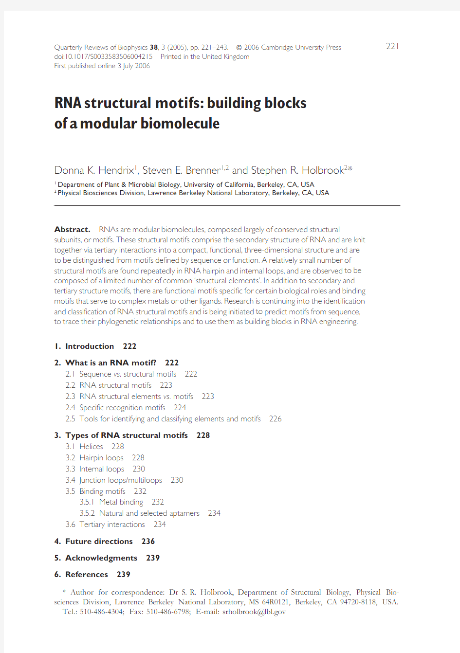
features.Figure 1shows an example of a complex RNA motif,the sarcin–ricin loop (Correll et al .
1999)and the structural elements from which it is composed.Another viewpoint is given in
Fig.2,which shows how an element,the U-turn,can occur in multiple motifs.
2.4Specific recognition motifs
In addition and in contrast to RNA structural motifs that occur ubiquitously in RNA biomol-
ecules and perform general structural or functional roles,there is a large class of RNA recognition
motifs that are the binding sites for speci?c proteins and other molecules.These speci?c rec-
ognition motifs may have well de?ned,stable and conserved 3D structures and sequence sig-
natures,but di?er necessarily from the general structural motifs in that they are narrowly
distributed within an organism to perform their singular recognition function,which would not
be possible if they were common motifs.Thus,these sequences may be broadly distributed
phylogenetically and may even have several variants within a genome,but they do not serve
general architectural roles in RNA folding and tertiary structure stabilization and do not interact
with many di?erent types of ligands.Examples of speci?c recognition motifs are TAR (Richter
et al .2002)and RBE (RRE)(Hung et al .2000;Lesnik et al .2002)motifs of HIV-1and the IRE
motifs found in mRNAs related to iron metabolism (Theil,2000;Pantopoulos,2004).Many
riboregulators,riboswitches,and aptamers form potentially highly structured regions for protein
or small molecule interaction,and mutations in these structures may result in a loss of regulation
Table 1.Characteristics of RNA structural elements and motifs
Characteristic
Element Loop motif Tertiary interaction motif Size
Small,local May span entire loop Multiple loops or stems involved Sequence conservation
Little or none Often have sequence preferences/isosteric Interaction sites Structural conservation
By de?nition Often conserved Evolutionarily conserved Features (i.e.pairing,stacking,turn)
Usually single feature Multiple features/elements Multiple in each interacting motif Occurrence Found within various motifs Not nested;may occur in tertiary interaction motifs May include multiple elements and motifs
224 D.K.Hendrix,S.E.Brenner and S.R.Holbrook
Table2.Some RNA structural elements
Element name(s)Description Found in Reference
U turn/Uridine turn/Pi turn A sharp bend in the phosphate-sugar backbone between the?rst and
second nucleotides,followed by characteristic stacking of the
second and third nucleotides.Original descriptions include
a stabilizing hydrogen bond between the?rst and third residues
Hairpin loops(e.g.GNRA,T Y C
loop)and internal loops
Kim&Sussman,1976;Quigley&
Rich,1976;Holbrook et al.1978;
Klosterman et al.2004
A-minor interaction The insertion of minor groove edges of an adenine into the minor
groove of neighboring helices.Four types have been identi?ed
Ribose zipper,kink-turn Nissen et al.2001
S-turn Two consecutive bends in the phosphate-sugar backbone
characterized by backbone distortions and inverted sugar puckers,
resulting in an‘S’shape Loop E motif,Sarcin–ricin loop Szewczak et al.1993;Wimberly
et al.1993;Correll et al.1999
Dinucleotide platform Two adjacent,covalently linked,co-planar residues that form a
non-Watson–Crick pairing
Internal loops,often
involved in a base triple
Cate et al.1996b;
Klosterman et al.2004
Base triples Three hydrogen-bonded,co-planar bases with two of the bases
sometimes forming a Watson–Crick pair or dinucleotide platform Loop E motif,Sarcin–ricin loop Nagaswamy et al.2002;
Klosterman et al.2004
Cross-strand stack A base on one strand stacks with a base on the opposing strand,
rather than stacking with the adjacent bases on its own strand
Internal loops,e.g.
bacterial loop E motif
Correll et al.1997
Non-canonical base pairs Two bases of any type interacting in a generally planar arrangement
can form hydrogen bonds in characteristic patterns
Double helices Leontis&Westhof,2001;
Nagaswamy et al.2002
Extruded helical single strand Two or three unpaired bases extruded from the main double helical
stack forming an independent stack
Internal and hairpin
loops
Klosterman et al.2004
Backbone rotamers Commonly occurring RNA backbone conformations Double helices,hairpin,internal
and junction loops
Duarte et al.2003;Hershkovitz
et al.2003;Murray et al.2003;
Schneider et al.2004
RNA
structural
motifs
225
and a corresponding disease state (Sobczak &Krzyzosiak,2002;Wong et al .2005).In the
remainder of this review we will focus on general RNA structural motifs,their identi?cation,
classi?cation,types,and roles.
2.5Tools for identifying and classifying elements and motifs
In recent years a variety of computational,database and graphics tools have been developed
that are extremely valuable in identifying,describing,characterizing and classifying RNA
motifs and elements.Descriptive base-pairing nomenclatures (Leontis &Westhof,2001;Lee
&Gutell,2004)and databases (Nagaswamy et al .2002)have been developed to annotate and
classify the wide variety of non-canonical base pairs observed in RNA structures.These have
been incorporated into an automated assignment and graphics program (Yang et al .2003)
for
Fig.1.The sarcin–ricin loop,depicted by elements from the rat 28S ribosomal RNA (PDB identi?er 483d).
The sarcin–ricin loop is an assembly of two secondary structure motifs:an internal loop (bulged G),
made up of residues a:2653-2657and a:2664-2667,and a hairpin loop (GNRA tetraloop)of residues
a:2659-2662.It can also be considered to be composed of several elements.These elements include cross-
strand stacking (in green and gray)with a:2655stacking on a:2664and a:2657on a:2665;a U-turn (in blue)
in the GNRA tetraloop;a base triple with a dinucleotide platform (orange);an S-turn (yellow).Not shown
are several non-canonical base pairs and the backbone-base hydrogen bonds.Note that the base triple
includes nucleotides that are also involved in cross-strand stacking.
226 D.K.Hendrix,S.E.Brenner and S.R.Holbrook
RNA structural motifs227
(a )
(b)
(c)
Fig.2.The U-turn element in multiple loops.3D representations are given on the left and corresponding 2D diagrams on the right.U-turns are colored red with a blue arrow indicating chain direction.(a)The U-turn in the T-loop of yeast tRNA phe(PDB identi?er6tna).(b)The U-turn in the GNRA loop from the23S ribosomal RNA of Haloarcula marismortui(PDB identi?er1jj2).(c)The U-turn in an internal loop, from an AMP-aptamer complex(PDB identi?er1am0).
228 D.K.Hendrix,S.E.Brenner and S.R.Holbrook
visualization of base-pairing and stacking patterns.A web server providing a sophisticated annotation of secondary structures and some tertiary interactions has been presented by Major and co-workers(Gendron et al.2001).Conformational analysis of the RNA backbone using reduced representations has also been used to characterize known motifs(Duarte&Pyle,1998; Duarte et al.2003;Hershkovitz et al.2003;Murray et al.2003)and even identify new motifs(Wadley&Pyle,2004).Cluster analysis of root-mean-square deviations between pairs of RNA fragments has been used as the basis for cluster analysis of RNA loop structures to group tetraloops into various families(Huang et al.2005).A graph theoretic approach has been used to search for substructure patterns in RNA structures using a vectorial approach(Harrison et al. 2003).Finally,the Structural Classi?cation of RNA(SCOR),database has been developed to organize and classify RNA structural motifs(Klosterman et al.2002;Tamura et al.2004).SCOR currently relies on manual classi?cation based on features recorded in the literature,computed, or observed by inspection.
3.Types of RNA structural motifs
Examination of RNA3D structures shows that they can be considered to be composed of a combination of recurring structural motifs joined by a small number of types of tertiary interaction motifs.An example of the secondary structure and tertiary interaction motifs in the P4–P6domain of the group I intron is shown in Fig.3.These motifs are commonly subgrouped by secondary structure type:double helices,hairpin loops,internal loops,or junction loops. Moreover,the interaction of RNA with metal ions,small molecules,proteins and other RNAs can also be characterized by a set of recurring motifs.These various types of RNA motifs are discussed in detail below.
3.1Helices
Although RNA is composed primarily of Watson–Crick base-paired A-form double helices, other helical forms have been observed.Although RNA double helices are generally thought to not have a sequence-structure relationship(Holbrook et al.1981),a comprehensive analysis with the large dataset currently available has not been completed.Recently,the structures of a high salt left-handed RNA duplex(Popenda et al.2004)and a mirror image or L-con?guration (Vallazza et al.2004)Spiegelmer RNA duplex were determined.While the left-handed RNA duplex of repeating(CG)units di?ered signi?cantly from its Z-DNA counterpart,the Spiegelmer RNA had a very similar structure to that of its right-handed,mirror-image structure even though crystallization conditions were quite di?erent.Structures of RNA quadruplexes have also been determined(Liu et al.2002;Pan et al.2003a,b).These structures include both guanine and adenine tetrads as well as bulged and looped out residues and generally di?er from their DNA homologs.
3.2Hairpin loops
Hairpin loops link the3k-and5k-ends of a double helix.Within the SCOR database classi?cation, structurally characterized hairpin or external loops must close with a Watson–Crick pairing and vary in length from2to14nucleotides.The most common and well studied of these are the tetraloops.Of these tetraloops,there are at least four that are characterized by their
RNA structural motifs229
(a)
(b)
Fig.3.Motifs in the P4–P6domain of the group I intron structure.Motifs are truly the structural units,or building blocks,of the Thermus thermophilus group I intron structure(PDB identi?er1gid).(a)Secondary structure motifs by color:fully-paired Watson–Crick helices in pink,internal loops in yellow,and hairpin loops in green.What remains(in gray)are junction loops,the3k and5k overhang ends,and a few residues that did not meet the strict de?nition of Watson-Crick pairings.(b)Tertiary interaction motifs by color: coaxial regions of helices in orange,ribose zippers in red,and the tetraloop–tetraloop receptor interaction in purple.Note that these tertiary interactions have signi?cant overlap.Also shown in both panels(a)and (b)are magnesium ions(cyan)and cobalt hexammine(dark blue).These metal ions are localized within critical tertiary interactions.
230 D.K.Hendrix,S.E.Brenner and S.R.Holbrook
sequence and conserved structures:the GNRA type,the UNCG type,the ANYA type and the(U/A)GNN type.In ribosomal RNAs about70%of tetraloops belong to either the GNRA or the UNCG families and are unusually stable thermodynamically compared to other tetraloop sequences(Antao et al.1991).The well-known GNRA tetraloop(Heus&Pardi,1991;Jucker &Pardi,1995a;Leontis&Westhof,2002;Correll&Swinger,2003)is the most frequently observed tetraloop in currently available RNA structures.The GNRA tetraloop is frequently used as part of a tertiary interaction motif in the formation of tetraloop–tetraloop receptor interactions.
Other hairpin loop motifs include the T-loop(Nagaswamy&Fox,2002)and D-loop motifs of tRNA(Quigley&Rich,1976),the lonepair triloop(Lee et al.2003),and the sarcin-ricin loop(Szewczak&Moore,1995).These and other RNA hairpin loop motifs are summarized in Table3.Depending on de?nition,the U-turn(Jucker&Pardi,1995a),and reversed U-turn(Kolk et al.1997),may be considered structural elements that are present in several motifs including the GNRA tetraloop and the T-loop.Examples of the T-loop hairpin motif and its U-turn subunit are shown in Fig.4.
3.3Internal loops
An internal loop separates double helical RNA into two segments by inclusion of residues that are not Watson–Crick paired in at least one strand of the duplex.Sometimes insertions on only one strand are de?ned as‘bulge loops’,and we include this as a special case of internal loops. Two types of internal loops can be distinguished:symmetric,with the same number of nucleo-tides inserted on both strands;and asymmetric,with a di?erent number of nucleotides inserted on the opposing strands.Non-canonical base pairing is common in internal loops.A frequently observed motif involves extension of double helical structure through continuous formation of non-Watson–Crick pairs in a symmetric internal loop.This double helical structure is distorted by unwinding,unstacking,and kinking formed by the non-canonical pairs.Fully paired and stacked internal loops of up to eight non-canonical pairs have been structurally observed (Vallurupalli&Moore,2003)(loop E).Fully paired internal loops can be well described using a standardized base-pairing nomenclature such as that developed by Leontis&Westhof (2001).Such nomenclature classi?cation and isostericity relationships are useful for prediction of these types of motifs from sequence and secondary structure(Leontis et al.2002;Lescoute et al.2005).
Certain asymmetric internal loop motifs have been identi?ed and characterized as resulting in sharp turns important for tertiary structure formation.These include the kink-turn(K-turn) (Klein et al.2001),reverse kink-turn(Strobel et al.2004)and hook-turn(Szep et al.2003).Some RNA internal loop motifs are summarized in Table4.
3.4Junction loops/multiloops
Junction loops are formed by the intersection of three or more double helices.These double helices are separated by single-strand sequences of zero or more residues.There are N linker (joining)sequences for N helices in a junction loop,although some of the linker sequences may be of zero length.Although junction loops have not been as systematically or extensively studied as the simpler hairpin and internal loops,some generalizations have been made for the more common three-way and four-way junctions(Lilley,1998,2000).Common examples of
junction loops are those in tRNA and the hammerhead ribozyme.Coaxial stacking of the helices
is a key feature of junction loops as observed in these and many other examples.It has been
proposed that coaxial (continuous)stacking in three helix junctions occurs opposite the longest
junction strand (Lescoute &Westhof,2005).The tendency for pairwise coaxial stacking of
helical arms,the importance of metal ion interactions in the induction of tertiary folding,and the
importance of hairpin or internal loop–loop interactions in stabilization of the tertiary structure
(Penedo et al .2004)are prominent features of junction loop architecture.
Table 3.Some RNA hairpin loop motifs
Motif name(s)
Description Reference Lonepair triloop Identi?ed by covariation analysis of 16S rRNA sequences and T-loop of tRNA.Characterized by a single base pair,either Watson–Crick or non-canonical,capped by a hairpin loop containing three nucleotides.Bases immediately 5k or 3k to the motif are NOT base paired to one another.Consensus sequences:Type R1:UGNRA;Type R2:UUYRA;Type R3:NRWAN-;Type R4:NRYAN-;Type R5:NCNUN-
Lee et al .2003GNRA tetraloop A common tetraloop found in ribosomal RNA,Group I intron,and Hammerhead ribozyme.The GNRA loop sequence is often closed by a C-G Watson–Crick https://www.doczj.com/doc/3214382562.html,monly folds into the ‘GNRA fold’of one base on the 5k stack and three in the 3k stack,and contains a U-turn.This fold is also formed by the tetraloop family of sequence UMAC
Heus &Pardi,1991;Jucker &Pardi,1995a;Correll &Swinger,2003UNCG tetraloop A stable tetraloop found in ribosomal and other functional https://www.doczj.com/doc/3214382562.html,monly forms the ‘UNCG fold’,with the U and C bases in the 5k stack,the G in the 3k stack and the N looped out.Also observed in a GUUA tetraloop
Cheong et al .1990ANYA tetraloop Identi?ed in aptamers binding to the MS2coat protein.It has 2common folds:one in a bound form,the other in the apo-form.The bound form has the 1st and 3rd bases in the 5k stack,and the 2nd and 4th bases looped out,interacting with protein.The apo-form has the 1st base in the 5k stack and the 4th base in the 3k stack,forming a Watson–Crick /Sugar Edge base pair
Convery et al .1998;Rowsell et al .1998;Klosterman et al .2004(U/A)GNN tetraloop Seen in RNase III endoribonucleases and the 18S rRNA of yeast,this tetraloop has ?rst and second bases in the 5k stack and third and fourth bases in the 3k stack
Butcher et al .1997b CUYG tetraloop One of the tetraloop sequences common in ribosomal RNAs,this sequence has been seen in two di?erent forms:as a di-loopin a solution structure,with the C and G Watson–Crick-paired,and as a hairpin loop in D.radiodurans 23S rRNA and T.theromphilus 16S rRNA
Woese et al .1990;Jucker &Pardi,1995b D-loop In tRNA,the D-loop contains the modi?ed base dihydrouracil.Is composed of 7–11bases,and inserts bases into the T-loop to form the tRNA T-loop:D-loop tertiary interaction
Quigley &Rich,1976;Holbrook et al .1978T-loop
The T-loop,originally characterized in tRNA is a 5-base hairpin closed by a trans-Watson–Crick/Hoogsteen base-pair interaction between bases N and N +4.Contains a classic U-turn between in the ?rst three bases.Consensus sequence is U(G/U)NR(A/U)Nagaswamy &Fox,2002;Krasilnikov &Mondragon,2003RNA structural motifs
231
3.5Binding motifs
A primary function of RNA is to bind ligands,either for structural stabilization,as co-
factors,substrates or signals.Ligand binding is critical for ribozyme,riboswitch and
splicing functions,as well as in mediating RNA–protein and RNA–RNA intermolecular
and tertiary interactions.RNA ligand-binding sites often demonstrate high selectivity and
speci?city,although there may be more than one motif capable of tightly binding a certain type
of ligand.
3.5.1Metal binding
Two types of metal ion interactions with RNA have been described:di?use ions that accumulate
near RNA due to the electrostatic ?eld while retaining their hydration sphere,and chelated ions
that are in direct contact with RNA at a speci?c location,and may have some waters of hydration
displaced by coordination with polar RNA atoms (Draper,2004).Numerous examples of site-
speci?c metal ion binding to RNA have been observed in RNA structures.One of the ?rst
of these was the extensively studied binding of metals to tRNA (Holbrook et al .1977;Quigley
et al .1978;Shi &Moore,2000).Although magnesium is considered the biologically relevant
metal,other divalent cations,lanthanides and metal hexammines have been observed as strongly
bound to tRNA.A database of metal ion-binding sites in RNA structures (MeRNA),has
been established to organize the metal-binding sites and identify RNA metal-binding motifs
(a )Yeast tRNA phe
T ΨC loop
1EHZ (b )RNase P 187 loop 1NBS
(c )RNase P
218 loop
1NBS
https://www.doczj.com/doc/3214382562.html,mon fold of the T-loop motif.(a )The T Y C loop of yeast phenylalanine transfer RNA (PDB
ID:1EHZ).(b ).A T-loop motif found in the crystal structure of the speci?city domain of Ribonuclease P
RNA (PDB ID 1NBS)at residue 187.(c ).A T-loop motif found in 1NBS at residue 218.A three-residue
U-turn is seen at the apex of the loop bounded by a non-Watson–Crick pair closing the T-loop.Cross-loop
hydrogen bonding is observed between base and backbone.Sharp turns are often observed for residues
at the 3k -end of the loop.Adenine nucleotides are colored red,guanine green,uracil light blue and cytosine
yellow.
232 D.K.Hendrix,S.E.Brenner and S.R.Holbrook
Table4.Some RNA internal loop motifs
Motif name Description Consensus
sequence5k-3k Reference
Bulged-G Found in the sarcin–ricin loop and eukaryotic loop E of5S ribosomal RNA,the loop is characterized by the backbone element‘S-turn’5k-GA-AY-3k
3k-AUGAY-5k
Correll et al.2003;
Szewczak&Moore,1995;
Wimberly et al.1993
Bacterial loop E A symmetric internal loop in bacterial5S ribosomal RNA,this motif is the binding site of the L25ribosomal protein,and is characterized by three cross-strand purine stacks and several
non-canonical base pairings 5k-GAGAGUA-3k
3k-AUGGUAG-5k
Correll et al.1997
Kink turn/K-turn Formed by two strands in a helix–internal loop–helix arrangement,this turn is named for the sharp bend,or kink,that is formed in the phosphodiester backbone of the strand,bringing
the minor groove side of the two surrounding helices together 5k-GCRNNGANG-3k
3k-CG—AGNC-5k
Klein et al.2001;
Lescoute et al.2005;
Vidovic et al.2000
Reverse Kink turn Like the kink turn,also a helix–internal loop–helix arrangement,but bending in the opposite direction,and thus toward the major grooves of the surrounding helices 5k-ACACAAACC-3k
3k-UG—AGGG-5k
Strobel et al.2004
Hook turn A sharp bend in a strand that is helical,A form-like,on its5k side,with a y180x turn in
backbone direction on the3k side that occurs between two residues,usually a sheared A-G
base pair
Szep et al.2003
C-loop This internal loop is made of two asymmetric strands and contains2(or more)base triples.
The longer strand usually begins with the nucleotide C which forms a base triple with the
?anking helix at its3k side.The nucleotide at the3k end of the longer strand forms a base
triple with the?anking helix at the5k end.The base(s)on the shorter strand are often looped
out 5k-C-CAC-U-3k
3k-G–C–A-5k
Ban et al.2000;Lescoute
et al.2005;Torres-Larios
et al.2002;Wimberly et al.
2000
Tetraloop receptor This conserved motif is made up of two C-G pairs and an internal loop(including a G-U pair).
The internal loop contains an adenosine platform and a looped-out U 5k-CC-UAAG-3k
3k-GGUA–U-5k
Cate et al.1996a
RNA
structural
motifs
233
234 D.K.Hendrix,S.E.Brenner and S.R.Holbrook
(Stefan et al.2005).As of August2005,9764metal sites have been identi?ed in256PDB entries and eight RNA metal ion-binding motifs identi?ed.
As with proteins,binding of metals or other ligands may either be to preformed sites or motifs,or through induction or selection of a speci?c binding https://www.doczj.com/doc/3214382562.html,rge structural rearrangements have been observed on metal binding(Wu&Tinoco,1998;Penedo et al. 2004)indicating that secondary structure is not always?xed prior to formation of tertiary interactions.
Monovalent cations,particularly sodium and potassium have also been observed to bind speci?cally to adenosine platforms(Cate et al.1996b;Klein et al.2004)and G-quadruplexes (Pan et al.2003a)among other elements.An especially striking example is a buried potassium ion surrounded by several phosphates that contributes a large binding free energy and allows a complex tertiary fold to be formed(Conn et al.2002).
Metal binding to RNA can serve to stabilize a speci?c3D structure(for example see Klein et al.2004),but also may perform a catalytic role(Wedekind&McKay,2003).In these cases,it is a combination of the3D fold induced by metal-ion binding and the chemical nature of the metal ion itself that is responsible for catalysis.
3.5.2Natural and selected aptamers
At least three classes of RNA molecules have been identi?ed that are capable of tight and speci?c binding to small organic ligands:the in vitro selected(SELEX)aptamers,the riboswitches found in the untranslated regions of mRNA,and certain functional RNAs such as the self-splicing group I introns that bind to guanine as a co-factor.3D structures exist for some members of each of these groups that illustrate the binding motifs(Patel&Suri,2000;Adams et al.2004;Batey et al.2004;Guo et al.2004;Serganov et al.2004).Guanine binding by group I intron and riboswitch have been shown to be mediated by a base triple sandwich(stacking)motif(Guo et al. 2004;Serganov et al.2004).
3.6Tertiary interactions
As with secondary structure,the tertiary structure of RNA biomolecules is dominated by a limited number of recurring types of interactions or motifs(Batey et al.1999).As enumerated in the SCOR database,these are:coaxial helices,kissing hairpin loops,the tetraloop–tetraloop receptor,the A-minor motif/patch,the tRNA D-loop:T-loop interaction,pseudoknots,and ribose zippers.Descriptions and references for each of these types of tertiary interaction are summarized in Table5.As shown in Figs5and6,coaxial helices and kissing hairpin loops both involve continuous base stacking,the?rst between helices either adjacent or connected by single base inserts or internal loops,and the second between two hairpin loops forming base pairs.Several of these modes of tertiary interaction such as the ribose zippers and the A-minor motif can be further subdivided into more speci?c sub-motifs(Nissen et al.2001; Tamura&Holbrook,2002),in these cases11and four subtypes respectively.Several ribose zippers identi?ed in the bacterial ribonuclease P RNA structure(Kazantsev et al.2005), including two subtypes are shown in Fig.7.Finally,it is clear that conformational changes can be induced on formation of tertiary interactions.Figure8shows the dramatic change in conformation of an isolated tetraloop receptor as compared to a tetraloop–tetraloop receptor interaction.
Table5.Some RNA tertiary interaction motifs
Motif name(s)Description Secondary structures Sequence preference Reference
Ribose Zipper Formed by hydrogen bonding between consecutive
backbone ribose2k hydroxyls from two distant regions
of the chain,interacting in an anti-parallel manner.
Classi?ed as canonical and6other types Double helix:
Hairpin or internal
loop
Antiparallel
5k-CC-3k(Stem)
3k-AA-5k(Loop)
O2k–O2k and base triples
(e.g.A-minor)
Cate et al.1996a;Tamura&
Holbrook,2002
A-Minor Motif/ A-patch A clustering of A-minor interactions,often decreasing in
type and order going from the5k to the3k direction
Internal loop,
Hairpin loop
Adenosines Nissen et al.2001
D-Loop:T-Loop Complex interaction between two conserved hairpins in
tRNA,includes interdigitated bases Hairpin loop:
Hairpin loop
Conserved sequences in
D-loop and T-loop motifs
Holbrook et al.1978;
Holbrook&Kim,1979
Tetraloop:Tetraloop receptor Conserved in Group I and II introns occuring between a
GNRA tetraloop(GNRA fold)and the receptor;an in-
ternal loop plus two C-G pairs.It is characterized by a
speci?c hydrogen bond pattern between the?rst A of the
tetraloop and the U.A of the receptor to form an A.U.A
triple;between the second A of the tetraloop and the
backbone of the receptor C and U;between the third A of
the tetraloop and the C:G pair of the receptor
Hairpin loop:
Internal loop
5k-CC-UAAG-3k
3k-GGUA–U-5k
Pley et al.1994;Cate et al.
1996a;Butcher et al.
1997a
Kissing Hairpin Loop The kissing hairpin complex is formed by base pairing
between single-stranded residues of two hairpin loops with
complementary sequences
Hairpin loop:
Hairpin loop
Self-complementary often
six nucleotides
Chang&Tinoco,1994;
Ennifar et al.2001
Coaxial Helices Interhelical stacking Nucleotide bases from two separate helices stack and align
axes to form a pseudo-continuous,coaxial helix.Coaxial
helices are highly stabilizing and are dominant in several
large RNA structures.
Interhelical stacking may occur via a single base or base
pair bridge between helices,resulting in continuous helical
stacking spanning multiple helices
Double helices often
across an internal
or junction loop
‘Bridging’nucleotide
between helices has a
preference for adenine
Kim et al.1974;
Cate et al.1996a
Pseudoknot When bases pair between nucleotide loops(hairpin or
internal)and bases outside the enclosing loop,they form a
pseudoknot.This structure often contains coaxial helices.
Can be very stable Hairpin loop:
Single strand
Complementary Shen&Tinoco,1995;
van Batenburg et al.2001
RNA
structural
motifs
235
Clearly,other motifs remain to be discovered and described,such as novel hairpin loop–loop interactions and base triple and quadruple interactions,but it is becoming more apparent with each new structure that most major classes of RNA tertiary interaction motifs have been ident-i?ed.A major challenge and opportunity to be undertaken is the prediction of RNA tertiary interactions from sequence alone.Detailed analysis of the known tertiary interaction motifs,together with an accurate secondary structure prediction and comparative sequence analysis should provide strong indications of the presence of speci?c tertiary interactions in RNA sequences.
4.Future directions
A comprehensive identi?cation,classi?cation and characterization of RNA structural elements,secondary structure motifs,tertiary interaction motifs,and binding motifs is critical
for
Fig.5.Coaxial interhelical stacking in the P4–P6domain of the group I intron.(a ).Coaxial stack between helices bulges and internal loops with non-Watson–Crick pairing as seen in the P4–P6domain of the group I intron (PDB ID 1GID).Non-stacked residues are shown in red.(b ).Crystallographic mobility parameters (B-factors)as observed in 1GID.The correspondence between stacking and reduced mobility is apparent.
236 D.K.Hendrix,S.E.Brenner and S.R.Holbrook
understanding RNA structure–function relationships,folding,evolution,engineering and design.Advances in RNA structure prediction and the identi?cation of RNA genes and genetic control elements in genomic DNA are dependent on our understanding of the modular nature of RNA and the subunits from which it is composed.
In order to obtain such an understanding,not only do we require additional examples of a wide variety of RNA structures,but also development of new and improved computational approaches for the identi?cation of structural motifs and the elements from which they are composed,for classi?cation leading to analysis of sequence and structural variation of these motifs,and for prediction of these motifs from sequences alone.In addition,evolutionary analysis of RNA elements and motifs,and studies relating motif structure to function are needed to place these structural features into a biological context.
In the last few years a strong trend has been established away from structural analysis of model compounds and molecular fragments,toward the structure determination of large,intact,biological RNA molecules (Holbrook,2005).Analysis of these large RNAs shows a combination of well-known structural motifs and the presence of potential novel motifs (Holbrook,2005).A future challenge is the con?rmation and characterization of these new motifs and forming an understanding of how these motifs are linked and interact to form functional
biological Fig.6.A kissing hairpin loop found in ribosomal RNA.Two hairpin loops forming a ‘kissing’interaction.A coaxial stack is formed by base pairing between the hairpins and their Watson–Crick stems (note the ‘minor groove’formed by the backbones of the hairpins).The blue hairpin is formed by residues 414–426and the orange hairpin by residues 2440–2449of 23S ribosomal RNA (1JJ2).The surrounding backbone of the ribosomal RNA is shown in gray.
RNA structural motifs 237
RNAs.The existing approaches and tools for identi?cation and analysis of RNA motifs have been described above,as have recent advances in the standardization of nomenclature and de?nitions.A working group,the RNA Ontology Consortium,has been formally assembled and assigned the task of establishing and improving standards in this area (see Leontis et al.2006).
Several areas of potentially great signi?cance are currently emerging from the study of RNA structure and RNA structural motifs.These include RNA engineering and design as an approach to nanotechnology (Chworos et al .2004;Guo,2005);the association of RNA structure with human disease (Michlewski &Krzyzosiak,2004;Barciszewska et al .2005;Darnell et al .2005)and its treatment through drug design (Tu et al .2005),siRNA,antisense,and ribozyme technology (Scanlon,2004);and ?nally the evolution of RNA and RNA motifs,their mutation patterns,
and
Fig.7.Ribose zipper motifs observed in Ribonuclease P RNA (PDB identi?er 2a64).Locations of ribose zipper tertiary interactions in the crystal structure of bacterial Ribonuclease P RNA are shown in the upper left.The following ?gures show close-up views of the ribose zippers with O2k -O2k and O2k -N3(base)hydrogen bonds indicated.All ribose zippers are of the canonical type except for 99–100;392–393which is classi?ed as a single ribose zipper due to the single ribose-base hydrogen bond.
238 D.K.Hendrix,S.E.Brenner and S.R.Holbrook
the conservation and identi?cation of functional non-coding RNAs (ncRNAs)in genomic sequences.
5.Acknowledgments
The authors acknowledge Dr Elizabeth Holbrook,Dr Peter Klosterman (U.C.Berkeley),and Dr Liliana Stefan (Lawrence Berkeley Laboratory)for assistance in preparation of this manuscript.S.R.H.was supported by National Institutes of Health grants 1R01GM66199and 1R01HG002665.S.E.B.was supported by NHGRI of the NIH grant K22HG00056and IR01GM66199.D.K.H.was supported by National Institutes of Health grant
1R01GM66199.
Fig.8.Conformational change in the tetraloop receptor on binding a GNRA tetraloop.Crystal structures of a ‘free’tetraloop receptor motif (1TLR)and the tertiary interaction formed between a GNRA tetraloop and receptor in the P4–P6domain of the group I intron (1GID).Shown are residues 1–9and 16–23of the free tetraloop and 220–228,148–155and 246–253of the complex.The tetraloop receptor sequence is variable and di?erent sequences are shown in the ?gure:(left GGCCUAAGA/UUAUGGCC,right GUCCUAAGU/AUAUGGAU).Bases are colored as red (A),cyan (U),green (G)and yellow (C)for the tetraloop receptor,while the tetraloop is in gray.Note the conformational change occurring in the receptor on tetraloop binding.
6.References
A DAMS ,P.L.,S TAHLEY ,M.R.,G ILL ,M.L.,K OSEK ,A.B.,
W ANG ,J.&S TROBEL ,S.A.(2004).Crystal structure of
a group I intron splicing intermediate.RNA 10,
1867–1887.
A NTAO ,V.P.,L AI ,S.Y.&T INOCO ,I.J.(1991).A ther-
modynamic study of unusually stable RNA and DNA
hairpins.Nucleic Acids Research 19,5901–5905.
B AN ,N.,N ISSEN ,P.,H ANSEN ,J.,M OORE ,P.B.&S TEITZ ,
T.A.(2000).The complete atomic structure of the
large ribosomal subunit at 2á4A
?resolution.Science 289,905–920.B ARCISZEWSKA ,M.Z.,S ZYMANSKI ,M.,W YSZKO ,E.,P AS ,J.,R YCHLEWSKI ,L.&B ARCISZEWSKA ,J.(2005).Lead tox-icity through the leadzyme.Mutation Research 589,103–110.B ATEY ,R.T.,G ILBERT ,S.D.&M ONTANGE ,R.K.(2004).Structure of a natural guanine-responsive riboswitch complexed with the metabolite hypoxanthine.Nature 432,411–415.B ATEY ,R.T.,R AMBO ,R.P.&D OUDNA ,J.A.(1999).Tertiary motifs in RNA structure and folding.Angewandte Chemie International Edition 38,2326–2343.
RNA structural motifs 239
B ERMAN,H.M.,O LSON,W.K.,B EVERIDGE, D.L., W ESTBROOK,J.,G ELBIN,A.,D EMENY,T.,H SIEH,S.-H., S RINIVASAN,A.R.&S CHNEIDER,B.(1992).The nucleic acid database:a comprehensive relational database of three-dimensional structures of nucleic acids.Biophysical Journal63,751–759.
B ERMAN,H.M.,W ESTBROOK,J.,F ENG,Z.,G ILLILAND,G., B HAT,T.N.,W EISSIG,H.,S HINDYALOV,I.N.&B OURNE, P.E.(2000).The Protein Data Bank.Nucleic Acids Research28,235–242.
B ROWN,J.W.(1999).The ribonuclease P database.Nucleic Acids Research27,314.
B UTCHER,S.E.,D IECKMANN,T.&F EIGON,J.(1997a). Solution structure of a GAAA tetraloop receptor RNA. EMBO Journal16,7490–7499.
B UTCHER,S.E.,D IECKMANN,T.&F EIGON,J.(1997b). Solution structure of the conserved16S-like ribosomal RNA UGAA tetraloop.Journal of Molecular Biology268, 348–358.
C ATE,J.H.,G OODING, A.R.,P ODELL, E.,Z HOU,K., G OLDEN, B.L.,K UNDROT, C.E.,C ECH,T.R.&
D OUDNA,J.A.(1996a).Crystal structure of a group I ribozyme domain:Principles of RNA packing.Science 273,1678–1685.
C ATE,J.H.,G OODING, A.R.,P ODELL, E.,Z HOU,K., G OLDEN, B.L.,S ZEWCZAK, A.A.,K UNDROT, C.E., C ECH,T.R.&
D OUDNA,J.A.(1996b).RNA tertiary structure mediation by adenosine platforms.Science273, 1696–1699.
C HANG,K.-Y.&T INOCO J R.,I.(1994).Characterization of a‘kissing’hairpin complex derived from the human immunode?ciency virus genome.Proceedings of the National Academy of Sciences USA91,8705–8709.
C HEONG,C.,V ARANI,G.&T INOCO J R.,I.(1990).Solution structure of an unusually stable RNA hairpin, 5k GGAC(UUCG)GUCC.Nature346,680–682.
C HWOROS,A.,S EVERCAN,I.,K OYFMAN,A.Y.,W EINKAM,P., O ROUDJEV,E.,H ANSMA,H.G.&J AEGER,L.(2004). Building programmable jigsaw puzzles with RNA. Science306,2068–2072.
C ONN,G.L.,G ITTIS,A.G.,L ATTMAN,E.E.,M ISRA,V.K. &
D RAPER,D.E.(2002).A compact RNA tertiary structure contains a buried backbone-K+complex. Journal of Molecular Biology318,963–973.
C ONVERY,M.A.,R OWSELL,S.,S TONEHOUSE,N.J., E LLINGTON,A.D.,H IRAO,I.,M URRAY,J.B.,P EABODY, D.S.,P HILLIPS,S.E.&S TOCKLEY,P.G.(1998).Crystal structure of an RNA aptamer-protein complex at2á8A?resolution.Nature Structural Biology5,133–139.
C ORRELL,C.C.,B ENEKEN,J.,P LANTINGA,M.J.,L UBBERS, M.&C HAN,Y.L.(2003).The common and the dis-tinctive features of the bulged-G motif based on a 1á04A?resolution RNA structure.Nucleic Acids Research 31,6806–6818.
C ORRELL,C.C.,F REEBORN,B.,M OORE,P.B.&S TEITZ, T.A.(1997).Metals,motifs,and recognition in the
crystal structure of a5S rRNA domain.Cell91, 705–712.
C ORRELL,C.C.&S WINGER,K.(2003).Common and distinctive features of GNRA tetraloops based on a GUAA tetraloop structure at1á4A?resolution.RNA9, 355–363.
C ORRELL,C.C.,W OOL,I.G.&M UNISHKIN,A.(1999).The two faces of the Escherichia coli23S rRNA sarcin/ricin domain:the structure at1á11A?resolution.Journal of Molecular Biology292,275–287.
D ARNELL,J.C.,F RASER,C.E.,M OSTOVETSKY,O.,S TEFANI, G.,J ONES,T.A.,
E DDY,S.R.&D ARNELL,R.B.(2005). Kissing complex RNAs mediate interaction between the Fragile-X mental retardation protein KH2domain and brain polyribosomes.Genes and Development19, 903–918.
D RAPER,D.E.(2004).A guide to ions and RNA structure. RNA10,335–343.
D UARTE,C.M.&P YLE,A.M.(1998).Stepping through an RNA structure:a novel approach to conformational analysis.Journal of Molecular Biology284,1465–1478.
D UARTE,C.M.,W ADLEY,L.M.&P YLE,A.M.(2003). RNA structure comparison,motif search and discovery using a reduced representation of RNA conformational space.Nucleic Acids Research31,4755–4761.
E HRESMANN,C.,B AUDIN,F.,M OUGEL,M.,R OMBY,P., E BEL,J.P.&E HRESMANN, B.(1987).Probing the structure of RNAs in solution.Nucleic Acids Research15, 9109–9128.
E NNIFAR,E.,W ALTER,P.,E HRESMANN,B.,E HRESMANN,C. &D UMAS,P.(2001).Crystal structures of coaxially stacked kissing complexes of the HIV-1RNA dimer-ization initiation site.Nature Structural Biology12, 1064–1068.
F ERA,D.,K IM,N.,S HIFFELDRIM,N.,Z ORN,J.,L ASERSON, U.,
G AN,H.H.&S CHLICK,T.(2004).RAG:RNA-As-Graphs web resource.BMC Bioinformatics5,88.
F URTIG,B.,R ICHTER,C.,W OHNERT,J.&S CHWALBE,H. (2003).NMR spectroscopy of RNA.ChemBioChem4, 936–962.
G AST,F.-U.(2003).A new structural motif in the left terminal domain of large viroids identi?ed by covari-ation analysis.Virus Genes26,19–23.
G ENDRON,P.,L EMIEUX,S.&M AJOR, F.(2001). Quantitative analysis of nucleic acid three-dimensional structures.Journal of Molecular Biology308,919–936.
G RIFFITHS-J ONES,S.,B ATEMAN, A.,M ARSHALL,M., K HANNA,A.&E DDY,S.R.(2003).Rfam:an RNA family database.Nucleic Acids Research31,439–441.
G UO,F.,G OODING,A.R.&C ECH,T.R.(2004).Structure of the Tetrahymena ribozyme:base triple sandwich and metal ion at the active site.Molecular Cell16351–362.
G UO,P.(2005).Bacterial virus phi29DNA-packaging motor and its potential applications in gene therapy and nanotechnology.Methods in Molecular Biology300, 285–324.
240 D.K.Hendrix,S.E.Brenner and S.R.Holbrook
七年级体育课蹲踞式起跑教学设计及教案 -CAL-FENGHAI-(2020YEAR-YICAI)_JINGBIAN
体育课教学设计及教案 年级:七年级授课教师:崔鹏飞 教学内容:蹲踞式起跑游戏 一、指导思想: “健康体魄是青少年为祖国和人民服务的基本前提,是中华民族旺盛生命力的体现。学校体育教育要树立健康第一的指导思想,切实加强体育工作”。短跑在体育教学中占有特殊地位,是中学田径教学的主要内容之一,通过短跑教学不仅能够提高学生的速度素质,还可以培养学生积极向上、勇于拼搏的精神,从而促进学生的全面发展。 二、教材分析 根据初一年级学生的锻炼特点和心理特点,有针对性的选择教材内容,培养学生进行体育锻炼的意识和提高学生心理健康水平。通过学习蹲踞式起跑技术,使学生掌握正确的起跑姿势;通过各种游戏练习,培养学生团结协作、积极向上的精神。 三、学生分析 A、有利因素:由于初一年级学生对体育活动的兴趣较高,学生的积极性和主动性是不难调动的。 B、由于初一年级学生心理素质不稳定、好动、自制力不强,还要在教学中教师多多提醒和引导,以确保教学活动的顺利进行。 四、教学目标 1.技能目标:通过学习,使学生掌握正确的蹲踞式起跑技术,发展学生的速度素质、灵敏素质和协调能力。
2.情感目标:培养学生团结协作、积极向上的精神,充分展现学生的反应能力。 五、教学重点、难点 重点:蹲踞式起跑“各就位”、“预备”姿势。 难点:起跑后的快速起动 六、教法:讲解示范练习 七、器材:助跑器 八、教学过程
开 始部分课堂常规 体育委员集 合整队,报 告人数 教师步入课堂,师生问好。 1.宣布本节课的内容任务及目标 2.提出课堂要求 3.安排见习生 1.全班成四 列横队背向 阳光站立 2.注意力集 中,保持安 静。 3.具备良好 的上课状 态。 组织: ♀♀♀♀♀♀♀♀♀♀ ♀♀♀♀♀♀♀♀♀♀ ♂♂♂♂♂♂♂♂♂♂ ♂♂♂♂♂♂♂♂♂♂ ☆ 要求:集合快静齐 3 准备部分游戏(锤 子、剪刀、 布) 1.师生互动,教师带领学生进行 练习。 2.口令指挥,声音洪亮,以良好 的精神风貌感染学生。 1.学生跟着 老师一起律 动。 2.学生在教 师带领下进 行游戏活 动。 1.组织: 要求:活动有序、一 致 2.组织:四列横队成 体操队形 要求:活动充分、动 作到位 9 基本部分1.蹲踞式起跑 技术教学 动作要领: 听到“各就 位”口令 后,两手臂 伸直,四指 并拢,拇指 张开成: “八”字 形,两手稍 比肩宽,放 在起跑线后 沿,左脚放 在起跑线后 约一脚或一 脚半长,右 膝跪在左脚 的中间部 位,身体中 心由两手臂 平均分配, 目视前下方 约40-50公 分,听到 “预备”口 令后,臀部 略抬起,高 1.依据教学图,给学生讲解蹲踞 式起跑及起跑后的快速起动动作 要领。 教师边讲解边进行分解动作及完 整动作的示范,让学生建立正确 的技术概念。 2.将学生分成四个小组,一二组 由教师指挥进行蹲踞式起跑的练 习,在练习中发现学生存在的问 题并予以纠正。三四组由小组长 带领进行练习。 1.学生成四 列横队,前 两列蹲下, 认真看教学 图,听老师 讲解动作要 领并看老师 示范完整技 术动作。 2.各小组听 从教师指 挥,练习蹲 踞式起跑。 1.组织:四列横队 要求:注意力集中, 认真听讲,仔细观看 老师示范 1.组织: ♀♀♀♀♀♀♀♀♀♀ ♂♂♂♂♂♂♂♂♂♂ 听口令,练习蹲踞式 起跑。 2.组织: 三四组进行练习 26
#论著# 前列腺癌与雄激素受体基因(CAG)n 重复多态性的关系 王钢 陈光椿 王晓慧 夏冰 张金山 卢建 =摘要> 目的 探讨前列腺癌(PC)与雄激素受体(AR)基因(CAG)n 重复多态性的关系。 方法 应用DNA 双链循环测序方法对34例PC 组织与癌旁正常组织、2例PC 患者外周血白细胞内的A R 基因(CAG)n 重复数进行测定。 结果 同一PC 患者的癌组织与癌旁正常组织(CA G)n 重复数相同;36例患者癌组织A R 基因(CAG)n 呈重复多态性,平均20.06,显著低于正常组织,差别有显著性意义(P <0.05);不同分化程度的癌组织(CAG)n 重复数差别无显著性意义(P >0.05)。 结论 A R 基因(CAG)n 重复数改变的体细胞突变在PC 癌细胞中罕见,该重复数的减少可能与PC 发病相关。 =关键词> 前列腺肿瘤; 癌; 受体,雄激素; 基因 Study on the polymorphism of (C AG)n repeats within androgen receptor gene in patients with prostate cancer WA N G Gang ,CH EN Guangchun,WA N G X iaohui,et al.Dep ar tment of Pathop hysiology , Second M illitary M edical University ,Shanghai 200433,China =Abstract > Objective T o study t he relationship between polymorphic (CAG)n repeats of androgen r eceptor(A R)gene and occurrence o f prostate cancer (P C). Methods (CA G)n repeats of both malig -nant cells and adjacent nonmalignant cells from 34paraffin -embedded PCs,and that of peripheral blood cells from 2PC patients w ere assessed using dsDN A cycle sequencing. Results T he number of (CAG )n re -peats in malignant cells w as equal to that in adjacent nonmalig nant cells from the same par affin -embedded PC;the mean of (CAG)n r epeats in the 36P C patients was 20.06w hich w as sig nificantly smaller than t hat of normal men (P <0.05),althoug h no significant difference in t he po lymorphic distributio n of (CA G)n was found among various grades of PC (P >0.05). Conclusions Shortening of (CAG )n re -peats w ithin AR gene may be associated with the occurrence of PC. =Key words > Prostate neoplasms; Carcinoma; R eceptor,androg en; G enes 基金项目:国家自然科学基金(39670300) 作者单位:200433上海,第二军医大学病理生理学教研室 前列腺癌(PC)为雄激素依赖性肿瘤,雄激素受体(AR)作为介导雄激素生物学效应的关键大分子在PC 的发生、发展过程中有重要地位。AR 由位于 X 染色体q 11-12的单拷贝AR 基因编码,后者含有8 个外显子和7个内含子。其中第一外显子内含有(CAG)n 三核苷酸重复/微卫星序列,该重复序列在人群中呈现出多态性[1] 。近年来研究发现,该多态 性重复序列与PC 发病之间存在相关性[2,3] ,但也 有不同结论 [4] 。我们在研究了中国正常男性AR 基 因(CAG)n 多态性的基础上[5],对该重复多态性与 PC 发病之间的关系进行探讨。 材料与方法 一、试剂 C -32P -ATP 系北京亚辉医学工程公司产品,DNA 测序试剂盒为美国Epicenter 公司产品。 二、标本 34例PC 病理石蜡切片标本及2例PC 患者外 周血标本,病理诊断为浸润性腺癌。其中高分化8例,中分化13例,低分化15例。 三、方法 1.组织细胞DNA 提取:石蜡切片经HE 染色后在显微镜下划分开癌组织和癌旁正常组织,以此为
快速跑教案 教学目标认知:通过快速跑的学习,发展学生的速度素质,提高机能水平。 技能:跑姿正确,协调。后蹬要充分,步频快,重心平稳。 情感:养成良好的习惯,团结友爱,互相帮助,培养坚强的意志品质。 教学程序教师活动学生活动执行情况 常规(2’)1、指定集合地点,检查着 装,接受学生体委报告。 2、宣布本课内容和教学目 标。 3、带领学生热身活动 1、按指定地点集合整 队,体委整队,报告人 数,向教师汇报。 2、注意听讲,记住学习 目标,自我分组定位。 ﹡﹡﹡﹡﹡﹡﹡﹡﹡﹡ ﹡﹡﹡﹡﹡﹡﹡﹡﹡﹡ ﹡﹡﹡﹡﹡﹡﹡﹡﹡﹡ ﹡﹡﹡﹡﹡﹡﹡﹡﹡﹡ ﹡ 要求:快、静、齐,注意力 集中。 准备活动1、慢跑。 2、徒手操(5节) 1)上肢运动 2)扩胸运动 3)全身运动 4)踢腿运动 5)跳跃运动 6)活动手腕、脚腕 3、教师讲明自编操的创编 原则,学生进行自编操 的创编和学习。 4、教师每组进行指导。 5、教师仔细观察,发现错 误进行个别指导。 (严格要求每个学生,使其 掌握正确的动作要领) 1、体委带领。 2、听教师的口令。 (充分活动,避免运动 损伤) 3、各小组长带领组内 学生讨论、自编, 每小组编两节。 4、小组进行表演,评 选出优秀小组。 5、学生认真听讲,模 仿动作。 (认真讲解学生提出的 问题并进行示范) ﹡﹡﹡﹡﹡﹡﹡﹡﹡﹡ ﹡﹡﹡﹡﹡﹡﹡﹡﹡﹡ ﹡﹡﹡﹡﹡﹡﹡﹡﹡﹡ ﹡﹡﹡﹡﹡﹡﹡﹡﹡﹡ ﹡
快速跑教法: 1、培养正确的跑姿练习 (1)原地摆臂练习,要求 自然协调。 1、慢跑一圈 2、教师讲解跑的技术动作 要领并示范。 3、教师指挥学生进行跑的 练习。 1)摆臂练习 2)小步跑练习 3)高抬腿练习 4)30米加速跑 学法: 1、学习正确的起跑 (1)以肩为轴,屈臂前 后摆动。 1、体委指挥。 2、学生认真听讲,认 真观察。 3、在教师统一指挥下 进行。 (充分活动,使各个关 节活动开,避免运动损 伤。) ﹡﹡﹡﹡﹡﹡﹡﹡﹡﹡ ﹡﹡﹡﹡﹡﹡﹡﹡﹡﹡ ﹡﹡﹡﹡﹡﹡﹡﹡﹡﹡ ﹡﹡﹡﹡﹡﹡﹡﹡﹡﹡ ﹡ (2)沿直线跑。(2)眼视前方,脚要尽 量落在一条直线上。 ﹡﹡﹡﹡﹡﹡﹡﹡﹡﹡ ﹡﹡﹡﹡﹡﹡﹡﹡﹡﹡ ﹡﹡﹡﹡﹡﹡﹡﹡﹡﹡ ﹡﹡﹡﹡﹡﹡﹡﹡﹡﹡ ﹡ (3)抢绳游戏(3)两人一组,看谁先 拿到绳子。 ﹡﹡﹡﹡﹡﹡﹡﹡﹡﹡ ﹡﹡﹡﹡﹡﹡﹡﹡﹡﹡ ﹡﹡﹡﹡﹡﹡﹡﹡﹡﹡ ﹡﹡﹡﹡﹡﹡﹡﹡﹡﹡ ﹡ (4)快速跑移物接力游戏。(4)每组第一人起跑至 场中,将支撑物移至场 边搭好后返回;第二人 再将支撑物移回场中, 依次进行 ﹡﹡﹡﹡﹡﹡﹡﹡﹡﹡ ﹡﹡﹡﹡﹡﹡﹡﹡﹡﹡ ﹡﹡﹡﹡﹡﹡﹡﹡﹡﹡ ﹡﹡﹡﹡﹡﹡﹡﹡﹡﹡ ﹡ 结束1、集体讲评、放松活动 2、归还器材、下课 1、放松、评价 2、下课 ﹡﹡﹡﹡﹡﹡﹡﹡﹡﹡ ﹡﹡﹡﹡﹡﹡﹡﹡﹡﹡ ﹡﹡﹡﹡﹡﹡﹡﹡﹡﹡ ﹡﹡﹡﹡﹡﹡﹡﹡﹡﹡ 器材:跳绳、 平均心率135 练习密度40
① 雄激素受体与核受体辅助抑制因子的关系 廖国庆3,汤恢焕,吕新生 (中南大学湘雅医院外科,长沙410008) [摘要] 目的:探讨雄激素受体(AR )与核受体辅助抑制因子(SMRT )是否能相互作用及其作用部位。方法:重 组构建AR ,SMRT 基因或其基因片段的质粒,体外转录,合成35S 标记融合蛋白,采用转染试验及哺乳动物细胞双杂交实验(transient transfection ,mammalian two 2hybrid test ),GST 沉淀试验(GST pull 2down assay ),间接免疫荧光观察 (indirect immunofluorescence staining )的方法,观察AR 与SMRT 的关系。结果:AR 具有内在的转录抑制活性,可与SMRT 直接作用,其作用部位:在AR 分子上位于配体结合区(LBD ),而DNA 结合区能增强这种作用;在SMRT 分子 上则位于羧基端核受体作用区(IDs ),在这个区域的L XXXIXXXI/L 功能基团突变后将会影响AR 与SMRT 的结合。结论:AR 通过其LBD 区与SMRT 分子中的ID2相互作用。 [关键词] 受体; 雄激素; SMRT ; 相互作用 [中图分类号] Q781 [文献标识码] A [文章编号] 167227347(2004)022******* Androgen receptor and SMRT L IAO Guo 2qing 3,TAN G Hui 2huan ,L U ?? Xin 2sheng (Depart ment of S urgery ,Xiangya Hospital ,Cent ral South U niversity ,Changsha 410008,Chi na ) Abstract : Objective To explore the interaction between androgen receptor (AR )and silencing mediator for retinoid and thyroid hormone receptor (SMR T )and their interaction site.Methods We recombined and constructed AR ,SMR T gene and gene fragments ,in vitro translated 35S fusion proteins to investigate the relationship between AR and SMR T using transient transfection ,mammalian two 2hy 2brid test ,GST pull 2down assay ,and indirect immunofluorescence staining.R esults AR possessed an intrinsic transcriptional repression activity and AR interacted directly with SMR T.One interactive sur 2face on AR was mapped to the ligand 2binding domain (LBD ),and the presence of DNA binding domain enhanced this interaction.The binding surface on SMR T was mapped to the carboxyl 2terminal nuclear receptor interacting domain (ID ),and mutation of the L XXXIXXXI/L corepressor motif within this do 2main interferred with the interaction.Conclusion LBD domain on the AR can interact with ID2motif on the SMR T. K ey w ords : receptor ; androgen ; SMR T ; interaction [J Cent South Univ (Med Sci ),2004,29(2):0157206] AR (androgen receptor )是核受体的成员之一,它不但与男性成熟有关,而且在前列腺癌进展中有重要作用[1]。SMR T (silencing mediator for retinoid and thyroid hormone receptor )是核受体辅助抑制因子,目前大量研究结果表明,SMR T 对许多核受体的活性,如ER ,PR ,等具有抑制作用,对平衡体内激素的消涨具有重要作用[2,3]。但AR 与SMR T 的关系如何,目前研究不多。本研究旨在探讨AR 能否与SMR T 作用及其作用部位。1 材料与方法 1.1 质粒和试剂 AR cDNA ,G al4D BD 与AR 的融 合基因片段(G 42AR A/B ,G 42AR DE ,G 42AR )分别由Dr.Chang (R ochester 大学医学中心,R ochester ,NY )和Dr.Lirim Shemshedini (T oled o 大学,T oled o ,Ohio )惠赠。G 4s 2AR ,G 42AR 是由本实验室构建,用全长人类AR cDNA 插入到pCMX 2G al Xho1和Asp718位点之间,PCR 扩增,由此构建1~500,1~660,501~660,501~919,和661~919AR 各片段,并插入到pCMX 2HA 载体 中,构建重组新的AR 及AR 片段质粒。人类SMR T e [4] 7 51①收稿日期:2003211220 作者简介:廖国庆(19622),男,湖南衡东人,博士,副教授,主要从事肿瘤外科的基础和临床研究。3通讯作 者,E 2mail :liaoguoqing @https://www.doczj.com/doc/3214382562.html, 中南大学学报(医学版) J Cent South Univ (Med Sci ) 2004,29(2)
教学设计 课题:短跑-快速跑 授课教师: 授课年级:初中七年级 一、指导思想: 本课以“健康第一”为指导思想。结合我校自己的特色,本着“以教师为主导、以学生为主体、以快乐为主题”依据学生的心理特点,结合本课教材内容,围绕五个学习领域,紧扣三个目标创设意境,给学生创造一个生动、活泼的学习氛围。通过教师的启发、引导让每位学生以饱满的热情,积极的姿态,投入到学练中,使学生在玩中学、学中玩,享受到在运动中做主人的快乐。从而促进学生创造性的学习,激发学生的运动兴趣。培养学生在学练中养成积极思考、勇于创新、主动参与的习惯,增强自信心,增进团体合作意识。 二、设计思路:按照《体育与健康》课程标准的要求,针对威胁人类生命头号杀手——心血管系统疾病的不断上升,选择跑单元为课例,围绕五个“学习领域”进行教学,探索提高学生心肺功能的新路。 二、设计思路 本课围绕两条线来设计,技术线:学习跑的方法,学习放松的方法,为终身体育打基础。心理路线:大家一起来,很兴奋,很鼓舞;大家关系很融洽,我很高兴,我运动,我健康,我快乐,我学到东西。 三、教学目标 1、运动参与目标:通过本课学习,培养学生自觉参与体育学习的习惯,激发兴趣。 2、运动技能目标:发展跑能力,提高心肺功能。 3、身体健康目标:通过本课各种活动,发展学生的灵敏、协调等能力。 4、心里健康目标:使学生在创造性情景教学活动中能培养学生积极思考、勇于创造、发现问题、解决问题的能力;勇于创新的优良品质。 5、社会适应目标:在友好和谐的课堂气氛中,体验合作的快乐,建立和谐的人际关系,培养合作精神。 四、重难点 1、初步掌握快速跑的基本技术动作。 2、步频和步幅的最佳结合。 五、课时计划
初中体育与健康课教案 课题:快速跑 课时:1课时 教学目标:1、认知目标:学会正确的快速跑姿势 2、技能目标:初步学会快速跑的基本技术,发展学生的速度、灵敏、协调的身 体素质。 3、情感目标:体验运动的乐趣,培养学生团结和协作的精神品质 教学的重点和难点: 重点:培养学生团结和协作的精神品质 难点:学会快速跑的基本技术 教学过程: 一、收心热身愉悦心身(开始部分) 1、师生问好,宣布本课内容。 2、热身活动。(学生一队一队的跑,先绕场跑——蛇行跑——绕场跑——8字形跑——绕场跑——蛇行跑——绕场跑。) 3、徒手操(2个8拍) a、扩胸运动 b、振臂运动 c、腹背运动 d、弓步压腿 e、活动小关节 组织:○——学生△——教师 ○○○○○○○○○○○ ○○ △ ○○ ○○○○○○○○○○○ 要求:动作到位有力,规范。 二、育心强体增智促技(基本部分) 活动1:游戏——运水接力赛 分组:打破男女的界限,平均分成六组 教学道具:6个雪碧瓶子,6个绿茶瓶子,6个小瓶子。 活动规则:听到老师的口令后,1个学生从大瓶子往小瓶子倒水,倒好后端着小瓶子跑到指定的位置,把水倒到绿茶瓶子里,跑回叫给下一个学生,继续,那一队先把绿茶瓶子倒满,拧好盖子,举起来获胜。罚最后一队全体抱头蹲2个。 起点终点○○○○○○| ??| 大瓶子小瓶子
教法引导:学生思考如何跑的快,还不让小瓶子里的水洒了。 活动2:快速跑的辅助练习 1、摆臂练习(有的学生跑姿不好看,一个很主要的原因就是摆臂不协调。) 动作要求:前不漏手,后不漏肘。自然、快速、有力。 2、原地高抬腿跑 动作要求:大腿高抬,快速有力。 教法与学法:教师讲解,做示范动作;学生集体做练习。 组织:○——学生△——教师 △ ○○○○○○○○○○○ ○○○○○○○○○○○活动3:快速跑 动作要求:(由下至上)a、前脚掌着地; b、大腿后蹬有力; c、摆臂自然协调有力; d、上体正直稍前倾; 教法与学法:教师讲解,做示范动作;学生分组做练习。 分组:每一排的学生为一组,分成4组。 练习内容: 1、反应练习2次,(正对+背对) 2、快速跑4次,按照动作要求去跑。(2次中等速度体会动作跑+1次快速协调跑+1次最大速度跑) 活动4:迎面击掌接力赛 分组:打破男女的界限,平均分成六组 活动规则:听到老师的口令后,第1个学生开始跑, 跑到指定位置返回, 和下一个学生击掌, 下一个学生跑, 第一个学生站到该队的队尾, 以后相同, 先跑完的队为第1名, 罚最后一队全体抱头蹲2个。 起点终点○○○○○○| | 三、稳定情绪恢复心身(结束部分) 1集合整队,做放松练习(深呼吸,+前后两个学生结合, 互相抖动一下手臂。+互相背抖动一下身体。) 2课堂小结(快速跑的技术动作要求4点。希望以后学生们在快速跑时向老师4点要求靠近,相信它能让你的跑姿更美,速度更快, 同学们在见。)
短跑加速跑教学设计 一、指导思想 本课教学依据新的《课程标准》“健康第一、以学生发展为中心”的理念,结合水平四学生的实际运动技能水平而设计的。以“打鸭子游戏”和“不倒翁”两个游戏为依托,结合本校体育场地特点,学校体育器材、设备的配置及学生身心发展的规律设计的。二、教材分析 跑的实用性较强,是人体基本活动能力,也是人们生活、运动中必不可少的基本活动技能。对于跑,学生已有一定的基础。本课的教学是提高学生快速奔跑的能力,尝试改进跑的技术动作的方法,引导学生从自身特点出发确定学练的重点,有针对性地从教师提供的多种学练方法中选择改进加速的练习方法,在教师的指导下进行合作练习。快速跑练习可发展速度、力量等身体素质。本节课的教学内容为“起跑加速跑”,运用游戏教学法替代了以往常用的重复训练法,使学生在游戏中学习体育技能,从而实现教学目标。在教学活动实施的过程中,及时总结学生的学习情况,提出问题,让学生探究解决问题的办法。培养学生分析问题,解决问题的能力。同时以游戏竞争为主线,让学生在玩中学、在学中玩,采用多种激励的教学手段,激发学生的学习兴趣,调动学生学习的主观能动性。三、教学对象 本节课根据学生年龄特点设计,本课学生是初二年级,性格活泼好动,喜欢游戏活动等特点,让他们进行友情分组和同质分组不同的教学形式。 四、教学目标 运动参与目标:让学生积极主动参与教学活动中来。运动技能目标:通过本节课的教学,使 85%-90%的学生初步掌握加速跑的技术动作。 身体健康目标:发展学生的身体协调能力,训练学生下肢力量素质,并且培养学生的目测能力、投准能力和空间感。 社会适应目标:在活动中让学生体验学习的乐趣,增强与他人的合作意识,提高社会适应能力,并在竞赛中培养学生的竞争意识。 五、教学重难点 本课的教学重点在于引导学生进行模仿练习,做到手、眼、脑同步,发展学生跑的能力,提高身体素质。激发学生的表现欲望及求胜信心,培养学生的竞争意识和团队合作精神。 六、教学流程 本节课以“打鸭子”游戏为开头带领学生走进一个欢快的课堂。在游戏中讲授短跑加速跑的动作要领。运用分组讨论将学生分成兴趣小组,展开相关讨论,让学生来组织自己小组的活动突出生本课堂培养学生的组织能力。在一系列游戏活动中完成教学任务,进而达成教学目标。(详细流程如下) —、开始部分 1.体委集合向老师报告人数。 2.师生问好。 3.老师宣布课的任务安排见习生。 教师活动
快速跑教学设计 体育特色课刘祥 教材分析: 快速跑是一项用很快的速度跑完一定距离的运动,不仅要在最短的时间发挥出最高速度,而且还要维持这种速度一鼓作气跑到终点。 1、学情分析:七年级新生由于来自不同的学校,基础不同,在技能水平上差异也非常大;对快速跑的的认识也不尽相同;如何实施体育教育需要教师认真思考的问题。 2、设计思路:针对学生的自然状况,以及快速跑教学的特点,结合新课标的要求.我设计了以下的教学思路。 ①注重引导让他们体验快速跑运动带给他们的身心改变以及超越自我的成功喜悦 ②教学内容的安排着重于能力的提高。 ③注重培养学生的自我分析能力、自主学习的能力,边学边用,阶梯式提高,逐步推进学生思考、改进。 ④严格要求,弘扬刻苦锻炼的精神。 ⑤教学过程多设计为游戏式、比赛式练习,既活跃课堂气氛,又激发学生学习的欲望。 ⑥教学过程、评价过程注重个体,因材施教、褒奖推进。 ⑦把思想品德、意志品质、心理健康教育贯穿于教学的全过程,抓住学生突发的闪光点实施教育。
七年级体育快速跑教案 绿化中学任课教师刘祥 学习内容:快速跑 学习目标: 通过学习使学生了解快速跑的技术动作要领,以及练习快速跑对提高身体素质的作用。 通过学炼提高快速反应能力与动作速率,提高肌肉协调用力与放松能力,增强下肢速度性力量与柔韧性。通过艰苦科学的练习过程,培养学生良好的意志品质,通过合作学习、研究性学习等培养学生学习能力与技巧,发展沟通与协作技巧。 重点:身体重心平稳,起伏不大 难点:步频和步幅的最佳组合 教学步骤: 一、课堂常规: 1、体育委员整队,报告人数。 2、师生相互问好,宣布课的内容。 组织:成四列横队密集站立。要求:快、静、齐。 二、调动情绪,激发兴趣阶段 1、慢跑(200*2圈) 组织:一路纵队行进 教师指导:带领学生按指定路线行进。 学生练习:积极配合,认真慢跑注意安全。 2、徒手操练习:扩胸振臂、臂绕环(前后绕环、交叉绕环)、协调性跳、扭髋、充分活动各关节 组织:四列横队成体操队形站立 教师指导:带领学生原地做徒手操 学生练习:认真练习,动作舒展 要求:充分活动开全身各个部位 三、学习体验阶段 (一)、导入新课:刘翔所从事的项目,成绩的取得需要什么?那么这节课我们就来进一步学习一下快速跑项目。 在学习快速跑之前,需要明白一些问题:决定跑速的主要因素有哪些?哪些练习能提高步幅,哪些练习能提高步频呢?让学生思考并体验。 教师讲解并给出练习的方法:步幅:弓箭步走、后蹬跑等。步频:小步跑、高抬腿等。 (二)、还有哪些因素影响跑速?让学生回答,根据回答进行练习。
耐久跑教案(第1 课时) 教 材 定时跑(男生6分钟、女生5分钟)) 教 学目标1、进一步掌握正确的呼吸方法和跑的节奏,发展耐力素质。 2、积极参与素质练习,提高学生的上肢力量。 3、体验课堂学练乐趣,克服练习枯燥、畏惧等心理情绪。 4、培养学生主动参与、团结协作精神和勇于克服困难的意志品质 教学重点、难点:正确的呼吸方法、跑的放松 课 序 教学策略学生活动组织形式和要求 准备部分一、教学常规 1、体育委员整队,报告人数。 2、向学生问好! 3、老师宣布本课主要内容和任 务。 二、热身部分 1、游戏:“老鹰捉小鸡” 2、热身操; 1、体育委员集合整队检查人数 并向老师汇报; 2、向老师问好!见习生随堂听 课; 3、明确教学目标和要求; 1、分组游戏; 2、听音乐跟教师模仿练习; 组织队形: 四列横队 要求:集合快、 静、齐, 精神饱 满; 组织队形:体操 队形 基本部分1、用马拉松运动项目故事引出本 课内容,询问学生知道的长跑 运动员有哪些?激发学生练习 兴趣和信心; 2、出示挂图,引导学生认真观察; 3、组织学生分组以中等速度在跑 中体会动作; 教法: 1)、教师讲解呼吸动作要领; 2)、原地练习呼吸节奏; 3)、踏步、齐步走、跑步中练习呼 吸节奏。要求:两步一呼、两 步一吸,与摆臂配合好。 4、教师巡回指导,强调正确的身 体知识; 5、组织学生图形跑: 男生6分钟、女生5分钟 6、组织学生讨论分享体验,教师 总结 1、学生认真听讲,把知道的有 关长跑运动员分享; 2、分组观察挂图,讨论总结正 确的跑步姿势、摆臂姿势, 并尝试练习;、、 3、在小组长的带领下,认真体 会动作; 4、相互交流体验; 5、在小组长的带领下讨论以 何种图形跑,相互鼓励,用 积极的语言提醒暗示; 6、积极参与讨论,制定今后 努力的目标 组织形式: 分组练习 练习要求: 遵守规则 积极参与 练习图形: 练习图形: [来 源:https://www.doczj.com/doc/3214382562.html,] 15m
快速跑 一、指导思想 本课以新课标“目标统领内容”的理念为依据,以“健康第一”和“终身体育”为指导思想,努力改革体育教学方法和思路。采用“观察体验──启发思维──合作互动”的教学策略,让学生在自主学习、探究学习、合作学习的平台上进行尝试与体验、思维与活动、自主与互动,以提升自己的能力,获得参与体育活动的乐趣,从而为更好地达成学习目标服务。 二、设计思路 新课程提出了五个领域,三个层次的课程目标体系,强调以目标的达成来统领教学内容和教学方法。快速跑属于七年级最基本的教材内容。它不仅是发展灵敏、速度、力量等体能的有效手段,而且是培养学生自信、果敢等良好心理品质的有效手段。同时,它作为人类的基本活动能力之一,在日常生活中,特别是在进行一些积极性身体活动或躲避灾难等方面,都有着极为重要的意义。但是由于田径项目的练习枯燥乏味,大多数学生参与跑等项目的活动兴趣不高,以前虽接触过快速跑教材但对快速跑的技术动作掌握情况不够理想。本课是七年级快速跑的第一次课,主要是让学生构建快速跑技术动作的正确概念,了解摆臂在跑步中的作用,以及快速跑过程中的上下肢协调配合的重要性。各种不同手位的体验跑让学生自己感受快速跑中最有利的手臂动作,然后经过针对练习巩固技术动作。通过不同要求的“跑格”练习让学生体会步幅的大小与手臂摆动幅度大小的关系,认识身体协调的重要。本课力求以学生的发展需要为中心,设计适合学生发展、利于学生综合能力培养和良好心理品质养成、对增强技能、增进健康有较强实效性的练习方法进行学习,运用多种学习形式来激发学生的学习兴趣,努力向五个领域目标靠近。 三、教法与学法设计 1、教法:提问法、启发法、演示法、讲解法、指导法、激励法、游戏法、比赛法 2、学法:自主、合作、体验、探究、讨论、评价等方法。 四、教学流程
七年级《快速跑》教学设计 一、教学内容 1、快速跑 二、教学理念 本课以《体育与健康课程标准》和义务教育课程标准实验教科书《体育与健康》为依据,以“健康第一”和“终身体育”为指导思想,针对七年级学生身理、心理特征,以发展学生的主动性、自主性、合作性、创造性为设计目标,让教师成为学习的主导,学生为主体,注重培养学生的团队精神和良好的行为习惯。 三、教材分析 短距离跑属于无氧代谢的周期性运动项目。其最大的特点是用最短的时间发挥最高速度,完成较短距离的运动,这就要求人体的运动器官、内脏器官在极短的时间内完成最大强度的工作。经常进行短距离跑的练习,对内脏器官、神经和肌肉系统的协调性以及提高机体在无氧条件下的工作能力都有显著的促进作用。本教材着重培养学生的正确的跑的姿势,重在发展学生的速度素质,并重视短跑对发展无氧耐力水平、上下肢力量、反应灵敏等方面的意义。 四、学情分析 刚进入初中的七年级同学,从小接触过快速跑的技术动作,有一定的技术动作基础,但快速反应能力和竞争意识不强,对于快速跑中身体产生的一些不适应反应的耐受度不够。这些是需要在课堂中需要解决的问题。 五、教学目标 1.认知目标:正确了解和认识快速跑的概念、动作过程和运动特点,了解短跑促进身体发展的作用。 2.技能目标:通过快速跑练习发展学生的速度素质,学生掌握并能够应用多种练习方法提高速度素质 和身体的灵敏性与协调性,增强学生体质,增进健康。 3.情感目标:体验并加深快速跑带给人勇于拼搏的精神和顽强的意志品质,并能够体现在自己的锻炼、 生活与学习中。 六、教材重难点 重点:快速反应与启动 难点:脚后蹬有力、前摆充分,手脚的协调配合 七、教学模式 热身导入——尝试感悟——辨析解疑——演练提高——拓展延伸——恢复评价
七年级体育《快速跑》教学设计 郑毅 一教学设计思路 热身活动设计了各种形式的花样跑,让学生通过积极活跃的心情主动地参与,使学生以轻松愉快的心理状态进入课的主体部分;本课的设计都是以学生发展为中心,充分展现学生的主体地位,在教学进程中以引导、提示、参与、游戏等教学方法,激发学生的运动和学习兴趣,体现实效性和趣味性;以发展学生积极主动,自主、创新和法制渗透为主要设计目标,强调学生个性发展和能力培养,使每个学生在认识上、情感上和态度上积极发展,为其终身体育奠定基础。 二教学分析 本课是一节老生常谈的《快速跑》教学设计,据了解我们这里周边的小学包括中心校都没有专职的体育教师,学生进入初中后我感觉学生的体育知识,运动技能非常贫乏,所以本课我只想解决一个问题:就是让学生在自身练习中找到影响快速跑的因素,进行必要的法制渗透教育,提升快速跑的方法。 热身活动一改以往枯燥,烦闷的跑步方法,设计了形式新颖的花样跑,让学生通过积极活跃地参与,以轻松愉快的心理状态进入课的主体部分。主体部分是快速跑教学,跑是每个人经常遇到的问题,而跑得快与慢则是个人的技术与运到能力的体现,短跑运动是一项技术活,在运动中想拿好成绩,就必须掌握一定的技术、技巧。在课的前半段我通过让学生运用不同的跑姿,进行快速跑的练习,找出其中影响快速跑因素。但同时也要让学生知道有些药物也可以影响跑速,这类药物可刺激人的神经,使人产生兴奋而达到提升跑速的目的,但这种取巧行为在比赛中是必须严格禁止的,进而对学生进行必要的法制渗透教育。人的一些潜意识行为,不留意的日常快跑,能有效的提升快速奔跑的能力,所以在教学中我特意安排游戏教学,让学生在快乐、有趣的游戏中快速奔跑,提升自己快速跑的能力。 三教学目标 1 通过教学,让学生了解影响快速跑的因素,引导学生在学习中学会创新,知道实践出真知中的道理,激发学生学习和锻炼的兴趣; 2 通过快速跑的学习,发展学生的速度素质,提高机能水平; 3 养成良好的体育锻炼习惯,团结友爱,互相帮助,培养学生坚强的意志品质。 4 在教学中寻找突破口,进行法制渗透教育。 四教学重点:影响快速跑的因素,法制渗透。 难点:提升快速跑的方法。
初中体育《快速跑》教案 二、教材分析: 根据《新课标》水平四的要求,径赛项目是学生比较喜爱的一项运动,这项内容充满了激情和观赏性,特别是快速跑项目。快速跑是一项用最快的速度跑完一定距离的运动,不仅要在最短的时间发挥出最高速度,而且还要维持这种速度一直跑到终点。在初一年级学习的基础上,进一步学习快速的动作方法。本次教学注重学生个性和全面发展,充分发挥学生的主体地位,使学生能够进一步掌握快速跑的技术要领并让学生通过观察、思考和实践,真正掌握快速跑技术。 三、学生分析: 我所担任的初二.8班教学班共有48人,男生30人、女生18人,学生课堂参与积极性高、活跃,身体素质好大多数学生都有竞争意识,好胜心强的特点。在课堂学习上能够互相学习、共同进步,非常团结。 四、教学目标和教学重点: 1、教学目标: (1)认知目标:通过学习让学生明确快速跑基本知识和技术要点。(2)技能目标:掌握快速跑的正确姿势,发展学生快速奔跑能力。(3)情感目标:培养学生[此文转于斐斐课件园 https://www.doczj.com/doc/3214382562.html,]的拼搏精神和时代的使命感,促进学生相互学习、团结向上的品质。
2、教学重点:掌握正确的快速跑姿势,发展快速奔跑能力。 教学难点:身体协调、控制重心。 五、教学策略 本课的教学指导思想主要采用教师启发、指导,学生反复练习的教学策略,发展学生的个性,充分发挥学生的主体作用,运用灵活多变手段,做到身心结合,努力达到教学目标。 全课的组织结构不拘泥于过分的统一规整,而以服务练习与教学为目标,力求合理、紧凑、流畅、新颖。 全课教学的内容及手段,用循序渐进和分组的方式进行教学,通过比赛、游戏和行为教育,激发学生自主参与教学,提高课堂效率。 六教学过程 (一)、开始部分 1、课堂常规(2’) 组织:学生四列横队要求:精神饱满、集中注意、不准穿拖鞋 2、准备活动(8’) (1)热身游戏:你说我答 教法:教师介绍游戏规则,游戏目的激发学生的参与兴趣。 组织:学生绕半个篮球场慢跑热身,最后用圆形队形结束并小结,为关节操练习自然过度。 (2)关节操 教法:教师口令体委领操学生练习
蹲踞式起跑 《蹲踞式起跑》教学设计 一、设计理念 本课积极贯彻“健康第一”的指导思想,依据初中一年级学生的身心特点,培养学生健康的意识和良好的体魄。尊重学生的基础能力,结合蹲踞式起跑教材特点,采用趣味化、游戏化的教学方法,重视学生的心理感受和情感体验,激发学习兴趣和参与锻炼的主动性。设置多种练习手段、辅助措施,强调同伴之间的合作与竞争,力求提高蹲踞式起跑学习的趣味性。同时,设计活泼的练习形式与竞赛活动,激发学生的学习兴趣和运动情感,培养学生养成良好的锻炼习惯。提高课堂教学的实效性。 二、教材、学情分析 蹲踞式起跑是发展学生基本活动能力(跑类)教材之一,是学生要掌握的基本技能和方法,是径赛类短跑教学内容的重要组成部分,所以该内容比较重要。 本课的教学对象是初中一年级学生,他们大多数素质较好、活泼好动,参与的积极性高,但动作技术又不能准确的完成;少数学生因体质单薄,基本活动能力较差,灵活性、协调性也不好,在做技术动作时显得没精神。初中一年级学生在小学阶段,对站立式起跑的技术有了一定的基础,由于他们对新事物的了解很快,对起跑的方法不再局限于站立式起跑,不会再满足于简单的起跑和活动形式。所以,本课将从激发学生兴趣入手,引出传统游戏与起跑练习相结合的活动方式,积极促进学生初步了解蹲踞式起跑的技术。 三、学习目标 1.感知蹲踞式起跑短距离跑中最快的起动动作方法。 2.体会起动时身体协调用力过程,发展身体反应速度和动作协调性。 3.参与“头上胯下传接球”游戏竞赛活动,获取同伴间协同互助的经验,具有勇于竞争的品质和合作进取的意识。 四、教学重点和难点 1.教学重点:初步学会蹲踞式起跑的动作。 2.教学难点:协调用力,快速起动。 五.主要教学环节 为了解决课堂的重难点,教学中采用循序渐进多种诱导手段和层层递进的练习步骤,促进学生逐层深入的学练动作技术。其主要教学环节与步骤有:学生体验,教师评析,教师示
快速跑单元设计 适用年 初中二年级 级 所需时 课内6课时,每周3节课,课外1课时 间 主题单元学习概述 快速跑是田径运动各个项目的基础,是径赛中距离最短、速度最快的项目,具体是指60米、100米、200米、400米跑。练习快速跑对发展奔跑能力,发展速度、力量、灵敏等素质有明显 效果。快速跑时,由于人体在大量缺氧条件下完成大强度的活动, 其生理表现为无氧代谢,因此,对运动系统和内脏器官的功能发展均有较大的锻炼价值,对培养学生勇敢、顽强、勇往直前的精 神均有积极作用。 快速跑单元教学我设计了四个专题:1、建立快速跑的概念;摆
臂+步频、步幅的学习2、学习途中跑技术;弯道跑技术3、蹲踞式起跑以及起跑后的加速跑;学习终点跑和全程跑技术。这三个专题各占2课时各个课时之间由易到难、环环相扣、层层递进。 快速跑的教学重点是起跑的快速起动技术和途中跑的支撑腿与 摆动腿的协调配合,其难点是后蹬腿有力、前摆充分和蹬摆结合技术的掌握。 主题单元规划思维导图 主题单元学习目标 1、运动参与:了解快速跑的锻炼价值,能主动积极参与快速跑 的学练,并在课余时间自主锻炼。 2、运动技能:掌握快速跑的起跑、加速跑、途中跑、冲刺跑以 及弯道跑的技术要领并在练习中有效地应用。发展速度、力量等全面的素质,掌握增强体能的自我锻炼方法。提高安全运动能力。 3、身体健康:通过多种方法的快速跑练习,发展无氧运动能力, 增强协调性,提高心肺代谢能力,全面发展体能,提高健康水平。 4、心理健康与社会适应:培养学生勇敢、顽强、果敢、吃苦耐 劳等意志品质和团结协作的集体主义精神;通过小组合作练习、 集体研讨、竞赛等方式,增强集体荣誉感、团队合作意识和探究 意识。
快速跑
中国书法艺术说课教案
今天我要说课的题目是中国书法艺术,下面我将从教材分析、教学方法、教学过程、课堂评价四个方面对这堂课进行设计。 一、教材分析: 本节课讲的是中国书法艺术主要是为了提高学生对书法基础知识的掌握,让学生开始对书法的入门学习有一定了解。 书法作为中国特有的一门线条艺术,在书写中与笔、墨、纸、砚相得益彰,是中国人民勤劳智慧的结晶,是举世公认的艺术奇葩。早在5000年以前的甲骨文就初露端倪,书法从文字产生到形成文字的书写体系,几经变革创造了多种体式的书写艺术。 1、教学目标: 使学生了解书法的发展史概况和特点及书法的总体情况,通过分析代表作品,获得如何欣赏书法作品的知识,并能作简单的书法练习。 2、教学重点与难点: (一)教学重点 了解中国书法的基础知识,掌握其基本特点,进行大量的书法练习。 (二)教学难点: 如何感受、认识书法作品中的线条美、结构美、气韵美。
3、教具准备: 粉笔,钢笔,书写纸等。 4、课时:一课时 二、教学方法: 要让学生在教学过程中有所收获,并达到一定的教学目标,在本节课的教学中,我将采用欣赏法、讲授法、练习法来设计本节课。 (1)欣赏法:通过幻灯片让学生欣赏大量优秀的书法作品,使学生对书法产生浓厚的兴趣。 (2)讲授法:讲解书法文字的发展简史,和形式特征,让学生对书法作进一步的了解和认识,通过对书法理论的了解,更深刻的认识书法,从而为以后的书法练习作重要铺垫! (3)练习法:为了使学生充分了解、认识书法名家名作的书法功底和技巧,请学生进行局部临摹练习。 三、教学过程: (一)组织教学 让学生准备好上课用的工具,如钢笔,书与纸等;做好上课准备,以便在以下的教学过程中有一个良好的学习气氛。